outer membrane protein profiles and multilocus enzyme
TRANSCRIPT

JOURNAL OF CLINICAL MICROBIOLOGY, OCt. 1992, p. 2632-26370095-1137/92/102632-06$02.00/0
Vol. 30, No. 10
Outer Membrane Protein Profiles and Multilocus EnzymeElectrophoresis Analysis for Differentiation of Clinical
Isolates of Proteus mirabilis and Proteus vulgarisTINA KAPPOS,' MICHAEL A. JOHN,2 ZAFAR HUSSAIN,2 AND MIGUEL A. VALVANO1*
Department ofMicrobiology and Infection Control, Victoria Hospital,2 and Department ofMicrobiology and Immunology, University of Western Ontario,
London, Ontario N6A 5CJ, Canada
Received 1 April 1992/Accepted 14 July 1992
Outer membrane protein (MP) profiles and multilocus enzyme electrophoresis (MEE) analysis were used astools for differentiating clinical isolates of Proteus spp. Fourteen distinct MP profiles were established bysodium dodecyl sulfate-urea polyacrylamide gel electrophoresis in 54 clinical isolates ofProteus spp. (44 strainsidentified as P. mirabilis and 10 strains identified as P. vulgaris). Forty-one isolates of P. mirabilis and eightisolates of P. vulgaris were grouped within six and three MP profiles, respectively. The remaining P. mirabilisand P. vulgaris isolates had unique profiles. MEE analysis was used to further discriminate among the strainsbelonging to the same MP groups. Thirty-five distinct electrophoretic types (ETs) were identified among P.mirabilis isolates. The isolates ofP. mirabiis from the four most common MP groups were subgrouped into 30ETs. All of the P. vulgaris strains had unique ETs. The results suggest that upon biochemical classification ofProteus isolates as P. mirabilis or P. vulgaris, further differentiation among strains of the same species can beobtained by the initial determination of MP profiles followed by MEE analysis of strains with identical MPs.
Members of the genus Proteus are commonly present inthe normal intestinal flora and become pathogenic only whenthey reach tissues outside the intestinal tract, particularlythe urinary and biliary tracts, wounds and bums, and alsothe peritoneum, meninges, and lungs (18). Proteus species,especially P. mirabilis and less frequently P. vulgaris, areoften isolated in cases of pyelonephritis (26). These infec-tions are often acquired in a hospital and are observed inpatients undergoing urologic manipulation or in patients withurinary tract obstruction (4, 31). Recurrent urinary tractinfections with Proteus species either may be the result ofreinfection with a new and different strain or may arise froma relapse because of failure to eliminate the original infectingstrain. Highly specific typing schemes for Proteus speciesare needed to determine whether either of these possibilitiesexplains recurrent infection in a specific patient and forepidemiological tracing of nosocomial strains.
Several methods for typing Proteus strains have beenreported in the literature. These involve the use of 0-specifictyping (13, 19), biotyping and phage sensitivity (9, 21),proticine production and sensitivity (24), and Dienes typing(25). In one study, a combination of proticine production andsensitivity with 0-specific typing and Dienes typing hasgiven the best results for discriminating among Proteusstrains (27). Other investigators have found the use ofbacteriophage typing useful in determining the epidemiologyof infections caused by P. mirabilis, whereas the Dienestyping reaction was only useful at times (9). Unfortunately,all these typing schemes are restricted to centers that pos-sess reference strains and panels of 0-specific antisera,bacteriophages, and/or proticine-producing strains.The study described here was designed to explore the use
of outer membrane protein (MP) profiles derived by sodiumdodecyl sulfate (SDS)-urea polyacrylamide gel electrophore-
* Corresponding author.
sis (PAGE) and multilocus enzyme electrophoresis (MEE)analysis as means for differentiating among clinical isolatesof Proteus spp. (10).
MATERIALS AND METHODS
Bacterial strains, media, and chemicals. Forty-four clinicalstrains identified as P. mirabilis and 10 strains identified asP. vulgaris were kindly donated by D. Colby, UniversityHospital, London, Ontario, Canada. All the strains werefrom different patients, and they were identified to thespecies level by conventional biochemical tests as describedpreviously (12, 18). Pure cultures were maintained on Luriaagar slants at room temperature for working purposes, andstock cultures were stored in 20% glycerol at -110°C. Allchemicals were purchased from Sigma Chemical Co., St.Louis, Mo.
Preparation of outer membranes. Outer membranes wereprepared by the method described by Achtman et al. (1),with some modifications. A loopful of culture taken fromLuria agar slants was used to inoculate a 1-ml Luria brothtube; this was followed by overnight incubation at 37°C. Atotal of 500 ,ul of this culture was added to tubes containing10 ml of Luria broth. Upon overnight incubation at 37°C, thecultures were centrifuged at 7,000 x g for 10 min. Thesedimented cells were then suspended in 2.5 ml of 10 mMTris-HCl (pH 7.0) and sonicated with an 80-s pulse by usinga Sonifier Cell Disruptor 350 (Branson Ultrasonics Corp.,Danbury, Conn.). After a brief centrifugation to pellet theunbroken cells, total membranes were pelleted by centrifu-gation at 40,000 x g for 30 min at 4°C. Pellets were carefullyand thoroughly resuspended in 500 pl of 20 mM Tris-HClwith 1.5% Sarkosyl (N-laurylsarcosine sodium salt) andwere incubated at room temperature for 20 min to solubilizelinear MPs. Outer membranes were recovered by anothercycle of centrifugation as described above, and the mem-
2632
on April 9, 2019 by guest
http://jcm.asm
.org/D
ownloaded from
TYPING OF CLINICAL PROTEUS ISOLATES 2633
TABLE 1. Enzyme electromorphs investigatedin the Proteus strains
Enzyme Abbreviation No. ofelectromorphs0
Aconitate hydratase ACO 2Adenosine deaminase ADA 6 (11)Adenylate kinase AK 3Alcohol dehydrogenase ADH 5 (7)Catalase CAT 3Fumarate hydratase FUM 5Glucose 6-phosphate dehydrogenase G6P 5 (1)Glutamic oxaloacetic transaminase GOT 5Glutamic pyruvate transaminase GPT 8 (6)Glyceraldehyde 3-phosphate G3P 4 (4)dehydrogenase
Hexokinase HEX 4Indophenyl oxidase IPO 6Isocitrate dehydrogenase IDH 5 (13)Leucine aminopeptidase LAP 6Nucleoside phosphorylase NSP 6Phosphoglucose isomerase PGI 7 (1)
a Includes null phenotypes. Values in parentheses are numbers of strainswith a null phenotype.
brane pellets were washed with distilled water and stored at-200C.A rapid method of obtaining total proteins was carried out
in a manner similar to that described by Senior and Voros(28). Briefly, 1.5-ml aliquots of an overnight culture werespun down, and cells were washed twice with phosphatesaline buffer. After the last wash, cells were resuspended in50 jtl of distilled water-50 ,ul of sample buffer and were usedfor gel electrophoresis (see below).PAGE. Polyacrylamide gels were cast by using acryl-
amide/bisacrylamide ratios of 30:0.8 and 4 M urea as de-scribed by Achtman et al. (1). Sample buffer consisted of0.125 M Tris-HCl (pH 6.8), 4% (wt/vol) SDS, 20% (vol/vol)glycerol, 10% (vol/vol) 2-mercaptoethanol, and 0.01% (wt/vol) bromophenol blue. Prior to electrophoresis, membranepellets were resuspended in 25 p,l of distilled water-25 p,l ofsample buffer. Either outer membrane preparations orwashed total cells were denatured by boiling for 5 min priorto loading onto the gels. Electrophoresis was carried out at20 mA of constant current under previously described con-ditions (29, 30), and slabs were stained with Coomassie blue.MEE. Prior to the determination of enzymes, Proteus
isolates were grown overnight on Columbia agar containing5% (vol/vol) washed horse erythrocytes, and the cells weresuspended in 8 ml of 0.2 mM Tris buffer (pH 8.0). After lysisby sonication and centrifugation at 15,000 x g for 15 min, thesupernatants were divided into aliquots in small screw-capvials and were stored frozen at -70°C. Electrophoresis wascarried out by using horizontal starch gels by a previouslydescribed method (23). Specimens were loaded onto the gel,with Whatman no. 3 filter paper inserted in a continuous slitcut into the gel. Eighteen lysates were run on each gel, withGSB (sample buffer containing bromophenol blue) on eitherside of the gel to show migration of the front of the bufferline. During electrophoresis, a constant voltage of 125 V wasmaintained and the gel was cooled with a pan of ice.Following electrophoresis, three of four horizontal slices (2mm thick) were cut from the gel with a thin wire andincubated individually at 37°C in various enzyme-stainingsolutions. The enzymes were stained as described previ-ously (8, 23) and are listed in Table 1. Comparisons of the
a bm A B C D E F G HI I A B C D E m
kDa97-66-
45-
31-
kDa-97-66
U J~~:-45
a 0 -
d* Amo-t21- ..-1
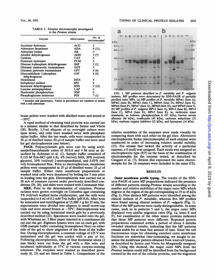
14-FIG. 1. MP patterns identified in P. mirabilis and P. vulganis
isolates. MP profiles were determined by SDS-PAGE of partiallypurified outer MPs. (a) MP profiles of P. mirabilis MPml (lane A),MPm2 (lane B), MPm3 (lane C), MPm4 (lane D), MPm5 (lane E),MPm6 (lane F), MPm7 (lane G), MPm8 (lane H), and MPm9 (lane I).(b) MP profiles of P. vulganis MPv1 (lane A), MPv2 (lane B), MPv3(lane C), MPv4 (lane D), MPv5 (lane E). m, molecular massstandards, as follows: phosphorylase b (97 kDa), bovine serumalbumin (66 kDa), ovalbumin (45 kDa), carbonic anhydrase (31kDa), soybean trypsin inhibitor (21 kDa), and lysozyme (14 kDa).
relative mobilities of the enzymes were made visually bycomparing them with each other on the gel slice. Alternativeelectrophoretic forms (electromorphs) of each enzyme werenumbered in order of increasing relative anodal mobility(15). For strains that lacked the activity of a particularenzyme, a 0 (null) was assigned. Each strain was assigned anelectrophoretic type (ET) on the basis of the combination ofelectromorphs for the enzyme tested, as described byCaugant et al. (7). Strains that expressed the same electro-morphs for the 16 enzymes were included in the same ETs.
RESULTS
Outer membrane profile typing. The results of the SDS-urea PAGE of outer MP preparations indicated the presenceof different patterns among Proteus strains according to thenumber and relative mobilities of the major outer MPs whichmigrate in the region of the gel corresponding to 35 to 45 kDa(Fig. 1). Nine distinct MP patterns were identified among theclinical isolates of P. mirabilis, whereas five MP profileswere found among clinical isolates of P. vulgaris (Fig. 1).Most of the MP patterns were easily distinguishable. In somecases, such as in patterns 5 and 6, the major outer MPsdisplayed very similar migration rates (Fig. la, lanes E andF), but examination of the other minor proteins indicatedthat these MP patterns were different. Repeated proteinpreparations examined within a period of about 6 monthsrevealed identical results, indicating that the MP patternsremain stable for at least that amount of time. Since the cellfractionation steps for obtaining enriched outer membranefractions are somewhat time-consuming, we sought to ex-amine the membrane pattern obtained from total cell lysates,as described by Senior and Voros for Morganella morganii(28). Using this method, the major outer MPs from theProteus isolates could still be identified from the backgroundcreated by the rest of the cellular proteins, and the migration
VOL. 30, 1992
4ft -
40.*.
-Aw mv 40
on April 9, 2019 by guest
http://jcm.asm
.org/D
ownloaded from

2634 KAPPOS ET AL.
TABLE 2. Distribution of MP profiles and ETs inP. mirabilis. and P. vulgaris
Species MP No. of No. of ET no.profile strains ETs
P. mirabilis MPml 25 18 2-4, 6-7, 15-17,19-21,24-25, 27-30, 34
MPm2 4 4 5, 9,11, 31MPm3 2 2 3, 22MPm4 4 3 10,18, 35MPm5 5 5 1, 8,14, 32, 33MPm6 1 1 13MPm7 1 1 26MPm8 1 1 23MPm9 1 1 12
P. vulgaris MPvl 4 4 2v, 5v, 8v, lOvMPv2 2 2 4v, 9vMPv3 1 1 7vMPv4 2 2 3v, 6vMPv5 1 1 lv
patterns of the major outer membranes were found to beidentical to those observed with the more purified outer MPpreparations (data not shown). However, the presence of alarge number of proteins sometimes made it difficult toclearly discern differences between strains with similar MPprofiles. The rapid method of protein preparation by usingboiled cells could be used as an alternative method ofexamining membrane patterns if facilities for preparing outermembranes are not available.
It is interesting that the MP patterns of P. mirabilis and P.vulgaris strains were different (Fig. 1). Therefore, MPm andMPv were chosen as the standard nomenclature to identifyMP patterns from P. mirabilis and P. vulgaris, respectively.The majority of the P. mirabilis isolates were grouped intofive MP patterns (Table 2). MPml, the most common patternamong the isolates of P. mirabilis, was found in 25 strains(57%) (Table 2 and Fig. la). Another 15 P. mirabilis strainswere assigned to MPm patterns 2, 3, 4, and 5 (Table 2 andFig. la, lanes B to E), whereas the remaining 4 isolates hadunique MP patterns (Fig. la, lanes F to I). In the case of P.vulganis, four strains were assigned to the pattern MPvl(Table 2 and Fig. lb, lane A). Patterns MPv2 and MPv4comprised two strains each, and the remaining two P.vulgaris isolates had unique MP patterns (Table 2 and Fig.lb, lanes B to E).MEE. All the clinical isolates of Proteus were also ana-
lyzed by MEE. We sought to use MEE to obtain an
independent confirmation of the validity of MP profiles forstrain differentiation and also to be able to subclassify thestrains of P. mirabilis and P. vulgaris belonging to the mostcommon MP profiles.The 16 enzymes chosen (Table 1) are known to occur in
Escherichia coli and Shigella species (15, 16) and were alsoexpected to be expressed by the Proteus isolates sincemembers of this genus belong to the family Enterobac-teriaceae. The conditions for enzyme analysis were thoseshown to be optimal for E. coli enzyme electrophoresis (16).All the enzymes assayed were detected in the majority of theProteus isolates examined. The number of electromorphsdetected for each enzyme among the P. mirabilis and P.vulgaris isolates is indicated in Table 1. CAT, ACO, AK,HEX, GOT, FUM, LAP, NSP, and IPO occurred in allisolates (enzyme abbreviations are listed in Table 1). Theremaining enzymes were not always detected in all of the
isolates. No enzyme was found to be characteristic of eitherP. mirabilis or P. vulgaris. IDH was not found in sevenisolates ofP. mirabilis and six isolates P. vulgaris. ADA wasnot found in eight and three isolates of P. mirabilis and P.vulgaris, respectively, and ADH was absent from sevenstrains (13%). It is not possible to conclude whether theseenzymes are indeed absent from certain Proteus strains or,alternatively, whether they are expressed as electromorphswhich cannot be resolved under the conditions used for theelectrophoresis.ETs consisting of a 16-digit number were generated for
each isolate by using the 16 enzymes assayed. The distribu-tions of ETs in P. mirabilis and P. vulgaris strains are givenin Tables 3 and 4, respectively. Thirty-five distinct ETs wereobserved among the 44 isolates of P. mirabilis screened(Table 3). Only a few P. mirabilis ETs were common to morethan one isolate, such as ET2 (two isolates), ET3 (fiveisolates), ET4 (four isolates), and ET10 (two isolates),whereas the remaining P. mirabilis strains each corre-sponded to a distinct ET (Table 3). In the case ofP. vulgaris,10 strains examined had 10 unique ETs (Table 4).The ETs expressed by isolates with the same MP profiles
in P. mirabilis and P. vulgaris are shown in Table 2. Thestrains with the MPml pattern expressed 18 ETs, indicatingthat this group is genetically heterogeneous. Altogether, 40isolates from the four most common MP groups of P.mirabilis were subgrouped into 30 ETs. Strains with uniqueMP patterns were also found to have unique ETs, except inthe case of ET3, which was reproducibly found in fourisolates with the MPml pattern and one isolate with theMPm3 pattern (Table 2) (data not shown). All P. vulgarisisolates had unique ETs, even though eight strains weregrouped into three MPv patterns (Table 2).
DISCUSSION
MP profile typing is a relatively simple and reliable typingtechnique which does not require any sophisticated equip-ment. The results reported here suggest that MP patterns canbe used for the differentiation of clinical isolates of Proteus.Most of the P. mirabilis isolates (90%) fell into five MPpatterns, and 8 of 10 P. vulgaris isolates were grouped intothree MP patterns. The remaining P. mirabilis and P. vul-garis isolates had unique MP patterns. At present, we do notknow whether the natural environment faced by these mi-croorganisms could play some role in the selection of strainswith specific MP patterns, the significance of which wouldrequire further investigation. However, it is likely that thedistribution of MP patterns reflects local epidemiologicalfactors.
Previous studies by other investigators have shown thatouter MP patterns correlate well with other indicators ofclonal descendence (1-3, 5, 14, 15, 20, 30). MP patterns havebeen used for typing clinical strains of E. coli (1, 2), Shigellaspp. (29), Haemophilus influenzae (3), Haemophilus pleuro-pulmoniae (14), Neisseria meningitidis (17), and Borreliaburgdorferi (5). Also, a protein typing system similar to thatdescribed in this report has recently been reported forclinical strains of M. morganii (28).Although the isolates examined in our study were obtained
from different patients, we initiated a prospective study withsequential isolates from the same patients with urinary tractinfections. Preliminary evidence shows that successive iso-lates of P. mirabilis obtained from two patients exhibited thesame patterns, suggesting that in each of those cases rein-fection with the same strain ofP. mirabilis occurred (11). We
J. CLIN. MICROBIOL.
on April 9, 2019 by guest
http://jcm.asm
.org/D
ownloaded from

VTYPING OF CLINICAL PROTEUS ISOLATES 2635
TABLE 3. ETs identified in 44 isolates of P. mirabilis
No. of Electromorph (isoenzyme)4ET no. No.aofstramns CAT ACO AK G3P G6P HEX IDH ADH GOT FIM LAP NSP ADA IPO PGI GPT
1 1 1 1 1 1 1 3 1 1 4 1 1 1 0 1 1 52 2 1 1 1 1 1 3 1 1 4 1 1 1 0 1 2 53 5 1 1 1 1 1 3 1 1 4 1 1 1 1 1 1 54 4 1 1 1 1 1 3 1 1 4 1 1 1 1 1 2 55 1 1 1 1 1 1 3 1 1 4 1 1 1 1 3 2 56 1 1 1 1 1 1 3 1 1 4 1 1 1 0 5 2 57 1 1 1 1 1 1 3 1 1 4 1 2 1 1 1 1 58 1 1 1 1 1 1 3 1 1 4 1 2 1 1 1 2 59 1 1 1 1 1 1 3 1 1 4 1 6 1 1 1 1 510 2 1 1 1 1 1 3 1 1 4 1 6 1 1 1 2 511 1 1 1 1 1 1 3 1 1 4 2 1 1 1 1 1 412 1 1 1 1 1 1 3 1 1 4 2 1 1 3 3 2 513 1 1 1 1 1 1 3 1 1 4 2 2 1 1 1 1 514 1 1 1 1 1 1 3 1 2 4 1 2 3 1 1 3 515 1 1 1 1 1 1 3 1 3 3 1 6 1 1 3 6 516 1 1 1 1 1 1 3 1 3 4 1 1 1 1 1 2 517 1 1 1 1 1 1 3 1 4 4 1 1 1 1 4 2 518 1 1 1 1 1 1 3 3 1 4 2 1 1 1 1 1 519 1 1 1 1 1 1 3 4 1 4 1 1 1 1 1 1 520 1 1 1 1 1 1 4 1 1 4 1 1 1 1 1 1 521 1 1 1 1 1 1 4 1 1 4 1 2 1 1 1 1 522 1 1 1 2 1 1 3 1 1 4 1 6 1 1 1 1 523 1 1 1 2 1 1 3 3 1 4 2 1 1 3 3 1 524 1 1 2 1 1 1 3 0 1 4 1 1 1 1 1 1 525 1 1 2 1 1 1 3 1 0 4 1 1 1 1 1 1 526 1 1 2 1 1 1 3 1 1 4 1 1 1 1 1 1 527 1 1 2 1 1 1 3 1 1 4 1 1 1 1 3 1 528 1 1 2 3 1 2 3 2 1 4 1 1 3 5 1 5 729 1 2 1 2 3 0 3 0 3 2 3 4 6 3 6 6 030 1 3 1 1 1 1 3 1 1 4 1 1 1 1 3 2 531 1 1 1 1 0 1 3 0 0 4 1 1 1 0 1 1 232 1 1 1 1 0 1 3 1 1 4 2 1 1 0 1 1 033 1 1 1 1 1 1 3 0 1 4 1 1 1 0 1 2 034 1 1 1 1 1 1 3 0 1 4 1 1 1 1 4 2 535 1 1 1 1 1 1 3 1 0 4 2 1 1 0 1 2 4a The abbreviation for each enzyme is listed in Table 1. Numerical assignment of electromorphs is described in the text.
thus propose that MP profiles may have clinical application members of a given species (7, 23). Thus, strains with thein the typing ofProteus species for cases of recurrent urinary same ETs are also closely related in other characteristicstract infections in patients with numerous complications and such as outer MP profiles, biotypes, and serotypes (6, 14, 15,also for typing isolates of epidemiological significance such 22). To our knowledge, this is the first report of an analysisas outbreak-related strains. of MEE patterns in Proteus strains. Our results suggest thatMEE has been used extensively to analyze genetic varia- both P. mirabilis and P. vulgaris exhibit high genetic vari-
tions in natural populations of bacteria since it gives a ability since 35 distinct ETs were identified in 44 strains ofP.representative measure of relatedness between individual mirabilis and 10 ETs were found in 10 strains of P. vulgans.
TABLE 4. ETs identified in 10 isolates of P. vulgaris
No.of ~~~~~~~~~~~Electromorph (isoenzyme)bET no.' No.ofnsris CAT ACO AK G3P G6P HEX IDH ADH GOT RIM LAP NSP ADA IPO PGI GPT
lv 1 1 1 1 1 4 2 0 0 4 1 2 1 1 1 3 02v 1 1 1 2 1 1 1 0 0 1 1 2 2 0 2 1 33v 1 1 1 2 1 1 1 0 0 1 1 3 1 3 2 1 64v 1 1 2 1 0 1 2 0 1 3 1 2 1 0 1 6 05v 1 1 1 1 2 3 2 3 1 2 5 1 5 2 2 1 46v 1 1 2 2 1 1 1 1 1 5 1 5 1 4 5 4 67v 1 1 2 2 1 1 2 0 1 2 4 2 1 3 1 5 38v 1 2 2 2 2 3 1 0 3 1 1 3 4 3 2 3 39v 1 1 2 2 0 1 2 0 0 2 1 2 1 0 1 0 0lOv 1 1 2 2 1 1 1 0 1 1 4 2 2 3 2 1 2a v indicates the ET number for P. vulgaris strains.b The abbreviation for each enzyme is listed in Table 1. Numerical assignment of electromorphs is described in the text.
VOL. 30, 1992
on April 9, 2019 by guest
http://jcm.asm
.org/D
ownloaded from

2636 KAPPOS ET AL.
The somewhat higher genetic variability found in isolates ofP. vulgaris could be related to the fact that P. vulgarisisolates are less frequently found in urinary tract infectionscaused by Proteus species (26). This could contribute to anapparently more pronounced variability in the ETs. Theanalysis of genetic variability in P. vulgaris strains requiresa larger number of isolates than we used in this study.The observation that the same ET was found in strains
with two distinct MP patterns may be interpreted as a case ofconvergent evolution or, alternatively, infectious transmis-sion of outer MP-determining genes. Similar observationswere reported to occur in genetically different strains of H.influenzae that expressed the same capsular antigens (20).The MP patterns taken alone may not be good indicators
of genetic variability, since several different ETs were dis-tinguished in strains classified as having the same MPpatterns. Results of this and previous studies (2, 3, 17, 20)have shown that MP patterns can, however, be used forclinical applications in an attempt to ascertain whether aparticular bacterial clone already identified is responsible forcontinued or recurrent infections in single patients or inoutbreaks. The large number of ETs identified among the P.mirabilis and P. vulgaris strains offers a more precise meansof differentiating Proteus strains of clinical interest. How-ever, it may be impractical to perform MEE in many clinicallaboratories because of the complexity of the procedure,which is labor intensive and time-consuming and whichrequires a considerable amount of standardization. There-fore, we propose that upon routine biochemical differentia-tion of specimens as P. mirabilis or P. vulgaris, clinicallyimportant strains can initially be distinguished by the use ofMP patterns, and if more refined differentiation is required,MEE analysis can be applied only to the strains withidentical MP profiles.At the moment we do not know how many different clonal
groups are to be expected in clinical strains of Proteus spp.Additional typing systems such as 0-specific and proticinetyping may be useful for comparing our results with thoseobtained by other methods used at other centers. Correlationof the prevalence of the most frequent patterns with antibi-otic resistance and potential virulence factors in Proteusstrains awaits further studies.
ACKNOWLEDGMENTS
This work was supported in part by a Vicepresident (Research)Award from the University of Western Ontario (to M.A.V.). T.K.was supported by the Medical Students Summer Training Program,University of Western Ontario.We express our gratitude to J. Welsh, L. Dafoe, and D. Moyles
for technical assistance.
REFERENCES1. Achtman, M., A. Mercer, B. Kusecek, A. Pohl, M. Heuzen-
roeder, W. Aaronson, A. Sutton, and R. P. Silver. 1983. Sixwidespread bacterial clones among Escherichia coli Kl isolates.Infect. Immun. 39:315-335.
2. Achtman, M., and G. Pluschke. 1986. Clonal analysis of descentand virulence among selected Escherichia coli. Annu. Rev.Microbiol. 40:185-210.
3. Barenkamp, S. J., R. S. Munson, and D. M. Granoff. 1981.Subtyping isolates of Haemophilus influenzae type b by outermembrane protein profiles. J. Infect. Dis. 143:668-676.
4. Bergeron, M. G. 1985. Urinary tract infection, p. 253-256. InL. A. Mandell and E. D. Ralph (ed.), Essentials of infectiousdiseases. Blackwell Scientific Publications, Boston.
5. Bundoc, V. G., and A. G. Barbour. 1989. Clonal polymorphismsof outer membrane protein OspB of Borrelia burgdorfeni. Infect.
Immun. 57:2733-2741.6. Caugant, D. A., B. R. Levin, I. 0rskov, F. 0rskov, C. Svanborg-
Eden, and R. K. Selander. 1985. Genetic diversity in relation toserotyping in Eschenichia coli. Infect. Immun. 49:407-413.
7. Caugant, D. A., B. R. Levin, and R. K Selander. 1981. Geneticdiversity and temporal variation in the E. coli of a human host.Genetics 98:467-490.
8. Harris, H., and D. A. Hopkinson. 1976. Handbook of enzymeelectrophoresis in human genetics. North-Holland PublishingCo., Amsterdam.
9. Hickman, F. W., and J. J. Farmer m. 1976. Differentiation ofProteus mirabilis by bacteriophage typing and the Dienes reac-tion. J. Clin. Microbiol. 3:350-358.
10. Kappos, T., M. A. John, Z. Hussain, and M. A. Valvano. 1991.Outer membrane protein profiles and multilocus enzyme elec-trophoresis analysis for differentiation among clinical isolates ofProteus, p. 374, C-196. Abstr. 91st Gen. Meet. Am. Soc.Microbiol. 1991. American Society for Microbiology, Washing-ton, D.C.
11. Kappos, T., and M. A. Valvano. Unpublished data.12. Kelly, M. T., D. J. Brenner, and J. J. Farmer IH. 1985.
Enterobacteriaceae, p. 263-277. In E. H. Lennette, A. Balows,W. J. Hausler, Jr., and H. J. Shadomy (ed.), Manual of clinicalmicrobiology. American Society for Microbiology, Washington,D.C.
13. Larsson, P., H. E. Andersson, and B. Norlen. 1978. Serotyping inepidemiological tracing of nosocomially acquired Proteus mi-rabilis in a geriatric ward. Infection 6:105-110.
14. Musser, J. M., V. J. Rapp, and R. K. Selander. 1987. Clonaldiversity in Haemophilus pleuropulmoniae. Infect. Immun.55:1207-1215.
15. Ochman, H., and R. K. Selander. 1984. Evidence for clonalpopulation structure in Escherichia coli. Proc. Natl. Acad. Sci.USA 81:198-201.
16. Ochman, H., T. S. Whittam, D. A. Caugant, and R. K. Selander.1983. Enzyme polymorphism and genetic population structurein Escherichia coli and Shigella. J. Gen. Microbiol. 129:2715-2726.
17. Olyhoek, T., B. A. Crowe, and M. Achtman. 1987. Clonalpopulation structure of Neisseria meningitidis serogroup Aisolated from epidemics and pandemics between 1915 and 1983.Rev. Infect. Dis. 9:665-691.
18. Penner, J. L. 1984. Proteus, p. 491-494. In N. R. Krieg and J. G.Holt (ed.), Bergey's manual of systematic bacteriology, vol. 1.The Williams & Wilkins Co., Baltimore.
19. Penner, J. L., and J. N. Hennessy. 1980. Separate 0-groupingschemes for serotyping clinical isolates of Proteus mirabilis andProteus vulgaris. J. Clin. Microbiol. 12:304-309.
20. Porras, O., D. A. Caugant, B. Gray, T. Lagergird, B. R. Levin,and C. Svanborg-Eden. 1986. Difference in structure betweentype b and nontypable Haemophilus influenzae populations.Infect. Immun. 53:79-89.
21. Schmidt, W. C., and C. D. Jeffries. 1974. Bacteriophage typingof Proteus mirabilis, Proteus vulgaris, and Proteus morganii.Appl. Microbiol. 27:47-53.
22. Selander, R. K., P. Beltran, N. H. Smith, R. Helmuth, F. A.Rubin, D. J. Kopecko, K. Ferris, B. D. Tall, A. Craviotto, andJ. M. Musser. 1990. Evolutionary genetic relationships of clonesof Salmonella serovars that cause human typhoid and otherenteric fevers. Infect. Immun. 58:2262-2275.
23. Selander, R. K., D. A. Caugant, H. Ochman, J. M. Musser,M. N. Gilmour, and T. S. Whittam. 1986. Methods of multilocusenzyme electrophoresis for bacterial population genetics andsystematics. Appl. Environ. Microbiol. 51:873-884.
24. Senior, B. W. 1977. Typing of Proteus strains by proticineproduction and sensitivity. J. Med. Microbiol. 10:7-17.
25. Senior, B. W. 1977. The Dienes phenomenon: identification ofthe determinants of compatibility. J. Gen. Microbiol. 102:235-244.
26. Senior, B. W. 1979. The special affinity of particular types ofProteus mirabilis for the urinary tract. J. Med. Microbiol.12:1-8.
27. Senior, B. W., and P. Larsson. 1983. A highly discriminatory
J. CLIN. MICROBIOL.
on April 9, 2019 by guest
http://jcm.asm
.org/D
ownloaded from

TYPING OF CLINICAL PROTEUS ISOLATES 2637
multityping scheme for Proteus mirabilis and Proteus vulgaris.J. Med. Microbiol. 16:193-202.
28. Senior, B. W., and S. Voros. 1990. Protein profile typing. A newmethod for typing Morganella morganii strains. J. Med. Micro-biol. 33:259-264.
29. Valvano, M. A., and C. L. Marolda. 1991. Relatedness of0-specific lipopolysaccharide side chain genes from strains ofShigella boydii type 12 belonging to two clonal groups and fromEscherichia coli 07:K1. Infect. Immun. 59:3917-3923.
30. Valvano, M. A., R. P. Silver, and J. H. Crosa. 1986. Occurrenceof chromosome- or plasmid-mediated aerobactin iron transportsystems and hemolysin production among clonal groups ofhuman invasive strains of Eschenichia coli Kl. Infect. Immun.52:192-199.
31. Warren, J. W., D. Damron, J. H. Tenney, J. M. Hoopes, B.Deforge, and H. L. Muncie. 1987. Fever, bacteremia, and deathas complications of bacteriuria in women with long-term ure-thral catheters. J. Infect. Dis. 155:1151-1158.
VOL. 30, 1992
on April 9, 2019 by guest
http://jcm.asm
.org/D
ownloaded from