our policy in intraventricular colloid cysts. experience ... · 54 a.v. ciurea et al...
TRANSCRIPT

54 A.V. Ciurea et al Intraventricular colloid cysts
Our policy in intraventricular colloid cysts. Experience of 31 operated cases.
A.V. Ciurea, F.M. Brehar, Al. Tascu, A. Iliescu, D. Talianu, R. Rizea
First Neurosurgical Department Emergency Clinical Hospital “Bagdasar-Arseni”, Bucharest
Abstract The colloid cyst of the third ventricle is a
benign tumor situated in the anterior part of the third ventricle. This lesion represents less than 1% of the primary brain tumor being more common in young adults. Because of its particular location, the colloid cyst can obstruct the Monro foramen, producing intermittent intracranian hypertension with headache, vomiting and visual disturbances. Thirty-one cases of colloid cysts have been operated using the microsurgical approach in the First Neurosurgical Department of Emergency Clinical Hospital “Bagdasar-Arseni” between January 1995 and December 2008. The age of the patients was between 17 and 46 years, with a medium age of 31 years. The follow-up period was between 9 months and 7 years. In three cases TTA approach has been performed. One of the cases developed a venous cerebral infarct after this procedure, but the patient had finally a good outcome. For 28 patients the transcortical approach has been performed. In all cases the total resection of the colloid cyst has been performed. Of all 31 cases, one case presented a transitory hemiparesis, two cases showed negativist behavior, and three cases had transitory memory disturbances. There was no intraventricular hemorrhage after colloid cyst resection in our series. In
conclusion, according to our policy, the microsurgical approach is the best treatment for third ventricular colloid cysts because of its main advantages compared with the endoscopic approach: the possibility of total resection of the cyst, the good control of the bleeding source during the procedure, and a better exposure of the anatomical landmarks.
Keywords: colloid cyst, obstructive hydrocephalus, microsurgical approach, neuro-endoscopy.
Introduction Colloid cysts are among the most
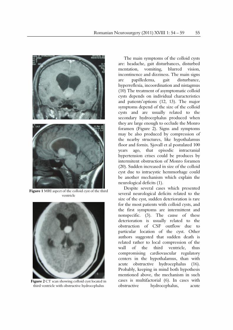
common benign tumors of the third ventricle and represents, according with literatury data, between 0.5 and 2% of all intracranial neoplasms (10, 11). A characteristic feature of the colloid cyst is its location at the anterior part of the third ventricle, which may produce the occlusion of the foramina of Monro, resulting in biventricular hydrocephalus (11). This particular location reflects in a specific clinical picture which may include rapid neurological deterioration and even sudden death. (9, 19). The vast majority of colloid cysts reported in the literature are symptomatic and were therefore treated. The size of the cysts which is clinically significant is more than 1,5 cm in diameter (Figure 1).
Romanian Neurosurgery (2011) XVIII 1: 54 – 59 55
Figure 1 MRI aspect of the colloid cyst of the third
ventricle
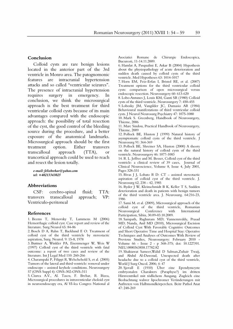
Figure 2 CT scan showing colloid cyst located in
third ventricle with obstructive hydrocephalus
The main symptoms of the colloid cysts are: headache, gait disturbances, disturbed mentation, vomiting, blurred vision, incontinence and dizziness. The main signs are papilledema, gait disturbance, hyperreflexia, incoordination and nistagmus (10) The treatment of asymptomatic colloid cysts depends on individual characteristics and patients’options (12, 13). The major symptoms depend of the size of the colloid cysts and are usually related to the secondary hydrocephalus produced when they are large enough to occlude the Monro foramen (Figure 2). Signs and symptoms may be also produced by compression of the nearby structures, like hypothalamus floor and fornix. Sjovall et al postulated 100 years ago, that episodic intracranial hypertension crises could be produces by intermitent obstruction of Monro foramen (20). Sudden increased in size of the colloid cyst due to intracystic hemmorhage could be another mechanism which explain the neurological deficits (1).
Despite several cases which presented several neurological deficits related to the size of the cyst, sudden deterioration is rare for the most patients with colloid cysts, and the first symptoms are intermittent and nonspecific. (3). The cause of these deterioration is usually related to the obstruction of CSF outflow due to particular location of the cyst. Other authors suggested that sudden death is related rather to local compression of the wall of the third ventricle, thus compromising cardiovascular regulatory centers in the hypothalamus, than with acute obstructive hydrocephalus (16). Probably, keeping in mind both hypothesis mentioned above, the mechanism in such cases is multifactorial (6). In cases with obstructive hydrocephalus, acute

56 A.V. Ciurea et al Our policy in intraventricular colloid cysts
deterioration might be provoked by lumbar puncture, therefore a CT scan is mandatory before performing such a procedure (3). In the presence of associated obstructive hydrocephalus, there are two managemnt options. If there is an acute symptomatic hydrocephalus, a ventricular drainage should be performed in emergency. If the ventricular enlargment is not accompanied by sings and symptoms of intracranial hypertension, the presence of hydrocephalus ventricles could be advantageous, offering to the surgeon a natural corridor and enough room to reach the cyst and to remove the lesion. This situation is favorable for the transcortical approach. Contrary, if the transcallosal route is chosen, one way is to implant a ventricular drain in order to avoid excessive retraction of the medial wall of the frontal lobe (11). In this paper the authors present their experience in 31 consecutive cases of colloid cysts operated using two microsurgical approaches.
Materials and methods Thirty one cases of colloid cysts have
been operated using the microsurgical approach in the First Neurosurgical Department between January 1995 and December 2008. The age of the patients was between 17 and 46 years, with a medium age of 31 years. There were 15 males and 16 females. The main symptoms were: headache in 28 cases (90,3%), ataxiain 12 cases (38,7%), VI-th nerve paresis in 9 cases (29,1%), memory impairment in 14 cases (45,2%), “ventricular seizures” in 18 cases (58,1%). Other clinical signs consist in gatism in 4 cases (12,9%), nystagmus in 5 cases (16,1%), bilateral dismetria in 4 cases (12,9%), epileptic seizures in 11 cases (35,5%), psychic impairment in 5 cases
(16,1%). Twenty-eight cases (90,4%) have preoperative hydrocephalus and 3 cases (9,6%) were without hydrocephalus The follow-up period was between 9 months and 7 years. One case died in the emergency room and diagnosis has been established during necropsy (case non-included in our series). Another case presented at the emergency room with intracranial hypertension syndrome. The CT scan showed signs of acute hydrocephalus. VP shunt has been performed in emergency and the patient’s symptoms improved significantly. The MR performed after surgical procedure identified a colloid cyst located in the third ventricle which had produced the obstructive hydrocephalus.
Treatment consisted in surgery in 31 cases (100%), respectively in tumour removal without VP shunt 29 cases (93,5%) and VP shunt (performed in emergency) and removal of the tumor in 2 cases (6,50%). There was no case for observation only.
In three cases, TTA approach has been performed. One of the cases developed a venous cerebral infarct after this procedure, but the patient had finally a good outcome.
For 28 patients, the transcortical approach has been performed. The advantages of this approach consist in the excellent exposure of the cyst and surrounding important anatomical landmarks (internal cerebral vein, anterior septal vein, talamo-striate vein, choroid plexus, etc.), the resection of the cyst totally inclusive the cyst’s capsule and a very good control of any bleeding point.
Results and discussions In all cases total removal of the colloid
cyst has been performed (Figure 3 and Figure 4).

Romanian Neurosurgery (2011) XVIII 1: 54 – 59 57
Figure 3A
Figure 3A Colloid cyst of the third ventricle.
B Postoperative aspect
Of all 31 cases, one case presented a transitory hemiparesis, two cases showed negativist behavior, and three cases had transitory memory disturbances. There was no intraventricular hemorrhage after colloid cyst resection in our series. Immediate complications consist in intracerebral hemorrhage 1 case (3,2%), epileptic seizures in 11 cases (35,4%), of which partial seizures in 7 cases (22,5%) and generalized seizures in 4 cases (12,9%).
Figure 4A
Figure 4A Colloid cyst of the third ventricle. B Postoperative MR image which shows total
resection of the cyst Late complications were: obstructive
hydrocephalus in 2 cases (6,4%), transitory left hemiparesis in 5 cases (16,1%), memory disorder in 8 cases (25,8%), negativism, apathy in 4 cases (12,9%).
Evolution of 31 cases operated were favorable in 30 cases (96,7%), worse in 1 case (3,3%) and no case of death (0 %).
Microsurgical approach should be the first treatment option. Either transvers transcallosal approach (TTA), or

58 A.V. Ciurea et al Our policy in intraventricular colloid cysts
transcortical approach could be used to reach and resect the lesion totally.
Both approaches are effective and usually safe, although neither is without complications. The initial steps of the transcallosal are perhaps a bit more difficult and require meticulous microsurgical techniques. We do not prefer it, however this is the procedure of choice if ventricles are not enlarged. It allows a good visualization of the structures inside the ventricular system. On the other hand the trancortical approach have several advantages compared with transvers transcallosal approach.
The minim invasive techniques consist in endoscopic, endoscope-assisted keyhole approaches and, less frequently nowadays, stereotactic aspiration of the cyst. Colloid cysts, especially in patients with normal ventricles after shunting, can be treated with simple stereotactic aspiration (2). It should be mentioned that the content could be too viscous and difficult to aspirate (15) and, furthermore, part of the capsule may be left behind, causing recurrence of the lesion in an unacceptably high rate (8). More recently, endoscopic approach represents reasonable alternatives. However, there are many cases when portions of the capsule are left behind so the risk of recurrence still remains. Despite of the technological advances, questions still remain regarding the advantages of open versus endoscopic in resection of colloid cysts. Comparing a series of a single institution, Horn et al. showed that in the endoscopic group the rate of residual or recurrent cysts was higher and the length of stay was slightly shorter (7). This study showed also, that severe complications such as hemiparesis and memory deficits can be associated with this minimal invasive
approach. Before the recent development of modern endoscopic technique, Kelly et al proposed another minimally invasive approach which consisted in placing stereotactically a tube retractor through a frontal burr hole. Other authors support the simultaneous approach of endoscopic and microsurgical strategies (4). As mentioned above, ventricular enlargement could be advantageous for the surgical approach, especially for transcortical approach. Contrary, if the transcallosal route is chosen, a ventricular drain should be used intra-operatively in order to avoid excessive retraction of the medial wall of the frontal lobe (11). Several authors who support the microsurgical approach of the colloid cyst of the third ventricle as the best therapeutically option (5, 14, and 18). M. Sami consider also that third ventricle colloid cyst has a perfect microsurgical indication (17). In our opinion the microsurgical approach is the best treatment for third ventricular colloid cysts because it offers several advantages compared with the endoscopic approach, such as the possibility of total resection of the cyst, the good control of the bleeding source during the procedure, and a better exposure of the anatomical landmarks. Our option is for the transcortical microsurgical approach because TTA has and important risk of memory deficit and could produce deep vein lesions.
The advantages of this approach consist in the excellent exposure of the cyst and surrounding important anatomical landmarks (internal cerebral vein, anterior septal vein, talamo-striate vein, choroid plexus, etc.), the resection of the cyst totally inclusive the cyst’s capsule and a very good control of any bleeding point.
Romanian Neurosurgery (2011) XVIII 1: 54 – 59 59
Conclusion Colloid cysts are rare benign lesions
located in the anterior part of the 3rd ventricle in Monro area. The patognomonic features are intracranial hypertension attacks and so called “ventricular seizures”. The presence of intracranial hypertension requires surgery in emergency. In conclusion, we think the microsurgical approach is the best treatment for third ventricular colloid cysts because of its main advantages compared with the endoscopic approach: the possibility of total resection of the cyst, the good control of the bleeding source during the procedure, and a better exposure of the anatomical landmarks. Microsurgical approach should be the first treatment option. Either transvers transcallosal approach (TTA), or trancortical approach could be used to reach and resect the lesion totally.
e-mail: [email protected] tel: +40213343025
Abbreviations CSF: cerebro-spinal fluid; TTA:
transvers transcallosal approach; VP: Ventriculo-peritoneal
References 1. Beems T, Menovsky T, Lammens M (2006) Hemorrhagic colloid cyst. Case report and review of the literature. Surg Neurol 65: 84-86 2. Bosch D A, Rahn T, Backlund E O: Treatment of colloid cyst of the third ventricle by stereotactic aspiration, Surg. Neurol. 9: 15-8, 1978 3. Buttner A, Winkler PA, Eisenmenger W, Weis W (1997) Colloid cyst of the third ventricle with fatal outcome: a report of two cases and review of the literature. Int J Legal Med 110: 260-266 4. Charampaki P, Filippi R, Welschehold S, et al. (2005) Tumors of the lateral and third ventricle: removal under endoscope – assisted keyhole conditions. Neurosurgery 57 (ONS Suppl 4): ONS-302-ONS-311 5. Ciurea A.V., Al. Tascu, F. Brehar, R. Rizea, Microsurgical procedures in intraventicular choloid cyst in neuroendoscopy era, Al VI-lea Congres National al
Asociatiei Romane de Chirurgie Endoscopica, Bucuresti, 11-14.11.2009. 6. Hamlat A, Pasqualini E, Askar B (2004) Hypothesis about the physiopathology of acute deterioration and sudden death caused by colloid cysts of the third ventricle. Med Hypotheses 63: 1014-1017 7. Horn EM, Feiz-Erfan I, Bristol RE, et al. (2007) Treatment options for the third ventricular colloid cysts: comparison of open microsurgical versus endoscopic resection. Neurosurgery 60: 613-620 8. Lobo-Antunes J, Louis KM, Ganti SR (1980) Colloid cysts of the third ventricle. Neurosurgery 7: 450-455 9. Lobosky JM, Vangilder JC, Damasio AR (1984) Behavioural manifestations of third ventricular colloid cysts. J Neurol Neurosurg Psychiatry 47: 1075-1080 10. Mark S. Greenberg, Handbook of Neurosurgery, Thieme, 2006 11. Marc Sindou, Practical Handbook of Neurosurgery, Thieme, 2009 12. Pollock BE, Huston J (1999) Natural history of asymptomatic colloid cysts of the third ventricle. J Neurosurg 91: 364-369 13. Pollock BE, Shreiner SA, Huston (2000) A theory on the natural history of colloid cysts of the third ventricle. Neurosurgery 46: 1077-1083 14. R. L. Jeffree and M. Besser, Colloid cyst of the third ventricle: a clinical review of 39 cases, Journal of Clinical Neuroscience, Volume 8, Issue 4, July 2001, Pages 328-331 15. Rivas J J, Lobato R D: CT – assisted stereotactic aspiration of colloid cyst of the third ventricle. J. Neurosurg 62, 238 – 42, 1985 16. Ryder J W, Kleinschmidt B K, Keller T S, Sudden deterioration and death in patients with benign tumors of the third ventricle area. J. Neurosurg. 64:216-23, 1986 17. Sami M. et al. (2009), Microsurgical approach of the colloid cyst of the third ventricle, Romanian Neurosurgical Conference with International Participation, Sibiu, 30.09-03.10.2009. 18. Sampath, Raghuram MD; Vannemreddy, Prasad MD; Nanda, Anil MD (2010), Microsurgical Excision of Colloid Cyst With Favorable Cognitive Outcomes and Short Operative Time and Hospital Stay: Operative Techniques and Analyses of Outcomes With Review of Previous Studies, Neurosurgery: February 2010 - Volume 66 - Issue 2 - p 368–375; doi: 10.1227/01. NEU.0000363858.17782.82 19. Shaktawat Sameer,Walid D Salman,Zuhair Twaij, and Abdul Al-Dawoud, Unexpected death after headache due to a colloid cyst of the third ventricle, World J Surg Oncol. 2006; 4: 47 20. Sjovall E (1910) Über eine Ependymcyste embryonalen Charakters (Paraphyse?) im dritten Hirnventrikel mit tödlichem Ausgang. Zugleich eine Beobachtung wahrer lipochromer Veränderungen mit Auftreten von Halbmondkörperchen. Beitr Pathol Anat 47: 248-269