our body’s defenses chapter 43. composition of mammalian blood
TRANSCRIPT

Our Body’s DefensesOur Body’s Defenses
Chapter 43
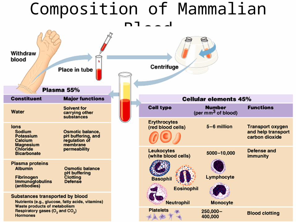
Composition of Mammalian Blood

Differentiation of blood cells
The Kidneys convert a plasma protein into a hormone called erythropoietin, which stimulates the bone marrow to produce erythrocytes
(Become macrophages“big eaters”)
(Phagocytes)
(Destroy larger invaders bySecreting destructive enzymes)
(release Histamine in response to tissue injury)
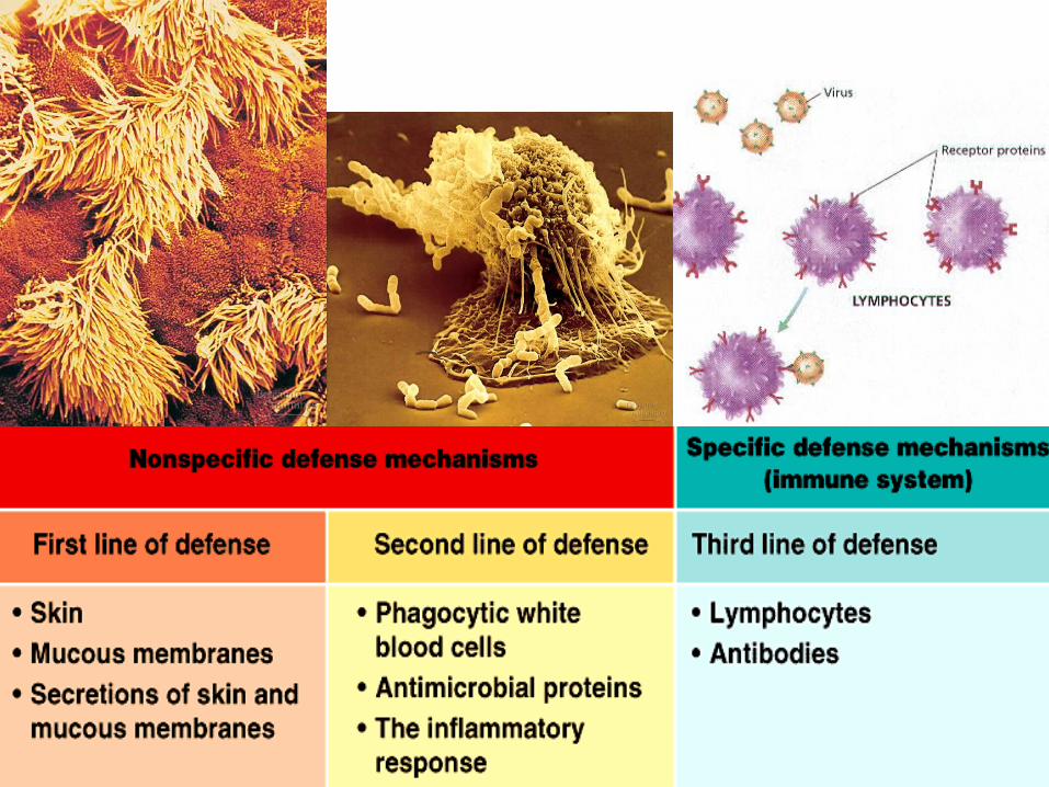

THE FIRST LINE OF DEFENSENon-Specific

The First Line of Defense The First Line of Defense Skin and Mucous membranesSkin and Mucous membranes
Physical barriers• Skin is a barrier when intact. Bacteria and viruses cannot
penetrate it• Mucous membranes of various internal tracts (G.I., Respiratory,
Urogenital) prevent entry of various microbes
Chemical barriers• The skin’s sebaceous glands and sweat glands maintain skin’s
pH at 3 to 5 – too acidic for “visiting” microbes – normal flora of skin are unaffected, but usually not harmful (opportunistic)
• Saliva, tears and mucous contain antimicrobial agents and enzymes like lysozymes
• Mucus in the upper respiratory tract, ear wax traps microbes • Stomach acid kills pathogens (although some survive - viruses
like Hepatitis A, certain bacteria like E.coli and parasitic worms)

THE SECOND LINE OF DEFENSENon-Specific a.k.a Innate

The Second Line of DefenseThe Second Line of DefensePhagocytic WBCs, Inflammatory response, Antimicrobial proteinsPhagocytic WBCs, Inflammatory response, Antimicrobial proteins
• Neutrophils, Eosinophils, Natural Killer cells, Monocytes (macrophages)
• Chemicals such as – Histamine released by mast cells and
basophils – pyrogens such as Interleukin 1– Chemokines such as Interleukins 1 and 2
• Complement system, interferons
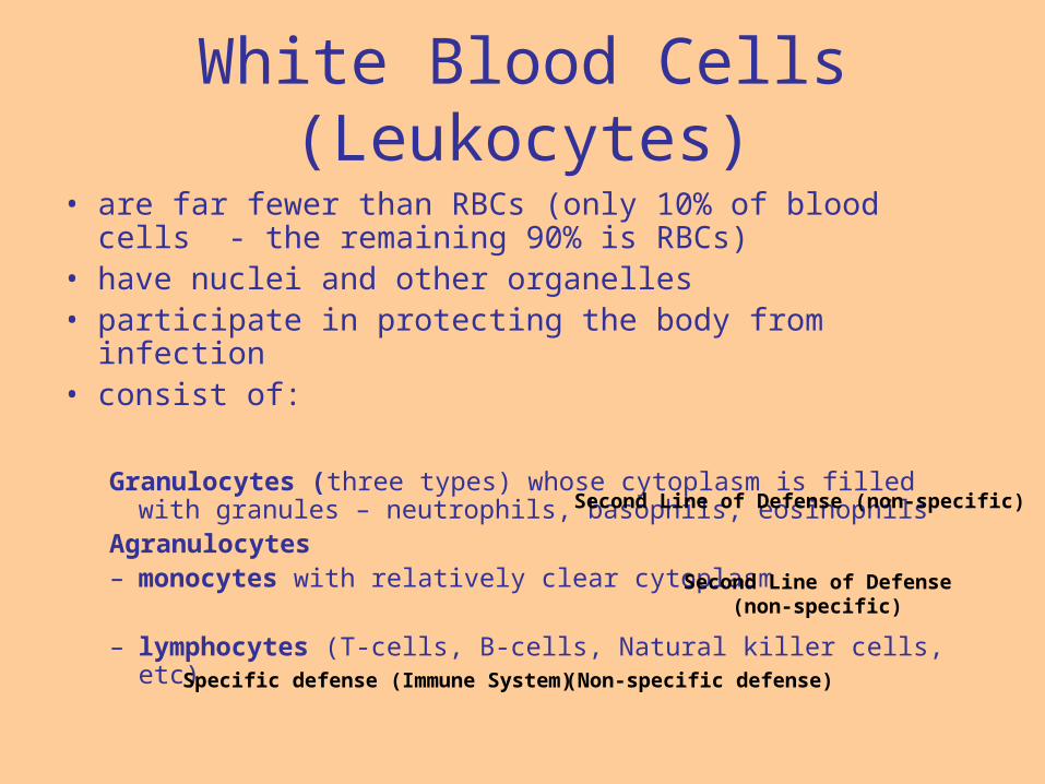
White Blood Cells (Leukocytes)
• are far fewer than RBCs (only 10% of blood cells - the remaining 90% is RBCs)
• have nuclei and other organelles• participate in protecting the body from infection • consist of:
Granulocytes (three types) whose cytoplasm is filled with granules – neutrophils, basophils, eosinophils
Agranulocytes– monocytes with relatively clear cytoplasm
– lymphocytes (T-cells, B-cells, Natural killer cells, etc)Specific defense (Immune System) (Non-specific defense)
Second Line of Defense (non-specific)
Second Line of Defense(non-specific)

Granulocytes vs. Agranulocytes
Granulocytes are a type of white Blood cell (Leukocyte) that attacks and destroys foreign substances. They have specific granules in their cytoplasm. The three different types of granulocytes have different types of specific granules. They are spherical in shape, contain nuclei.
- neutrophils, - eosinophils, and - Basophils
Monocytes and macrophages are agranulocytes.
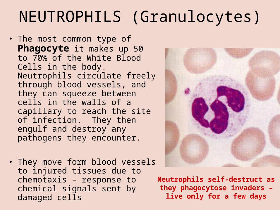
NEUTROPHILS (Granulocytes)• The most common type of
Phagocyte it makes up 50 to 70% of the White Blood Cells in the body. Neutrophils circulate freely through blood vessels, and they can squeeze between cells in the walls of a capillary to reach the site of infection. They then engulf and destroy any pathogens they encounter.
• They move form blood vessels to injured tissues due to chemotaxis – response to chemical signals sent by damaged cells
Neutrophils self-destruct as they phagocytose invaders – live only
for a few days

EOSINOPHILS (Granulocytes)• About 1.5% of leukocytes
• Attack larger parasites such as blood flukes (Platyhelminthes)
• Adhere to the external wall of parasite and release destructive enzymes
• Do not have good phagocytic skills
• their numbers increase sharply in certain diseases, especially infections by parasitic worms.

Basophils (Granulocytes)• Basophils comprise less than 1% of
normal blood leukocytes
• Not phagocytic
• Basophils leave the blood and accumulate at the site of infection or other inflammation.
• There they discharge the contents of their granules, releasing histamine and heparin.
• Histamine triggers blood vessel dilation – so more leukocytes can get to the site of injury and heparin is an anticoagulant that helps the blood flow easily to site of injury.
•The number of basophils The number of basophils increases during infection.increases during infection.

MONOCYTES (Agranulocytes)
• Only constitute 5% of the leukocytes, but very effective
• Long-lived, excellent phagocytes
• Some microbes can evade them
• They circulate in the blood for some time, then they migrate into body tissues and become macrophages
Monocytes or Macrophages also release IL1 which induces fever and also stimulates an immune response

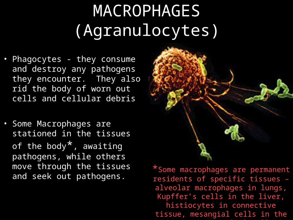
MACROPHAGES (Agranulocytes)
• Phagocytes - they consume and destroy any pathogens they encounter. They also rid the body of worn out cells and cellular debris
• Some Macrophages are stationed in the tissues of the
body*, awaiting pathogens, while others move through the tissues and seek out pathogens.
*Some macrophages are permanent residents of specific tissues – alveolar
macrophages in lungs, Kupffer’s cells in the liver, histiocytes in connective tissue,
mesangial cells in the kidney and microglial cells in the brain.

Pus
• Accumulates at site of infection
• Consists of dead phagocytic cells (like neutrophils and macrophages)
• Also contains fluids and proteins that leaked from capillaries
• Absorbed by body eventually
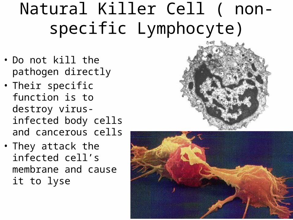
Natural Killer Cell ( non-specific Lymphocyte)
• Do not kill the pathogen directly
• Their specific function is to destroy virus-infected body cells and cancerous cells
• They attack the infected cell’s membrane and cause it to lyse

Summary of Functions of WBCs
• Leukocytes can squeeze between cells lining walls of blood vessels by diapedesis and attack bacteria and debris.
• Neutrophils and monocytes are phagocytic, with monocytes engulfing the larger particles.
• Eosinophils moderate allergic reactions as well as defend against parasitic infections.
• Basophils migrate to damaged tissues and release histamine to promote inflammation and heparin to inhibit blood clotting.
• Lymphocytes are the major players in specific immune reactions and some produce antibodies.

The Second Line of Defense in Action
The Inflammatory Response IS A NONSPECIFIC DEFENSE REACTION OF THE BODY TO TISSUE DAMAGE.
1. Despite the initial defenses of the skin and mucous membranes, pathogens sometimes enter the body.
2. When pathogens enter the body, the immune system has a second line of defense. The body's second line of defense acts when tissues are injured.
3. The mast cells found in connective tissues and basophils release a chemical called HISTAMINE, when injured - which starts a series of changes called the inflammatory response.
4. Histamine increases blood flow to the injured area and increases the permeability of the surrounding capillaries, as a result, fluid and white blood cells (WBC) leak from blood vessels into nearby tissue.

6. Pathogens are attacked by phagocytic white blood cells (leukocytes) such as Neutrophils and Monocytes in response to chemokines – chemical signals
7. Certain toxins released by pathogens may raise body temperature, but leukocytes can do the same by releasing molecules called pyrogens – fevers inhibit microbial growth, speed up chemical reactions and tissue repair
8. Antimicrobial agents collectively called the complement system lyse invading cells
9. Inteferons are proteins secreted by virus-infected cells that limit cell-to-cell spread of the virus
The Second Line of Defense in Action – cont’d.

Mast Cells (Not WBC, but helps with the body’s Defenses)
• A mast cell (or mastocyte) is a resident cell of several types of tissues and contains many granules rich in histamine and heparin.
• Although best known for their role in allergy and anaphylaxis, mast cells play an important protective role as well, being intimately involved in wound healing and defense against pathogens.

The Allergic Response is initiated by plasma cells that
produce antibodies for specific allergens. Some of
the antibodies can be attached to special cells called mast cells, that
produce histamine. This causes most of the
symptoms associated with allergies.
THE FINAL LINE OF DEFENCESpecific Immunity

LYMPHOCYTESLYMPHOCYTES ARE WBCs THAT ACTIVATE THE
SPECIFIC IMMUNE RESPONSE.
There are TWO Main Types of Lymphocytes: - B Cells and - T Cells. (NKCs are also lymphocytes, but non-specific, usually involved in second line of defense)
These WBCs accumulate in the Lymph and Lymph Nodes, where they clean out the lymph fluid by eating and destroying and foreign cells.
Lymphocytes are also found in the Spleen and Blood.
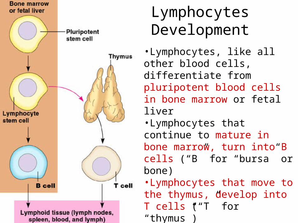
Lymphocytes Development
•Lymphocytes, like all other blood cells, differentiate from pluripotent blood cells in bone marrow or fetal liver•Lymphocytes that continue to mature in bone marrow, turn into B cells (“B” for “bursa” or bone)•Lymphocytes that move to the thymus, develop into T cells (“T” for “thymus”)•Both B and T cells populate lymph nodes, the spleen and other organs of the lymphatic system

B Cells and T Cells
• Circulate in blood and lymph but concentrated in lymph nodes, spleen and other lymphatic regions
• Both cells deal with specific microbes or foreign bodies – specificity!

The Origin of the word Bursa• In birds, the bursa of
Fabricius is the site of hematopoiesis. It is a specialized lymphatic organ necessary for B cell development in birds.
• Mammals generally do not have an equivalent organ; the bone marrow is often both the site of hematopoiesis and B cell development.

The lymphatic systemThe lymphatic systemConsists of organs, ducts, and nodes. It transports a watery Consists of organs, ducts, and nodes. It transports a watery
clear fluid called lymph. This fluid distributes immune cells clear fluid called lymph. This fluid distributes immune cells and other factors throughout the body. It also interacts and other factors throughout the body. It also interacts with the blood circulatory system to drain fluid from cells with the blood circulatory system to drain fluid from cells and tissues. The lymphatic system contains immune cells and tissues. The lymphatic system contains immune cells called called lymphocyteslymphocytes, which protect the body against , which protect the body against antigens (viruses, bacteria, etc.) that invade the body. antigens (viruses, bacteria, etc.) that invade the body.
Main functions of lymphatic system:Main functions of lymphatic system:
• to collect and return interstitial fluid, including plasma to collect and return interstitial fluid, including plasma protein to the blood, and thus help maintain fluid balance, protein to the blood, and thus help maintain fluid balance,
• to defend the body against disease by producing to defend the body against disease by producing lymphocytes, lymphocytes,
• to absorb lipids from the intestine and transport them to to absorb lipids from the intestine and transport them to the blood. the blood.
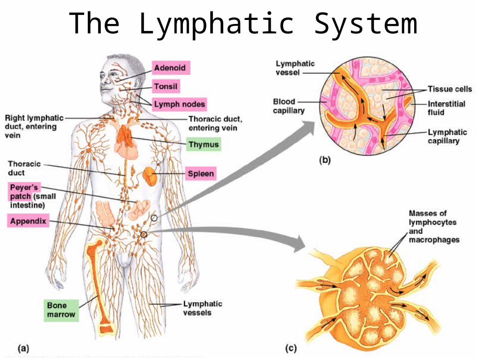
The Lymphatic System
So how does this 3So how does this 3rdrd line of line of defense or specific immunity defense or specific immunity
work?work?
Cell-surface proteins
• All cells, bacteria, viruses, fungi, yeast, etc. have proteins on their surface.
• These proteins may help with facilitated diffusion, active transport, cell-to-cell communication, etc.
• But some of these proteins are used for cell-to-cell recognition.
• Foreign proteins that initiate the immune response are called ANTIGENS
• These antigens are found on viruses, bacteria, parasitic worms, pollen, fungi, protozoa, etc.

Specific Immunity – The Final Line of DefenseSpecific Immunity – The Final Line of Defense
• Antigens cause B lymphocytes to produce ANTIBODIES
• The term antigen is a contraction of ANTIBODY GENERATOR
• Each antigen has a specific shape and several specific sides or sub-shapes called epitopesepitopes.
• The antibodies that B cells produce bind to specific antigens and specific epitopes of the antigens only (Shape matters)

Antibodies• Group of Globular proteins found in blood serum called Immunoglobulins (Ig)
• Each is made up of 4 polypeptide chains – 2 identical light and 2 identical heavy chains, held together with disulfide bridges
• Some amino acid regions of the chains are constant and found in every antibody, whereas others are vary between different antibodies
• The variable regions that form the arms of the Y-shaped molecule are the antigen binding sites
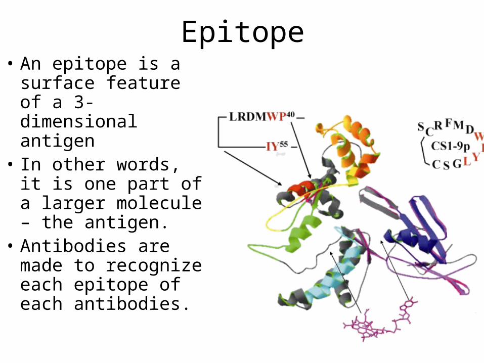
Epitope• An epitope is a
surface feature of a 3-dimensional antigen
• In other words, it is one part of a larger molecule – the antigen.
• Antibodies are made to recognize each epitope of each antibodies.


How Antibodies workThe binding of antibodies to antigens tags foreign cells and molecules for
destruction by phagocytes, or the complement system of proteins.
Opsonization: to make pathogen
ready for phagocytosis
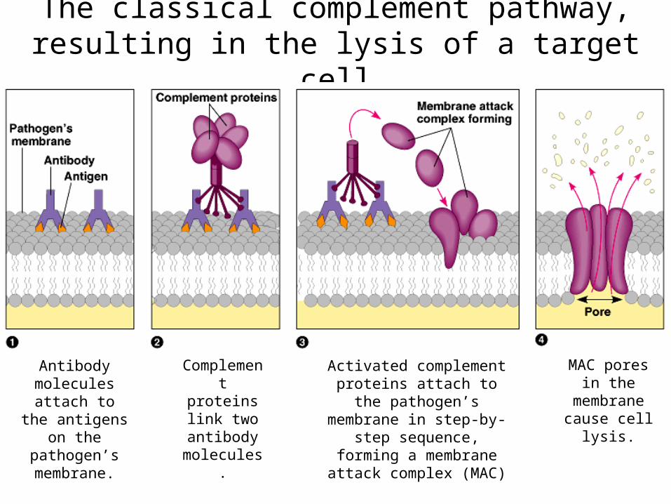
The classical complement pathway, resulting in the lysis of a target cell
Antibody molecules
attach to the antigens on the
pathogen’s membrane.
Complement proteins link two antibody molecules.
Activated complement proteins attach to the
pathogen’s membrane in step-by-step sequence,
forming a membrane attack complex (MAC)
MAC pores in the membrane
cause cell lysis.

Two Groups of Antibodies
• Polyclonal antibodies are a mixed group of antibodies – each one binds to a separate epitope of an antigen. Antibodies made in our bodies are usually polyclonal.
• Monoclonal Antibodies – are a group of antibodies that recognize and bind to only ONE epitope of an antigen. They are made in labs, using mice, cell cultures, etc.
The most common example of antigens and antibodies
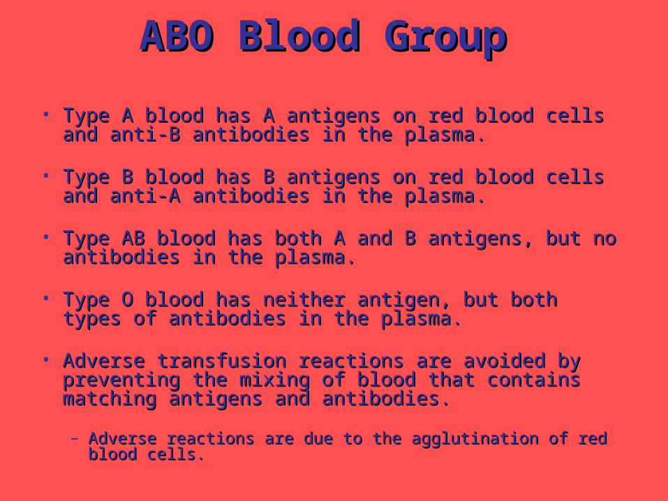
ABO Blood GroupABO Blood Group
• Type A blood has A antigens on red blood cells and anti-B Type A blood has A antigens on red blood cells and anti-B antibodies in the plasma.antibodies in the plasma.
• Type B blood has B antigens on red blood cells and anti-A Type B blood has B antigens on red blood cells and anti-A antibodies in the plasma.antibodies in the plasma.
• Type AB blood has both A and B antigens, but no Type AB blood has both A and B antigens, but no antibodies in the plasma.antibodies in the plasma.
• Type O blood has neither antigen, but both types of Type O blood has neither antigen, but both types of antibodies in the plasma.antibodies in the plasma.
• Adverse transfusion reactions are avoided by preventing Adverse transfusion reactions are avoided by preventing the mixing of blood that contains matching antigens and the mixing of blood that contains matching antigens and antibodies.antibodies.
– Adverse reactions are due to the agglutination of red blood cells.Adverse reactions are due to the agglutination of red blood cells.
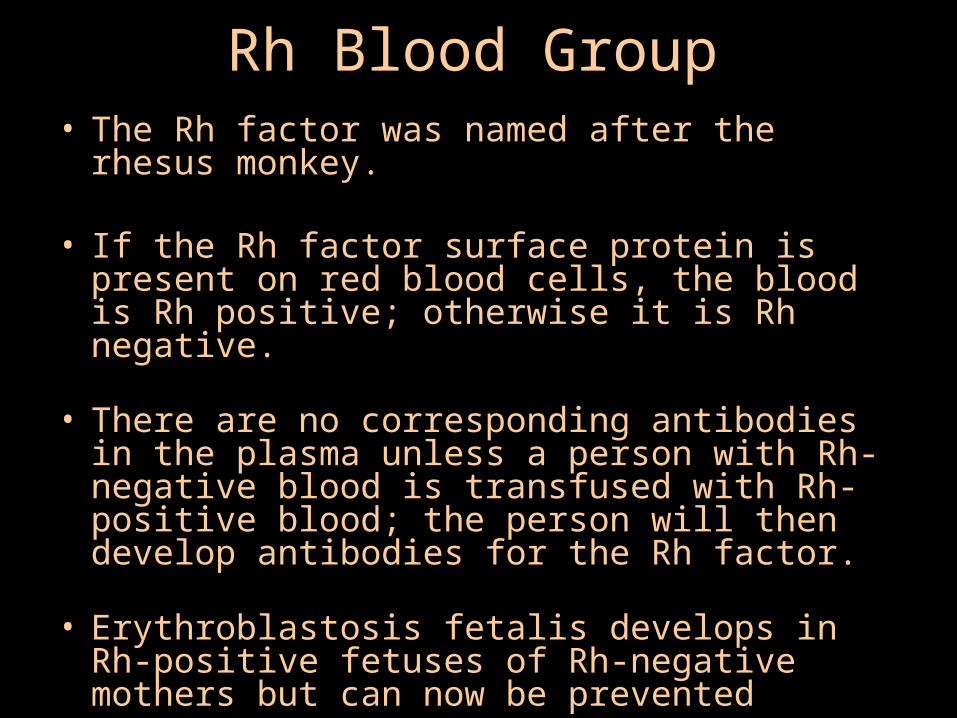
Rh Blood Group• The Rh factor was named after the rhesus
monkey.
• If the Rh factor surface protein is present on red blood cells, the blood is Rh positive; otherwise it is Rh negative.
• There are no corresponding antibodies in the plasma unless a person with Rh-negative blood is transfused with Rh-positive blood; the person will then develop antibodies for the Rh factor.
• Erythroblastosis fetalis develops in Rh-positive fetuses of Rh-negative mothers but can now be prevented

“Self” Markers or MHC• All our cells have surface glycoproteins called MHC
(Major Histocompatibility Complex) glycoproteins. They are also known as HLA for Human Leukocyte Antigens
• There are 2 classes of MHC – Class I MHC and Class II MHC– Class I MHC glycoproteins are found on all nucleated
cells (almost all body cells) non-professional antigen-non-professional antigen-presenting cellspresenting cells
– Class II MHC glycoproteins are found only on a few cell types like macrophages, B cells, activated T cells and the cells that make up the interior of the thymus professional antigen-presenting cellsprofessional antigen-presenting cells

MHCs and Chromosome 6
• The genes for both class I and class II MHC proteins are on chromosome 6
• There are hundreds of different alleles for the MHC genes, so no 2 people will have the same type of MHC glycoproteins on their cells, unless they are identical twins
• This leads to tissue rejection after organ transplants (the “histo” in histocompatibility means tissue)
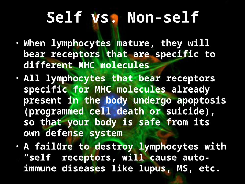
Self vs. Non-self
• When lymphocytes mature, they will bear receptors that are specific to different MHC molecules
• All lymphocytes that bear receptors specific for MHC molecules already present in the body undergo apoptosis (programmed cell death or suicide), so that your body is safe from its own defense system
• A failure to destroy lymphocytes with “self” receptors, will cause auto-immune diseases like lupus, MS, etc.

What do the MHCs do?What do the MHCs do?
• The job of MHC molecules is to “present” The job of MHC molecules is to “present” antigens that cells may be housing due to an antigens that cells may be housing due to an infection or intrusion by foreign tissues to T infection or intrusion by foreign tissues to T cellscells
Remember: An antigen is a foreign molecule Remember: An antigen is a foreign molecule that does not belong to the host cellthat does not belong to the host cell
• The antigens are “read” by T cells – there are The antigens are “read” by T cells – there are 2 types of T cells: 2 types of T cells: – Helper T cells and Helper T cells and – Cytotoxic T cellsCytotoxic T cells
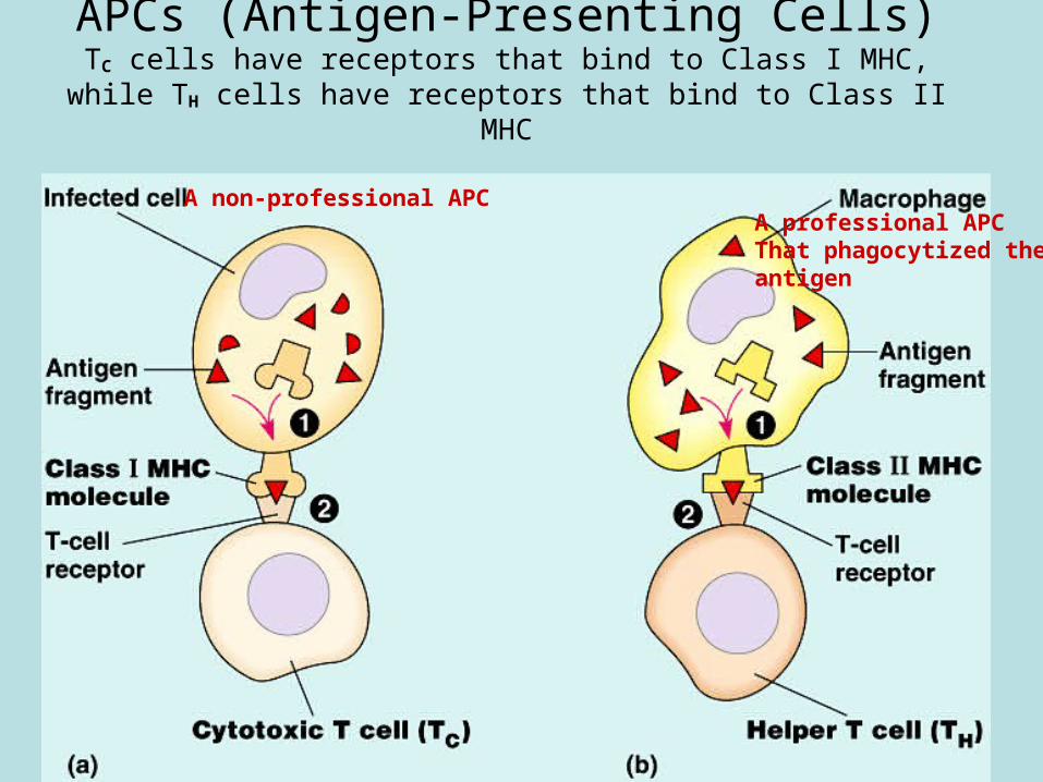
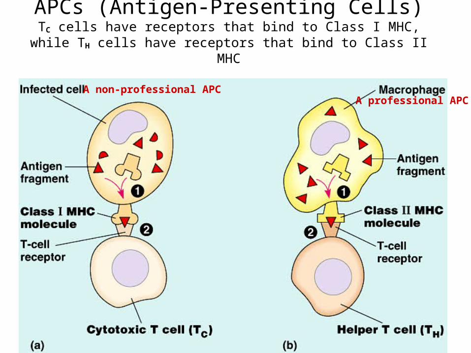
APCs (Antigen-Presenting Cells)TC cells have receptors that bind to Class I MHC, while TH cells have
receptors that bind to Class II MHC
A professional APCThat phagocytized theantigen
A non-professional APC
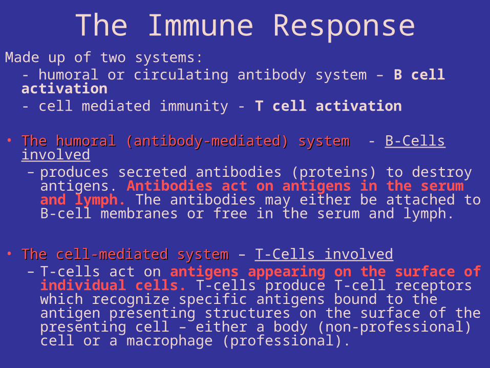
The Immune ResponseMade up of two systems:
- humoral or circulating antibody system – B cell activation- cell mediated immunity - T cell activation
• The humoral (antibody-mediated) systemThe humoral (antibody-mediated) system - B-Cells involved– produces secreted antibodies (proteins) to destroy antigens.
Antibodies act on antigens in the serum and lymph. The antibodies may either be attached to B-cell membranes or free in the serum and lymph.
• The cell-mediated systemThe cell-mediated system – T-Cells involved– T-cells act on antigens appearing on the surface of
individual cells. T-cells produce T-cell receptors which recognize specific antigens bound to the antigen presenting structures on the surface of the presenting cell – either a body (non-professional) cell or a macrophage (professional).
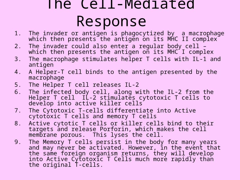
The Cell-Mediated Response 1. The invader or antigen is phagocytized by a macrophage which
then presents the antigen on its MHC II complex2. The invader could also enter a regular body cell – which then
presents the antigen on its MHC I complex3. The macrophage stimulates helper T cells with IL-1 and antigen 4. A Helper-T cell binds to the antigen presented by the macrophage5. The Helper T cell releases IL-26. The infected body cell, along with the IL-2 from the Helper T cell
IL-2 stimulates cytotoxic T cells to develop into active killer cells7. The Cytotoxic T-cells differentiate into Active cytotoxic T cells and
memory T cells8. Active cytotic T cells or killer cells bind to their targets and release
Porforin, which makes the cell membrane porous. This lyses the cell.
9. The Memory T cells persist in the body for many years and may never be activated. However, in the event that the same foreign organism returns, they will develop into Active Cytotoxic T Cells much more rapidly than the original T-cells.

The Suppressor T Cell
A special T Cell called the Suppressor T cell turns off the whole immune response when the threat has passed – conserve energy!
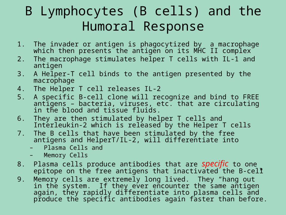
B Lymphocytes (B cells) and the Humoral Response
1. The invader or antigen is phagocytized by a macrophage which then presents the antigen on its MHC II complex
2. The macrophage stimulates helper T cells with IL-1 and antigen 3. A Helper-T cell binds to the antigen presented by the macrophage4. The Helper T cell releases IL-25. A specific B-cell clone will recognize and bind to FREE antigens –
bacteria, viruses, etc. that are circulating in the blood and tissue fluids.
6. They are then stimulated by helper T cells and Interleukin-2 which is released by the Helper T cells
7. The B cells that have been stimulated by the free antigens and HelperT/IL-2, will differentiate into
– Plasma Cells and – Memory Cells
8. Plasma cells produce antibodies that are specific to one epitope on the free antigens that inactivated the B-cell
9. Memory cells are extremely long lived. They “hang out” in the system. If they ever encounter the same antigen again, they rapidly differentiate into plasma cells and produce the specific antibodies again faster than before.
Memory B and Memory T cellsMemory B and Memory T cells
• Memory Cells persist in the body for many Memory Cells persist in the body for many years and may never be activated.years and may never be activated.
• However, in the event that the same However, in the event that the same foreign organism returns, they will develop foreign organism returns, they will develop into Plasma Cells much more rapidly than into Plasma Cells much more rapidly than the original B-Cells or T-cells.the original B-Cells or T-cells.
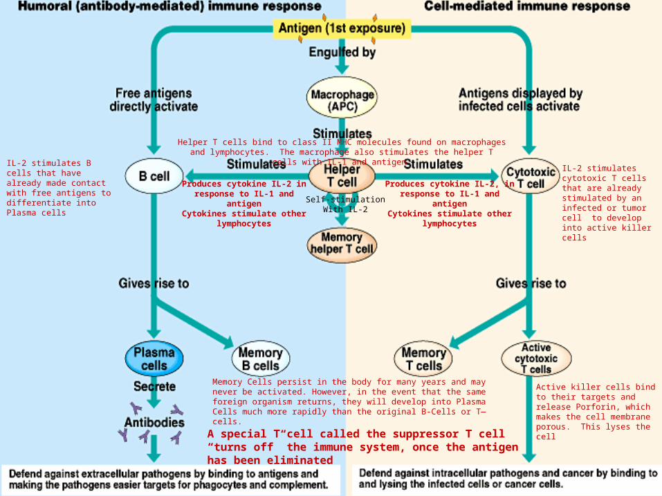
Produces cytokine IL-2 in response to IL-1 and antigen
Cytokines stimulate other lymphocytes
Helper T cells bind to class II MHC molecules found on macrophages and lymphocytes. The macrophage also stimulates the helper T cells with IL-1 and antigen
IL-2 stimulates B cells that have already made contact with free antigens to differentiate into Plasma cells
Produces cytokine IL-2, in response to IL-1 and antigen
Cytokines stimulate other lymphocytes
IL-2 stimulates cytotoxic T cells that are already stimulated by an infected or tumor cell to develop into active killer cells
Self-stimulationWith IL-2
A special T cell called the suppressor T cell “turns off” the immune system, once the antigen has been eliminated
Active killer cells bind to their targets and release Porforin, which makes the cell membrane porous. This lyses the cell
Memory Cells persist in the body for many years and may never be activated. However, in the event that the same foreign organism returns, they will develop into Plasma Cells much more rapidly than the original B-Cells or T—cells.
APCs (Antigen-Presenting Cells)TC cells have receptors that bind to Class I MHC, while TH cells have
receptors that bind to Class II MHC
A professional APCA non-professional APC

Immune Responses
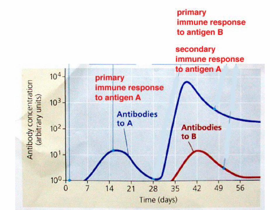
• When B or T cells become activated the first time, their actions constitute a primary immune response, after which some cells remain as memory cells.
• If the same antigen is encountered again, more numerous memory cells can mount a more rapid response, known as the secondary immune response.
– The ability to produce a secondary immune response may be long-lasting.

IMMUNITY
• Active immunity is achieved by recovering from an infectious disease such as measles
• Active immunization can be acquired artificially through vaccination. Vaccines can be made of:
- Inactivated bacterial toxins (tetanus)- Killed microbes (flu, cholera, bubonic plague, and hepatitis A)- Parts of microbes (HPV)- Living but weak (attenuated) microbes (measles, mumps, polio)
These can no longer cause disease, but can act as antigens and stimulate the immune response to produce antibodies and more importantly, memory cells.
• Passive immunity is what is transferred from one person to another – such as from mother to her fetus or from a nursing mother to her child. It can also be transferred from one animal immune to a disease, to another, through an injection.

Blood Groups and Transfusions Blood Groups and Transfusions Antigens and AntibodiesAntigens and Antibodies
• Clumping of red blood cells following Clumping of red blood cells following transfusion is called agglutination.transfusion is called agglutination.
• Agglutination is due to the interaction of Agglutination is due to the interaction of proteins on the surfaces of red blood cells proteins on the surfaces of red blood cells (antigens) with certain antibodies carried in (antigens) with certain antibodies carried in the plasma.the plasma.
• Only a few of the antigens on red blood cells Only a few of the antigens on red blood cells produce transfusion reactions.produce transfusion reactions.
– These include the ABO group and Rh group.These include the ABO group and Rh group.

HemostasisHemostasis• Hemostasis refers to the stoppage Hemostasis refers to the stoppage
of bleeding.of bleeding.
• Following injury to a vessel, three Following injury to a vessel, three steps occur in hemostasis: steps occur in hemostasis:
1.1. blood vessel spasm, blood vessel spasm,
2.2. platelet plug formation, and platelet plug formation, and
3.3. blood coagulation.blood coagulation.

1.1. Blood Vessel SpasmBlood Vessel Spasm
• Cutting a blood vessel causes the muscle Cutting a blood vessel causes the muscle in its walls to contract in a reflex, or in its walls to contract in a reflex, or engage in vasospasm.engage in vasospasm.
• This reflex lasts only a few minutes, but it This reflex lasts only a few minutes, but it lasts long enough to initiate the second lasts long enough to initiate the second and third steps of hemostasis.and third steps of hemostasis.

2.2. Platelet Plug FormationPlatelet Plug Formation
• Platelets stick to the exposed edges of Platelets stick to the exposed edges of damaged blood vessels, forming a net with damaged blood vessels, forming a net with spiny processes protruding from their spiny processes protruding from their membranes.membranes.
• A platelet plug is most effective on a small A platelet plug is most effective on a small vessel.vessel.
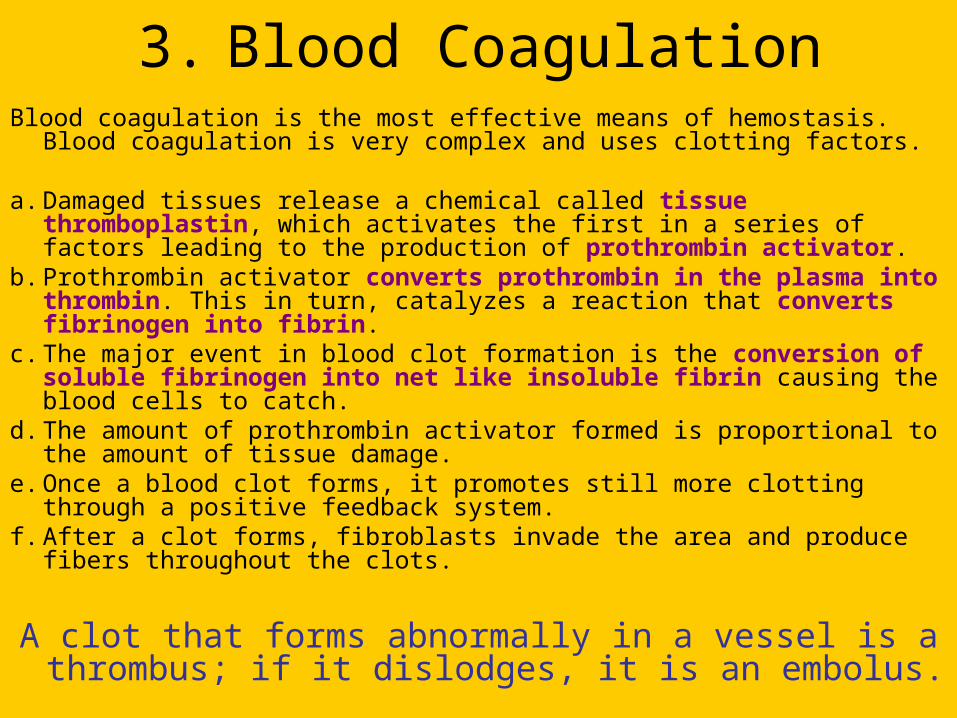
3. Blood CoagulationBlood coagulation is the most effective means of hemostasis. Blood
coagulation is very complex and uses clotting factors.
a. Damaged tissues release a chemical called tissue thromboplastin, which activates the first in a series of factors leading to the production of prothrombin activator.
b. Prothrombin activator converts prothrombin in the plasma into thrombin. This in turn, catalyzes a reaction that converts fibrinogen into fibrin.
c. The major event in blood clot formation is the conversion of soluble fibrinogen into net like insoluble fibrin causing the blood cells to catch.
d. The amount of prothrombin activator formed is proportional to the amount of tissue damage.
e. Once a blood clot forms, it promotes still more clotting through a positive feedback system.
f. After a clot forms, fibroblasts invade the area and produce fibers throughout the clots.
A clot that forms abnormally in a vessel is a thrombus; if it dislodges, it is an embolus.
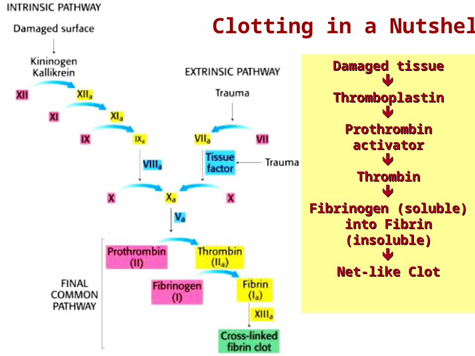
Damaged tissueDamaged tissue
ThromboplastinThromboplastin
Prothrombin activatorProthrombin activator
ThrombinThrombin
Fibrinogen (soluble) into Fibrinogen (soluble) into Fibrin (insoluble)Fibrin (insoluble)
Net-like ClotNet-like Clot
Clotting in a Nutshell


THE END