osteoporosis of pregnancy and lactation - porthosp.nhs.uk · transient osteoporosis of the hip a...
TRANSCRIPT
Osteoporosis of pregnancy and lactation Dr Beth Curtis
SpR Rheumatology, Queen Alexandra Hospital
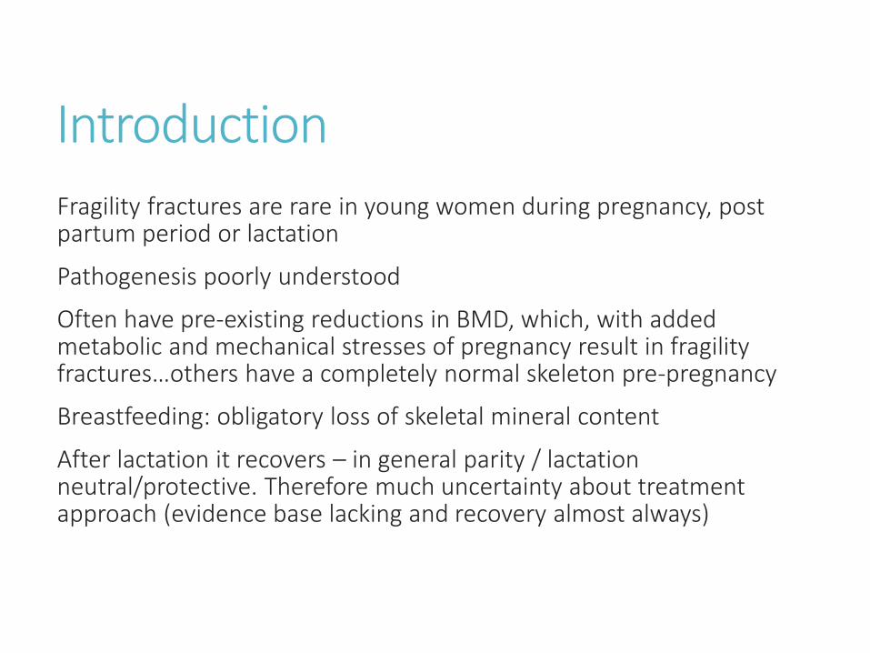
Introduction Fragility fractures are rare in young women during pregnancy, post partum period or lactation
Pathogenesis poorly understood
Often have pre-existing reductions in BMD, which, with added metabolic and mechanical stresses of pregnancy result in fragility fractures…others have a completely normal skeleton pre-pregnancy
Breastfeeding: obligatory loss of skeletal mineral content
After lactation it recovers – in general parity / lactation neutral/protective. Therefore much uncertainty about treatment approach (evidence base lacking and recovery almost always)

Case Study Mrs B 32 yr old accountant
PMH: one wrist fracture age 12 roller skating, one episode of urticarial, no cause found
First pregnancy aged 29
R Sacroiliac pain for 2 weeks, resolved
In labour, severe back pain lasting one day
10 weeks post partum, severe back pain, could not turn over in bed
Stopped breast feeding, pain settled over 6 weeks

Case Study Mrs B Second pregnancy aged 32
10 weeks post partum: severe back pain
Stopped breast feeding
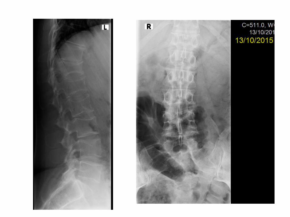
Multiple vertebral fractures T10-L4
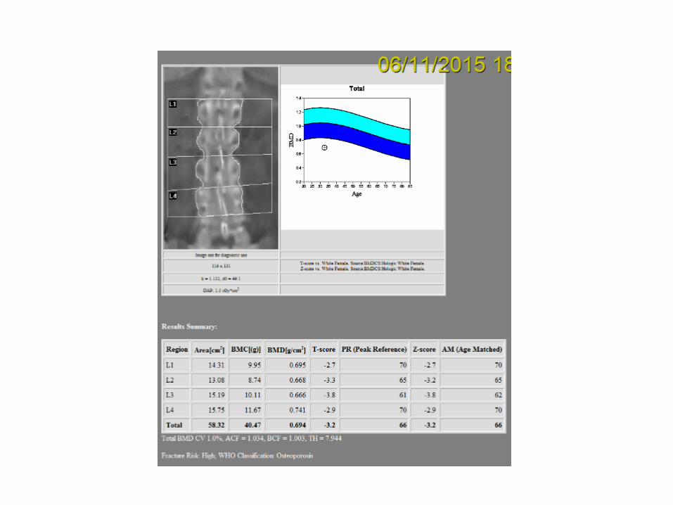
DXA: osteoporosis
No RFS: normal diet, exercise, sunlight exposure, drank 200ml milk per day, no steroids, normal BMI, normal menarche, non smoked, no alcohol.
No FH

Case Study Mrs B
O/E: wt 74.4kg, height 1.71m, BP 104/96
Normal HS, pulses, chest, abdomen, no splinter haemorrhages or lymphadenopathy, joints normal. Normal sclerae.
Ix: PTH 2.7, vit D 30nmol/l, FBC, U+E, LFT, CRP, Igs, protein, cortisol, TSH, glucose, HbA1c, FSH, LH, oestradiol normal, TTG neg, mast cell tryptase N (?mastocytosis), 9am cortisol
Urine: NAD, urinary cortisol and BJP Normal

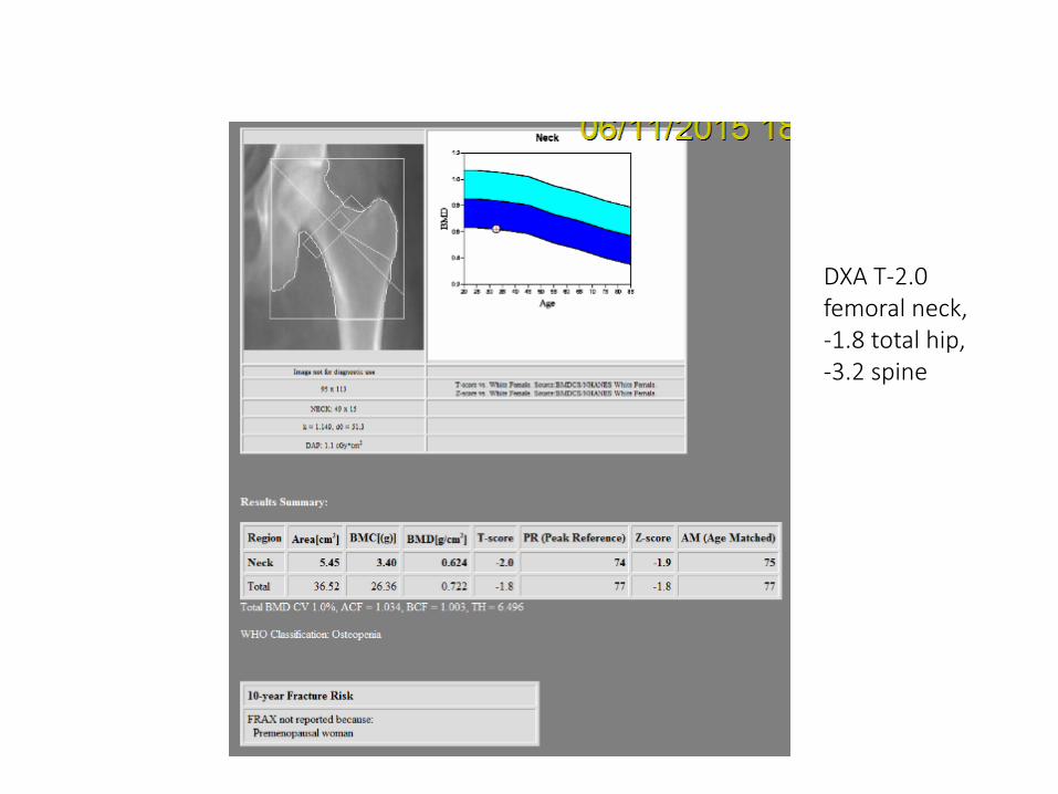
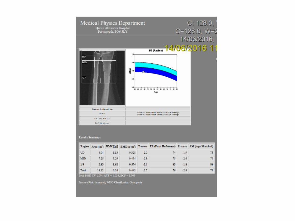
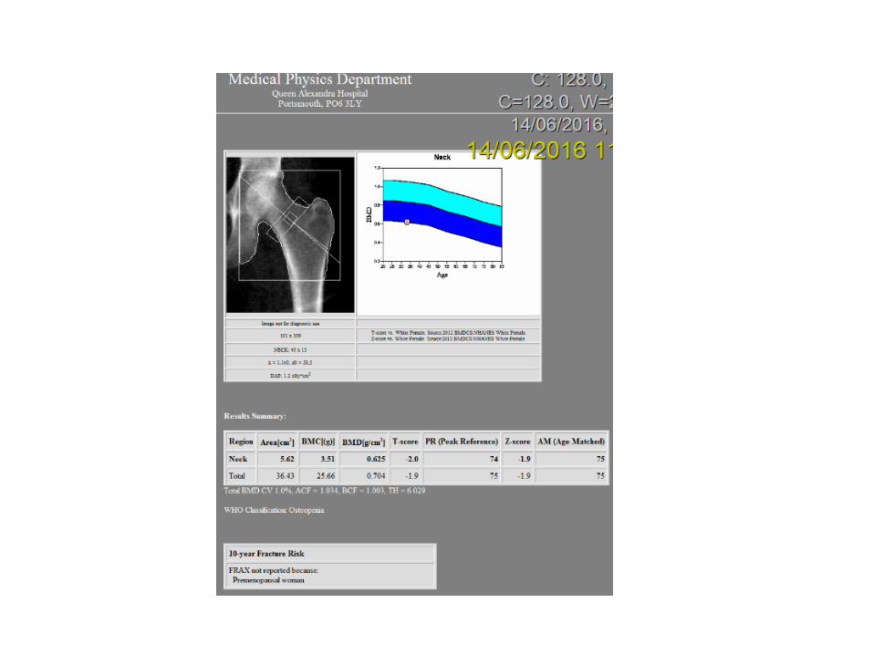
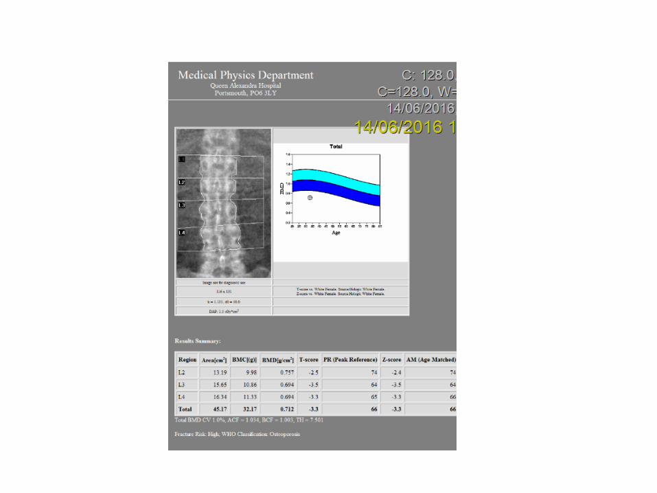
DXA T-2.0 femoral neck, -1.8 total hip, -3.2 spine

Case Study Mrs B- management
Vitamin D replacement 3200 units daily for 12 weeks
Vitamin D 400-800 units daily in winter months
Repeat DXA in 6 months
Gentle weight bearing exercise
Support: NOS contacts for OP in pregnancy
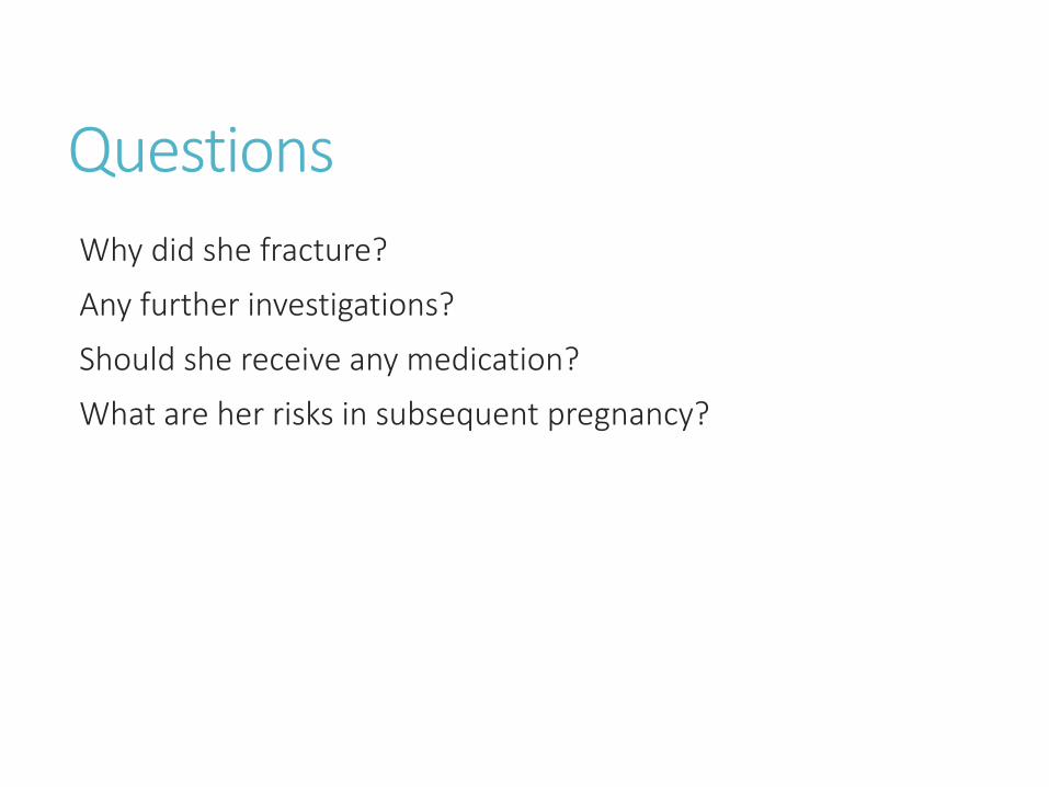
Questions
Why did she fracture?
Any further investigations?
Should she receive any medication?
What are her risks in subsequent pregnancy?

The maternal skeleton in pregnancy
Average fetus has 30g calcium in skeleton at term
80% fetal calcium deposition happens in the 3rd trimester.
Mother must provide 100-150mg/day of calcium in 3rd trimester, 300-500mg in last 6/52
Efficiency of intestinal calcium absorption doubled from week 12 – positive calcium balance mid pregnancy
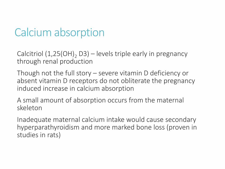
Calcium absorption
Calcitriol (1,25(OH)2 D3) – levels triple early in pregnancy through renal production
Though not the full story – severe vitamin D deficiency or absent vitamin D receptors do not obliterate the pregnancy induced increase in calcium absorption
A small amount of absorption occurs from the maternal skeleton
Inadequate maternal calcium intake would cause secondary hyperparathyroidism and more marked bone loss (proven in studies in rats)
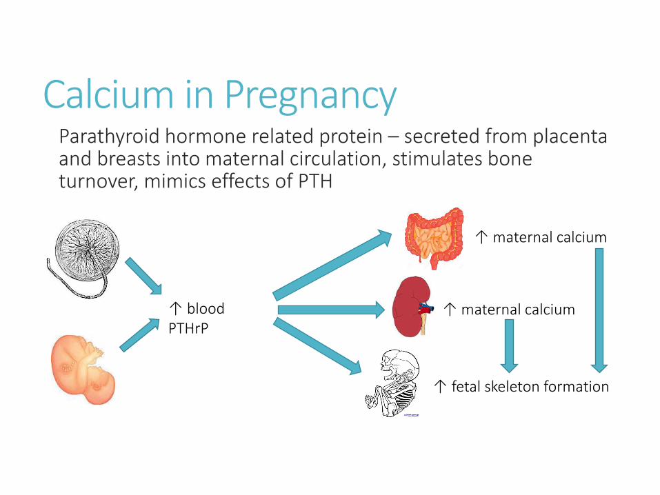
Calcium in Pregnancy Parathyroid hormone related protein – secreted from placenta and breasts into maternal circulation, stimulates bone turnover, mimics effects of PTH
↑ blood PTHrP
↑ maternal calcium
↑ maternal calcium
↑ fetal skeleton formation
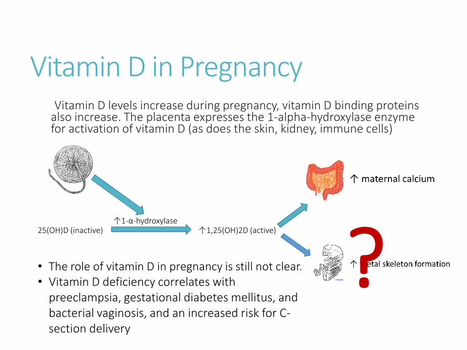
Vitamin D in Pregnancy
↑1,25(OH)2D (active) 25(OH)D (inactive) ↑1-α-hydroxylase
? • The role of vitamin D in pregnancy is still not clear. • Vitamin D deficiency correlates with
preeclampsia, gestational diabetes mellitus, and bacterial vaginosis, and an increased risk for C-section delivery
Vitamin D levels increase during pregnancy, vitamin D binding proteins also increase. The placenta expresses the 1-alpha-hydroxylase enzyme for activation of vitamin D (as does the skin, kidney, immune cells)

Chakhtoura et al, BMJ Open 2016
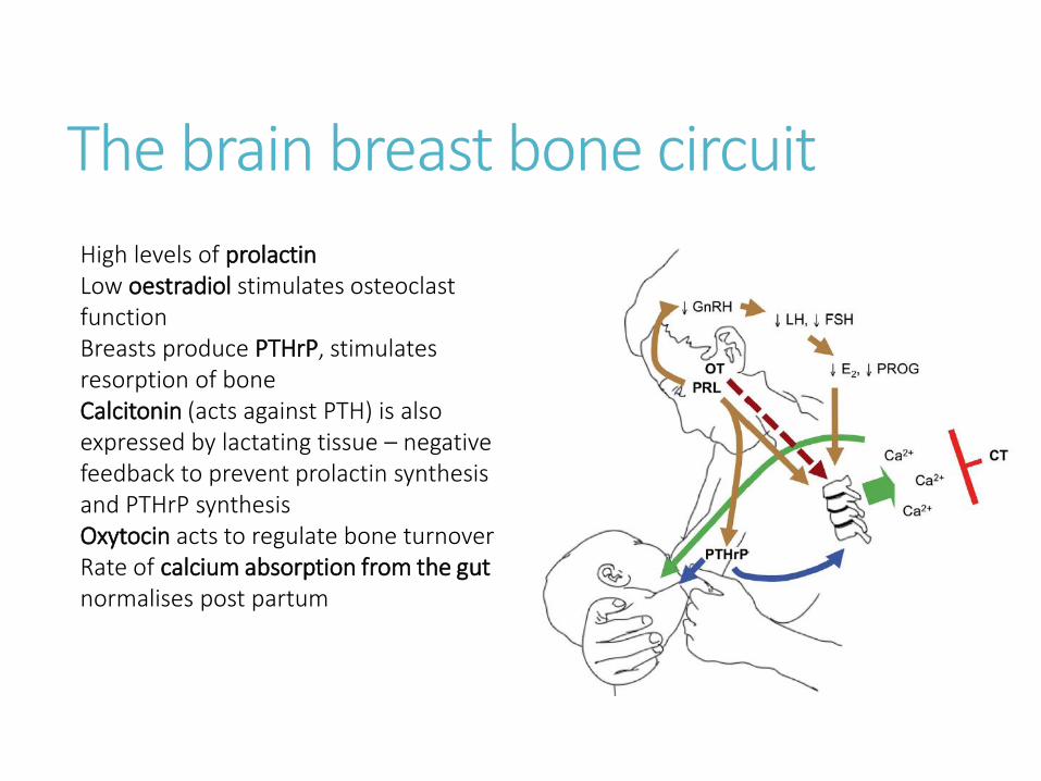
The brain breast bone circuit High levels of prolactin Low oestradiol stimulates osteoclast function Breasts produce PTHrP, stimulates resorption of bone Calcitonin (acts against PTH) is also expressed by lactating tissue – negative feedback to prevent prolactin synthesis and PTHrP synthesis Oxytocin acts to regulate bone turnover Rate of calcium absorption from the gut normalises post partum

The brain breast bone circuit So…. Increased PTHrP and low estradiol have synergistic effects to increase skeletal resorption Bone resorption markers go up in lactation There is 5-10% loss of trabecular BMD in first 3-6 months (1-3% per month…. In post-menopausal women a loss of 1-2% per yr is considered rapid), much greater than in pregnancy when 3-5% lost The loss has been shown to be even greater in adolescent women Also in women nursing twins…this is pre-programmed and is independent of diet. Therefore there are temporary reductions in skeletal strength
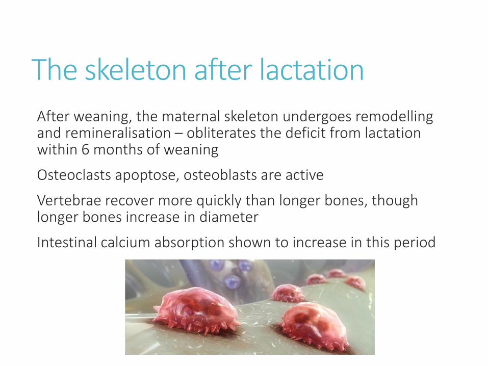
The skeleton after lactation
After weaning, the maternal skeleton undergoes remodelling and remineralisation – obliterates the deficit from lactation within 6 months of weaning
Osteoclasts apoptose, osteoblasts are active
Vertebrae recover more quickly than longer bones, though longer bones increase in diameter
Intestinal calcium absorption shown to increase in this period
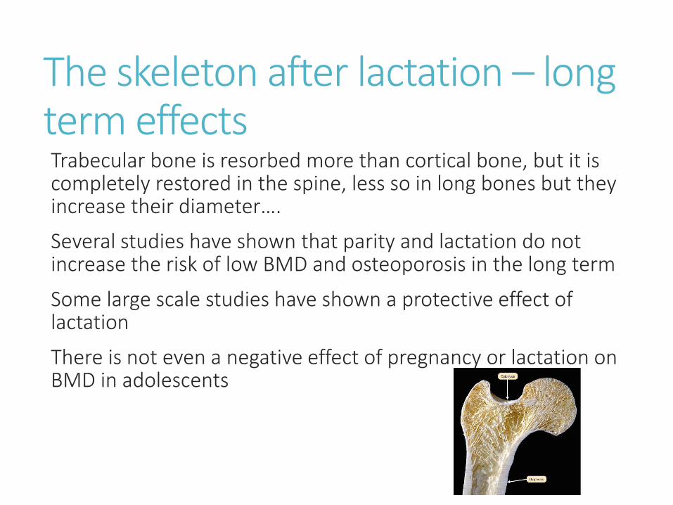
The skeleton after lactation – long term effects Trabecular bone is resorbed more than cortical bone, but it is completely restored in the spine, less so in long bones but they increase their diameter….
Several studies have shown that parity and lactation do not increase the risk of low BMD and osteoporosis in the long term
Some large scale studies have shown a protective effect of lactation
There is not even a negative effect of pregnancy or lactation on BMD in adolescents

Vertebral fractures during pregnancy / lactation 75% go undetected…most women who fracture are otherwise healthy…and we do not generally have pre pregnancy DXA
Pregnancy – wt gain at least 12kg, lordotic posture
Predisposing factors are often present: mild osteogenesis imperfecta, hypercalciuria, Cushing’s syndrome, RA, premature ovarian failure, vitamin D deficiency, dairy intolerance, anorexia, low body wt, petite frame, longstanding oligoamenorrhoea, steroid use, certain anti-convulsants, heparin use, depo-Provera, chemotherapy, alcohol and smoking, lack of physical activity
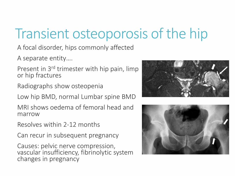
Transient osteoporosis of the hip A focal disorder, hips commonly affected
A separate entity….
Present in 3rd trimester with hip pain, limp or hip fractures
Radiographs show osteopenia
Low hip BMD, normal Lumbar spine BMD
MRI shows oedema of femoral head and marrow
Resolves within 2-12 months
Can recur in subsequent pregnancy
Causes: pelvic nerve compression, vascular insufficiency, fibrinolytic system changes in pregnancy

Long term outlook
Fractures typically occur in a first pregnancy
They typically do not recur
Parity does not increase risk of fractures
….this suggests that the predisposing factor is often present before pregnancy and has been corrected later
It may be appropriate to discourage breastfeeding in women who are known to be predisposed to skeletal fragility... But there is not enough evidence to say it is contraindicated

Investigations
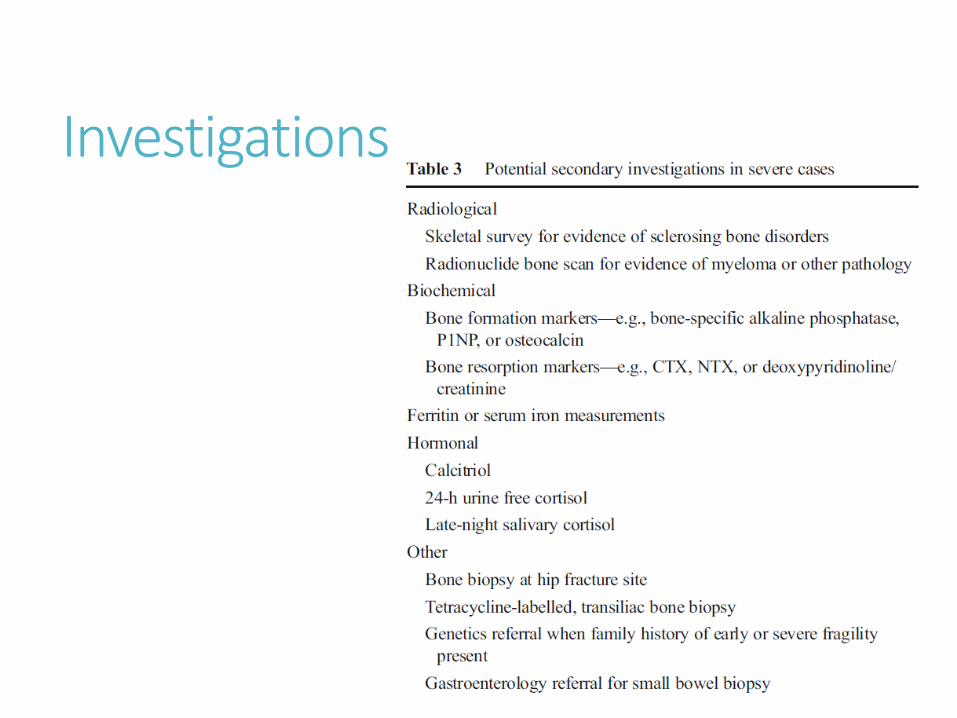
Investigations
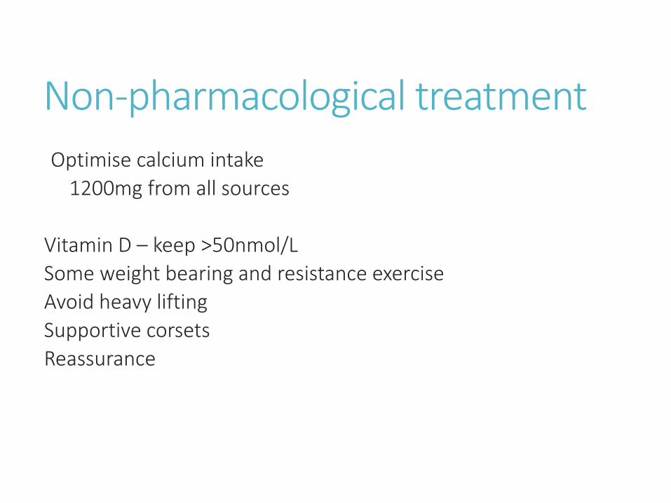
Non-pharmacological treatment
Optimise calcium intake
1200mg from all sources
Vitamin D – keep >50nmol/L
Some weight bearing and resistance exercise
Avoid heavy lifting
Supportive corsets
Reassurance

Pharmacologic / surgical treatment
BMD normally increases during the 6-12 months after weaning…by 10-20%
Therefore appropriate to delay pharmacological treatment for 12-18 months until the extent of recovery established
Treatment on a case by case basis: safety concerns about long term treatment with calcitonin (cancer risk), bisphosphonates (concerns re. fetal bone development), strontium, teriparatide, denosumab (crosses placenta)
If treatment is needed (BMD not improving significantly on its own), generally bisphosphonate or denosumab for 4-5 yrs is used
Vertebroplasty and kyphoplasty occasionally used – overall efficacy uncertain

Revisitation of case…. Why did she fracture? Not clear… vit D deficiency? Inherited disorder?
Any further investigations? Bone turnover markers? Skeletal survey (sclerosing bone disorders)? Look further for nutritional deficiencies… gastro and dietician referral? Genetics referral?
Should she receive any medication? Not yet…perhaps if next DXA at 12 months shows subsequent improvement
What are her risks in subsequent pregnancy? Probably minimal but she developed fractures after her second pregnancy…
Any other suggestions?
Conclusions
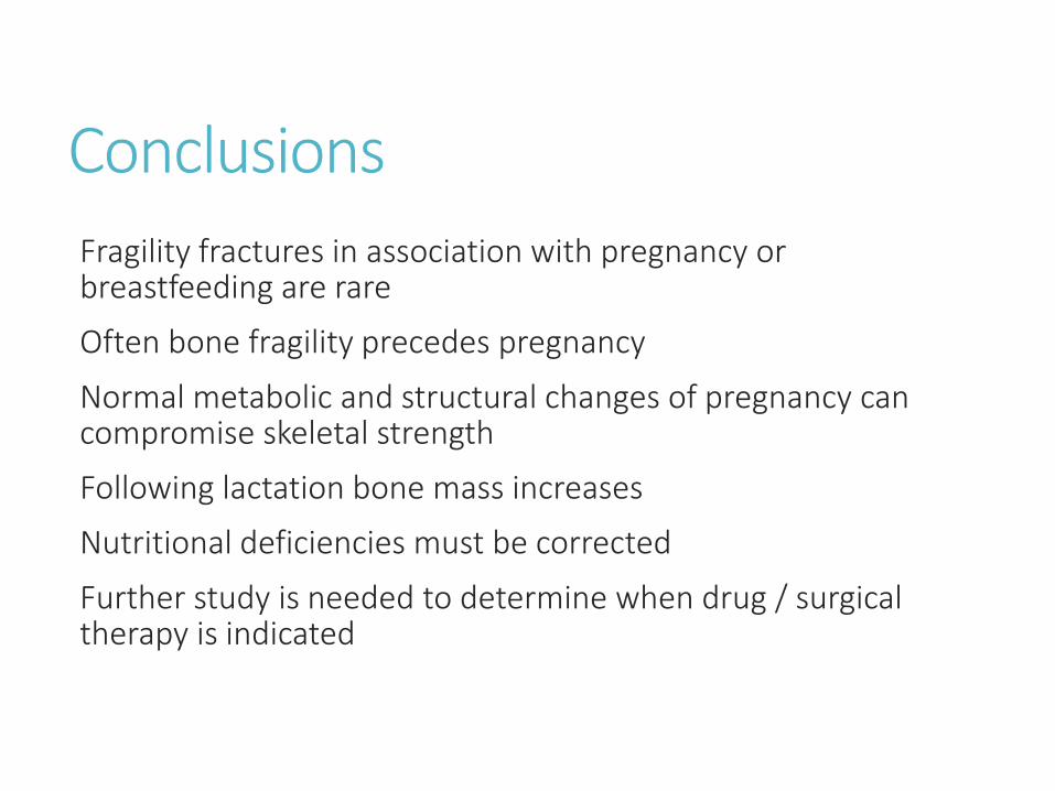
Fragility fractures in association with pregnancy or breastfeeding are rare
Often bone fragility precedes pregnancy
Normal metabolic and structural changes of pregnancy can compromise skeletal strength
Following lactation bone mass increases
Nutritional deficiencies must be corrected
Further study is needed to determine when drug / surgical therapy is indicated


References
http://www.niams.nih.gov/health_info/Bone/Bone_Health/Pregnancy/default.asp
www.uptodate.com
https://www.nos.org.uk/health-professionals/~/document.doc?id=405
Kovacs CS and Ralston SH; Presentation and management of osteoporosis presenting in association with pregnancy or lactation. Osteoporosis Int (2015) 26: 2223-2241

Osteoporosis of pregnancy and lactation Dr Beth Curtis
SpR Rheumatology, Queen Alexandra Hospital