orthopedics chronic problems nptc530t fall 2004. osteoarthritis incidence >90% by age 45 50% of...
TRANSCRIPT

Orthopedics
Chronic problems
NPTC530T
Fall 2004

Osteoarthritis
Incidence >90% by age 45 50% of those over 65 will have symptomatic
arthritis Overall women = men
Before 45yo men>women After 55 yo women>men

Symptoms
Cardinal sx joint pain on weight bearing Stiffness <15 min in the AM
Swelling less common Neuro sxs only secondary to vessel or nerve
impingement

Etiology
Seems to go along with aging and other factors contribute Previous trauma Infection Obesity Inflammatory arthritis Mechanical misalignment

Lab
No abnormalities in blood or urine Sed rate wnl Joint fluid essentially nl

Xray
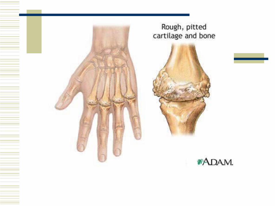
Loss of joint space Osteophytes at margin Normal cartilage OA on xray does not rule out other types of
arthritis


This is typically the first joint affected by osteoarthritisLook at the base of your thumb, do you see any bony hypertrophy?

A normal joint surface
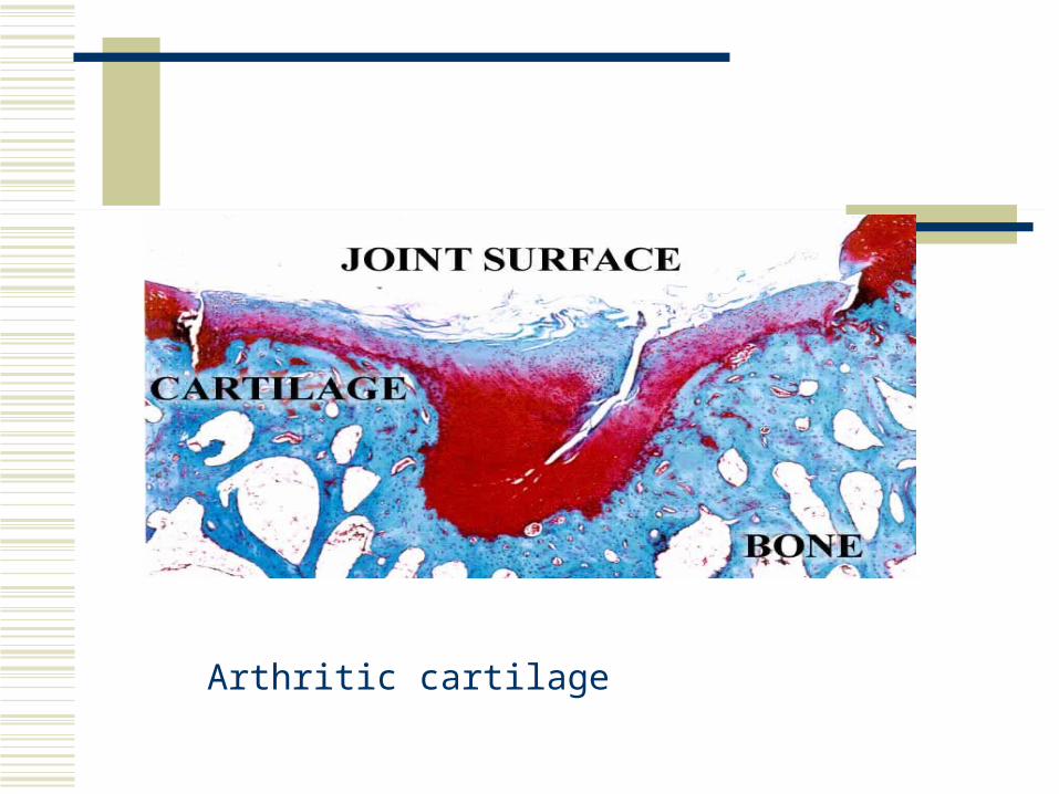
Arthritic cartilage

PE
Osteoarthritis typically affects certain joints Dip Pip Base of thumb Spine Hips Knees NOT the mcps, elbow, wrists, shoulders

Distsribution is usually asymmetrical Early on may be swollen, red

Osteoarthritis of the hand

Treatment
To lessen discomfort and retard progression NSAIDs Weight reduction Exercise Possibly steroid injection synavisc


Erosive OA
An OA that resembles RA, usually in elderly white caucasian women
Characterized by pain, tenderness, and swelling in hands but not mcps
Labs wnl, xray typical of OA May be severe but burns out quickly
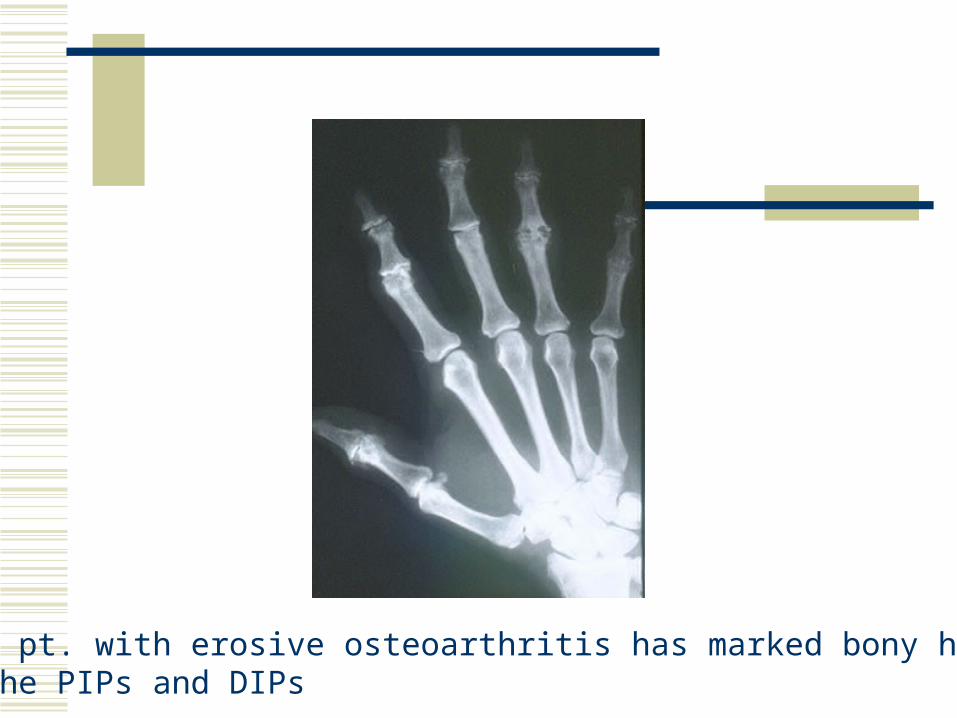
This pt. with erosive osteoarthritis has marked bony hypertrophy of the PIPs and DIPs

Rheumatoid Arthritis
Most common chronic inflammatory polyarthritis (always multiple joints, usually hands and feet)
Cause unknown Synovial thickening and joint damage Extra articular manifestations common


Labs
none are specific but reinforce the clinical impressionNormochromic, normocytic anemia in 40%, no response to
iron+ RF in 50% at 6 months 75% at 10 months20% will have – RFElevated sed rateAspiration of joint fluid will show elevated WBCs

+ RF may show with psoriasis, inflammatory diseases, TB, SLE, SBE, chronic active hepatitis


Younger patients
More gradual onset 67% female Less systemic features Usually begins in hands or feet 75% will have +RF Average sed 28 mm 17% with fever 20% with weight loss

Differential diagnosis in young
SLE Mixed connnective tissue disease Systemic Sjogrens Vasculitis Fibromyalgia Hypothyroidism Spondyloarthropathies Bacterial endocarditis

Differential diagnsosis in elderly
Previous diseases + PMR Erosive OA Gout, pseudogout Seronegative syndromes

2 types of rheumatoid arthritis in elderly Carryover RA Elderly onset
Average of onset 55yoDisease behaves differently than in younger groupsThose with severe disease tend to die early

Elderly onset RA
More abrupt onset More systemic features May present with shoulder involvement 57% female Fever 8% 33% with weight loss Average ESR 56m +RF 89% Diagnosis may be confusing, easy to confuse with other
illnesses/conditions

Symptoms
May have prodrome of months of fatigue, stiffness, weight loss, fever, vague arthralgias before developing the multiple inflamed joints
AM stiffness usually >30 minutes Will have joint pain, swelling, erythema, warmth Joint pattern
Never dips, sometimes pips, always mcps Swelling of mcps may lead to loss of valleys

PIPs markedly swollen with faint swelling of the MCPs

Rheumatoid nodules and deformity
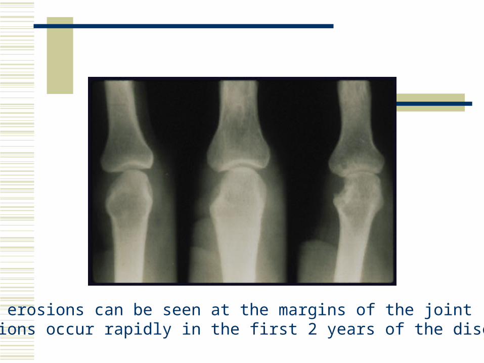
Bony erosions can be seen at the margins of the jointErosions occur rapidly in the first 2 years of the disease

Typical visible changes include ulnar deviation of the fingers at the MCP joints, hyperextension or hyperflexion of the MCP and PIP joints, flexion contractures of the elbows, and subluxation of the carpal bones and toes (cocked —up).

X ray
Xray may show joint destruction, erosions and spurring
Helpful to monitor disease

Extra articular Disease
Much more problematic Weight loss Anemia fever Rheumatoid nodules/granulomas Sjorgrens syndrome Episcleritis RA lungs – pleurisy, pleural effusions Splenomegaly lymphadenopathy

Although the joints are almost always the principal focus of the rheumatoid arthritis, other organ systems may also be involved. Extra-articular manifestations of rheumatoid arthritis occur most often in seropositive patients with more severe joint disease. Interestingly, extra-articular manifestations can occur in later stages of the disease when there is little active synovitis ("burnt-out" disease). In contrast to the predilection of rheumatoid arthritis for women, extra-articular manifestations of the disease are more common in men

Cardiopulmonary Disease There are several pulmonary manifestations of rheumatoid arthritis, including pleurisy with or without effusion, intrapulmonary nodules, rheumatoid pneumoconiosis (Caplan's syndrome), diffuse interstitial fibrosis, and rarely, bronchiolitis obliterans pneumothorax. On pulmonary function testing, there commonly is a restrictive ventilatory defect with reduced lung volumes and a decreased diffusing capacity for carbon monoxide. Although mostly asymptomatic, of greatest concern is distinguishing these manifestations from infection and tumor. Pericarditis is the most common cardiac manifestation

Ocular Disease Keratoconjunctivitis of Sjogren's syndrome is the most common ocular manifestation of rheumatoid arthritis. Sicca (dry eyes) is a common complaint. Episcleritis occurs occasionally and is manifested by mild pain and intense redness of the affected eye. Scleritis and corneal ulcerations are rare but more serious problems.

Neurologic Disease The most common neurologic manifestation of rheumatoid arthritis is a mild, primarily sensory peripheral neuropathy, usually more marked in the lower extremities. Entrapment neuropathies (e.g., carpal tunnel syndrome and tarsal tunnel syndrome) sometimes occur in patients with rheumatoid arthritis because of compression of a peripheral nerve by inflamed edematous tissue. Cervical myelopathy secondary to atlantoaxial subluxation is an uncommon but particularly worrisome complication potentially causing permanent, even fatal neurologic damage.(top of section)

Sjogren's Syndrome Approximately 10 to 15% of patients with rheumatoid arthritis, mostly women develop Sjogren's syndrome, a chronic inflammatory disorder characterized by lymphocytic infiltration of lacrimal and salivary glands. This leads to impaired secretion of saliva and tears and results in the sicca complex: dry mouth (xerostomia) and dry eyes (keratoconjunctivitis sicca). Patients with Sjogren's syndrome have a variable expression of disease in other exocrine glands. This is manifested clinically as dry skin, decreased perspiration, dry vaginal membranes, or a nonproductive cough. Commonly, there is also a polyclonal lymphoproliferative reaction characterized by lymphadenopathy and splenomegaly. This can mimic and rarely transform into a malignant lymphoma
Rheumatoid Nodules
The subcutaneous nodule is the most characteristic extra-articular lesion of the disease. Nodules occur in 20 to 30% of cases, almost exclusively in seropositive patients. They are located most commonly on the extensor surfaces of the arms and elbows but are also prone to develop at pressure points on the feet and knees. Rarely, nodules may arise in visceral organs, such as the lungs, the heart, or the sclera of the eye. (learn more about rheumatoid nodules in case report #6)(top of section)
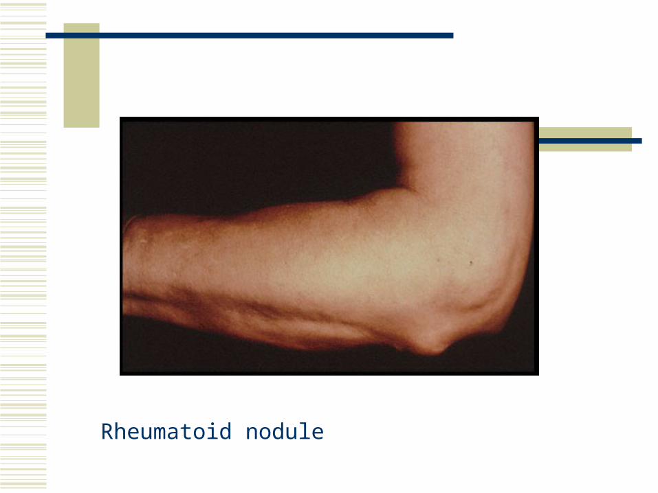
Rheumatoid nodule

Rheumatoid Vasculitis The most common clinical manifestations of vasculitis are small digital infarcts along the nailbeds. The abrupt onset of an ischemic mononeuropathy (mononeuritis multiplex) or progressive scleritis is typical of rheumatoid vasculitis. The syndrome ordinarily emerges after years of seropositive, persistently active rheumatoid arthritis; however, vasculitis may occur when joints are inactive.

A rheumatoid vasculitis resulting in gangrene

The course of rheumatoid arthritis cannot be predicted in a given patient. Several patterns of activity have been described:
•a spontaneous remission particularly in the seronegative patient within the first •6 months of symptoms (less than 10%) •recurrent explosive attacks followed by periods of quiescence most commonly •in the early phases •the usual pattern of persistent and progressive disease activity that waxes and •wanes in intensity.
Disability is higher among patients with rheumatoid arthritis with 60% being unable to work 10 years after the onset of their disease. Recent studies have demonstrated an increased mortality in rheumatoid patients. Median life expectancy was shortened an average of 7 years for men and 3 years for women compared to control populations. In more than 5000 patients with rheumatoid arthritis from four centers, the mortality rate was two times greater than in the control population. Patients at higher risk for shortened survival are those with systemic extra-articular involvement, low functional capacity, low socioeconomic status, low education, and prednisone use.

Treatment
Important to treat aggressively and in early stages to minimize joint destruction
Therefore important to diagnose early In past used to “step up” only when sx’s
uncontrolled sometimes missing window of opportunity

Treatment
NSAIDS still basic therapy for RA All are essentially the same
Problems with NSAID therapy in the elderly Gastropathy Complicated diverticular disease Renal insufficciency Drug interactions

Cox-2 inhibitors Corticosteroids Anti-malarials Gold therapy Penicillamine Sulfasalazine Methotrexate Arava, Remicade (disease modifying agents)
Rest PT Exercise

Analgesic Drugs Pain caused by inflammation is best treated with an anti-inflammatory drug, although occasionally the addition of acetaminophen can be helpful. Chronic narcotic therapy is not used routinely due to side effects such as diminished mental status, hyper somnolence, constipation, and dependency. Furthermore, they have no anti-inflammatory activity. They may be needed for patients with severe joint destruction who are not surgical candidates.


Which joints are affected in RA?

Look at the articular spaces – Can you see the margins clearly?

Typical deformity of RA

http://www.hopkins-arthritis.org/edu/acr/acr.html#class_rheum
This excellent site details the ACR (American College ofRheumatology) clinical classification criteria. Please take a look – you won’t believe how detailed it is!
SLESystemic Lupus Erythematosus
Chronic inflammatory disease of multiple organs
Primarily a disease of younger people Women age 20-40
15% will have onset later in life Older persons will have a milder disease

Symptoms
Range from fulminant febrile illness to asx with only abnormal labs
Most common presentation is that of multiple constitutional sxs-- fever and malaise, fatigue and weight loss

Systemic: fever, chills, fatigue, anorexia, weight loss Skin: butterfly rash on face but may be anywhere,
photosensitivity, frontal alopecia, palmar rash MS: polyarthritis Eyes: conjunctivitis, retinal lesions Lung: pleurisy, rubs, effusions CV: pericarditis, endocarditis, cardiomyopathy GI: abd. pain secondary to vasculitis, dysphagia,
Hepatosplenomegaly

Butterfly rash

Neuro: anxiety, memory loss, minor psychoses, depression, seizures
Raynauds: 25% Renal: present in most,
Protenuria, hematuria Nephritis develops early, may lead to HTN


Lab
ANA – sensitive and + in most with disease but not diagnostic
Dec. WBCs, anemia, false + RPR Elevated SGOT Low titres and speckled ANA patterns
usually mean a mixed connective tissue disease ( mild SLE)

double-stranded DNA antigen (anti-dsDNA) and antibody to Sm nuclear antigen (anti-Sm) may be helpful in patients who do not meet the diagnostic criteria for lupus

The ACR recommends that primary care physicians consider a rheumatology referral for patients with characteristic signs and symptoms of systemic lupus erythematosus and a positive ANA test, particularly if these patients have more than mild or stable disease.2

Diagnostic Criteria for Lupus
No one diagnosis or PE finding will tell you thePerson has lupus. Instead meeting a certain # of diagnosticcriteria is involved.


Treatment
Will need chronic care NSAIDs, rest Antimalarials – plaquenil (need regular
ophth. Exams) Immunosupressives Sun avoidance

http://www.aafp.org/afp/20031201/2179.html
Excellent article
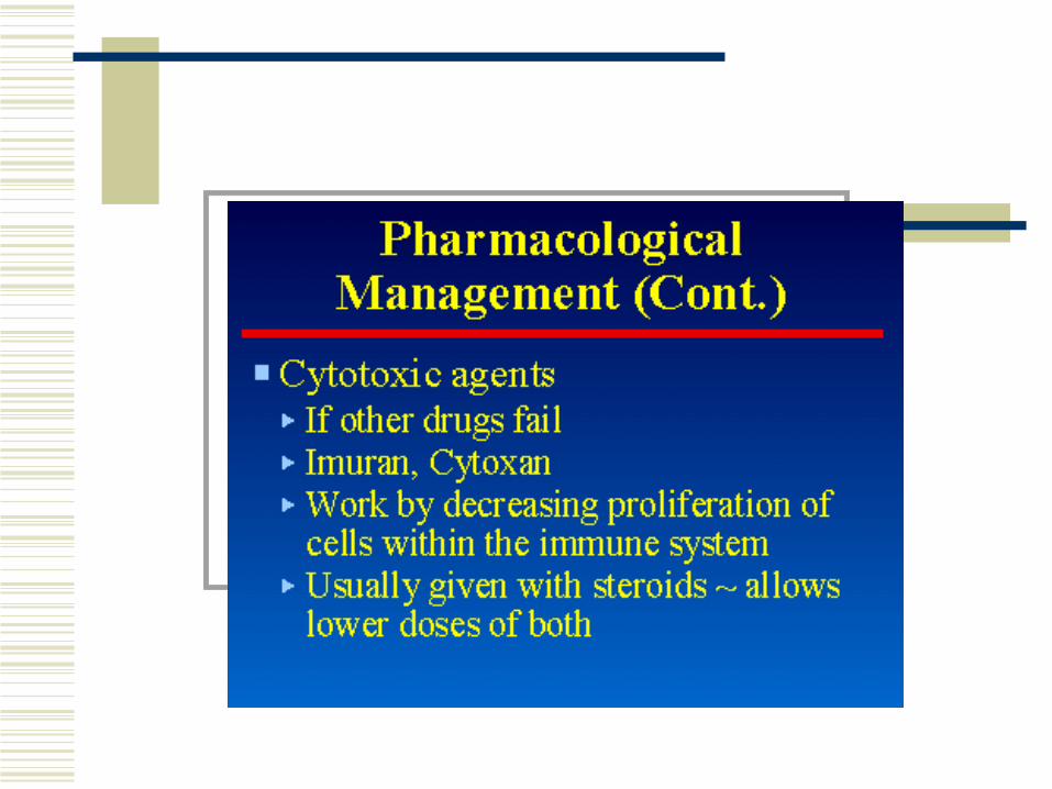
Ankylosing Spondylitis
Inflammatory arthritis affecting the axial skeleton (spine, sacroiliac joints)
Predominantly male 3:1 Incidence increases after age 40 Will “burn out” in the elderly but are then
left with spine abnormalities

Symptoms
Posture – flattened lumbar lordosis Curving throacic kyphosis Rigid spine, unable to turn neck Walk with knees bent to see ahead Associated with iritis, aortic enlargement
Pain lessens with activity Sxs persist > 3 months Pain worse with rest Onset insidious

PE
Pathophysiology: AS most commonly affects the SI joints and the axial skeleton. Involvement of the SI joints is required to establish the diagnosis. Hip and shoulder joints are affected less frequently. Peripheral joint involvement is least common.
The initial presentation generally occurs in the SI joints and is followed by involvement of the discovertebral, apophyseal, costovertebral, and costotransverse joints and the paravertebral ligaments

Chronic involvement of the spine eventually can lead to decreases in ROM and fusion of the vertebral bodies. Involvement of the cervical and upper thoracic spine can lead to fusion of the neck in a stooped forward-flexed position (see Images 1-2). This position can significantly limit the patient's ability to ambulate and look straight ahead.

Focus the physical examination on active ROM and passive ROM of the axial and peripheral joints. Tenderness in the SI joints is common.
•Screen for extra-articular manifestations by performing specific examinations (eg, ophthalmologic, cardiac, and gastrointestinal examinations).

Labs
Genetic component + HLA B27 in 90% - rheumatoid factor

Xrays
Classic “bamboo spine” secondary to fusion

Treatment
PT exercise In advanced disease no NSAIDs Treat early pain symptomatically

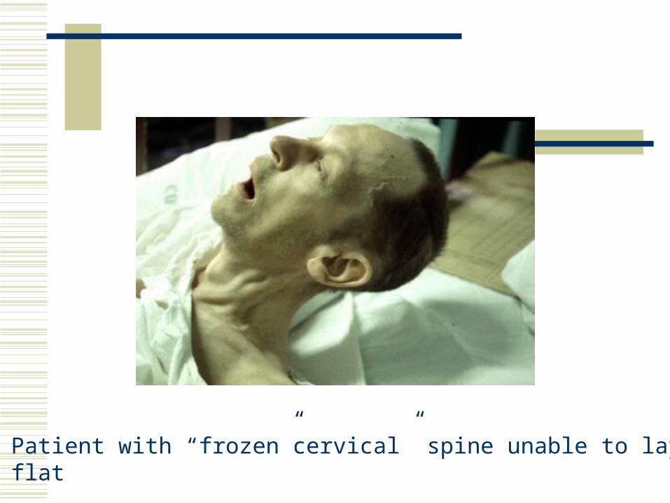
Patient with “frozen”cervical” spine unable to lay headflat

Complications
•Patients with a history of AS who report any recent trauma or• an increased level of back or neck pain should be fully •evaluated for the possibility of a vertebral fracture and •subsequent spinal instability

Neck fracture in a patient with ankylosing spondylitis
•Many patients with advanced disease have fusion of the spine.• As discussed above, if these patients report any change in •position or movement of the spine, they should be assumed to •have a spinal fracture since this is the only method for the spine •to move. Patients should be treated cautiously until fracture• has been ruled out. If spinal fracture is present, surgical •stabilization may be necessary.•Symptoms generally

Reiters
Most frequent cause of arthritis in young men
Male predominance 10:1
Symptoms
Classic triad Arthritis (usually of lower extremities, asymm
Urethritis (no response to abs)
Conjunctivitis, iritis

Symptoms
Keratoderma blenorrhagica - Psoriasis like rash
Balanitis Oral ulcers Heel pain Nail dystrophy Dactylitis (sausage toes)


Conjunctivitis

Urethritis

Labs
80% + HLA B27 Leukocytosis, elevated ESR RF – ANA -

X rays
Normal early in disease Later may resemble RA

Course
Acute phase subsides within weeks May recur in different parts over > 10 yrs May be mild chronicity Good prognosis without deformity, disability

http://www.aafp.org/afp/990800ap/499.html
Great article on Reiter’s

Polymyalgia Rheumatica (PMR)
Common in older patients, nearly as common as RA
Usual onset at > 60 years Definition: a clinical syndrome characterized
by aching and morning stiffness in the proximal portions of the extremities and torso
Patient will c/o pain or aching symmetrically in neck, shoulder girdle and pelvic girdle
Profound morning stiffness No inflammation Exam will be normal Associated with fever, weight loss

Labs
ESR will be very elevated, sometimes over 100
Possible normocytic anemia Possible elevated LFTs
Disease very significant because of its’ association with Temporal Arteritis

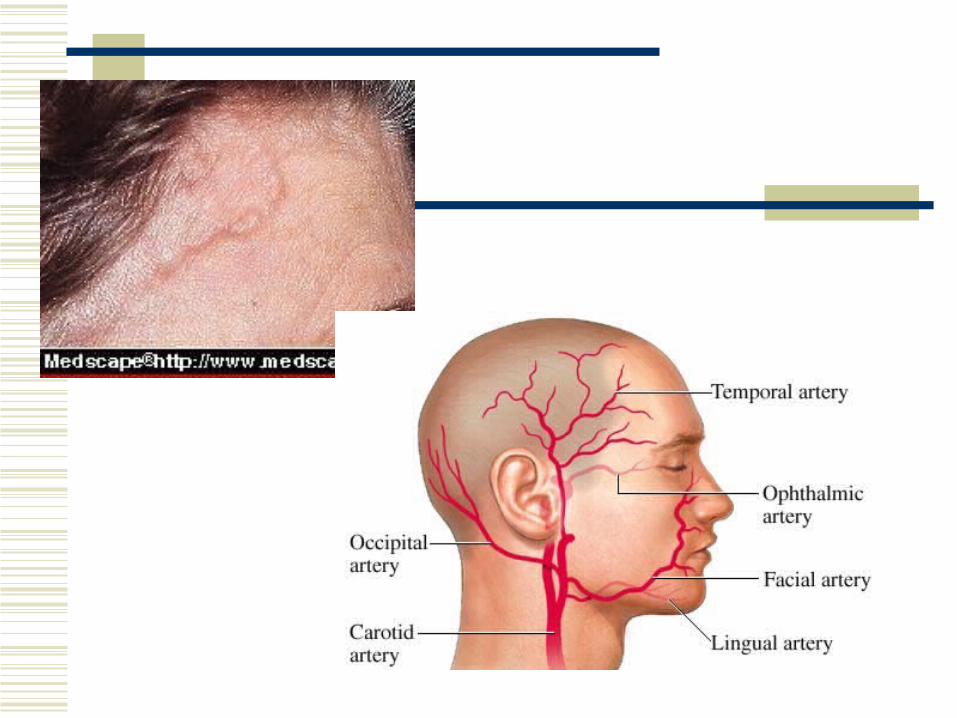
Temporal Arteritis
A clinical syndrome characterized by inflammation of the cranial arteries, most often producing headache and at times, blindness
Mean age at onset 75 yo Women predominate 2:1
Signs and Symptoms
Headache 90% Jaw claudication Impaired vision, diplopia Thickening of temporal arteries Elevated sed rate, anemia Need biopsy for definitive diagnosis
PE
Look at general appearance Examine head and neck Vision!!!!!

Labs
CBC Sed rate C-reactive protein Kidney, liver function

PMR & TA
40-60% of patients with TA have PMR 0-80% of those with PMR have evidence of
TA PMR may appear at any time in the course of
TA

Treatment
Corticosteroid therapy only rx that is proven effective
High doses (60mg qd) reduced very slowly every 2 weeks. May last up to 2 years
Disease usually “gone” at that point

Complications
Vision loss Thoracic/abdominal aneurysms

Gout
Most common form of acute arthritis in elderly
Occurs secondary to crystal deposition in the joint
May be acute or chronic and may have extra articular manifestations

The peak incidence of acute gout occurs between 30 and 50 years of age Approximately 90 percent of first attacks are monoarticular. In more than one half of patients with acute gout, the first metatarsophalangeal joint is the initial joint involved, a condition known as podagra. Joint involvement (in order of decreasing frequency) includes the metatarsophalangeal joint, the instep/forefoot, the ankle, the knee, the wrist and the fingers.

Gout in women occurs exclusively after menopause. Women develop gout at an older age than men and have twice the prevalence of hypertension, renal insufficiency and exposure to diuretics. The onset of gout before age 30 in men or before menopause in women is atypical and raises concern about an associated inherited enzyme defect or renal disease.

Signs and symptoms
Acute – warm, red, very painful swollen joint usually in lower extremities
Chronic – aching in various joints and AM stiffness
Extra articular manifestations Tophi Renal calculi
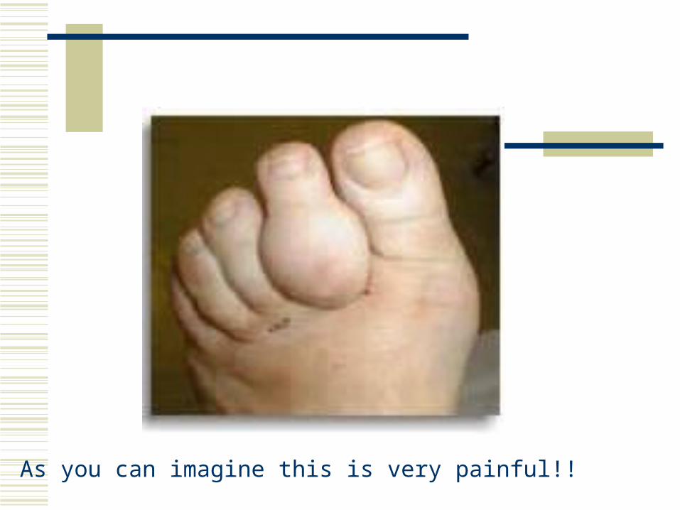
As you can imagine this is very painful!!

Labs
Serum uric acid >9 does not diagnose but correlates with high risk
Poss elevated ESR Definitive dx only with joint aspiration

Acute Gout The four treatment options available for the acute gouty attack are NSAIDs, colchicine, corticosteroids and analgesics

NSAIDs are the preferred therapy for the treatment of patients without complications. Indomethacin (Indocin) was the first NSAID used for gout, but other NSAIDs, including ibuprofen (Motrin), naproxen (Naprosyn), sulindac (Clinoril), piroxicam (Feldene) and ketoprofen (Orudis) are also effective in the treatment of acute gout. Maximum dosages should be given immediately after the onset of symptoms or at the time of diagnosis and continued for 24 hours after complete resolution of the acute attack, then tapered quickly over two to three days.

Colchicine, an antimitotic drug derived from the roots of the herb Colchicum autumnale, is one of the oldest treatments for gout. Although colchicine is effective in treating acute gout, 80 percent of patients experience gastrointestinal side effects, including nausea, vomiting and diarrhea, at therapeutic dosages. Furthermore, colchicine is less effective once an acute attack has persisted for a few days.

Corticosteroids, administered intra-articularly, intravenously, intramuscularly or orally, have been shown to be effective in the treatment of acute gout.

Meds
Indocin 50 mg tid X 1 week doc Colchicine >5 mg q 2 hrs until effective may
lead to severe diarrhea If attacks recur, tophii are present, or serum
UA >9 use allopurinol 100-200 mg qd to prophylax

Pagets disease
A localized disorder of bone remodeling of unknown cause, characterized by increased osteoclast activity, along with compensatory osteoblast activity, resulting in disorganized bone formation, pain and deformity


Prevalence
Average age at diagnosis, 58 Rare before 40 Begins to appear in middle age Affects 10% of those older than 80 Typical patient Caucasian of European
descent

Usually multiple bones involved and any bone can be involved
70% of pts asx and will have unexplained elevation of serum Alk phos
Bone or joint pain possible

Diagnosis
Xray Bone scan

The tibia is very large in relation to the fibula

Therapy
Relief of pain Inhibit osteoclasts
Calcitonin biphosphonates

Osteoporosis
Definition: systemic skeletal disease characterized by decreased bone mass and deterioration of bone tissue leading to increased bone fragility and susceptibility to fracture

Osteoporosis lead to…
250,000 hip fractures per year 240,000 wrist fractures per year 500,000 vertebral fractures per year Government expense of 10 billion dollars per
year Decreased mobility, decreased
independence, pain syndromes and disfigurement

Kyphotic changes of osteoporosis

Example of a vertebral compression fracture


Risk Factors for Osteoporosis
20% of Caucasian women >70 have it 40% of Caucasian women >80 have it Thin Positive family History Increased ETOH and caffeine Smoking Steroids, thyroid replacement, anticonvulsants Positive family history of kidney stones (Ca++
wasting)

Indications for Bone Densitrometry
Women at menopause Persons with major risk factors Osteopenia by plain X-ray Persons with fractures Major risk factors Secondary causes

Normal vs osteoporotic bone

WHO Criteria
Osteopneia – 1-2.5 SD below mean Osteoporosis - >2.3 SD below mean Severe osteoporosis – non violent fracture

Treatment
Calcium – need lifelong supplementation 1000mg qd if post menopausal on ERT 1500 mg qd if has osteoporosis or not on ERT
Need dietary Ca++ too Vit D Adults 400u qd/ Seniors 800u qd Exercise!!!

ERT – reduces the increased osteoblastic and osteoclastic activity present in older women
Decreases risk of fracture by 25% The dose may have an effect on therapeutic response Duration required to show benefit is 7 or more years Timing of therapy has only a moderate influence on
outcome Tamoxifen and Raloxifen also improve bone density

Osteoclastic Inhibition
Alendronate (Fosomax)10 mg qd or 70mg/week
Calcitonin (Miacalcin)nasal spray use 200 IU qd or subq 50IU qd Some pain relief with calcitonin

Failure to address osteoporosis

Low Back Pain
Incidence Lifetime probability of 70-80% Most people will improve without treatment Most people will have multiple recurrences Referral is not usually warranted

Etiology?????? Soft tissue injury, overuse, deconditioning Herniated intervertebral disc Impingement of nerve Compression fractures Rheumatic diseases OA Metastatic disease, spinal tumor, infection spondylolisthesis


This disc is protruding into the nerve

Other causes of back pain
Spinal stenosis – typically in the elderly Ureteral colic Pyelonephritis AAA Pancreatitis Peptic ulcer Mood disorders

Differential Diagnoses Acute MS pain Recurrent chronic MS pain Pain from other organ systems Somatiform disorders

3 main questions to address… Is there a serious systemic illness causing the
back pain? Is there any neurologic compromise? Are there any psychosocial issues complicating
the pain?
And how will you know if these problems exist?
Your history and physical!!!
There is usually very little need of any diagnostic testing beyond H & P. You will justify not doing additional testing by the completeness of your H & P. Patients are often very adamant that they need xrays or a MRI – and you need to justify why you are not doing them.

History Oldcart Pertinent ROS Onset, hx of pain Occupation, activity Treatment to date Previous workup, dx Other health problems

PE Affect, Gait, general appearance Palpate back Observe back flexion Knee, ankle reflexes Strength: HT walking, big toe dorsiflexion Straight leg raising (SLR) Abd, chest, possibly pelvic and rectal

Labs…(select carefully – most patients don’t need any) Chem panel ESR Rheumatologic testing PSA Serum protein immunoelectrphoresis

Imaging Xrays MRI Bone scan

Treatment If suspect Cauda Equina Syndrome - ---
Send to ED!
And how would you know or suspect they had cauda equina syndrome?
Almost everything else (including herniated discs) Education Xrays – probably never need, consider MRI in 8 weeks if
no improvement 1-2 days max supine Exercise – overall, flexibility, abd tone Consider PT NSAIDs (and use enough) (but not if they’re old)


Great book to recommend to patients with back pain – it really works and offers remedies or relief of acute spasm
Robin McKenzie Treat Your Own Back