orthopedic pearls and pitfalls - ucsf medical education - hendey, greg ortho.pdf · orthopedic...
TRANSCRIPT

Orthopedic Pearls and PitfallsGregory W. Hendey, MD, FACEPProfessor of Clinical Emergency MedicineUCSF Fresno

Objectives
� To discuss high-risk orthopedic injuries� To identify common pitfalls in emergency
orthopedic care� To elucidate clinical pearls to avoid pitfalls
and complications

Case 1:
� 25 yo M cyclist who missed a small jump, complains of L ankle pain
� Ankle deformity, pulses intact


Anesthesia options:
� Procedural sedation� US-guided Sciatic nerve block– Just above popliteal fossa– Entire lower leg except medial skin
(saphenous)

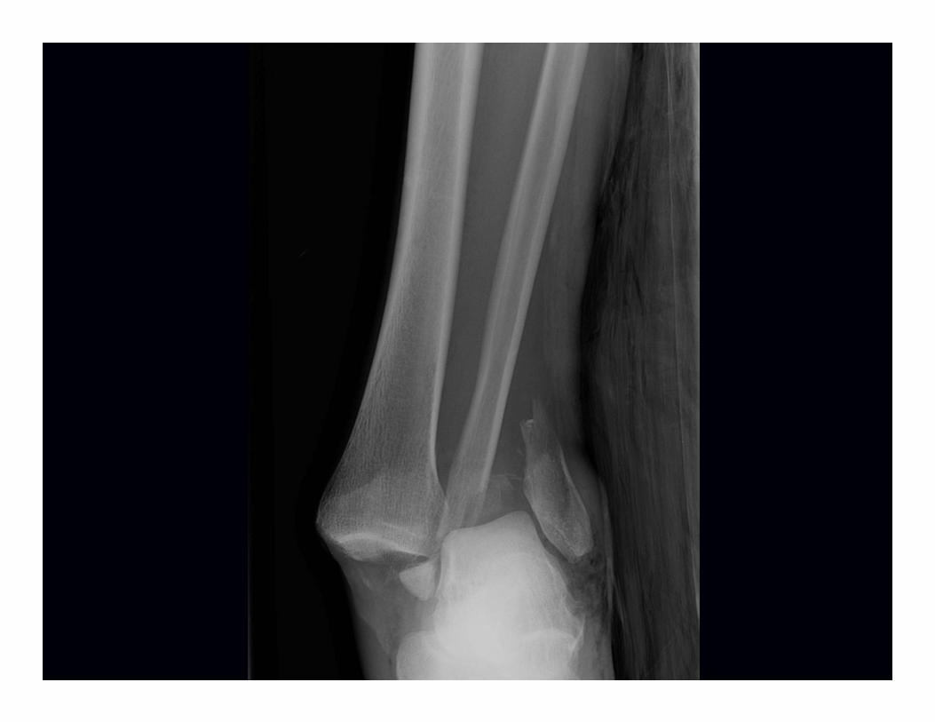
Ankle fracture or dislocation
� Reduction: –Flex the knee–Hand behind heel, and dorsal foot–Anterior traction, plantar flexed–Then 90o dorsiflexion–May need to exaggerate fracture
deformity initially

Pearls:
� Think of ways to numb the injured part (instead of the whole patient)– Hematoma block– Intra-articular injection– Nerve block
� Flex the knee for reduction of an ankle fracture or dislocation

Case 2:
� 4 yo M pulled by arm, won’t use it� Elbow in slight flexion, pronated

Nursemaid’s elbow
� Radial head subluxation, annular ligament
� Age 1-5 (usually 2-3 yo)� X-ray?
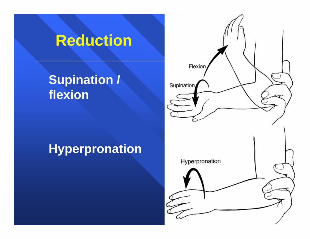
Reduction
� Supination / flexion
� Hyperpronation

Studies:
� Macias, Pediatrics, 1998:– Prospective, randomized, 90 kids– Supination/flexion vs hyperpronation– Pronation (95% vs 77%, 1st attempt)
� McDonald, Acad Emerg Med, 1999:– Prospective, randomized, 135 kids– Pronation (80% vs 69%, 1st attempt)
� Bek, Eur J Emerg Med, 2009:– 66 kids (94% vs 69%, 1st attempt)

Pearl:
� Hyperpronation

Case 3:
� 75 yo F fell onto her L side� Pain in L hip with weight bearing


What next?
a) Hip contusion—d/c homeb) CT scanc) MRId) Sign out to next doc

Occult hip fracture
� Common, and clinically important� Bone Scan (?) vs CT vs MRI–MRI is most supported by evidence–All three are superior to plain films–Local resources dictate choice

MRI
� Frihagen, Acta Orthop, 2005:–100 pts, hip trauma, neg plain films–All had MRI–46 Hip Fx–27 other fractures (mostly pelvic)–30 had surgery

MRI:

�Can CT exclude hip fx?– Rapid advances in technology– As good as MRI?

MRI vs CT:
� Lubovsky, Injury, 2005:– 6 pts with suspected fx, negative Xrays– All had MR and CT (slice?)– 5 of 6 had fx. CT “misdx’d” three.• Greater tuberosity fx in 3 who had
inter-trochanteric fx by MRI
� Case series (not yet published)– 4 cases, neg 64-slice CT, pos MRI

82 yo FFell on L hipPlain films negative:

CT:

MRI:

Pearl:
� MRI still the gold standard for excluding occult hip fracture
� What if I only have CT?

Occult scaphoid fracture?
� Patient with snuffbox tenderness, negative X-ray?

Scaphoid fracture
� One of most commonly missed fx� Most common carpal fracture– 10-20% occult
� Delayed complications:– Non-union– Avascular necrosis

Frequent occult fractures +
Frequent complications =
Thumb spica splint and follow-up

Best way to find occult fx?
� Bone scan: – Traditional, tried and true
� MRI: – Better than bone scan, multiple studies– Gold standard
� CT: – New technology, as good as MRI?

MRI vs CT:
� Memarsadeghi, Radiology, 2006:–29 pts, neg Xray, had CT (4), MRI–Gold std: plain films at 6 wks–11 scaphoid fx–MR found 11/11, CT found 8/11

CT for scaphoid fracture:
� 2 small studies used CT– Ty (2008), Cruickshank (2007)
� MRI only in selected cases� Reported no missed scaphoid fx

Remaining questions:
� Is modern CT as good as MRI?
� Should I order MR or CT today instead of splint and f/u?

Case 4:
� 56 yo F in MVA� c/o R shoulder pain� No deformity, slightly swollen� Markedly decreased ROM


Posterior Dislocation vs Normal

Posterior Dislocation vs Normal

Posterior Shoulder Dislocation
� 1-2% of shoulder dislocations� Blunt trauma, Seizure, Electrocution� 50% initially missed (?)– Less obvious clinically, radiographically
Anesthesia:
� Procedural sedation– Propofol–Midazolam / fentanyl– Ketamine
� Intra-articular injection
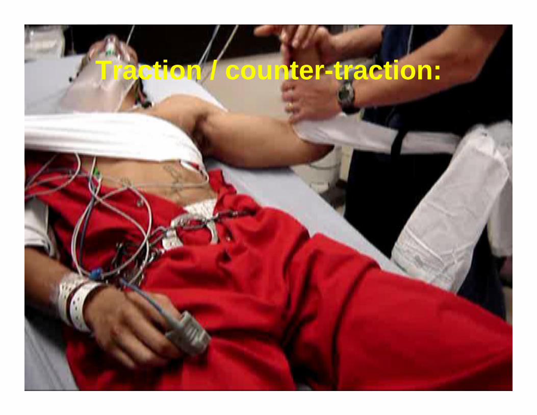
Traction / counter-traction:

Pearls:
� Watch out for posterior dislocation – (2 views, including Y-scapula)
� Intra-articular injection

Case 5:
� 50 yo male fell from a ladder� Right elbow appears dislocated� Absent radial/ulnar pulses


What next?
a) Reduce the dislocationb) Order an angiogramc) Order CT angiod) Sign out to next doc

Immediate limb threat
� Reduce the dislocation� Post-reduction exam– Still no pulses
� CT angio


Pearls:
� Careful vascular exam� Reduce immediately for limb threat� CT angio

Summary:
� Numb the injury, not the whole patient� Hyperpronation for Nursemaid’s� MRI occult hip, scaphoid� Posterior shoulder is tricky� Reduce fractures/dislocations with
limb threat� CT angio for vascular injury

� Thank you!
� Fair Use Notice: This presentation and syllabus may contain copyrighted material the use of which has not always been specifically authorized by the copyright owner. It is being made available in an effort to advance the understanding of medical, scientific, and other issues. It is believed that this constitutes a 'fair use' of any such copyrighted material as provided for in section 107 of the US Copyright Law. In accordance with Title 17 U.S.C. Section 107, the material on this site is distributed without profit to those who have expressed a prior interest in receiving the included information for research and educational purposes. If you wish to use copyrighted material from this site for purposes of your own that go beyond 'fair use', you must obtain permission from the copyright owner.