orise lesson plan: utilizing x-ray diffraction and
TRANSCRIPT

Utilizing X-Ray Diffraction and Computer Simulation to Determine the Structure of the Protein Lysozyme
Submitted by: Alaina Kilpatrick, Neil Snedeker, and Darlene Rutledge Appalachian Regional Commission/Oak Ridge National Laboratory
Summer Math-Science-Technology Institute
Grade: 9-12 grade Chemistry
Lesson Duration: 200-250 minutes Background Information: This activity comes in two main parts. The first portion involves forcing the protein lysozyme to crystallize, and the second is to use computer modeling (Molecular Dynamic Simulation) to visualize/analyze the dynamic structure of the protein. Many remaining sections in this lesson plan will contain those divisions.
Lysozyme Crystallization The successful crystallization of any protein requires specific conditions (concentrations, pH, salt, temperatures, etc) such that the water molecules will separate from the protein and force the proteins to crystalize [Additions of detergents, organic solvents and other minor components may also be essential to slow down the rate of protein precipitation in an effort to produce large, perfect crystals.] Finding suitable conditions for the crystallization of a newly isolated and characterized protein could take anywhere from a couple of weeks to several months to many years. The driving forces for crystallization are still very much unknown. In this project you will screen crystallization conditions for the enzyme lysozyme, which has been well characterized with respect to crystallization properties. You will use the hanging and sitting drop methods that rely on vapor diffusion, in which a drop containing lysozyme/precipitant solution is allowed to equilibrate in a closed system containing a reservoir of precipitant. With vapor diffusion, the sample is at 50% of the concentration of the precipitant compared to the reservoir solution and is less than that required for protein crystallization. Thus because the precipitant is the major solute present, vapor diffusion in the closed system results in the net transfer of water from the protein solution to the reservoir, until the precipitant concentration is the same in both solutions. Upon equilibration this transfer of water ceases and the resultant protein solution stays at the optimal precipitant concentration for crystallization. We will survey the range of conditions over which lysozyme might crystallize, with NaCl as precipitant, and screen several enzyme concentrations at two different pHs. The precipitant (well) conditions will be as indicated in table 1 (where we will make our observations/crystal count). We will calculate the volume of each component of the reservoir solution required to give the correct concentration in the well, given the stock solutions, deionized H2O and desired final volume of 1 ml in the well. At the end of we will have 24 wells, each with 1 ml of precipitant solution. (Follow Table 2) We will set up duplicate conditions – one set as hanging drops with circular glass cover slips and the second as sitting drops with micro-bridges. The steps required for these techniques are given on the following pages.

We will observe our crystallizations trays over the course of the project. We will write down our observation in Table 2 to 4. Observations will be reported as:
- Clear drop - Precipitated drop (spikes) - Multiple crystals - Few isolated crystals
Our ultimate objective is to determine the best crystallization conditions. “Best conditions” being defined as the conditions that produce the largest, well-shaped crystals.
Finally we will practice fishing/scooping crystal out of the crystallization drops.
Molecular Dynamic Simulations
Introduction to VMD: VMD (Visual Molecular Dynamics) is a molecular visualization and analysis program designed for biological systems such as proteins, nucleic acids, lipid bilayer assemblies, etc. It is developed by the Theoretical and Computational Biophysics Group at the University of Illinois at Urbana-Champaign. Among molecular graphics programs, VMD is unique in its ability to efficiently operate on multi-gigabyte molecular dynamics trajectories, its interoperability with a large number of molecular dynamics simulation packages, and its integration of structure and sequence information. Key features of VMD include:
General 3-D molecular visualization with extensive drawing and coloring methods Extensive atom selection syntax for choosing subsets of atoms for display Visualization of dynamic molecular data Visualization of volumetric data Supports all major molecular data file formats No limits on the number of molecules or trajectory frames, except available memory Molecular analysis commands Rendering high-resolution, publication-quality molecule images Movie making capability Building and preparing systems for molecular dynamics simulations Interactive molecular dynamics simulations Extensions to the Tcl/Python scripting languages Extensible source code written in C and C++
Lesson Objectives: The student will… • Identify what distinguishes the primary, secondary, tertiary, and quaternary structure of a
protein. • Discuss x-ray crystallography • Grow protein crystals and evaluate crystal structure most appropriate for x-ray crystallography • Discuss how diffraction patterns determine static protein structure • Discuss/Explore Molecular Dynamics procedures (webquest) • Explore the Protein Data Bank • Choose an alternative assessment (poster, shadow box, skit, etc) to present a specific protein from
the Protein Data Bank that has been studied

Materials Needed:
Lysozyme Crystallization Molecular Dynamic
Simulations
End-of-Unit Project
• Nitrile gloves • Goggles • Precision balance • Weighing boat • Graduated cylinders • Beakers • Micropipettes
(P10 uL, P200 uL, P1000 uL) • Micropipette tips • Linbro plate • Grease and applicator (Plastic syringe) • 22mm Coverslips • Tweezers / Forceps • Microscope • Crystal support (loop) • Crystal handling tools • TRIS solution • NaCl solution • NaAcetate solution • Lysozyme stock solution
• Webquest (computers for students)
• Computer • Free
downloadable Software
• Student handout • Rubric for
expectations and self-evaluation
For those not familiar with Linbro plates, here are some pictures. The sitting drops plate can be bought with the pedestal already in the well (Figure 1). These instructions are for Linbro plates that do not have the pedestal already in the wells and require the micro-bridge seen in Figure 2 so that only one tray is used for 24 reservoirs. Figure 1 Figure 2

Instructional Process: LYSOZYME CRYSTALLIZATION
Lysozyme pH/[NaCl] screening crystallization conditions pH screening
NaCl concentration screening 0.6 M 0.8 M 1.0 M 1.2 M 1.4 M 1.6 M
pH 4.2 50 mM NaAcetate
100 µl NaAc 150 µl NaCl 750 µl H2O
100 µl NaAc 200 µl NaCl 700 µl H2O
100 µl NaAc 250 µl NaCl 650 µl H2O
100 µl NaAc 300 µl NaCl 600 µl H2O
100 µl NaAc 350 µl NaCl 550 µl H2O
100 µl NaAc 400 µl NaCl 500 µl H2O
pH 4.2 50 mM NaAcetate
100 µl NaAc 150 µl NaCl 750 µl H2O
100 µl NaAc 200 µl NaCl 700 µl H2O
100 µl NaAc 250 µl NaCl 650 µl H2O
100 µl NaAc 300 µl NaCl 600 µl H2O
100 µl NaAc 350 µl NaCl 550 µl H2O
100 µl NaAc 400 µl NaCl 500 µl H2O
pH 7.0, 100 mM Tris-HCl
100 µl TRIS 150 µl NaCl 750 µl H2O
100 µl TRIS 200 µl NaCl 700 µl H2O
100 µl TRIS 250 µl NaCl 650 µl H2O
100 µl TRIS 300 µl NaCl 600 µl H2O
100 µl TRIS 350 µl NaCl 550 µl H2O
100 µl TRIS 400 µl NaCl 500 µl H2O
pH 7.0, 100 mM Tris-HCl
100 µl TRIS 150 µl NaCl 750 µl H2O
100 µl TRIS 200 µl NaCl 700 µl H2O
100 µl TRIS 250 µl NaCl 650 µl H2O
100 µl TRIS 300 µl NaCl 600 µl H2O
100 µl TRIS 350 µl NaCl 550 µl H2O
100 µl TRIS 400 µl NaCl 500 µl H2O
Table 1. Lysozyme crystallization screen conditions with required volume of each constituent.
Rows 1 and 3: hanging drop Rows 2 and 4: sitting drop
------------------------------------------------------------------------------------------
The Hanging Drop Method The hanging drop vapor diffusion method is one of the most popular techniques for crystallization condition screening and optimization. It is economical with protein and is a reasonably fast procedure to carry out. Step 1. Apply a thin layer of grease around the rim of each well (reservoir) in the limbro plate (Rows A to D, columns 1 to 6). (These can be ordered pre-greased) Step 2. Fill in each well (reservoir) with the corresponding volume of reagents (TRIS or Na Acetate, NaCl, water) (Table 1). Mix each well.

Step 3. Use forceps to pick up the cover slips. Lay out six on top of linbro box lid. Slightly off-set them from the center for easier pick up. Do not touch. Step 4. Pipette 3 µl of lysozyme stock onto the center of the first cover slip. Add 3 µl of the precipitant from the first well (A1) to the first drop. (This is colored red for easy viewing.)
Step 5. Pick up the cover slip with forceps and invert, over the first well. The grease will form a seal between the cover slip and top of well.
Step 6. Repeat steps 4 and 5 for wells A2 to A6. Step 7. Repeat steps 4 to 6 for row C.

Sitting Drops – Using Micro-bridges
Sitting drop vapor diffusion is another popular method for crystallization condition screening and optimization. It allows you to use large volumes and is an even easier setup than hanging drop. Step 1. Using forceps or fingers, place micro-bridges into row B, wells B1 to B6 and row D, wells D1 to D6 of the greased limbro plate.
Step 2. Pipette 10 µl of lysozyme stock into the depression at the center of each micro-bridge. Add 10 µl of the precipitant from well B1 to the lysozyme in the micro-bridge in B1. Step 3. Pick up a clean cover slip with forceps and invert it over the well. The grease will form a seal between the slip and top of well.
Step 4. Repeat steps 2 and 3 for wells B2 to B6. Step 5. Repeat steps 1 to 5 for wells in row D, D1 to D6. Step6. Cover the tray with the linbro plate lid. Label lid and tray. OBSERVING the crystals: The hanging drops and sitting drops can both be viewed under the microscope straight from the Linbro trays keeping the seal in place. Use the data sheet below to record observations.
Observations will be reported as: - Clear drop - Precipitated drop (spikes) - Multiple crystals - Few isolated crystals

Lysozyme Crystallization – Observations Name ________________________________Date___________ DATA SHEET DAY # pH screen-ing
NaCl concentration screening 0.6 M 0.8 M 1.0 M 1.2 M 1.4 M 1.6 M
pH 4.2 50 mM NaAcetate
pH 4.2 50 mM NaAcetate
pH 7.0, 100 mM Tris-HCl
pH 7.0, 100 mM Tris-HCl
Table 2. Crystallization observation ---------------------------------------------------------------------------------------------------------------------------- DAY # pH screen-ing
NaCl concentration screening 0.6 M 0.8 M 1.0 M 1.2 M 1.4 M 1.6 M
pH 4.2 50 mM NaAcetate
pH 4.2 50 mM NaAcetate
pH 7.0, 100 mM Tris-HCl
pH 7.0, 100 mM Tris-HCl
Table 3. Crystallization observation ---------------------------------------------------------------------------------------------------------------------------- DAY # pH screen-ing
NaCl concentration screening 0.6 M 0.8 M 1.0 M 1.2 M 1.4 M 1.6 M
pH 4.2 50 mM NaAcetate
pH 4.2 50 mM NaAcetate
pH 7.0, 100 mM Tris-HCl
pH 7.0, 100 mM Tris-HCl
Table 4. Crystallization observation

Instructional Process (cont.): MOLECULAR DYNAMICS SIMULATIONS It is at this point in the unit that funding, resources, and time limit further hands-on procedures for students. X-ray diffraction machines would be used to take many “pictures” that are processed and sent to Molecular Dynamics Researchers to further study motions of the protein. Teachers can use the web quest to show students what would happen next with the specimens they created. (Web quest is found in the attachments section) Assessment/Follow Up: (These will all be found in the attachments section.)
• Pre-test—Teachers should administer pre-test prior to instruction. • Web quest • Discussion / questions • Post-test • End-of-Unit Project
Key Vocabulary: (Note: Students should already be familiar with transcription, translation, and possibly mutation giving the primary structure of proteins, which is where this unit begins.)
Lysozyme Crystallization Computer Modeling / Simulation
• Polypeptide chain • Primary structure • Secondary Structure
(alpha helix / beta sheet) • Tertiary structure • Quaternary structure • Crystal structure • X-ray diffraction • Crystallography • Goniometer
• Visualization • Molecular dynamics • Simulation
Safety and Cleanup Required: Glass lids from the Linbro plates break easily and should be disposed of properly.
Non-Hazardous Laboratory Glass and Plastic Non-hazardous laboratory glass and plastic waste includes items not contaminated with biohazardous material that could puncture a plastic bag:
• micropipette tips • serological pipettes • test tubes • swabs/sticks • non-contaminated broken glass, razor blades, fragile glass items
including glass Pasteur pipettes, glass slides and cover slips
Package non-hazardous lab glass and plastic waste items in sturdy cardboard boxes. Empty chemical containers (including pipette tips and centrifuge tubes) can be packaged as non-hazardous lab glass. Use any cardboard box, provided the box is sturdy and will not weigh more than 25 pounds when full. Label boxes with the room number and PI name and seal with "Laboratory Glass" tape. If printed tape is not available, seal the box with other packaging tape and clearly label as "Laboratory Glass." Place the Laboratory Glass box next to the regular trash container for

custodial pick-up and disposal via municipal waste. Boxes and tape are available in the Chemistry stockroom and from several UW vendors, and tape is available from Biochemistry stores. Alignment with the Next Generation Science Standards:
• HS-LS1 From Molecules to Organisms: Structures and Processes • HS-LS1-1 Construct an explanation based on evidence for how the structure of DNA
determines the structure of proteins which carry out the essential functions of life through systems of specialized cells.
Science and Engineering Practices: Developing and Using Models, Planning and Carrying out Investigations, Constructing Explanations and Designing Solutions, Scientific Investigations use a Variety of Methods
• HS-ETS1 Engineering Design • HS-ETS1-2 Design a solution to a complex real world problem by breaking it down into
smaller, more manageable problems that can be solved through engineering. • HS-ETS1-3 Evaluate a solution to a complex real-world problem based on prioritized
criteria and trade-offs that account for a range of constraints, including cost, safety, reliability, and aesthetics, as well as possible social, cultural and environmental impacts.
• HS-ETS1-4 Use computer simulation to model the impact of proposed solutions to a complex real-world problem with numerous criteria and constraints on interactions within and between systems relevant to the problem.
Science and Engineering Practices: Asking questions and defining problems, constructing explanations and designing solutions

Additional Teacher Information: Crystallization of Lysozyme
Stock solutions: 4M NaCl – 200 ml Weigh 46.72 g of NaCl (4 x MWNaCl x 0.2 = 4 x 58.4 x 0.2 = 46.72) Dissolve in 180 ml deionized water (use a graduated cylinder) Adjust to 200 ml upon complete dissolution (remove the magnetic stirring bar for accurate adjustment!) Filter and store in glass bottle 0.5M Sodium Acetate, pH 4.2 – 100 ml Weigh 6.8 g of Sodium Acetate 3H2O (0.5 x MWNaAcetate x 0.1 = 0.5 x 136.08 x 0.1 = 6.8) Dissolve in 80 ml deionized water (use a graduated cylinder) Insert a calibrated pH meter probe Add Glacial Acetic Acid drop wise until pH = 4.2. Adjust to 100 ml with deionized water (remove the magnetic stirring bar for accurate adjustment!) Check final pH Filter and store in glass bottle 1M Tris-HCl, pH 7.0 – 100 ml Weigh 12.1 g of Tris Base (1 x MWTris x 0.1 = 1 x 121.14 x 0.1 = 12.1) Dissolve in 80 ml deionized water (use a graduated cylinder) Insert a calibrated pH meter probe Add HCl drop wise until pH 7.0 (be aware 7.0 is at the border of the buffer range, i.e pH will drop faster when you get close to 7.00) Adjust to 100 ml with deionized water (remove the magnetic stirring bar for accurate adjustment!) Check final pH Filter and store in glass bottle Lysozyme at 50, 25, 12.5 mg/ml in deionized water Attachments:
• Pre- and Post-test • Web quest • Questions after viewing crystals • Project requirements handout • Project rubric • Answer Keys

Protein Crystal Structure Pre/Post Test Name:____________________________ Date:______________
1. _______ Proteins are long chains of A. amino acids. B. nucleotides.
C. fatty acids. D. sugar molecules.
2. _______ Proteins do all of the following things in the body except
A. carry genetic information. B. speed up chemical reactions. C. digest food. D. carry oxygen in blood. E. defend against microorganisms.
3. _______ These proteins are biological catalysts:
A. transport proteins. B. structural proteins. C. enzymes.
4. _______ Which is the most commonly used technique to determine protein structures?
A. Nuclear magnetic resonance (NMR) spectroscopy B. Computer modeling C. X-ray crystallography D. Magnetic resonance imaging (MRI) E. Microscopy
5. _______ The amino acid sequence of a polypeptide chain determines its ____ structure.
A. Primary B. Secondary
C. Tertiary D. Quaternary
6. _______ The level of polypeptide folding in which the primary sequence coils around itself, stabilized by regularly spaced hydrogen bonds, is called:
A. primary structure. B. alpha helix. C. beta sheet. D. tertiary structure. E. quaternary structure.
7. _______ X-ray diffraction can only be done on samples that are in crystal form?
A. True B. False 8. _______ Each type of protein has a unique shape?
A. True B. False

9. _______ X-ray diffraction can be used to build a 3D model of A. DNA. B. RNA. C. viruses. D. proteins. E. all of the above.
10. _______ In an X-ray diffraction measurement, a ________ is mounted on a goniometer and gradually rotates while being bombarded with X-rays, producing a diffraction pattern of regularly spaced spots known as reflections.
A. Crystal B. Solution C. Grid
11. _______ In a crystal, proteins are arranged periodically in
A. 1 dimension. B. 3 dimensions.
C. 4 dimensions. D. none of the above.
12. _______ What does the picture to the right show?
A. Protein crystal B. Uric acid crystal causing gout C. Protein structure D. Protein-DNA complex
13. _______ X-rays are composed of
A. atoms. B. electrons. C. photons.
14. _______ The picture below shows
A. a diffraction pattern from a protein crystal. B. the arrangement of the protein in a crystal.
15. _______ Protein molecules are static structures?
A. True B. False
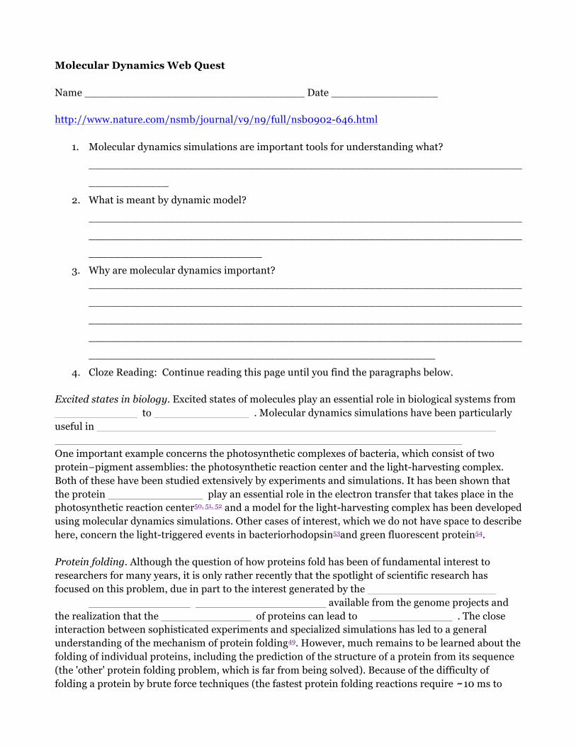
Molecular Dynamics Web Quest Name _________________________________ Date ________________ http://www.nature.com/nsmb/journal/v9/n9/full/nsb0902-646.html
1. Molecular dynamics simulations are important tools for understanding what?
_________________________________________________________________
____________
2. What is meant by dynamic model?
_________________________________________________________________
_________________________________________________________________
__________________________ 3. Why are molecular dynamics important?
_________________________________________________________________
_________________________________________________________________
_________________________________________________________________
_________________________________________________________________
____________________________________________________
4. Cloze Reading: Continue reading this page until you find the paragraphs below.
Excited states in biology. Excited states of molecules play an essential role in biological systems from to . Molecular dynamics simulations have been particularly useful in One important example concerns the photosynthetic complexes of bacteria, which consist of two protein−pigment assemblies: the photosynthetic reaction center and the light-harvesting complex. Both of these have been studied extensively by experiments and simulations. It has been shown that the protein play an essential role in the electron transfer that takes place in the photosynthetic reaction center50, 51, 52 and a model for the light-harvesting complex has been developed using molecular dynamics simulations. Other cases of interest, which we do not have space to describe here, concern the light-triggered events in bacteriorhodopsin53and green fluorescent protein54. Protein folding. Although the question of how proteins fold has been of fundamental interest to researchers for many years, it is only rather recently that the spotlight of scientific research has focused on this problem, due in part to the interest generated by the available from the genome projects and the realization that the of proteins can lead to . The close interaction between sophisticated experiments and specialized simulations has led to a general understanding of the mechanism of protein folding49. However, much remains to be learned about the folding of individual proteins, including the prediction of the structure of a protein from its sequence (the 'other' protein folding problem, which is far from being solved). Because of the difficulty of folding a protein by brute force techniques (the fastest protein folding reactions require 10 ms to

complete, which is at the limit of accessible simulation times), simplified models — for example, lattice models49 and C off-lattice models55 — have been used to obtain insights concerning the mechanism of protein folding. Recently, it has become possible to follow the folding and unfolding of model
1. View Figure 4. Identify this picture and why it is significant. _______________________________________
_______________________________________
_______________________________________
_______________________________________
_______________________________________
_______________________________________
_______________________________________
_______________________________________________________
2. Cloze Reading: Conclusions Molecular dynamics simulations of biological macromolecules have provided many insights concerning since the first protein was studied over years ago. With continuing advances in the methodology and the speed of computers, which is still doubling every months or less (according to law), molecular dynamics studies are being extended to larger systems, greater conformational changes and longer time scales. This makes possible the that have particular functional implications and to obtain information that is not accessible from experiment. The results available today make clear that the applications of molecular dynamics will play an even more important role for our understanding of biology in the future.
Once structure of a protein is determined, we can study it to determine function, find molecules that interact, and eventually (hopefully) use those molecules to correct malfunctioning proteins. Drug discovery is only one field that follows molecular dynamics in this research. http://www.news-medical.net/health/Polypharmacology-the-future-of-drug-discovery.aspx Polypharmacology: the future of drug discovery?
3. What is polypharmacology? _____________________________________________________________________________
4. What are the concerns regarding patient safety, and how is it handled?
___________________________________________________________________________________________________________________________________________________________________________________________________

_____________________________________________________________________________________________________________________
Questions after viewing crystal Name ____________________________________Date______________
1. In the space provided, draw and describe from your field of view a single crystal that would be ideal to use in the next step of x–ray diffraction. (High quality drawings J)
2. In the space provided, draw and describe from your field of view a single crystal that is a precipitated crystal that obviously grown too quickly.
3. What conditions must be present for some solutions to precipitate the conditions too quickly?
(Review Tables 1-4)
End-of-Unit Project Assignment Name__________________________________ Date_______________
1. Choose your protein. 2. You will create one of the following to showcase the protein you chose:
a. A shadow box (diorama) b. Brochure c. Poster d. Skit/song
3. Your project must include the following sections/information: a. RCSB PDB-4 figure code b. Picture or model of protein c. What is its CLASSIFICATION? d. In what organism is it found? e. Highlights of your protein. (This should be the largest section of your project.) Note: It
is recommended to add PDB 101 after the name of your protein when searching on the internet. It is an excellent source.
• When the protein was discovered/Who identified the protein • Go into detail about the normal function of your protein, ex. What is
your protein responsible for? Does it work along or closely with other proteins?
• Any other interesting facts about it. • What happens when things go wrong with the protein? (Are there
diseases or disorders associated with this protein not working properly?) • What current research is being done using your protein?
f. Create a work cited section

Suggested Rubric for groups assigned Building A Structure: Protein Project Student Name: _______________________________ Date: _________________________
CATEGORY 4 3 2 1 Scientific Knowledge
Explanations by all group members indicate a clear and accurate understanding of scientific principles underlying the construction and modifications.
Explanations by all group members indicate a relatively accurate understanding of scientific principles underlying the construction and modifications.
Explanations by most group members indicate relatively accurate understanding of scientific principles underlying the construction and modifications.
Explanations by several members of the group do not illustrate much understanding of scientific principles underlying the construction and modifications.
Information Gathering
Accurate information taken from several sources in a systematic manner.
Accurate information taken from a couple of sources in a systematic manner.
Accurate information taken from a couple of sources but not systematically.
Information taken from only one source and/or information not accurate.
Construction - Care Taken
Great care taken in construction process so that the structure is neat, attractive and follows plans accurately.
Construction was careful and accurate for the most part, but 1-2 details could have been refined for a more attractive product.
Construction accurately followed the plans, but 3-4 details could have been refined for a more attractive product.
Construction appears careless or haphazard. Many details need refinement for a strong or attractive product.

Answer Key for Webquest: Molecular Dynamics Webquest Name _________________________________ date _______ http://www.nature.com/nsmb/journal/v9/n9/full/nsb0902-646.html
1. Molecular dynamics simulations are important tools for understanding what? the physical basis of the structure and function of biological macromolecules.
2. What is meant by dynamic model?
the internal motions and resulting conformational changes play an essential role in their function
3. Why are molecular dynamics important?
a) Simulations can provide the ultimate detail concerning individual particle motions as a function of time. Thus, they can be used to address specific questions about the properties of a model system, often more easily than experiments on the actual system.
b) although the potentials used in simulations are approximate, they are completely under the control of the user, so that by removing or altering specific contributions their role in determining a given property can be examined.
4. Cloze Reading: Continue reading this page until you find the paragraphs below.
Excited states in biology. Excited states of molecules play an essential role in biological systems from vision to photosynthesis . Molecular dynamics simulations have been particularly useful in increasing our understanding of the coupling between the excited state behavior and the electron and/or energy transfer that results. One important example concerns the photosynthetic complexes of bacteria, which consist of two protein−pigment assemblies: the photosynthetic reaction center and the light-harvesting complex. Both of these have been studied extensively by experiments and simulations. It has been shown that the protein motions play an essential role in the electron transfer that takes place in the photosynthetic reaction center50, 51, 52 and a model for the light-harvesting complex has been developed using molecular dynamics simulations. Other cases of interest, which we do not have space to describe here, concern the light-triggered events in bacteriorhodopsin53and green fluorescent protein54. Protein folding. Although the question of how proteins fold has been of fundamental interest to researchers for many years, it is only rather recently that the spotlight of scientific research has focused on this problem, due in part to the interest generated by the numerous protein sequences available from the genome projects and the realization that the misfolding of proteins can lead to disease . The close interaction between sophisticated experiments and specialized simulations has led to a general understanding of the mechanism of protein folding49. However, much remains to be learned about the folding of individual proteins, including the prediction of the structure of a protein from its sequence (the 'other' protein folding problem, which is far from being solved). Because of the difficulty of folding a protein by brute force techniques (the fastest protein folding reactions require 10 ms to complete, which is at the limit of accessible simulation times), simplified models — for example, lattice models49 and C off-lattice models55 — have been used to obtain insights concerning

the mechanism of protein folding. Recently, it has become possible to follow the folding and unfolding of model
5. View Figure 4. Identify this picture and why it is significant. The permeation of the water molecules (red and gray) through the aquaporin tetramer. It is important because with modern computers, the simulation time is extended to a range from 100 ns to s, making it possible to study biological phenomena as they happen. A striking recent result is that, by running multiple simulations of 10 ns duration, the 'real time' visualization of water molecule migrating through a model of the aquaporin channel has been achieved (Fig. 4).
6. Close Reading: Conclusions
Molecular dynamics simulations of biological macromolecules have provided many insights concerning the internal motions of these systems since the first protein was studied over 25 years ago. With continuing advances in the methodology and the speed of computers, which is still doubling every eighteen months or less (according to Moore's law), molecular dynamics studies are being extended to larger systems, greater conformational changes and longer time scales. This makes possible the investigation of motions that have particular functional implications and to obtain information that is not accessible from experiment. The results available today make clear that the applications of molecular dynamics will play an even more important role for our understanding of biology in the future.
7. What is polypharmacology? It involves the pharmaceutical compounds that exert an effect on
multiple targets in the body. 8. What are the concerns regarding patient safety, and how is it handled? polypharmacology has
the potential to cause problems when it is not used correctly, or insufficient information is known about the activity of the drug. This is primarily as a result of adverse effects that result from secondary drug targets. The safe and effective use of medications with polypharmacological properties requires extensive data collation to ensure the best results. This process included computer models, synthetic chemistry, pharmacological testing and clinical trials before it can be implemented in widespread practice.
Answer Key for Pre / Post-test
1. A 2. A 3. C 4. C 5. A
6. B 7. A 8. A 9. E 10. A
11. B 12. A 13. C 14. A 15. B