original research paper influence of ph and heat …
TRANSCRIPT
The Annals of the University Dunarea de Jos of Galati Fascicle VI – Food Technology (2015), 39(2), 64-76
ORIGINAL RESEARCH PAPER
INFLUENCE OF pH AND HEAT TREATMENT ON β-LACTOGLOBULIN - OLEIC ACID COMPLEX
ANA-MARIA SIMION (CIUCIU)1*, IULIANA APRODU, PETRU ALEXE,
NICOLETA STĂNCIUC
Faculty of Food Science and Engineering, Dunarea de Jos University of Galati, 111 Domnească St. 800201, Galati, Romania
*Corresponding author: [email protected]
Received on 1st September 2015 Revised on 8th October 2015
One of the major concerns of food technologists is to produce healthier products with specific functionalities. The potential use of β-lactoglobulin as a supplement for new functional products is encouraging due to its nutritional and functional characteristics. The aim of this work was to obtain β-lactoglobulin-oleic acid complexes at different pH values (5.0, 6.0, and 7.0) and to test their stability at different temperatures (25-85°C) such as to allow identifying their potential use in a variety of food products. The complexes were characterized through different fluorescence spectroscopy based techniques: phase diagram, intrinsic and extrinsic fluorescence, along with fluorescence quenching experiments. Results showed the presence of more than two structurally distinct species with intermediates as induced by thermal treatment. The heat treatment at temperatures higher than 70°C caused an increase in both intrinsic and ANS fluorescence intensity. Acrylamide quenching showed no significant differences between the values of Stern-Volmer constants as function of temperature for pH 5.0, suggesting that no significant changes occurred in the Trp microenvironments. Quenching experiments with KI lead to decreases in Stern-Volmer constants in the temperature range 25-70°C, suggesting protein folding, whereas at higher temperatures a small increase was observed suggesting unfolding and an increased accessibility of the fluorophore to the quencher for all pH values. Keywords: β-lactoglobulin, oleic acid, complex, pH
Introduction β-lactoglobulin (βLG) is the dominant whey protein, and one of the most studied proteins in the field of food chemistry. It consists of 162 amino acid residues, and it is composed mainly of β-sheet motifs (Brownlow et al., 1997). Due to the excellent nutritional and functional characteristics βLG may represent a healthy choice in the formulation of new foods and beverages. The amino acid content of βLG is important as a fuel for muscle growth and also a source of cysteine, which is
Simion (Ciuciu) et al. / AUDJG – Food Technology (2015), 39(2), 64-76
65
important for glutathione synthesis (de Wit, 1989). Moreover, this protein and its peptide fragments are continuously capturing the imagination of food scientists and technologists, mostly because of their bioactivities (Chatterton et al., 2006). βLG has the ability to bind and transport lipophilic bioactive nutrients such as fatty acids, retinol and vitamin D (Qin et al., 1998; Wang et al., 1999; Simionet al., 2015). Its high stability at low pH helps protecting hydrophobic molecules during passage through the stomach, in order to deliver the ligands to the specific receptors of the intestine (Cho et al., 1994). Because of its binding properties, βLG is of great interest for use in nutraceuticals delivery (Spontonet al., 2014; Livney, 2010). Among the compounds that βLG has affinity for, fatty acids are particularly important because they may prevent disorders such as cardiovascular diseases, hypertension, obesity, neurological or psychiatric disorders (Simion et al., 2015; Yashodhara et al., 2009). There is a need to develop new food grade carriers for hydrophobic micronutrients such as to allow developing new highly functional food ingredients or nutraceuticals. Chemical and physical treatments used in food industry induce conformational and structural changes in βLG molecules, affecting the functional properties. One of the βLG properties is related to its pH dependent oligomeric association (Bhattacharjee & Das, 2000). At pH values between 5.2 and 7.0 βLG occurs as stable dimmer, as octamer at pH values between 3.5 and 5.2, while at pH 3.0 and above 7.5 it occurs as a monomer with two-cysteine residues per monomer (de Wit, 1989). Around its isoelectric pH 5.1, βLG is known to form tetramers or higher oligomers (McKenzie, 1971). In our previous work, we studied the complex formed at pH 8.0 along with thermal treatment, but in order to allow efficient use of the complex in different food products, further research at different pH values combined with heat-treatment is needed. Therefore, in the present study, the structural changes of βLG-Oleic Acid (OA) complex at different pH (5.0, 6.0 and 7.0) and temperatures (25-85°C) have been investigated. The obtained information on the βLG-OA complex might be of importance for the food industry in order to obtain new functional products with multiple health benefits. Material and methods Materials Bovine βLG (99% purity), oleic acid (OA), 1-anilino-8-naphtalenesulphonic acid (ANS), potassium iodide (KI) and acrylamide were obtained from Sigma (Sigma-Aldrich Co, St. Louis, MO). The protein was used without further purification. All other chemicals used were of analytical grade.
βLG - oleic acid complex formulation The βLG-OA complex was prepared by simple mixing the two components in Tris-HCl buffer at pH 5.0, 6.0 and 7.0. The final protein/OA molar ratio was 1:10. The blends were mixed with a vortex, and the OA excess was removed by centrifugation (10000 × g, 4°C, 10 min).

Simion (Ciuciu) et al. / AUDJG – Food Technology (2015), 39(2), 64-76
66
Heat-treatment For thermal treatments, the complex solutions were filled in Eppendorf tubes (Eppendorf AG, Hamburg, Germany). The experiments were conducted in a thermostatic water bath (Digibath-2 BAD 4, RaypaTrade, Barcelona, Spain) at temperatures ranging from 25 to 85°C for 10 min. After heating, the tubes were immediately cooled in ice to prevent further denaturation. Fluorescence spectroscopy techniques A LS-55 luminescence spectrometer (PerkinElmer Life Sciences, Shelton, CT, USA) with a quartz cell of 10 mm path length was used to characterize the changes induced by temperature and pH in the protein-fatty acid complexes. Fluorescence techniques, such as phase diagram method, intrinsic fluorescence spectroscopy, synchronous fluorescence, fluorescence quenching experiments, and ANS binding, were used. All fluorescence spectroscopy experiments were performed as described earlier by Simion et al. (2015). All experiments were performed in triplicate and the standard deviation was lower than 3.5%. Results and discussion Phase diagram The structural changes of the βLG-OA complex as a consequence of thermal treatment were studied at different pH values. As can be seen in Figure 1, regardless of pH values, a nonlinear correlation was obtained in the experiments. Correlations between I365 versus I320 in the phase diagram were described by third-order polynomial functions for all pH values. These nonlinear correlations indicate the presence of more than two structurally distinct species that became populated as a consequence of the thermal treatment. The temperature increase caused rearrangements within the βLG-OA complex, inducing the transition of the protein molecules from the native state to an unfolded/refolded one, via three intermediate states. High temperatures are expected to induce conformational changes associated with folding and aggregation of the molecules (Nistor et al., 2014). Our results indicated the sequential character of structural changes, suggesting the presence of partially folded species and intermediates. When we studied the same complex at pH value 8.0, a linear dependence was observed indicating the presence of two different conformations without the presence of other intermediates (Simion et al., 2015). Our results showed that the βLG-OA complex was stable in the temperature range 25-70°C, but when the temperature was further increased to 75°C and above, the unfolding of polypeptide chains occurred. At 75°C, the fluorescence intensity values were the highest for pH values of 5.0 and 7.0, suggesting that, at this temperature, rearrangements in the complex molecules lead to exposure of hydrophobic residues. For pH 6.0 the highest fluorescence intensity was registered at 80°C. Further thermal treatment led to molecules aggregation and consequently to the burial of the hydrophobic residues, and significant decrease in

Simion (Ciuciu) et al. / AUDJG – Food Technology (2015), 39(2), 64-76
67
fluorescence intensity. It seems that pH values have an important role in the βLG-OA complex structural changes induced by heat treatment.
Figure 1. Phase diagram analysis of heat-induced conformational changes of βLG-OA complex pH 5.0 (a), pH 6.0 (b) and pH 7.0 (c) based on intrinsic fluorescence intensity values measured at wavelengths 320 and 365 nm. The temperature values are indicated in the vicinity of the corresponding symbol. Three independent tests were carried out in each case and SD was lower than 2.5%.
(a)
(b)
(c)

Simion (Ciuciu) et al. / AUDJG – Food Technology (2015), 39(2), 64-76
68
Intrinsic fluorescence The intrinsic fluorescence of indole chromophores in tryptophan (Trp) residues is sensitive to their microenvironment, and, as a consequence, fluorescence spectroscopy can be used to study conformational changes of proteins (Chen and Barkley, 1998). The emission band of βLG is the result of its two Trp and four Tyr residues (Liang and Subirade, 2012). This kind of information is useful for a detailed description of the structural properties of the βLG-OA complex, and was employed in the present study for a better understanding of the pH and heat treatment induced structural changes. When heating the βLG-OA complex at pH 5.0, the fluorescence intensity (IF) initially decreased in the temperature range 25 to 70°C, while at 75°C an important increase was observed followed by a decrease of about 27% at 80°C, and 37% at 85°C. When excited at 292 nm, the βLG-OA complex at pH 5.0 showed a 3 nm red-shift of the λmax when heating at 75°C (338.5 nm) compared to 25°C (335 nm) (Figure 2a). On the other hand, only a small red-shift of 0.5 nm was observed at the excitation at 274 nm (data not shown) when heating the complex at 75°C (334 nm) compared to 25°C (333.5 nm). The observed red-shifts can be explained by the changes occurring in the tertiary structure of βLG (Perez et al., 2014). Furthermore, thermal treatment led to a decrease in IF suggesting burial of the hydrophobic residues and monomers aggregation. Similar changes were observed for pH values 7.0 and 6.0. When heated at 75°C, at pH 7.0, a 3 nm red-shift in λmax was observed when compared to the un-treated solutions (Figure 2c). When exciting at 292 nm the βLG-OA complex, (pH 6.0) the λmax at 80°C was 358.2 nm while at 25°C was 335 nm (Figure 2b).The same behavior was observed at 274 nm: the protein heated at 80°C showed λmax at 339.5 nm, whereas for the protein treated at 25°C the λmax was 333 nm (data not shown). Red-shifts of about 5-6 nm were observed at both wavelengths suggesting higher exposure of the hydrophobic residues at temperatures ranging from 75 to 80°C. Further increase of temperature led to aggregation and burial of hydrophobic residues for both pH values. A fully exposed tryptophan residue is expected to have λmax over 350 nm (Bhattacharjee & Das, 2000). This indicates that thermal treatment does not completely unfold the βLG molecule of the complex, its Trp residues being somewhat protected from the aqueous environment. It can be concluded that, at temperatures ranging from 75 to 80°C, the molecules partially unfold with an exposure of hydrophobic regions to the aqueous environment, but, at even higher temperatures, those residues became protected from the polar environment. These results are consistent with those obtained for the phase diagram, pointing out the important effect of the pH along with heat-treatment that lead to folding/unfolding events in the βLG-OA complex.

Simion (Ciuciu) et al. / AUDJG – Food Technology (2015), 39(2), 64-76
69
Figure 2. Structural changes of βLG-OA complex monitored by emission spectrum at pH values 5.0 (a), 6.0 (b) and 7.0 (c). The excitation wavelength was 292 nm. Three independent tests were carried out in each case and SD was lower than 3.5%. Synchronous fluorescence The synchronous fluorescence technique is used to study the microenvironment of amino acid residues in biomolecules. The shifts in λmax correspond to changes in
(a)
(b)
(c)

Simion (Ciuciu) et al. / AUDJG – Food Technology (2015), 39(2), 64-76
70
polarity around the chromophore molecules. When considering ∆λ = 60 nm (Figure 3) we can observe red-shifts of 2-3 nm for Trp residues, regardless of pH values studied, while for ∆λ = 15 nm, blue-shifts of 3-4 nm for Tyr residues were observed (Figure 4). The maximum IF values for ∆λ = 60 nm were obtained at 75°C for pH 5.0 (Figure 3a) and 7.0 (Figure 3c), while for pH 6.0, the maximum IF was obtained at 80°C (Figure 3b).
Figure 3. Synchronous fluorescence spectra (Δλ = 60 nm) of βLG-OA complex at different temperatures and pH values: 5.0 (a), 6.0 (b), 7.0 (c). Three independent tests were carried out in each case and SD was lower than 3.0%.
(b)
(c)
(a)
Simion (Ciuciu) et al. / AUDJG – Food Technology (2015), 39(2), 64-76
71
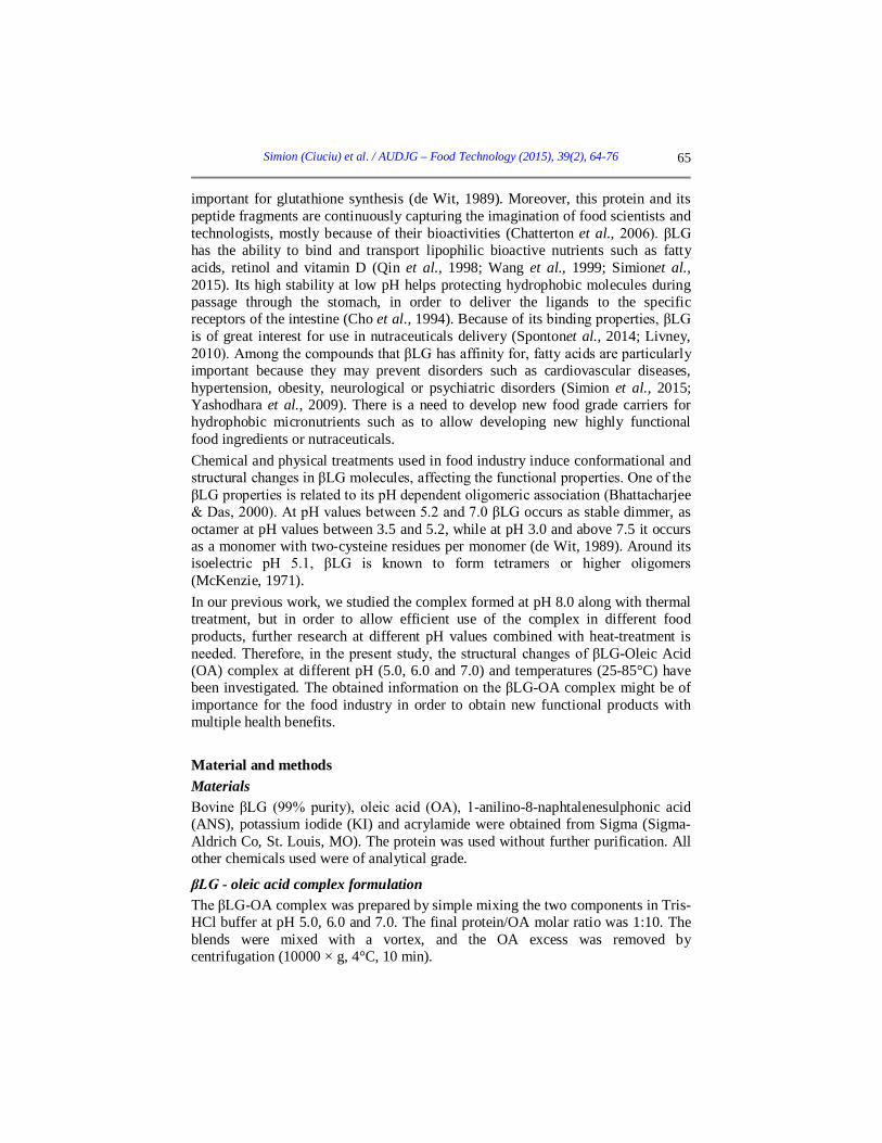
The results suggested that heat treatment induced conformational changes in the βLG-OA complex with Trp exposure and Tyr burial at 75-80°C. Further thermal treatment led to a decrease in IF suggesting a decrease in the polarity around both Trp and Tyr. Also we can observe an increase in the hydrophobicity around those fluorescent residues.
Figure 4. Synchronous fluorescence spectra (Δλ = 15) nm of β-LG-OA complex at different temperature and pH values: pH 5.0 (a), 6.0 (b), 7.0 (c). Three independent tests were carried out in each case and SD was lower than 3.0%.
(a)
(b)
(c)
Simion (Ciuciu) et al. / AUDJG – Food Technology (2015), 39(2), 64-76
72
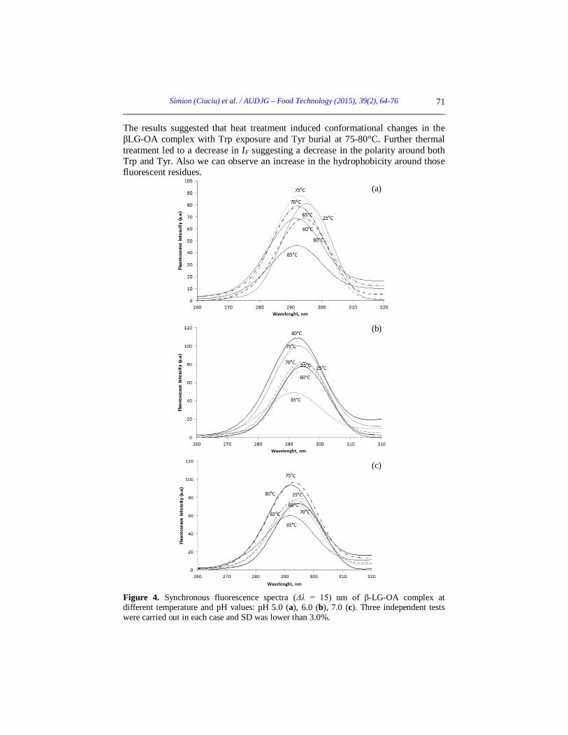
Fluorescence quenching experiments Each Trp residue in the native protein is characterized by unique micro-environmental properties (Somogyi et al., 2007). The temperature induced conformational changes of the βLG-OA complex at three pH values were also investigated by using acrylamide and KI quenching experiments. Ionic quenchers, such as KI, can extinguish fluorescence emitted by fluorophores located at the surface or near the molecule surface. Acrylamide can penetrate both hydrophobic and hydrophilic regions (Bódis et al., 2013). A linear plot between F/F0 and quencher concentration [Q] indicates that all Trp residues are equally accessible to the quencher. Our results led to a linear plot indicating that the fluorescence quenching took place on a simple collisional basis. Stern-Volmer constants (KSV) were obtained from the slope of each linear relationship. As it can be seen in Table 1, no significant statistical differences (p<0.1) were observed when heating the complex at pH 5.0, suggesting that at this pH value the accessibility of Trp residues to the quencher was not significantly modified by heat treatment. On the other hand, a decrease from 2.54±0.12 to 1.62±0.07 was observed at pH 6.0, when the temperature increased from 25°C to 75°C, and from 2.44±0.16 to 1.44±0.03 at pH 7.0. Significant increases to 3.26±0.01 for pH 6.0 and to 3.64±0.01 for pH 7.0 at temperatures higher than 75°C were observed. The increase in the Stern-Volmer constants at higher temperatures (75-80°C) seems to be caused by the βLG-OA complex unfolding. Then, at higher temperatures for pH 5.0 and 7.0, a decrease was observed suggesting a refolding of the βLG-OA complex. These results are in agreement with those obtained for the intrinsic fluorescence.
Table 1. Stern Volmer quenching constants (Ksv) in the different conformational stages of heat treated complex and at different pH values Temperature
(°C) KSV
L mol-1 KSV
L mol-1 Acrylamide KI
pH 5.0 pH 6.0 pH 7.0 pH 5.0 pH 6.0 pH 7.0 25 2.60±0.06 2.54±0.12 2.44±0.16 2.94±0.03 1.23±0.14 1.02±0.1 60 2.43±0.02 2.19±0.04 3.04±0.09 0.99±0.01 0.82±0.02 0.39±0.01 65 2.63±0.03 1.88±0.13 2.82±0.14 0.83±0.16 0.5±0.01 0.48±0.06 70 2.19±0.2 1.97±0.03 2.82±0.02 1.33±0.23 0.6±0.07 0.91±0.02 75 2.57±0.21 1.62±0.07 1.44±0.03 0.91±0.07 2.46±0.25 1.33±0.01 80 2.99±0.01 3.15±0.05 3.64±0.01 1.03±0.02 0.77±0.03 0.93±0.06 85 2.57±0.16 3.26±0.01 1.33±0.02 1.1±0.01 1.03±0.12 1.02±0.03
When quenching with KI, the Stern-Volmer plot was also linear with higher values of the constants at 25°C. These results may be correlated with the exposure of Trp residues and the lower accessibility induced by heat treatment. The KSV values highlighted a sequential denaturation process involving the molecules folding in the temperature range 25-65°C, followed by unfolding at higher temperatures for
Simion (Ciuciu) et al. / AUDJG – Food Technology (2015), 39(2), 64-76
73
all complexes at all pH values. The higher values for KSV at temperatures above 70°C suggest that local conformational changes took place involving Trp19 residue at the complex surface (Liang and Subirade, 2012). Values for the Stern-Volmer constants were, as expected, significantly lower compared to acrylamide quenching.
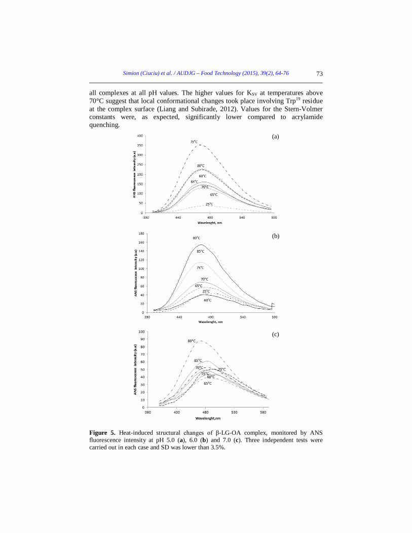
Figure 5. Heat-induced structural changes of β-LG-OA complex, monitored by ANS fluorescence intensity at pH 5.0 (a), 6.0 (b) and 7.0 (c). Three independent tests were carried out in each case and SD was lower than 3.5%.
(a)
(b)
(c)
Simion (Ciuciu) et al. / AUDJG – Food Technology (2015), 39(2), 64-76
74
ANS binding measurements Information about the changes of protein conformation and variations of the accessible hydrophobic areas are offered by the fluorescence measurements on the βLG-OA-ANS complex. Two binding sites for ANS were reported for βLG, an external one near a hydrophobic patch and an internal one in the hydrophobic core of the protein (Collini et al., 2000). ANS binding to the βLG-OA complex was influenced by thermal treatment, especially at temperatures above 70°C. The ANS fluorescence increased by 91% at 75°C for pH 5.0, if compared to the unheated complex (Figure 5a). A similar behaviour was observed for pH 6.0 and 7.0, with an increase of 75% and 50%, respectively, at 80°C (Figure 5b and 5c). Results are similar to those obtained in our previous work when we studied the same complex at pH 8.0 (Simion et al., 2015), although changes have been observed for the maximum fluorescence intensity values. Heat treatment induced changes in the βLG-OA complex resulting in a higher accessibility to ANS binding of hydrophobic regions, at temperatures ranging from 75 to 80°C (Perez et al., 2014). At higher temperatures the hydrophobic regions became less accessible being buried in the core of the complex. Heat treatment up to 70°C caused significant blue-shifts for all pH values, ranging between 6 and 15 nm compared to untreated complex. At those temperatures some non-polar groups may become exposed to water, thus increasing the hydrophobic surface of the protein, as explained by Ptitsyn (1996). Conclusions Results obtained in this study provide some insights into the pH and heat-dependent structural changes of the βLG-OA complex. Fluorescence spectroscopy experiments were carried out to monitor the pH and heat-induced changes in the βLG-OA complex. Based on the phase diagram and fluorescence spectra measurements, we suggest the presence of several different molecular species induced by pH and thermal-treatment. The heat treatment caused an increase in intrinsic fluorescence, ANS fluorescence intensity and Stern-Volmer quenching constants for temperatures higher than 70°C. The complex stability at thermal treatment appears to be pH dependent, pointing toward the importance of electrostatic interactions, most likely combined with hydrophobic interactions. These results may lead to a better understanding of the pH and heat-induced conformational changes of the βLG-OA complex. Acknowledgements The work has been funded by the Sectoral Operational Programme Human Resources Development 2007-2013 of the Ministry of European Funds through the Financial Agreement POSDRU/159/1.5/S/132397.

Simion (Ciuciu) et al. / AUDJG – Food Technology (2015), 39(2), 64-76
75
References Bhattacharjee, C. & Das, K.P. 2000. Thermal unfolding and refolding of β-lactoglobulin:
An intrinsic and extrinsic fluorescence study. European Journal of Biochemistry, 267, 3957-3964.
Bódis E., Raics K., Nyitrai M., Majer Z. & Lukács A. 2013. Fluorescence lifetime distributions report on protein destabilisation in quenching experiments. Journal of Photochemistry and Photobiology B: Biology, 129, 108-114.
Brownlow, S., Cabral, J.H.M., Cooper, R., Flwoer, D.R., Yewdall, S.J., Polikarpov, I., North, A.C.T. & Sawyer, L. 1997. Bovine β-lactoglobulin at 1.8 Å resolution-still an enigmatic lipocalin. Structure, 5, 481-495.
Chatterton, D.E.W., Smithers G., Roupas, P. & Brodkorb, A. 2006. Bioactivity of β- lactoglobulin and α-lactalbumin—Technological implications for processing. International Dairy Journal, 16, 1229-1240.
Chen, Y. & Barkley, M.D. 1998. Toward understanding tryptophan fluorescence in proteins. Biochemistry, 37, 9976–9982.
Cho, Y., Batt, C.A. & Sawyer, L. 1994. Probing the retinol-binding site of bovine β-lactoglobuline. Journal of Biological Chemistry, 269, 11102-11107.
de Wit, J. N. 1989. Functional properties of whey proteins. In Developments of dairy chemistry. In P. F. Fox (Ed.). Functional milk proteins (Vol. 4). London: Applied Science.
Liang, L. & Subirade, M. 2012. Study of the acid and thermal stability of β-lactoglobulin-ligand complexes using fluorescence quenching. Food Chemistry, 132, 2023-2029.
Livney, Y. D. 2010. Milk proteins as vehicles for bioactives. Current Opinion in Colloid & Interface Science, 15, 73–83.
Collini, M., D’Alfonso L. & Baldini, G. 2000. “New insight on beta-lactoglobulin binding sites by 1-anilinonaphtalene-8-sulfonate fluorescence decay”. Protein Science, 9, 1968-1974.
McKenzie, H.A. 1971. In Milk Proteins, Chemistry and Molecular Biology, Vol. 2 (McKenzie, H.A., ed.). 257-330. Academic Press, New York.
Nistor, V.O., Stanciuc, N., Aprodu, I. & Botez, E. 2014. New insights into heat induced structural changes of pectin methylesterase on fluorescence spectroscopy and molecular modeling basis. Spectrochimica Acta Part A: Molecular and Biomolecular Spectroscopy, 128, 15-21.
Perez, A., Andermatten, R., Rubiolo, A. &Santiago, L. 2014. β-Lactoglobulin heat-induced aggregates as carriers of polyunsaturated fatty acids. Food Chemistry, 158, 66-72.
Ptitsyn, O. 1996. How molten is the molten globule? Nature Structural & Molecular Biology, 3(6), 488–490.
Qin, B.Y., Creamer, L.K., Baker, E.N. & Jameson, G.B. 1998. 12-Bromododecanoic acid binds inside the calyx of bovine b-lactoglobulin. FEBS Letters, 438, 272–278.
Simion (Ciuciu), A.M., Aprodu, I., Dumitrascu, L., Bahrim, G.E., Alexe, P. & Stanciuc, N. 2015. Probing thermal stability of the β-lactoglobulin-oleic acid complex by fluorescence spectroscopy and molecular modeling. Journal of Molecular Structure, 1095, 26-33.
Somogyi, B., Nyitrai, M. & Hild, G. 2007. Steady-state quenching of fluorescence to study protein structure and dynamics, in: V. Uversky, E. Permyakov (Eds.), Methods in

Simion (Ciuciu) et al. / AUDJG – Food Technology (2015), 39(2), 64-76
76
Protein Structure and Stability Analysis, Nova Science Publisher Inc., New York, pp. 153–185.
Sponton, O.E., Perez, A.A., Carrara, C. & Santiago, L.G. 2014. Effect of limited enzymatic hydrolysis on linoleic acid binding properties of β-lactoglobulin. Food Chemistry, 146, 577–582.
Wang, Q., Allen, J.C. & Swaisgood, H.E. 1999. Binding of lipophilic nutrients to β-lactoglobulin prepared by bioselective adsorption. Journal Dairy Science, 82, 257–264.
Yashodara, B.M., Umakanth, S.J., Pappachan, M., Bhat, S.K., Kamath, R. & Choo, B.H. 2009. Omega-3 fatty acids: a comprehensive review of their role in health and disease. Postgraduate Medical Journal, 85, 84-90.