original article novel piperazine induces apoptosis in u937 cells
TRANSCRIPT

Introduction The evasion of apoptosis has been character-ized as one of the six major hallmarks of cancer [1]. In cancerous cells, the normal program-ming to undergo apoptosis may not be activated due to the nonreception of proapoptotic signals, the decrease or lack of synthesis of proapop-totic signals, the increase in the synthesis of antiapoptotic signals or a combination of these [2]. As multiple gain- or loss-of-function muta-tions, from spontaneous, cytotoxic, or UV-induced mutation, viral infection or uncorrected misreading during transcription, are incorpo-rated into the defective genomes, these evasive cells may subsequently acquire invasive and/or metastatic ability, ultimately leading to cancer and the possible demise of the host. Strategies have been ongoing in many laborato-ries to look for new agents, such as piperazine derivatives, which can function as anti-cancer drugs [1, 3]. Historically, most piperazine de-rivatives have been used as antihelminthics or antibiotics [4-11]. However, it has been re-cently demonstrated that some piperazine de-
rivatives are capable of inducing apoptosis in some cancer cells [10]. This report provides novel evidence that 1,4-bis-(4-(1H-benzo[d] imi-dazol-2-yl-phenyl)) piperazine (BIPP) (Figure 1A) induces apoptosis in leukemia cells without affecting normal monocytes. We further charac-terize that BIPP utilizes intrinsic apoptotic sig-naling mechanisms to induce leukemia cell death. Materials and methods Cell culture U937 myeloid leukemia cells and K562 erythro-leukemia cells were acquired from ATCC, grown in RPMI 1640 media (Cellgro, Mediatech, Inc.) supplemented with 10% Fetal Bovine Serum (Atlanta Biologicals) and 1% Penicillin-Streptomycin (Gibco, Invitrogen Corporation) and maintained at 37oC, 5% CO2. Ficoll monocyte extraction Ficoll-Paque PREMIUM reagents were obtained from GE Healthcare and monocytes isolation
Int J Biochem Mol Biol 2011;2(1):78-88 www.ijbmb.org /ISSN:2152-4114/IJBMB11010003
Original Article Novel piperazine induces apoptosis in U937 cells Josiah J. Sampson, III1, Isaac O. Donkor2, Tien L. Huang3, Samuel E. Adunyah1 1Department of Biochemistry and Cancer Biology, Meharry Medical College, Nashville, TN, 37208, USA; 2Department of Pharmaceutical Sciences, The University of Tennessee Health Science Center, Memphis, Tennessee, 38163, USA; 3College of Pharmacy, Xavier University of Louisiana, New Orleans, LA, 70125, USA. Received January 12, 2011; accepted January 26, 2011; Epub January 30, 2011; Published February 15, 2011 Abstract: The effect of 1,4-bis-(4-(1H-benzo[d]imidazol-2-yl-phenyl)) piperazine (BIPP), a newly synthesized piperazine derivative, on U937 leukemia cell viability was investigated. We show that BIPP induces dose-responsive apoptotic cell death in U937 cells by intrinsic mechanisms of apoptosis. Maximum apoptotic effect of BIPP on U937 cells was observed at 12.8µM. BIPP-induced apoptosis was evident by characteristics such as altered annexin-V binding, cas-pase activation, loss of mitochondrial membrane potential (MMP) and cytochrome c release. BIPP also differentially activates initiator and effector caspases combined with the loss of MMP strongly suggesting that BIPP causes an intrinsic apoptosis in U937 leukemia cells. Due to our observations that BIPP induces leukemia cell death without significantly affecting normal cells, our data suggests that it may be a potential therapeutic agent for human myeloid leukemia. Keywords: Piperazine, apoptosis, cancer, caspase, mitochondria, membrane potential
Novel piperazine induces apoptosis in U937 cells
79 Int J Biochem Mol Biol 2011:2(1):78-88
protocol was followed. Primary monocytes (peripheral blood mononuclear cells) were iso-lated from whole blood obtained from New York Blood Center. Using the Ficoll-Paque extraction technique, the isolated monocytes were main-tained in RPMI -1640 culture medium in the presence of 10 % FBS and 1% pen/strep antibi-otic and allowed an hour to distress and re-cover. Subsequently, the cells were either un-treated or treated with 12.8µM BIPP and fol-lowed for viability and caspase activation. Preparation of BIPP BIPP was synthesized in the laboratories of Drs. Isaac Donkor and Tien L Huang and solubilized in 14.1M DMSO from Sigma and stored at a stock concentration of 10µg/ml at -20oC. Trypan blue exclusion cell viability assay U937 cells, K562 cells, or primary isolated monocytes were treated with 12.8µM BIPP as specified under figure legend and the ability of the compound to induce apoptosis was evalu-ated at various time points. Untreated cells were maintained in an identical manner and
used as a control. 10µL of cell culture was added to 90µL of trypan blue exclusion dye and examined under microscope. Viable cell popula-tion was estimated by hemocytometer. DNA ladder detection Apoptosis was induced in 1x10⁶ cells/mL U937 leukemia cells by treating with various concen-trations of BIPP for desired time points. Sam-ples were prepared according to Quick Apop-totic DNA Ladder Detection Kit protocol from MBL International Corporation. Next, 1% aga-rose gel electrophoresis at 5V for 2 hours was used for DNA separation. DNA ladder or bands were visualized under UV light and photo-graphed. Colorimetric caspase assays All caspase assay kits were acquired from MBL International Corporation and the manufac-turer’s protocols were followed to detect cas-pase activity. U937 cells or monocytes were untreated or treated with various concentra-tions of BIPP for desired time points. Cells were counted and pelleted to achieve a density of
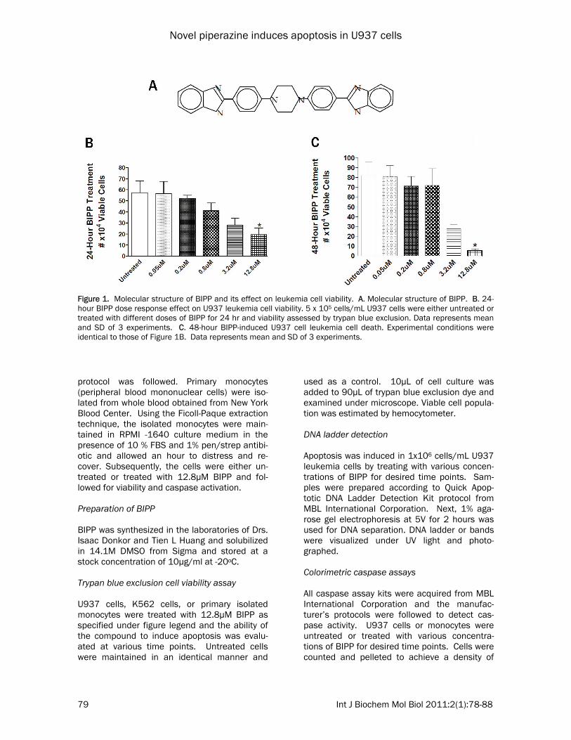
Figure 1. Molecular structure of BIPP and its effect on leukemia cell viability. A. Molecular structure of BIPP. B. 24-hour BIPP dose response effect on U937 leukemia cell viability. 5 x 10⁵ cells/mL U937 cells were either untreated or treated with different doses of BIPP for 24 hr and viability assessed by trypan blue exclusion. Data represents mean and SD of 3 experiments. C. 48-hour BIPP-induced U937 cell leukemia cell death. Experimental conditions were identical to those of Figure 1B. Data represents mean and SD of 3 experiments.
Novel piperazine induces apoptosis in U937 cells
80 Int J Biochem Mol Biol 2011:2(1):78-88
2x10⁶ cells/mL. Cells were then suspended in 50µl of chilled lysis buffer provided in the assay kit from MBL and incubated on ice for 10 min-utes. Protein concentration was performed by BCA Protein Assay from Pierce Protein Products (now Thermo Fisher Scientific) and 150µg of protein was diluted in 50µl of cell lysis buffer. Next, 50µl of 2x reaction buffer (containing 10mM DTT) was then added to each sample and followed by the addition of 5µl of 4mM x-pNA substrate. The mixture was incubated at 37oC for 2 hours after which the samples were read at 405nm by plate reader. Annexin apoptosis detection assay Assay kit was acquired from MBL International Corporation and the manufacturer’s protocol was followed. Specifically, 2x10⁵ U937 cells were either untreated or treated with 12.8µM BIPP for desired time points. Annexin binding data was collected using fluorescence micros-copy and plate reader. Reactive oxygen species (ROS) assay Image-iT Live Green Reactive Oxygen Species Detection Kit from Invitrogen Detection Tech-nologies was acquired and protocol followed. About 2x10⁵ U937 cells were either untreated or treated with various concentrations of BIPP for desired time points. Samples were washed three times with PBS and fluorescence recorded at 495/529nm for estimation of ROS levels. Nitric oxide assay Nitric Oxide Colorimetric Assay Kit from MBL International Corporation was acquired and pro-tocol followed. About 2x10⁵ U937 cells were either untreated or treated with various concen-trations of BIPP for desired time points and ab-sorbance was read at 540nm for estimation of NO levels. Mitochondrial membrane potential (MMP) as-say Mitochondrial Membrane Potential Detection Kit from Stratagene (now AgilentTechnologies, Santa Clara, CA) was acquired and protocol fol-lowed. About 1x10⁶ U937 cells were either un-treated or treated with various concentrations of BIPP for desired time points. Fluorescence measured red – 585/590nm, green –
510/527nm using fluorescence plate reader. Cytochrome c assay Cytochrome c ELISA Kit from MBL International Corporation was acquired and protocol followed. Isolation of cytosolic and mitochondrial fractions was done according to the protocols provided in the kit and certified by the vendor. Briefly, about 5x10⁶ U937 cells were either untreated or treated with various concentrations of BIPP for desired time points. The cells were collected, washed in cold PBS and homogenized in a ho-mogenizing buffer provided with the kit. The homogenate was centrifuged at 10,000 x g for 60 minutes at 4o C and the supernatant was collected and used as cytosolic fraction. The pellet was resuspended in ice cold buffer-2 (also provided by the kit) and sonicated with Ultra sonicator (BRANSON; SONIFER), for three times (20 second each) on ice. The homogenate was centrifuged at 10,000 for 30 minutes at 4oC. The supernatant was collected and used as mitochondria fraction. The samples (cytosolic and mitochondrial fractions) were assayed separately for Cytochrome C using the Cyto-chrome C ELISA kit provided by the vendor and samples were read on plate reader at 450nm. Luminescent cell viability assay CellTiter-Glo Luminescent Cell Viability Assay from Promega was acquired and protocol fol-lowed. 5x10⁶ U937 cells were either untreated or treated with various concentrations of BIPP for desired time points. Cell viability was as-sessed as outlined in the protocol. ADP/ATP ratio assay ApoGlow Assay Kit from Cambrex Bio Science Rockland, Inc. was acquired and protocol fol-lowed. 5x10⁶ U937 cells were untreated or treated with various concentrations of BIPP for desired time points and ADP/ATP ratio meas-ured. Data from three experiments were aver-aged for each experiment. Result BIPP decreases cell viability In this experiment, the ability of BIPP to cause reduction of the viable U937 leukemia cell populations in vitro was examined. As shown in

Novel piperazine induces apoptosis in U937 cells
81 Int J Biochem Mol Biol 2011:2(1):78-88
Figures 1B and C, we investigated the effects of various concentrations of BIPP on cell viability over several time points using the viability of untreated cells as control in order to find the inhibitory concentration of BIPP that produces a >50% (IC50) decrease in U937 cell viability. The data in Figure 1B shows that BIPP induces a dose-responsive decline in U937 leukemia cell viability and as seen in Figure 1C, BIPP in-duces dose-dependent decline in U937 cell vi-ability during a 48-hour treatment. Again, maxi-mum effect of BIPP is seen at 12.8µM. Similar results were seen in K562 erythroleukemia cells (data not shown). The data also shows that at BIPP concentration of 12.8µM produced maxi-mum reduction in cell viability and that the re-duction is sustained through a 96 hour time period exemplified by an IC50 in cell population density. BIPP induces annexin-V binding One of the defining characteristics of apoptosis is Annexin-V binding [12, 13]. We used SYTOX
green fluorescent dye from MBL International Corporation to stain the treated cells. This al-lowed us to discriminate apoptotic cells from necrotic cells. We stimulated U937 cells with 12.8µM BIPP to induce cell death and com-pared the results to those in untreated cells. At the 48-hr time point, cells were analyzed for apoptotic death by this differential fluorescent detection. As shown in Figures 2A, apoptotic cells fluoresce red while necrotic cells fluoresce green. The ratio of the red/green fluorescence, Figure 2B and C, shows an approximate two-fold increase in annexin-V binding in BIPP-treated cells compared to vehicle treated cells suggest-ing BIPP induces apoptosis in the U937 cells. BIPP induces DNA fragmentation Next, to provide further evidence that BIPP induces apoptosis, we probed for another characteristic of apoptosis, DNA fragmentation. DNA is frag-mented during apoptosis due to the activation of various downstream protease activity such as caspase-6 and caspase-activated DNase (CAD)
Figure 2. Time Course of BIPP-induced Annexin V binding during apoptosis. 2 x 10⁵ cells/mL U937 leukemia cells were ei-ther untreated or treated with 12.8µM BIPP for up to 48-hours. A. BIPP in-duces apoptosis but not necrosis. Cells were un-treated or treated with 12.8µM BIPP for up to 48 hours. Red stain indi-cates apoptotic cells while green stain indi-cates necrotic cells. B. Shows tallied increase in apoptotic cells after 48-hour treatment. C. Dem-onstrates amount of red/green fluorescence ratio measured during 48-hr period. Data represents a mean plus SD of 3 experi-ments.

Novel piperazine induces apoptosis in U937 cells
82 Int J Biochem Mol Biol 2011:2(1):78-88
[14, 15]. These degrading enzymes are acti-vated downstream by the prior activation of cas-pase-3. Upon their activation, they translocate to the nucleus where they cut the linker DNA between nucleosomes into approximately 200bp segments [16]. As shown in Figure 3A, apoptosis was induced in U937 leukemia cells and DNA fragmentation was seen at each concentra-tion (5nM – 12.8µM) of BIPP tested. BIPP induces caspase activation These experiments were performed to deter-mine which, if any, of the various caspases in the cell are being activated in response to stimulation with BIPP further supporting apop-totic activation. In Figure 3A, U937 cells were either untreated or treated with 12.8µM BIPP for 96 hours and then analyzed for caspase activation. BIPP significantly increased activa-tion of the initiator caspase-9 and the effector caspase-3. However, there was as slight de-crease in caspase-8 activity (Figure 3A) but no change in the activities of in initiator caspases-2 and -10 (data not shown), which would ordinar-ily be activated due to some receptor-ligand binding initiating the caspase signal cascade
from outside the cell. The activation of the ef-fector caspase-3 without activation of the initia-tor caspases aforementioned is strongly sugges-tive of intrinsic apoptosis [17]. BIPP stimulates generation of reactive oxygen and nitrogen species After establishing that BIPP triggers an intrinsic apoptotic response, we next decided to investi-gate whether BIPP treatment leads to genera-tion of reactive oxygen species (ROS) and reac-tive nitrogen species (RNS), which may contrib-ute to apoptosis. The rationale is that during intrinsic apoptosis, genomic damage from UV or g-induced radiation or the presence of a cyto-toxic drug results in the emergence of a proapoptotic signal that is sent to the mitochon-dria causing the disruption of the outer mem-brane integrity. One of the initial post-BIPP treatment events noticed, was the accumulation of radical species: reactive oxygen and nitrogen. The charged radicals may then cause oxidative damage to many subcellular structures [18 - 20]. As seen in Figures 4A and B, cells were treated with 12.8µM BIPP for 48 hours followed by measurement of the accumulated ROS and
Figure 3. BIPP-induced caspase activation and DNA fragmentation. A. BIPP induces differential activation of cas-pases. 2 x 10⁶ cells/mL U937 leukemia cells were untreated or treated with 12.8µM BIPP and caspase activity was measured as indicated in the Materials and Methods. B. BIPP causes DNA fragmentation. 1 x 10⁶ cells/mL U937 leukemia cells were either untreated or treated with varying doses (Lanes: 1 - untreated; 2 - 0.05µM; 3-0.2µM; 4 - 0.8µM, 5 - 3.2µM, 6 - 12.8µM) of BIPP. DNA fragmentation as evidence for apoptosis is shown by the stained bands on 1% agarose gel electrophoregram.

Novel piperazine induces apoptosis in U937 cells
83 Int J Biochem Mol Biol 2011:2(1):78-88
RNS. An untreated sample was used as nega-tive control while sodium butyrate-treated cells were used as a positive control for comparison [21]. Clearly, significant increase in both radical species (ROS and RNS) was apparent in BIPP treated cells when compared to the untreated U937 sample.
BIPP causes loss of MMP The buildup of oxygen radicals triggers series of events that cause the disruption of outer mito-chondrial membrane (OMM) and a subsequent loss of components necessary for the mainte-nance of MMP [19, 20, 22]. This further accen-
Figure 4. BIPP-induced changes in mitochon-drial microenvironment. U937 leukemia cells were either untreated or treated with 12.8µM BIPP for 48 hours and ROS, RNS, mitochondrial membrane potential, cytochrome c release and ADP/ATP ratios levels were all measured as indicated under Ma-terial and Methods. 2 x 10⁵ cells/mL were used to measure ROS and RNS. 1 x 10⁶ cells/mL were used for MMP. 5 x 10⁶ cells/mL were used for MTS, cytochrome c and ADP/ATP ratio. Data represents mean plus SD of 3 experi-ments. A. BIPP-induced ROS generation. B. BIPP-induced RNS generation. C. BIPP-induced decline in mitochondrial mem-brane potential. D. BIPP-induced mitochondrial cytochrome c release. E. BIPP-induced inhibi-tion of ATP synthesis. F. BIPP-induced increase in ADP/ATP ratio attesting to decline in ATP synthe-sis.

Novel piperazine induces apoptosis in U937 cells
84 Int J Biochem Mol Biol 2011:2(1):78-88
tuates the apoptotic response as the cells abil-ity to make ATP is further jeopardized and the decrease in MMP is thought to begin stimulat-ing the release of cytochrome c from the mito-chondria which is used to form apoptosomes [19]. In Figure 4C, cells were untreated or treated for 48 hours with 12.8µM BIPP, then stained to measure MMP levels. Results in treated samples were compared to those in untreated samples. Clearly, BIPP causes signifi-cant decline in MMP in these cells. BIPP stimulates mitochondria cytochrome c protein release For the apoptotic response to continue, a neces-sary component of the apoptosome, cytochrome c, must be released from the mitochondria into the cytoplasm [19, 20]. Decline in mitochondrial membrane potential and disruption of the OMM leads to cytochrome c release to the cytoplasm and then utilized in apoptosome formation. In Figure 4D, after 12.8µM BIPP treatment for 48 hours there is a significant release in cyto-chrome c from the mitochondria to the cytosol. BIPP reduces ATP synthesis All of the aforementioned changes to the mito-chondrial microenvironment ultimately affect the cell’s ability to synthesize ATP. The deple-tion of ATP hinders the cell’s ability to undergo metabolic processes and exacerbates the apop-totic response. To obtain data to support this, cells were untreated or treated with 12.8µM BIPP for 48hrs. Since only viable and prolifera-tion cells with intact oxidative phosphorylation systems synthesize ATP, we employed a lumi-nescent cell viability assay (MTS) from Promega to indirectly assess ATP level. A direct ADP/ATP ratio was measured using an adenylate nucleo-tide ratio assay obtained from Cambrex Bio Sci-ence. The results from both experiments dem-onstrate that ATP synthesis is markedly de-creased after treatment with BIPP, shown in Figures 4E and F. BIPP fails to induce cell viability changes and apoptosis in human monocytes Because U937 leukemia cells are cancerous it was important to investigate the effect of BIPP on normal human cells to ascertain whether BIPP has similar or dissimilar effects in normal and transformed cells. To achieve that goal we
examined the effect of BIPP on primary human monocytes (PBMC). Primary monocytes were isolated from whole blood and either untreated or treated with 12.8µM BIPP for 24-hours. Based on results from trypan blue exclusion assay (Figure 5A), it was concluded that there was no significant loss in monocytic cell viability in response to BIPP treatment. This data was further supported by the outcome of MTS assay for monocytes viability (Figure 5B). Similarly, BIPP failed to induce significant change in both caspases-9 and -3 activities (Figure 5C). Taken together, the results in Figure 5 further suggest that the killing/apoptotic effect of BIPP is more prominent in cancerous cells than noncancer-ous cells. The apparent reasons why normal cells were less sensitive to BIPP remains un-known at this point and require future investiga-tion. Discussion Piperazine-based compounds have recently been receiving increasing attention due to the chemical advancements they provide in drug delivery. Knowing that the evasion of apoptosis is one of the predominant ways that cells be-come oncogenic [23] and that this characteris-tic is one of the most difficult targets in design-ing an appropriate cancer treatment [18], we wanted to stimulate U937 cells into eliciting an apoptotic response by treating them with BIPP, a newly synthesized piperazine derivative. We expect the drug to exhibit toxicity primarily to cancerous cells based on previously published data [4 – 8, 11]. Concerns of toxicity have been a problem in designing an efficient and permis-sible drug [3, 4]. Our rationale for using BIPP, being that the drug will induce DNA damage in cancerous and noncancerous cells but noncan-cerous cells should be less susceptible and more capable of correcting or resisting the dam-age. This is because they are still able to halt the cell cycle to analyze its condition or initialize the apoptotic response. Cancerous cells, with their increased mitotic rate, however, would be more susceptible to the induced DNA damage as they often do not halt the cell cycle to correct DNA damage and thus be more susceptible to apoptosis. We determined that BIPP has the ability to in-duce apoptosis in U937 leukemia cells. We demonstrate that there is a dose-dependent decrease in viable cells at each time point ex-

Novel piperazine induces apoptosis in U937 cells
85 Int J Biochem Mol Biol 2011:2(1):78-88
amined. We observed that at 24 hours there is a linear concentration-dependent sensitivity of the cells to the drug as the drug concentration used increased (Fig 1B) and that the effect of the drug corresponding to an IC>50% was achieved using 12.8 uM concentration. To de-termine the mechanism by which BIPP kills U937 cells, we conducted several experiments to determine whether the type of death seen was apoptosis as opposed to other forms of cell death; most notably necrosis. Caspase activation is one of the defining char-acteristics of apoptosis [1]. Caspases are nor-mally inactive zymogens inside the cell until some stimulus is received which causes a se-ries of events leading to their eventual activa-tion. Once activated, these proteases act to activate other caspases, to expand their effect, and to dismantle the cell in an orderly fashion to be lastly discarded by activated phagocytic cells [16]. Extrinsic apoptosis involves the binding of a ligand to its cognate receptor triggering the cytoplasmic end of the receptor to recruit adap-tor proteins and certain initiator caspases most notably caspase-8, ultimately forming a death-inducing signaling complex (DISC) [24, 25]. Since the drug is quite lipophilic, we believe that
it passively diffuses into the cell through the plasma membrane. After 96-hour BIPP treat-ment of U937 cells, there was a noticeable in-crease in the activation of caspase-3 and -9 (Figure 3A) but not caspase-2, -8, and -10 (data not shown). This suggests an intrinsic mito-chondrial involvement in the induction of apop-tosis of these cells following BIPP treatment. The activation of the effector caspase-3 without activation of the initiator caspases is strongly suggestive of intrinsic apoptosis [17]. Thus, our data suggests that BIPP triggers intrinsic apop-tosis in U937 leukemia cells. This lack of extrinsic initiation of apoptosis, di-rects us toward investigating an intrinsic activa-tion of apoptosis. Intrinsic apoptosis normally begins with DNA damage that is caused by UV or g-irradiation or loss of inner mitochondrial membrane integrity [22, 26], spontaneous mu-tation or cytotoxic drug treatment. Whilst it is currently unknown whether BIPP induces DNA damage or affect the DNA repair mechanisms in the leukemia cells once inside the cell this drug signals the cell to initiate the apoptotic re-sponse. Bands are visualized that correspond to the production of DNA fragments in each of the induced samples. This damage results in a
Figure 5. Lack of BIPP-induced apoptosis in primary monocytes. Primary monocytes were isolated from whole blood using ficoll extraction. 3 x 10⁶ cells/mL of iso-lated primary mono-cytes were either un-treated or treated with 12.8µM BIPP for 24 hours and examined for viability, using trypan blue exclusion dye (A) or MTS assay (B), caspase-9 and -3 activation assay (C), and cytochrome c release (D) as de-scribed under Materi-als and Methods.

Novel piperazine induces apoptosis in U937 cells
86 Int J Biochem Mol Biol 2011:2(1):78-88
signal sent forth to the cytoplasmic portion of the cell to begin apoptosis. The fact that BIPP induces DNA fragmentation is evidenced by the presence of DNA laddering, which occurred at all of the BIPP concentrations that were investi-gated (Figure 3B). To verify that the reduction in viable cells was due to apoptosis and not necrosis we looked for another characteristics of apoptosis, namely, annexin-V binding. During apoptosis, enzymes are activated that are responsible for externaliz-ing phosphotidyl serine (PS) from the cytoplas-mic side of the plasma membrane to its ex-tracellular surface [12]. PS residues can be marked and visualized by using a fluorescent annexin-V conjugate. Our data indicates that treatment of U937 cells with 12.8µM BIPP re-sults in an increase in the amount of annexin-V binding signifying the induction of apoptotic cell death (Figures 2A, B and C). Similar results were obtained when K562 cells were studied (data not shown). We also probed for the generation of ROS and RNS since these radicals serve as some of the internal signals for initiation of the apoptotic process. Even though their production is inher-ent in normal metabolic function, their genera-tion is increased due to some cellular stressing event. Further implication of a buildup of the reactive radicals is the reduction of MMP asso-ciated with a decrease in ATP synthesis. Treat-ment of the U937 cells with 12.8µM BIPP for 48 hours resulted in a significant increase in ROS and RNS radical formation (Figure 4A and B). Next, we studied the effect of BIPP on MMP. As expected, after a 48-hour treatment with 12.8µM BIPP a marked decrease in MMP was observed (Figure 4C). Further evidence of the sequential effect of BIPP-induced apoptosis in U937 cells was ob-tained when the ATP and ADP content of the cells post-BIPP treatment was measured. Using both indirect and direct approaches as indi-cated under methods we measured ATP levels. It was determined that the amount of ATP gen-erated by BIPP treated U937 cells was signifi-cantly lower than the amount of ATP generated by untreated cells. Next, the amount of ADP in the cells post BIPP treatment was directly quan-tified. A dramatic increase in ADP buildup after a 48-hour treatment of the cells with BIPP was observed compared to untreated cells (Figure
4F). This is consistent with the fact that BIPP-treated cells display decreases in oxidative phosphorylation and MMP. Cytochrome c is a peripheral protein loosely associated with the outer side of the inner mito-chondrial membrane. Its release requires per-meabilization of the OMM. The free cytochrome c becomes part of a functioning apoptosome after combining with Apaf-1 and caspase-9. BIPP treatment enhanced the release of cyto-chrome c into cytosol (Figure 4D). Taken to-gether, our data strongly suggest that BIPP in-duces apoptosis in U937 cells and that the apoptotic response is intrinsic in nature. In order to determine whether BIPP will elicit similar effects on noncancerous cells, we exam-ined the effects of the drug on primary human monocytes by assessed changes in cell viability, caspase activation, and cytochrome c release in untreated and BIPP-treated monocytes. As shown in Figure 5, BIPP causes little yet no sig-nificant induction of cell killing or apoptosis in the normal cells. The results taken from these experiments; no change in viable cells as seen with trypan blue exclusion, no significant change in proliferation seen through MTS, no significant caspase-9 or -3 activation, and no significant cytochrome c release from the mito-chondria to the cytoplasm clearly suggest that BIPP may differentially target leukemia cells while sparing normal hematopoietic cells. Clearly, decline in mitochondrial membrane po-tential associated with cytochrome c release and capases-3 activation is triggered by BIPP during apoptosis in U937 leukemia cells. In con-clusion, we have shown that BIPP induces U937 leukemia cell death by intrinsic apoptosis mechanisms and that BIPP has little yet no sig-nificant effect on viability of human monocytes. Therefore, we propose that BIPP may be a novel piperazine derivative that may be an effective anticancer agent against myeloid monocytic leukemia cells without cytotoxic effects on nor-mal monocytes.
Acknowledgement The work was supported with funds from NCI/NIH grant # 5U54 CA091480 grant to Dr. Sam-uel E. Adunyah, Co-PI of MMC-VICC cancer part-nership. Mr. Josiah Sampson, III was supported with funds from NHLBI T32 training grant # NIH

Novel piperazine induces apoptosis in U937 cells
87 Int J Biochem Mol Biol 2011:2(1):78-88
5 T32 HL007737-17. Lastly, we want to ac-knowledge Dr. Richard Akomeah for his techni-cal advice during the early stages of this project. Please address correspondence to: Dr. Samuel E. Adunyah, Department of Biochemistry and Cancer Biology, Meharry Medical College, 1005 Dr. D.B. Todd Blvd., Nashville, TN, 37208, USA. Tel: 615-327-6345, E-mail: [email protected] References [1] Hanahan, Douglas and Robert A. Weinberg, The
Hallmarks of Cancer, Cell, 2000, Vol. 100, January 7, pp. 57-70.
[2] Ferreira, Carlos G., Mirjam Epping, Frank A.E. Kruyt, and Giuseppe Giaccone, Apoptosis: Tar-get of Cancer Therapy, Clinical Cancer Re-search, 2002, July, Vol. 8, 2024-2034.
[3] Cushion, Melanie T., Peter D. Walzer, Margaret S. Collins, Sandra Rebholz, Jean-Jacques Van-den Eynde, Annie Mayence, Tien L. Huang, Highly Active Anti-Pneumocystis carinii Com-pounds in a Library of Novel Piperazine-Linked Bisbenzamidines and Related Compounds, 2004, Antimicrobial Agents and Chemotherapy, Vol. 48, No. 11, Nov., pp. 4209-4216.
[4] Committee For Veterinary Medicinal Products Final Report, Piperazine Summary Report, 2001, www.emea.europa.eu/pdfs/vet/mrls/080701en.pdf.
[5] Mayence, Annie, Jean Jacques Vanden Eynde, Louis LeCour, Jr., Larry A. Walker, Babu L. Tek-wani, Tien L. Huang, Piperazine-linked bisben-zamidines: a novel class of antileishmanial agents, European Journal of Medicinal Chemis-try, 2004, 39, pp. 547-553.
[6] Guo, Can-Cheng, Rong-Biao Tong, and Ke-Lai Li, Choroalkyl piperazine and nitrogen mustard porphyrins: synthesis and anticancer activity, Bioorganic and Medicinal Chemistry, 2004, 12, 2469-2475.
[7] Gillet, Reynald, Pierre Jeannesson, Houcine Sefraoui, Marie-Luce Arnould-Guerin, Serge Kirkiacharian, Jean-Claude Jardillier, Francois Pieri, Piperazine derivatives of butyric acid as differentiating agents in human leukemic cells, Cancer Chemotherapy and Pharmacology, 1998, 41: 252-255.
[8] Zornig, Martin, Annie-Odile Hueber, Wiebke Baum, Gerard Evan, Apoptosis regulators and their role in tumorigenesis, Biochimica et Bio-physica Acta, 2001, Vol. 1551, F1-F37.
[9] Eilon, Gabriel F., Jirong Gu, Lewis M. Slater, Kaoru Hara and John W. Jacobs, Tumor apop-tosis induced by epoxide-containing piperazi-nes, a new class of anti-cancer agents, Cancer Chemotherapy and Pharmacology, 2000, 45: 183-191.
[10] Yi, E.Y., E.J. Jeong, H.S. Song, M.S. Lee, D.W. Kang, J.H. Joo, H.S. Kwon, S.H. Lee, S.K. Park,
S.G. Chung, E.H. Cho, Y.J. Kim, Anti-angiogenic and anti-tumor apoptotic activities of SJ-8002, a new piperazine derivative, International Jour-nal of Oncology, 2004, 25: 365-372.
[11] Zhivotovsky, Boris and Sten Orrenius, Defects in the apoptotic machinery of cancer cells: role in drug resistance, Seminars in Cancer Biology, 2003, Vol. 13, pp. 125-134.
[12] Kenis, Heidi, Hugo van Genderen, Abdel Ben-naghmouch, Hilde A. Rinia, Peter Frederik, Ja-gat Narula, Leo Hofstra and Chris P.M. Reu-telingsperger, Cell Surface-expressed Phos-phatidylserine and Annexin A5 Open a Novel Portal of Cell Entry, Journal of Biological Chem-istry, 2004, Vol. 279, No. 50, December 10, pp. 52623-52629.
[13] Gidon-Jeangirard, Carole, Be´ne´dicte Hugel, Vincent Holl, Florence Toti, Jean-Louis Lap-lanche, Dominique Meyer, J.M. Freyssinet, An-nexin V Delays Apoptosis While Exerting an External Constraint Preventing the Release of CD41 and PrPc1 Membrane Particles in a Hu-man T Lymphocyte Model, Journal of Immunol-ogy, 1999, Vol. 162, pp. 5712-5718.
[14] Warby, Simon C., Crystal N. Doty, Rona K. Gra-ham, Jeffrey B. Carroll, Yu-Zhou Yang, Roshni R. Singaraja, et al., Activated caspase-6 and cas-pase-6-cleaved fragments of huntingtin specifi-cally colocalize in the nucleus, Human Molecu-lar Genetics, 2008, Vol. 17, No. 15 pp. 2390-2404.
[15] Cao, Guodong, Wei Pei, Jing Lan, R. Anne Stetler, Yumin Luo, Tetsuya Nagayama, Steven H. Graham, et al., Caspase-Activated DNase/DNA Fragmentation Factor 40 Mediates Apop-totic DNA Fragmentation in Transient Cerebral Ischemia and in Neuronal Cultures, The Journal of Neuroscience, 2001, July 1, Vol. 21, No. 13, pp. 4678-4690.
[16] Creagh, E.M. and S. J. Martin, Caspases: cellu-lar demolition experts, Biochemical Society Transactions, 2001, Vol. 29, part 6, pp. 696-702.
[17] Salganik, Rudolf I., The Benefits and Hazards of Antioxidants: Controlling Apoptosis and Other Protective Mechanisms in Cancer Patients and the Human Population, Journal of the American College of Nutrition, 2001, Vol. 20 No. 5 464S-472S.
[18] Cummings, Jeff, Tim H. Ward, Malcolm Ran-som, Caroline Dive, Apoptosis pathway-targeted drugs – from the bench to the clinic, Biochimica et Biophysics Acta, 2004, Vol. 1705, pp.53-66.
[19] Henry-Mowatt, Judith, Caroline Dive, Jean-Claude Martinou and Dominic James, Role of mitochondrial membrane permeabilization in apoptosis and cancer, Oncogene, 2004, Vol. 23, pp. 2850-2860.
[20] Razavi, Habib M., Joel A. Hamilton, Qingping Feng, Modulation of apoptosis by nitric oxide: implications in myocardial ischemia and heart failure, Pharmacology & Therapeutics, 2005,

Novel piperazine induces apoptosis in U937 cells
88 Int J Biochem Mol Biol 2011:2(1):78-88
Vol. 106, pp. 147-162. [21] Li, C.J. and T.H. Elsasser, Butyrate-induced
apoptosis and cell cycle arrest in bovine kidney epithelial cells: Involvement of caspase and proteasome pathways, Journal of Animal Sci-ence, 2005, Vol. 83, pp. 89-97.
[22] Denning, M.F., Y Wang, S Tibudan, S Alkan, BJ Nickoloff and J-Z Qin, Caspase activation and disruption of mitochondrial membrane poten-tial during UV radiation-induced apoptosis of human keratinocytes requires activation of protein kinase C, Cell Death and Differentiation, 2002, Vol. 9, pp. 40-52.
[23] Brown, J. Martin and Laura D. Attardi, The role of apoptosis in cancer development and treat-ment response, Nature Reviews Cancer, 2005,
Vol. 5, March, pp. 231-237. [24] Denicourt, Catherine and Steven F. Dowdy,
Targeting Apoptotic Pathways in Cancer Cells, Science, 2004, Vol. 305, pp. 1411-1413.
[25] Zhivotovsky, Boris and Sten Orrenius, Defects in the apoptotic machinery of cancer cells: role in drug resistance, Seminars in Cancer Biology, 2003, Vol. 13, pp. 125-134.
[26] Haupt, Susan, Michael Berger, Zehavit Gold-berg and Ygal Haupt, Apoptosis – the p53 net-work, Journal of Cell Science, 2003, Vol. 116, pp. 4077-4085.