original article - indian journal of ophthalmology · original article evaluation of ... 6-, and...
TRANSCRIPT

Original Article
Evaluation of intrastromal corneal ring segments for treatment of keratoconus with a mechanical implantation technique
Zeki Tunc, Firat Helvacioglu, Sadik Sencan
IJO_344_11R8
Access this article onlineWebsite: www.ijo.inDOI: *** PMID: *****
Quick Response Code:
Purpose: To evaluate the clinical outcomes of intrastromal corneal ring segment (ICRS) implantation in patients with keratoconus using a mechanical implantation technique. Materials and Methods: Thirty eyes of 17 patients with keratoconus were enrolled. ICRSs (Keraring) were implanted after dissection of the tunnel using Tunc’s specially designed dissector under suction. A complete ophthalmic examination was performed, including uncorrected distance visual acuity (UDVA), corrected distance visual acuity (CDVA), spherical equivalent, keratometric readings, inferosuperior asymmetry index (ISAI), and ultrasound pachymetry. All 3-, 6-, and 12-month follow-ups were completed, and statistical analysis was performed. Results: The mean preoperative UDVA for all eyes was 1.36 ± 0.64 logMAR. At 12 months, the mean UDVA was 0.51 ± 0.28 logMAR (P = 0.001), and the mean preoperative CDVA was 0.57 ± 0.29 logMAR, which improved to 0.23 ± 0.18 (P = 0.001) at 1 year. There was a significant reduction in spherical equivalent refractive error from –6.42 ± 4.69 diopters (D) preoperatively to –1.26 ± 1.45 D (P = 0.001) at 1 year. In the same period, the mean K-readings improved from 49.38 ± 3.72 D to 44.43 ± 3.13 D (P = 0.001), and the mean ISAI improved from 7.92 ± 3.12 to 4.21 ± 1.96 (P = 0.003). No significant changes in mean central corneal thickness were observed postoperatively. There were no major complications during and or after surgery. Conclusion: ICRS implantation using a unique mechanical dissection technique is a safe and effective treatment for keratoconus. All parameters improved by the 1-year follow-up.
Key words: Intrastromal corneal ring segments, keraring, keratoconus
Department of Ophthalmology, Maltepe University School of Medicine, Istanbul, Turkey
Correspondence to: Dr. Zeki Tunc, Kultur mah Kucuk Camlik Sit A1 Blok D3 Etiler 34340 Istanbul, Turkey. E-mail: [email protected]
Manuscript received: 09.07.11; Revision accepted: 23.01.12
Keratoconus is a progressive, noninflammatory, bilateral corneal ectasia with an estimated prevalence of 1 in 2000.[1] In this condition, the cornea assumes a conical shape as a result of noninflammatory progressive corneal thinning. The thinning and protrusion in keratoconus induces irregular astigmatism with or without myopia, resulting in mild to marked impairment in the quantity and quality of vision.[2,3] Although only one eye may be affected initially, this progressive disorder ultimately affects both eyes.[4]
Treatment options for early stages of keratoconus include spectacles and contact lenses. In more advanced cases with severe corneal irregular astigmatism and stromal opacities, lamellar or penetrating keratoplasty should be considered.[5] Recently, intrastromal corneal ring segments (ICRS) and corneal collagen cross-linking have added a new dimension to the management of keratoconus. Furthermore, long-term data on ICRS procedures indicate the possibility of deferring or even replacing keratoplasty in keratoconus patients.[6,7]
Corneal tunnelization for ring segment insertion can be performed by mechanical dissection or by femtosecond laser technology. For mechanical dissection, there is already a 6 and 7 mm semiautomated dissector that operates under suction (Intacs, Addition Technology). However, in this study, 5 mm optical zone rings (Keraring) were implanted after dissection of the tunnel by using a semiautomated dissector operating
under suction [Fig. 1], which was designed by Dr. Tunc, in patients with keratoconus.
Materials and MethodsThis prospective, non-comparative study was approved by the Ethics Board Committee and followed the tenets of the Declaration of Helsinki. All patients agreed to participate in the study and to return for the postoperative examinations. Written consent was obtained from every patient after the purpose and procedures of the study had been fully explained. Inclusion criteria were clear central corneas and no visual dysfunctions other than keratoconus. A corneal thickness of 400 µm at the site of segment implantation was considered the minimum
Figure 1: Intrastromal dissector for a 5.0 mm diameter implantation zone with suction ring
AOP*** 13
acceptable for the study. All patients had clear central corneas and contact lens intolerance (rigid gas-permeable contact lenses intolerance, frequent contact lens displacement, unsatisfactory visual acuity with contact lenses). Exclusion criteria were advanced keratoconus with leukoma or inferior corneal thinning less than 400 µm and additional severe ocular and systemic pathologies (e.g., history of herpes keratitis, diagnosed autoimmune disease, and systemic connective tissue disease, glaucoma, cataract, diabetic retinopathy, and age-related macular degeneration). All operations were performed by the same surgeon (ZT) at the Department of Ophthalmology, Maltepe University School of Medicine, Istanbul, Turkey.
During each surgery session, only one eye of each patient was implanted with the Keraring; if patients needed ring segment implantations for both eyes, each eye was implanted during a different session. A complete ophthalmic examination was performed preoperatively and postoperatively, including uncorrected distance visual acuity (UDVA), corrected distance visual acuity (CDVA), spherical equivalent (SE), keratometric (K) readings, the inferosuperior asymmetry index (ISAI), and ultrasound pachymeter. ISAI was calculated as the difference between the dioptric powers at 3 mm, of the corneal geometric center using EyeSys Vision, Inc V 4.5. Posterior ectasia and corneal thickness were measured using the Orbscan II Slit Scanning Corneal Topography/Pachymetry System (version 3.10.27, Orbtek Inc.) and the DGH-500 ultrasound pachymeter (DGH, Paghette 2). The ICRSs’ placements were analyzed by Fourier-domain optical coherence tomography (OCT) (Optovue RTVue with Cornea/Anterior Module (CAM). Visual acuity was measured using Snellen notation and then converted to logMAR for statistical analysis.
Keratoconus patients were graded according to the Amsler–Krumeich classification which includes the following stages:[7]
Stage I• Eccentric steeping.• Myopia and/or induced astigmatism <5.00 D.• Mean central K readings <48.00 D.
Stage II• Myopia and/or induced astigmatism from 5.00 to 8.00 D.• Mean central K readings <53.00 D.• Absence of scarring.• Minimum corneal thickness >400 µm.
Stage III• Myopia and/or induced astigmatism from 8.00 to 10.00 D.• Mean central K readings >53.00 D.• Absence of scarring.• Minimum corneal thickness 300 to 400 µm.
Stage IV• Refraction not measurable.• Mean central K readings >55.00 D.• Central corneal scarring.• Minimum corneal thickness 200 µm.
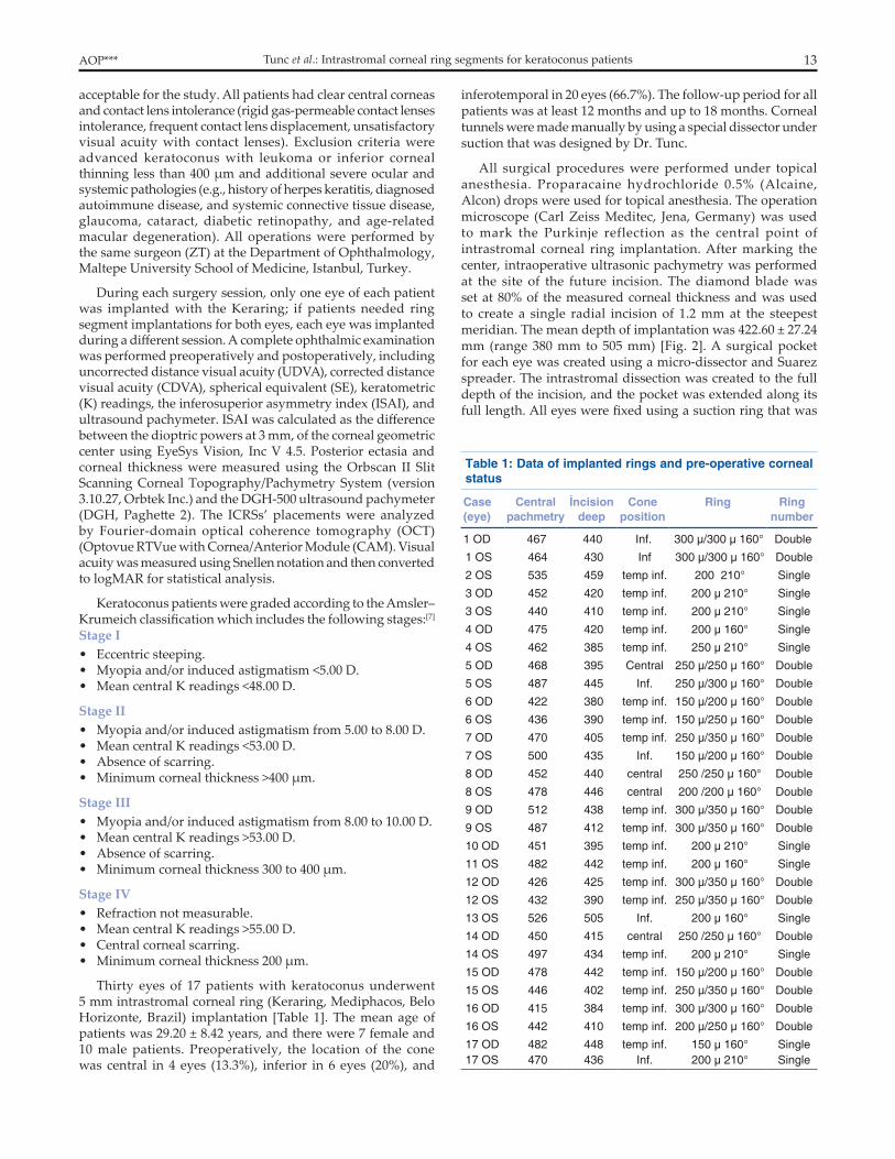
Thirty eyes of 17 patients with keratoconus underwent 5 mm intrastromal corneal ring (Keraring, Mediphacos, Belo Horizonte, Brazil) implantation [Table 1]. The mean age of patients was 29.20 ± 8.42 years, and there were 7 female and 10 male patients. Preoperatively, the location of the cone was central in 4 eyes (13.3%), inferior in 6 eyes (20%), and
inferotemporal in 20 eyes (66.7%). The follow-up period for all patients was at least 12 months and up to 18 months. Corneal tunnels were made manually by using a special dissector under suction that was designed by Dr. Tunc.
All surgical procedures were performed under topical anesthesia. Proparacaine hydrochloride 0.5% (Alcaine, Alcon) drops were used for topical anesthesia. The operation microscope (Carl Zeiss Meditec, Jena, Germany) was used to mark the Purkinje reflection as the central point of intrastromal corneal ring implantation. After marking the center, intraoperative ultrasonic pachymetry was performed at the site of the future incision. The diamond blade was set at 80% of the measured corneal thickness and was used to create a single radial incision of 1.2 mm at the steepest meridian. The mean depth of implantation was 422.60 ± 27.24 mm (range 380 mm to 505 mm) [Fig. 2]. A surgical pocket for each eye was created using a micro-dissector and Suarez spreader. The intrastromal dissection was created to the full depth of the incision, and the pocket was extended along its full length. All eyes were fixed using a suction ring that was
Table 1: Data of implanted rings and pre-operative corneal status
Case (eye)
Central pachmetry
İncision deep
Cone position
Ring Ring number
1 OD 467 440 Inf. 300 µ/300 µ 160° Double
1 OS 464 430 Inf 300 µ/300 µ 160° Double
2 OS 535 459 temp inf. 200 210° Single
3 OD 452 420 temp inf. 200 µ 210° Single
3 OS 440 410 temp inf. 200 µ 210° Single
4 OD 475 420 temp inf. 200 µ 160° Single
4 OS 462 385 temp inf. 250 µ 210° Single
5 OD 468 395 Central 250 µ/250 µ 160° Double
5 OS 487 445 Inf. 250 µ/300 µ 160° Double
6 OD 422 380 temp inf. 150 µ/200 µ 160° Double
6 OS 436 390 temp inf. 150 µ/250 µ 160° Double
7 OD 470 405 temp inf. 250 µ/350 µ 160° Double
7 OS 500 435 Inf. 150 µ/200 µ 160° Double
8 OD 452 440 central 250 /250 µ 160° Double
8 OS 478 446 central 200 /200 µ 160° Double
9 OD 512 438 temp inf. 300 µ/350 µ 160° Double
9 OS 487 412 temp inf. 300 µ/350 µ 160° Double
10 OD 451 395 temp inf. 200 µ 210° Single
11 OS 482 442 temp inf. 200 µ 160° Single
12 OD 426 425 temp inf. 300 µ/350 µ 160° Double
12 OS 432 390 temp inf. 250 µ/350 µ 160° Double
13 OS 526 505 Inf. 200 µ 160° Single
14 OD 450 415 central 250 /250 µ 160° Double
14 OS 497 434 temp inf. 200 µ 210° Single
15 OD 478 442 temp inf. 150 µ/200 µ 160° Double
15 OS 446 402 temp inf. 250 µ/350 µ 160° Double
16 OD 415 384 temp inf. 300 µ/300 µ 160° Double
16 OS 442 410 temp inf. 200 µ/250 µ 160° Double
17 OD 482 448 temp inf. 150 µ 160° Single17 OS 470 436 Inf. 200 µ 210° Single
Tunc et al.: Intrastromal corneal ring segments for keratoconus patients
14 Indian Journal of Ophthalmology Vol. ??? No. ???
placed around the limbus and was guided by the previously marked geometric center of the cornea. The tunnels were created by using left and right special dissectors. As a result, two 180-degree or one 240-degree semicircular dissections into the stroma with an approximate diameter of 5 mm were achieved. Fixating the eye under suction during intrastromal dissection was essential to create the tunnel depth, which was decided upon preoperatively. The suction ring not only served to fix eye, but also prevented uncontrolled movement of the dissector during corneal tunnelization, which is called “semiautomated dissector”. After the suction device, which was the same device used in Intacs segment implantation, was removed, the 1 or 2 Keraring segments were inserted into each of the semicircular channels using Albertazzi forceps [Fig. 3a–c]. The 5-mm diameter Keraring with appropriate arc length and thickness was implanted in each eye according to the manufacturer’s nomogram [Fig. 4]. The decision to do asymmetrical implantation was made according to the corneal topography.[8] The radial incision was closed with 1 embedded 10-0 nylon suture. All operations were uneventful. The corneal suture was removed 1 week after surgery to minimize the
potential for induced astigmatism and to minimize the risk of keratitis. Postoperative medications included a topical tobramycin 0.3% (Tobrex, Alcon), fluorometholone 5% (Flarex, Alcon), and hydroxypropyl methylcellulose 0.003% (Tears Naturale Free, Alcon), 4 times a day for 2 weeks.
Statistical MethodStatistical calculations were performed with the NCSS 2007 program for Windows. In addition to the standard descriptive statistical calculations [mean, standard deviation, and median inter quartile range (IQR)], Friedman’s repeated measures test was used to determine the differences in measurement at each time point, and postoperative Dunn’s multiple comparisons post-hoc tests were used for pairwise comparisons. The statistical significance level was established at P < 0.05.
ResultsThirty eyes of 17 patients with keratoconus grading; grade I, 8 eyes (26.6%); grade II, 14 eyes (46.8%); grade III, 8 eyes (26.6%), underwent Keraring implantation. There were no intraoperative complications such as anterior chamber perforation; postoperatively, all eyes showed excellent corneal tolerance with intrastromal segments. Some of the eyes showed subconjunctival hemorrhage due to the suction ring of the dissector, which dissolved spontaneously and completely after 1 week.
The mean UDVA increased significantly from 1.36 ± 0.64 logMAR preoperatively to 0.57 ± 0.3 logMAR (n = 30) (P = 0.001) three months after implantation, and at 12 months the mean UDVA was 0.51 ± 0.28 logMAR (n = 30) (P = 0.001) [Fig. 5a]. The mean preoperative CDVA was 0.57 ± 0.29 logMAR. The mean CDVA improved to 0.3 ± 0.27 logMAR (n = 30) (P = 0.001) at three months after implantation. One-year postoperatively, the mean CDVA was 0.23 ± 0.18 logMAR (n = 30) (P = 0.001) [Fig. 5b]. There was a significant reduction in spherical equivalent refractive error from –6.42 ± 4.69 diopters (D) preoperatively to –1.26 ± 1.45 (P = 0.001) at 1 year [Table 2]. In the same period, the mean K readings improved from 49.38 ± 3.72 D to 44.43 ± 3.13 D (P = 0.001) and the mean ISAI from 7.92 ± 3.12 to 4.21 ± 1.96 (P = 0.001) [Fig. 6]. No significant changes in preoperative mean central corneal thickness (466.80 ± 29.42
Figure 3: (a): A slit-lamp photograph of an eye with keratoconus after implantation of double 160° Keraring segment, (b): Single 160° Keraring segment, (c): Single 210° Keraring segment
a b c
Figure 2: Fourier-domain optical coherence tomography (OCT) showing the precise cross-sectional visualization and ICRS at the adequate depth of the cornea

AOP*** 15
Tabl
e 2:
Pre
oper
ativ
e an
d po
stop
erat
ive
3-, 6
-, an
d 12
-mon
th fo
llow
-up
data
Pre
oper
ativ
e3
Mon
ths
Pos
tope
rativ
e6
Mon
ths
Pos
tope
rativ
e12
Mon
ths
Pos
tope
rativ
e
Cas
e (e
ye)
UD
VA
lo
g-M
AR
CD
VA
lo
g-M
AR
RS
RC
SE
(D)
AvK
(D
)U
DV
A
log-
MA
R
CD
VA
lo
g-M
AR
RS
RC
SE
(D)
AvK
(D
)U
DV
A
log-
MA
R
CD
VA
lo
g-M
AR
RS
RC
SE
(D
)A
vK
(D)
UD
VA
lo
gMA
RC
DV
A
logM
AR
RS
RC
SE
(D
)A
vK
(D)
1 O
D2
1−5
−9−9
.555
.92
0.6
0.4
−1−9
.50.
6250
.34
0.3
0.1
−9.5
−9.5
−0.8
738
.57
0.3
0.1
−9.5
−9.5
−0.5
238
.57
1 O
S1
0.7
−5−8
−954
.59
0.5
0.2
−9−9
−1.5
44.6
90.
50.
4−9
−9−0
.236
.97
0.5
0.4
−9−9
−1.7
536
.97
2 O
S0.
70.
5−0
.75
−5−3
.25
47.4
20.
40.
181.
5−1
.50.
7543
.78
0.1
01
−1.2
50.
3744
.72
0.1
01
−1.5
0.25
44.6
7
3 O
D2
0.5
2−7
.75
−1.8
752
.52
0.5
0.18
0.5
−2−0
.549
.90.
50.
180.
5−1
.75
−0.3
749
.80.
50.
180.
5−1
.75
−0.3
749
.2
3 O
S2
0.3
2−6
.75
−1.3
751
.40.
40.
181
−20
49.5
90.
40.
180.
75−2
−0.2
549
.46
0.4
0.18
0.75
−2−0
.25
49.3
2
4 O
D1
0.2
0−1
.75
−0.8
742
.47
0.18
00
−1−0
.542
.19
0.18
00
−1−0
.542
.23
0.18
00
−1−0
.542
.38
4 O
S2
0.7
−1.2
5−6
.5−4
.550
.48
0.5
0.18
−1−1
.51.
7545
.73
0.5
0.18
−1−1
.5−1
.75
45.8
60.
50.
18−1
−1.7
5−1
.87
45.7
4
5 O
D2
1−4
−6−7
49.9
80.
70.
4−1
.25
−1.5
−243
.46
0.6
0.4
−1−1
.5−1
.75
43.3
80.
60.
4−1
.25
−1.5
−243
.2
5 O
S1.
60.
7−4
.25
−4.2
5−6
.37
48.7
10.
40.
1−1
−1.7
5−1
.87
44.3
60.
40.
1−1
−1.7
5−1
.87
44.2
60.
40.
1−1
.5−1
.75
−2.3
744
.19
6 O
D1.
30.
5−2
.5−3
.25
−4.1
244
.93
0.8
0.18
−1.2
5−2
.25
−2.3
744
.04
0.8
0.18
−1.2
5−2
.5−2
.544
.12
0.8
0.18
−1.2
5−2
.25
−2.3
744
.16
6 O
S1.
30.
3−4
−4−6
47.4
90.
80.
2−1
.5−3
−344
.33
0.8
0.2
−1−2
.75
−2.3
744
.02
0.8
0.2
−1.5
−2.7
5−2
.87
44.2
3
7 O
D2
1−8
−7.5
−11.
2552
.21
0.7
0.75
−1.7
5−0
.12
46.4
21
0.7
0.75
−1.7
5−0
.12
46.5
41
0.6
0.75
−1.7
5−0
.12
46.4
2
7 O
S0.
20.
4−3
−4−5
47.6
40.
10
00.
5−0
.25
45.2
80.
10
00.
5−0
.25
45.3
20.
10
00.
5−0
.25
45.2
8
8 O
D2
0.5
−3.5
−7.5
−7.2
552
.47
1.3
0.4
−0.5
3.25
−2.1
241
.34
10.
4−0
.5−3
.25
−2.1
241
.42
10.
4−0
.75
−3.2
5−2
.37
41.6
4
8 O
S1
0.4
−2.5
−5−5
48.7
90.
80.
2−0
.5−1
.75
−1.3
740
.29
0.6
0.2
−0.5
1.5
−1.2
540
.12
0.6
0.2
−0.2
5−1
.75
1.12
40.2
9
9 O
D2
0.5
−8−8
.5−1
2.25
46.6
20.
80.
40.
25−1
.5−0
.541
.55
0.8
0.4
0.25
−2−0
.75
41.7
60.
80.
4−0
.25
−1.5
142
.14
9 O
S1
0.4
−8−6
−11
48.5
70.
80.
6−1
−2.5
−2.2
542
.37
0.8
0.6
−1−2
.5−2
.25
42.4
30.
80.
6−1
.5−2
.5−2
.75
42.8
9
10 O
D2
0.7
0.75
−8.7
5−3
.62
49.3
50.
50.
40.
75−0
.75
0.37
48.3
20.
50.
40.
75−0
.75
0.37
47.9
30.
50.
40.
75−0
.75
0.37
47.9
8
11 O
S0.
40.
32
−3.5
0.25
47.5
50.
40.
30.
75−0
.75
0.37
46.3
0.4
0.3
0.75
−0.7
50.
3746
.54
0.4
0.2
0.5
−10
46.7
4
12 O
D2
1−1
1−1
2−1
759
.62
10.
8−1
.25
−1.5
−51
.62
10.
8−1
.25
−1.5
−252
.35
10.
8−1
.25
−1.5
−252
.35
12 O
S2
1−9
−8.5
−13.
2552
.85
0.5
0.3
1.5
−0.7
51.
1246
.32
0.5
0.3
1.5
−0.7
51.
1247
.67
0.6
0.3
1.5
−0.7
51.
1247
.67
13 O
S0.
40.
23
−3.5
1.25
47.3
40.
20.
81
−10.
546
.87
0.2
01.
25−1
0.75
46.9
40.
20
1.25
−10.
7546
.82
14 O
D1
0.7
−4.5
−7.5
−8.2
547
.99
0.8
0.6
−1−2
−244
.37
0.8
0.6
−1−1
.5−1
.75
44.1
70.
70.
5−0
.75
−1.5
1.5
44.0
5
14 O
S0.
50.
3−0
.75
−3.5
−2.2
541
.66
0.4
0.2
0.5
−1.7
5−0
.37
40.3
90.
40.
20.
5−1
.5−0
.25
40.2
60.
40.
20.
5−1
.5−0
.25
40.2
1
15 O
D1.
30.
2−4
.75
−5.5
−747
.36
0.8
0.1
−2.5
−4−4
.544
.44
0.8
0.1
−2−4
444
.20.
80.
1−2
−4−4
44.1
4
15 O
S2
1−9
−8−1
352
.42
0.8
0.4
−4.7
5−4
−6.7
549
.64
0.8
0.4
−3.5
−4.5
−5.7
549
.12
0.8
0.4
−3.5
−4.5
−5.7
549
.28
16 O
D2
1−1
0−8
.5−1
4.25
49.9
40.
50.
4−1
−11.
546
.88
0.5
0.3
−0.5
−1−1
46.2
40.
50.
3−0
.5−1
−146
.32
16 O
S1
0.7
−3−6
.5−6
.25
47.4
30.
10.
10
0.5
−0.2
543
.63
0.1
0.1
0−0
.5−0
.25
43.4
20.
10.
10
−0.5
−0.2
543
.45
17 O
D0.
50.
21.
5−3
−0.7
546
.67
00
00
046
.18
00
00
046
.16
00
00
046
.18
17 O
S0.
50.
2−0
.5−4
.52.
7548
.89
0.4
0.2
−0.7
5−1
.75
−1.6
247
.66
0.4
0.2
−0.5
1.75
−1.3
747
.62
0.4
0.2
−0.5
−1.7
51.
3747
.66
UD
VA
: Unc
orre
cted
dis
tanc
e vi
sual
acu
ity, C
DV
A: C
orre
cted
dis
tanc
e vi
sual
acu
ity, R
S =
Ref
ract
ion
sphe
ric, R
C =
Ref
ract
ion
cylin
der,
SE
: Sph
eric
al e
quiv
alen
t; A
vK: A
vera
ge k
erat
omet
ric re
adin
gs, D
: Dio
pter
Tunc et al.: Intrastromal corneal ring segments for keratoconus patients

16 Indian Journal of Ophthalmology Vol. ??? No. ???
µm) were observed 1 year postoperatively (465.33 ± 34.86 µm) (P = 0.538). The means, standard deviation and median inter quartile range (IQR) of all data are shown in Table 3. Significant improvements were observed postoperatively for UDVA and CDVA, and corneal ectasia, keratometry, SE, and ISAI were significantly reduced. Moreover, according to Dunn’s multiple comparisons tests, 3, 6, 12-month postoperative UDVA, CDVA, keratometry, and SE values showed significantly improved results from the preoperative values. Additionally, 3, 6, and 12-month results showed that there was no significant difference in refraction stability [Table 4]. None of the patients needed to use rigid gas-permeable or soft (toric) contact lens after the operations.
The patients were subdivided into two groups: those with one (group 1, n = 11) and two (group 2, n = 19) segment implantations. The outcomes were further analyzed for these groups. The preoperative central pachymeter and incision depth of the groups were 479.27 ± 30.17 and 459.57 ± 27.18 µm, respectively, and 432.18 ± 32.83 and 417.05 22.54 µm, respectively, and statistical analysis showed no significant
difference for these parameters (P1 = 0.077 P2 = 0.146). The preoperative mean UDVA of groups 1 and 2 were 1.09 ± 0.73 and 1.51 ± 0.54 logMAR, respectively, which were not significantly different (P = 0.085). The preoperative mean CDVA of groups 1 and 2 were 0.37 ± 0.19 and 0.68 ± 0.28 logMAR, respectively (P = 0.003). The preoperative mean spherical equivalent refractive errors of the groups 1 and 2 were −1.79 ± 1.71 D and −9.09 ± 3.62 D, respectively, and group 2 had a significantly higher refractive error than group 1 (P = 0.001). The preoperative mean cylindrical refractive errors of the groups 1 and 2 were −4.95 ± 2.20 D and −6.81 ± 2.18 D, respectively, and group 2 had a significantly higher refractive error than group 1 (P = 0.033). The preoperative mean K readings of the groups were 47.79 ± 3.37 D and 50.29 ± 3.68 D, respectively. The mean K readings of group 2 were slightly but not significantly higher than the K readings of group 1 (P = 0.076). The preoperative mean ISAI of the groups were 7.33 ± 3.36 and 8.25 ± 3.00 (P = 0.449), respectively. Improvements in all of the parameters in both groups were observed at 12 months [Tables 2 and 4]. The 12-month postoperative mean UDVA and CDVA of group 1 and 2 were 0.32 ± 0.17 and 0.63 ± 0.27 logMAR, respectively, and 0.14 ± 0.12 and 0.32 ± 0.21 logMAR, respectively (P1 = 0.002, P2 = 0.016). The 12-month postoperative mean spherical equivalent refractive errors of the groups 1 and 2 were −0.34 ± 0.77 D and −1.73 ± 1.54 D, respectively (P = 0.014). The 12-month postoperative mean cylindrical refractive errors of the groups 1 and 2 were −1.27 ± 0.58 D and −2.72 ± 2.54 D, respectively (P = 0.075). The 12-month postoperative mean K readings of the groups were 46.08 ± 2.78 D and 44.06 ± 3.55 D, respectively (P = 0.118). The 12-month postoperative mean ISAI of the groups were 3.77 ± 2.47 and 5.107 ± 2.35, respectively (P = 0.166).
DiscussionSeveral possible alternatives to manage keratoconus have been reported in the literature, including scleral fitted gas-permeable contact lenses, inferior eccentric penetrating grafts, corneal wedge excision, penetrating keratoplasty, and deep lamellar keratoplasty. ICRSs were designed to achieve a refractive adjustment by flattening the central corneal curvature while maintaining clarity in the central optical zone. Because of the removable and tissue-saving nature of this technique, its
Figure 5: Graphs showing the comparison of (a) uncorrected distance visual acuity (UDVA) and (b) corrected distance visual acuity (CDVA) values between preoperative and 3-, 6-, and 12-month postoperative follow-ups
a b
Figure 4: Keraring segment implantation nomogram, which describes the implantation of one or two segments according to the localization of the ectasia in the corneal topography map

AOP*** 17
application could be expanded to patients with corneal thinning disorders. Several reports have demonstrated the efficacy of Intacs in correcting keratoconic eyes,[9-12] and preliminary studies show encouraging results in eyes with post-LASIK corneal ectasia.[6,7,13-15] The magnitude of this flattening effect is in direct proportion to the thickness of the implant and in inverse proportion to its diameter.[16] Furthermore, soft ectasia corneas show more flattening effect than healthy corneas.[10] The goal of implanting intracorneal rings for keratoconus is to reduce the corneal steepening, which results in a favorable visual outcome and eliminates or delays the need for corneal grafting.
The three main ICRS on the market are Intacs (Addition Technology, Inc.), Ferrara (Ferrara Ophthalmics Ltd.), and Keraring (Mediphacos Ltd.). Kerarings are made of PMMA and are characterized by a triangular cross-section that induces a flattening effect on the cornea. Their apical diameter is 5 mm and the flat basis width is 0.6 mm with variable thickness (0.15- to 0.35-mm with 0.05-mm steps) and arc length (90 degrees, 160 degrees, and 210 degrees). The optical zone provided by Keraring segments is 5.0 mm in diameter.
During ICRS implantation, the tunnel is created by 1 of 2 techniques: mechanical dissection or use of a femtosecond laser.[17] The traditional mechanical technique for tunnel creation can cause complications. These include epithelial defects at the keratotomy site, anterior and posterior perforations during channel creation, extension of the incision toward the central visual axis or limbus, shallow placement and/or uneven placement of ICRS, infectious keratitis with the introduction of the epithelial cells into the channel during the channel dissection, asymmetric placement, persistent incisional gaping, decentration, stromal thinning, and corneal stromal edema around the incision and channel from surgical manipulation.[12,18-20] Kanellopoulos et al. reported a 35% rate of postoperative complications such as segment movement, exposure, and corneal melt with the mechanical tunnel dissection method.[19] Although the femtosecond laser has several advantages that could reduce these complications because of the more precise localization of the channel achieved by manipulating its dimensions, depth, diameter, and width, Alio et al. reported no significant difference in the risk of extrusion between the use of femtosecond laser and mechanical dissection. Of extrusion cases (18 Intacs, 10 Keraring) of their study, 15 had tunnel creation by femtosecond laser and 13 by mechanical dissection.[21]
The surgical technique for tunnel creation in Keraring and Ferrara procedures differs from the mechanical tunnel creation technique used in Intacs. While a suction device is used in Intacs, the tunnels of Keraring and Ferrara are prepared by the Ferrara double spatula. Implantation of ICRS without the use of a suction device is a more surgeon-dependent technique which has increased risk of complications while the surgeon is in the learning curve of the procedure. Additionally, Kwitko and Severo reported decentration of Ferrara ICRS in 3.9% of cases, segment extrusion in 19.6%, and bacterial keratitis in 1.9%.[22] The authors suggest that most of the complications related to surgical technique were caused by the surgeon’s learning curve and the differing healing processes of keratoconic corneas. Siganos et al. reported superficial implantation and asymmetric placement of the segments in 7.7% of cases. A vacuum was not used to create the channels in their series, and these complications were associated with implantation of the Ferrara rings.[23]
Table 3: Statistical calculations of preoperative and postoperative 3. 6. and 12 month follow-up data with Friedman’s test measurements
UDVA (logMAR) CDVA (logMAR) SE(D) AvK(D) ISAI
Mean ± SD Median (IQR)
Mean ± SD Median (IQR)
Mean ± SD Median (IQR)
Mean ± SD Median (IQR)
Mean ± SD Median (IQR)
Preoperative 1.36 ± 0.64 1.3 (0.85-2)
0.57 ± 0.29 0.5 (0.3-0.85)
−6.42 ± 4.69 −6.13 (−10.25-2.5)
49.38 ± 3.72 48.75 (47.39-52.31)
7.92 ± 3.12 7.34 (5.35-10.08)
3 Mo Postoperative
0.57 ± 0.3 0.5 (0.4-0.8)
0.3 ± 0.22 0.2 (0.18-0.4)
−1.22 ± 1.63 −1 (−2-0.06) 45.41 ± 2.98 44.99 (43.55-47.27)
5.03 ± 2.81 4.8 (2.98-6.75)
6 Mo Postoperative
0.53 ± 0.29 0.5 (0.35-0.8)
0.26 ± 0.21 0.2 (0.1-0.4)
−1.09 ± 1.44 −0.81 (−1.94-0.16)
44.79 ± 3.45 44.49 (42.33-47.28)
4.67 ± 2.53 4.84 (2.85-6.08)
12 Mo Postoperative
0.51 ± 0.28 0.5 (0.35-0.8)
0.23 ± 0.18 0.2 (0.1-0.4)
−1.26 ± 1.45 −1 (−2.19-0.25)
44.43 ± 3.13 44.21 (42.64-46.58)
4.21 ± 1.96 4.54 (3.29-5.37)
Fr 74.4 56.9 49.7 53.11 49.3P 0.0001 0.0001 0.0001 0.0001 0.0001
ISAI: Inferosuperior asymmetry index. Statistical significance level was established at P<0.05
Figure 6: The topography of the eye with keratoconus (top left) and 3 months after (bottom left) ICRS implantation and the difference map
Tunc et al.: Intrastromal corneal ring segments for keratoconus patients

18 Indian Journal of Ophthalmology Vol. ??? No. ???
Table 4: Evaluation of the postoperative results and the refraction stability with Dunn’s Multiple Comparisons Tests
Dunn’s multiple comparisons tests
UDVA (logMAR)
CDVA (logMAR)
SE(D) AvK(D) ISAI
Preoperative vs 3 Mo
0.0001 0.0001 0.0001 0.0001 0.0001
Preoperative vs 6 Mo
0.0001 0.0001 0.0001 0.0001 0.0001
Preoperative vs 12 Mo
0.0001 0.0001 0.0001 0.0001 0.0001
3 Mo vs 6 Mo 0.031 0.196 0.047 0.145 0.144
3 Mo vs 12 Mo 0.021 0.108 0.794 0.158 0.1266 Mo vs 12 Mo 0.326 0.083 0.065 0.610 0.336
Statistical significance level was established at P<0.05
Making the tunnels in the 5 mm optical zone gives the surgeon the ability to correct higher amount of refractive error with thinner ICRS implantation. But this procedure requires precision because the cornea in this area is thinner than the periphery of the cornea and is close to optical axis. Recently, Kubaloglu et al. used suction ring (Moria) to minimize decentration during the implantation of Keraring segments.[24] Unfortunately, this suction ring only fixes the eye but cannot prevent uncontrolled movements of the dissector. Although the operations were performed by a very experienced surgeon, corneal perforation and superficial segment implantation were observed in 6% of the cases that had mechanical implantation.[25] We believe that fixating the eye under suction during intrastromal dissection is essential to be able to create a tunnel at the adequate depth preoperatively. In our cases, the intrastromal dissection was created to the full depth of the incision. The suction ring not only served to fix the eye, but also prevented uncontrolled movement of the dissector during rotational movement (counterclockwise and clockwise) by holding the dissector (like the semiautomated suction ring of Intacs). All operations were uneventful. There was no incidence of delayed complications. The integrity of the cornea was well preserved in all eyes, and there was no extrusion of the rings. In contrast, standard mechanical stromal dissection of the Keraring could cause a higher rate of extrusion.[22]
Our postoperative results reveal a significant reduction in the magnitude of corneal steepening, an increase in topographical regularity, and an improvement in the UDVA due to the improved ISAI and simultaneous partial correction of the residual myopia. Other studies also found a significant improvement in CDVA, UDVA, and K readings after ICRS implantation with a mechanical and femtosecond laser tunnel creation. In a study by Coskunseven et al., 15.68% of 50 eyes had improved CDVA after ICRS implantation; the mean keratometry decreased from 50.6 D to 47.5 D and the mean SE from −5.6 D to −2.4 D at 1 year.[26] Also in a study by Kubaloglu et al. that compared the outcomes of Keraring ICRS implantation with a mechanical and femtosecond laser tunnel creation, the UDVA and CDVA improved in 86% and 88% of eyes, respectively; the mean maximum K value decreased from 53.5 D to 48.9 D and the mean SE from −5.05 to −1.87 D at 1 year in femtosecond laser group. The UDVA and CDVA improved in 88% and 84% of eyes, respectively; the mean maximum K
value decreased from 54.1 D to 43.8 D and the mean SE from −5.75 to 0.75 D at 1 year in the mechanical group.[25]
Kymionis et al. demonstrated the long-term stability of Intacs segments. In their study, 28 patients (36 eyes) had initially participated in a clinical trial for the safety and efficacy of Intacs implantation in keratoconic patients. In five patients (seven eyes), Intacs segments were removed three to 12 months after implantation because of patients’ dissatisfaction (lack of improvement of the patients’ data) without any late postoperative complication (all of them underwent uneventful PK). Mechanical implantation technique was used for all of the patients in this study and seven eyes (19.44%) had Intacs removal. These patients were then excluded from the study group. Although no evidence about the extrusion of the segments was shown in this study, these patients dissatisfaction might be related to implantation problems. Refractive and visual acuity outcomes showed significant improvements in this study and spherical equivalent error was statistically significantly reduced (pre-Intacs, mean ± SD: −5.54 ± 5.02 diopters [D]; range: −12.50–3.63 D to 3.02 ± 2.65 D; range: −8.25 to 1.88 D), uncorrected visual acuity was improved in 13 eyes (77%) compared with preoperative levels. BSCVA was maintained to the pre-Intacs level; its level in six eyes was (35%), whereas 10 eyes were 59%. BSCVA experienced a gain of one up to 8 lines at the last follow-up examination.[27]
In a study of Shabayek et al., ICRS implantation was performed by femtosecond laser for keratoconus correction. No surgical complications such as anterior chamber perforation extrusion, migration, or visualization around the incision or the tunnels occurred. Implantation was difficult in only 1 (4.76%) eye and a 10/0 nylon suture was needed to close the wound, which was removed after 10 days. This eye was excluded from the statistical analysis due to lower segment explantation after late localized infectious keratitis. As a result of this study, Keraring implantation significantly increased UCVA from 0.06 to 0.3, BSCVA from 0.54 to 0.7, and decreased the spherical equivalent by 2.28 diopters (D) and the average keratometric values (K value) by 2.24 D. There was no significant difference between the 3 and 6 months follow-up.[28]
Our study has potential limitations, such as the small sample of treated eyes, the lack of higher-order aberration analysis and the lack of a comparative group. However, the results are similar to those in a keratoconus study in which ICRS were used for treatment.[25-28] Similarly, in our study the UDVA and CDVA improved in 90% and 93.3% of eyes, respectively; the mean maximum K value decreased from 49.4 D to 44.4 D and the mean SE from −6.42 to −1.26 D at 1 year. The technique was also successfully used in the treatment of patients with post-LASIK ectasia.[29] As previously described for the post-LASIK ectasia, Tunc’s dissector for the preparation of the tunnel facilitates the procedure and adds to the safety of the surgery in patients with keratoconus. Further follow-up and additional cases are needed to draw final conclusions about the efficacy of this surgical technique.
References1. Rabinowitz YS. Keratoconus. Surv Ophthalmol 1998;42:297-319.2. Krachmer JH, Feder RS, Belin MW. Keratoconus and related
noninflammatory corneal thinning disorders. Surv Ophthalmol 1984;28:293-322.

AOP*** 19
3. Tomidokoro A, Oshika T, Amano S, Higaki S, Maeda N, Miyata K. Changes in anterior and posterior corneal curvatures in keratoconus. Ophthalmology 2000;107:1328-32.
4. Rabinowitz YS, Nesburn AB, McDonnell PJ. Videokeratography of the fellow eye in unilateral keratoconus. Ophthalmology 1993;100:181-6.
5. Colin J, Cochener B, Savary G, Malet F. Correcting keratoconus with intracorneal rings. J Cataract Refract Surg 2000;26:1117-22.
6. Alió J, Salem T, Artola A, Osman A. Intracorneal rings to correct corneal ectasia after laser in situ keratomileusis. J Cataract Refract Surg 2002;28:1568-74.
7. Lovisolo CF, Calossi A, Ottone AC. Intrastromal inserts in keratoconus and ectatic corneal conditions. In: Lovisolo CF, Fleming JF, Pesando PM, editors. Intrastromal Corneal Ring Segments. Canelli AT, Italy: Fabiano Editore; 2000. p. 95-163.
8. Alió JL, Shabayek MH, Belda JI, Correas P, Feijoo E. Analysis of results related to good and bad outcome of Intacs implantation for correction of keratoconus. J Cataract Refract Surg 2006;32:756-61.
9. Colin J, Cochener B, Savary G, Malet F, Holmes-Higgin D. INTACS inserts for treating keratoconus: One-year results. Ophthalmology 2001;108:1409-14.
10. Tunc Z, Deveci N, Sener B, Bahcecioglu H. [Corneal ring segments (INTACS) for the treatment of asymmetrical astigmatism of the keratoconus. Follow-up after 2 years.]. J Fr Ophtalmol 2003;26:824-30.
11. Siganos CS, Kymionis GD, Kartakis N, Theodorakis MA, Astyrakakis N, Pallikaris IG. Management of keratoconus with INTACS. Am J Ophthalmol 2003;135:64-70.
12. Boxer Wachler BS, Christie JP, Chandra NS, Chou B, Korn T, Nepomuceno R. INTACS for keratoconus. Ophthalmology 2003;110:1031-40.
13. Kymionis GD, Siganos CS, Kounis G, Astyrakakis N, Kalyvianaki MI, Pallikaris IG. Management of post-LASIK corneal ectasia with INTACS inserts: one-year results. Arch Ophthalmol 2003;12:322-6.
14. Pokroy R, Levinger S, Hirsh A. Single INTACS segment for post-laser in situ keratomileusis keratectasia. J Cataract Refract Surg 2004;30:1685-95.
15. Siganos CS, Kymionis GD, Astyrakakis N, Pallikaris IG. Management of corneal ectasia after laser in situ keratomileusis with INTACS. J Refract Surg 2002;18:43-6.
16. Patel S, Marshall J, Fitzke FW. Model for deriving the optical performance of the myopic eye corrected with an intracorneal ring. J Refract Surg 1995;11:248-52.
17. Ertan A, Colin J. Intracorneal rings for keratoconus and keratectasia. J Cataract Refract Surg 2007;33:1303-14.
18. Ruckhofer J, Stoiber J, Alzner E, Grabner G; for the Multicenter
European Corneal Correction Assessment Study Group. One year result of European multicenter study of intrastromal corneal ring segments. J Cataract Refract Surg 2001;27:287-96.
19. Kanellopoulos AJ, Lawrence H, Perry HD, Donnenfeld ED. Modified intracorneal ring segment implantations (INTACS) for the management of moderate to advanced keratoconus: Efficacy and complications. Cornea 2006;25:29-33.
20. Hellstedt T, Makela J, Uusitalo R, Emre S, Uusitalo R. Treating keratoconus with Intacs corneal ring segments. J Refract Surg 2005;21:236-46.
21. Ferrer C, Alió JL, Montañés AU, Pérez-Santonja JJ, del Rio MA, de Toledo JA, et al. Causes of intrastromal corneal ring segment explantation: Clinicopathologic correlation analysis. J Cataract Refract Surg 2010;36:970-7.
22. Kwitko S, Severo NS. Ferrara intracorneal ring segments for keratoconus. J Cataract Refract Surg 2004;30:812-20.
23. Siganos D, Ferrara P, Chatzinikolas K, Bessis N, Papastergiou G. Ferrara intrastromal corneal rings for the correction of keratoconus. J Cataract Refract Surg 2002;28:1947-51.
24. Kubaloglu A, Cinar Y, Sogutlu Sari E, Koytak A, Ozdemir B, Ozerturk Y. Comparison of 2 intrastromal corneal ring segment models in the management of keratoconus. J Cataract Refract Surg 2010;36:978-85.
25. Kubaloglu A, Sogutlu Sari E, Cinar Y, Cingu K, Koytak A, Coskun E, et al. Comparison of mechanical and femtosecond laser tunnel creation for intrastromal corneal ring segment implantation in keratoconus. J Cataract Refract Surg 2010;36:1556-61.
26. Coskunseven E, Kymionis GD, Tsiklis NS, Atun S, Arslan E, Jankov MR, et al. One-year results of intrastromal corneal ring segment implantation (KeraRing) using femtosecond laser in patients with keratoconus. Am J Ophthalmol 2008;145:775-9.
27. Kymionis GD, Siganos CS, Tsiklis NS, Anastasakis A, Yoo SH, Pallikaris AI, et al. Long-term Follow-up of Intacs in Keratoconus. Am J Ophthalmol 2007;143:236-44.
28. Shabayek MH, Alió JL. Intrastromal corneal ring segment implantation by femtosecond laser for keratoconus correction. Ophthalmology 2007;114:1643-52.
29. Tunc Z, Helvacioglu F, Sencan S. Evaluation of Keraring segments for treatment of post-LASIK ectasia patients with a mechanical implantation technique. Indian J Ophthalmol 2011;59:437-43.
Cite this article as: Citation will be included before issue gets online***
Source of Support: Nil. Conflict of Interest: None declared.
Tunc et al.: Intrastromal corneal ring segments for keratoconus patients