original article enhances insulin transcription and secretion...
TRANSCRIPT

WldS Enhances Insulin Transcription and Secretion viaa SIRT1-Dependent Pathway and ImprovesGlucose HomeostasisJingxia Wu,
1Fang Zhang,
1Menghong Yan,
1Dongmei Wu,
1Qiujing Yu,
1Yi Zhang,
1Ben Zhou,
1
Michael W. McBurney,2and Qiwei Zhai
1
OBJECTIVE—WldS (Wallerian degeneration slow), a fusionprotein from a spontaneous mutation containing full-lengthnicotinamide mononucleotide adenylyltransferase 1, has NADbiosynthesis activity and protects axon from degeneration ro-bustly. NAD biosynthesis is also implicated in insulin secretionin b-cells. The aim of this study was to investigate the effect ofWldS on b-cells and glucose homeostasis.
RESEARCH DESIGN AND METHODS—Using the WldS mice,we measured the expression of WldS in pancreas and analyzedthe effect of WldS on glucose homeostasis. The direct effectof WldS on insulin transcription and secretion and the relatedmechanisms was measured in isolated islets or b-cell lines. Silentinformation regulator 1 (SIRT1), an NAD-dependent proteindeacetylase, is involved in insulin secretion. Thus, WldS micewith SIRT1 deficiency were generated to study whether the SIRT1-dependent pathway is involved.
RESULTS—WldS is highly expressed in the pancreas andimproves glucose homeostasis. WldS mice are resistant to high-fat diet–induced glucose intolerance and streptozotocin (STZ)-induced hyperglycemia. WldS increases insulin transcriptiondependent on its NAD biosynthesis activity and enhances insulinsecretion. SIRT1 is required for the improved insulin transcription,secretion, and resistance to STZ-induced hyperglycemia caused byWldS. Moreover, WldS associates with SIRT1 and increases NADlevels in the pancreas, causing the enhanced SIRT1 activity todownregulate uncoupling protein 2 (UCP2) expression and upre-gulate ATP levels.
CONCLUSIONS—Our results demonstrate that WldS combinesan insulinotropic effect with protection against b-cell failureand suggest that enhancing NAD biosynthesis in b-cells to in-crease SIRT1 activity could be a potential therapeutic ap-proach for diabetes. Diabetes 60:3197–3207, 2011
Glucose homeostasis is largely maintained by thepancreatic b-cells, which secrete insulin in re-sponse to elevated ATP levels as a result ofglucose metabolism (1). b-Cell dysfunction often
leads to diabetes (2,3). However, the underlying mechanismsinvolved in the preservation of b-cell function remain to befully understood.
Silent information regulator 1 (SIRT1), an NAD-dependentprotein deacetylase, regulates various biological processesincluding glucose homeostasis (4–6). SIRT1 controls thegluconeogenic/glycolytic pathways in the liver (7) and im-proves insulin sensitivity under insulin-resistant conditionsin C2C12 myotubes (8). In b-cells, SIRT1 promotes theexpression of NeuroD and MafA, two factors essential forthe transcription of insulin, by deacetylating and activatingFoxo1 to protect against oxidative stress (9). Moreover,glucose-stimulated insulin secretion (GSIS) in islets ofSIRT1 knockout mice is blunted (10), whereas GSIS isenhanced in b-cell–specific SIRT1-overexpressing mice (11).Nevertheless, how SIRT1 is regulated to modulate insulinsecretion and improve glucose homeostasis remains to befurther explored.
Wallerian degeneration is an experimental model of axondegeneration. Remarkably, this degeneration is dramaticallyslowed in Wallerian degeneration slow (WldS) mice, aspontaneous mutant mouse strain (12). The protectivefunction is attributed to a chimeric gene resulting froman 85 kb tandem triplication in chromosome 4 (13). Thechimeric gene, WldS, encodes an N-terminal 70 aminoacids fragment of ubiquitination factor E4B (Ube4b) fusedto nicotinamide mononucleotide adenylyltransferase 1(NMNAT1), a crucial enzyme for NAD biosynthesis (14,15).The NAD biosynthesis activity is pivotal for the neuronalprotective function of WldS, which maintains the cellularNAD levels in a steady state and prevents NAD decline ininjured axons (16,17). It has been implicated that NADbiosynthesis is also involved in b-cell function. The activityof SIRT1 on GSIS in b-cell–specific SIRT1-overexpressingmice decreases with age, which probably is the result of adecline in systemic NAD biosynthesis (18). Haplodeficiencyof nicotinamide phosphoribosyltransferase (NAMPT) infemale mice, but not male, causes the decrease of NADbiosynthesis and defect of GSIS, and the extracellular formof NAMPT seems to be the main causation (19). Thus,whether the enhancement of NAD biosynthesis mediated byWldS increases GSIS and improves glucose homeostasisremains to be elucidated.
In this study, we found that WldS mice showed improvedglucose homeostasis even under high-fat (HF) diet and
From the 1Key Laboratory of Nutrition and Metabolism, Institute for NutritionalSciences, Shanghai Institutes for Biological Sciences, Chinese Academy ofSciences, Graduate School of the Chinese Academy of Sciences, Shanghai,China; and the 2Center for Cancer Therapeutics, Ottawa Hospital ResearchInstitute, and Department of Medicine, University of Ottawa, Ottawa, Ontario,Canada.
Corresponding author: Qiwei Zhai, [email protected] 22 February 2011 and accepted 16 September 2011.DOI: 10.2337/db11-0232This article contains Supplementary Data online at http://diabetes
.diabetesjournals.org/lookup/suppl/doi:10.2337/db11-0232/-/DC1.� 2011 by the American Diabetes Association. Readers may use this article as
long as the work is properly cited, the use is educational and not for profit,and the work is not altered. See http://creativecommons.org/licenses/by-nc-nd/3.0/ for details.
diabetes.diabetesjournals.org DIABETES, VOL. 60, DECEMBER 2011 3197
ORIGINAL ARTICLE

streptozotocin (STZ)-challenged states and that WldS
regulated insulin transcription and secretion dependent onSIRT1. Moreover, WldS associated with SIRT1 and increasedNAD levels in the pancreas, which led to the enhancedSIRT1 activity to downregulate uncoupling protein 2 (UCP2)and upregulate ATP levels. Thus enhancing NAD bio-synthesis in b-cells to increase SIRT1 activity might be apotential therapeutic approach for diabetes.
RESEARCH DESIGN AND METHODS
Animals. All mice were maintained and used in accordance with the guidelinesof the Institutional Animal Care and Use Committees at the Institute for Nu-tritional Sciences. WldS mice (C57BL/6 background) were purchased fromHarlan. C57BL/6 mice and imprinting control region (ICR) mice were pur-chased from Slac (Shanghai, China). SIRT1+/2 mice (129/Sv background) weredescribed previously (20). SIRT1+/2 mice were crossed with ICR mice to getSIRT1+/2 mice on an outbred genetic background. We then mated WldS micewith SIRT1+/2 mice (129/ICR background) and intercrossed the double-heterozygous SIRT1+/2WldS+/2mice to obtain SIRT1+/+ WldS2/2, SIRT1+/+ WldS+/+,SIRT12/2 WldS+/+, and SIRT12/2 WldS2/2 mice. The genotyping for WldS andSIRT1 was carried out as described previously (20,21).Islet morphology analysis. The islet area and the islet cell density weredetermined as described previously (22). All measurements were made usinga fluorescence microscope (BX61; Olympus) and Image-Pro Plus software(version 5.0.1; Media Cybernetics). At least four mice and 40 islets per mousewere studied for each group.Immunofluorescence. Pancreatic fresh-frozen sections of 16-week-old miceor fixed cells were incubated overnight at 4°C with indicated antibodies in-cluding anti-mouse insulin antibody (Sigma), anti-WldS antibody (a gift fromDr. Michael P. Coleman, The Babraham Institute), and rabbit anti-Myc antibody(Santa Cruz Biotechnology) and then detected with Alexa Fluor 555 goat anti-rabbit IgG and/or Alexa Fluor 488 goat anti-mouse IgG antibodies (MolecularProbes). DAPI (Sigma) was used to stain the nuclei. Immunofluorescenceimages were obtained on a Zeiss LSM 510 META confocal microscope or anOlympus IX51 fluorescence microscope.Glucose, insulin, C-peptide, homeostasis model of insulin resistance,
and homeostasis model of b-cell measurements. Fed glucose and insulinlevels were measured about 3 h after light on. Fasted glucose, insulin, andC-peptide levels were measured after 17-h fasting. The refed blood glucoselevels were measured in the mice refed for 2 h after a 17-h fasting. Tail bloodwas collected to measure glucose with a glucometer (FreeStyle), to measureinsulin levels by a radioimmunoassay kit (Beijing North Institute of BiologicalTechnology, China), or to measure C-peptide by an ELISA kit (Millipore). Ho-meostasis model of insulin resistance (HOMA-IR) and homeostasis model ofb-cell function values were obtained using the HOMA Calculator v2.2 (23).Glucose tolerance and insulin tolerance tests. Glucose tolerance tests wereperformed on male mice fasted 13 h. Glucose concentrations were measuredin blood collected by venous bleeding from the tail vein at the indicated timesafter an intraperitoneal injection of glucose (2 g/kg body wt). Insulin tolerancetests were performed on male mice fasted for 4 h (9:00–13:00). Glucose levelswere likewise measured from tail blood after an intraperitoneal injection at0.75 units/kg body weight of human insulin (Eli Lilly).HF diet–induced obesity mouse model. Five-week-old WldS and wild-typemice were randomly assigned and fed with either normal chow containing 10kcal% fat or HF diet containing 45 kcal% fat (Research Diets). After mice werefed for 12 weeks, fat and lean mass were measured in conscious animals bya minispec mq serial NMR spectrometer (Bruker). Serum total cholesterol,HDL-cholesterol, and LDL-cholesterol levels were determined by enzymaticassays on an Olympus automated analyzer.Multiple low-dose STZ (MLDS) treatment in vivo. Ten-week-old malemice were injected intraperitoneally for 5 consecutive days with 40 mg/kg STZ(Sigma) freshly prepared in cold 0.1 mol/L citrate buffer (pH 4.5) as describedpreviously (24). Blood glucose was monitored weekly, and the mice wereconsidered diabetic when their blood glucose levels were over 250 mg/dL in 2consecutive weeks. Pancreata were frozen in liquid nitrogen and processed forfrozen sections and immunohistochemistry. The percentage of insulin areawas quantified as described previously (25).Immunohistochemistry. After being fixed with ice-cold acetone, frozensections were treated with 3% H2O2 to block the endogenous peroxidase. Anti-mouse insulin antibody was from Sigma. Biotinylated goat anti-mouse IgG(Jackson ImmuoResearch) and Streptavidin-HRP (Zymed) were used to am-plify the signal, and diaminobenzidine (DAB) was used as chromogen.Plasmids. pCMV-WldS and pCMV-WldS-F116S were constructed as describedpreviously (26). pCMV-NMNAT1 and pCMV-NMNAT1-F28S were constructedby insertion of NMNAT1 or NMNAT1-F28S cDNA fragment amplified from
pCMV-WldS or pCMV-WldS-F116S into pCMV-tag3A vector (Stratagene), re-spectively, using the primers including GCTGAAGCTTGATCACAGAGTGGAA-TGGTTG and CAGCGGATCCTATGGACTCATCCAAGAAG. pEGFP (enhancedgreen fluorescent protein)-WldS was constructed by insertion of WldS cDNAfragment amplified from pCMV-WldS into pEGFP-C3 vector (Clontech) usingthe primers including CTGCAAGCTTATGGAGGAGCTGAGCGCTGAC and
FIG. 1. WldSprotein is highly expressed in pancreatic b-cells and has no
effect on the morphology of islets. A: WldSprotein levels of the in-
dicated tissues were determined by Western blot. Tubulin was used asa loading control. WAT, white adipose tissue; BAT, brown adipose tis-sue. B: Quantification of Wld
Sprotein levels in A. Except where in-
dicated, in this and all other figures, error bars represent SD. C: Theexpression of Wld
Sin frozen pancreatic sections of the indicated geno-
types was measured by immunofluorescence using antibodies againstWld
Sand insulin. DAPI was used to visualize the nuclei. Scale bar, 20mm.
D: Hematoxylin and eosin staining of the pancreatic sections. Scale bar,40 mm. E and F: Percentage of islet area (E) and islet cell density (F) in16-week-old Wld
Smice were similar to wild-type (WT) mice (n = 4 for
each genotype). (A high-quality digital representation of this figure isavailable in the online issue.)
WldS IMPROVES GLUCOSE HOMEOSTASIS
3198 DIABETES, VOL. 60, DECEMBER 2011 diabetes.diabetesjournals.org

FIG. 2. WldSmice show increased serum insulin levels, improved glucose tolerance, normal insulin clearance rate, and normal insulin sensitivity.
A: Serum insulin levels of WldSmice (n = 9) were significantly higher than those of wild-type (WT) (n = 10) no matter whether they were fed ad
libitum or fasted overnight. Except where indicated, in this and all other figures, *P < 0.05 and **P < 0.01. B: Blood glucose levels of 13-week-oldWld
Smice (n = 16) were significantly lower than those of wild-type (n = 24) when fasted overnight or refed for 2 h after fasting. C: HOMA-b
(homeostasis model of b-cell function) index of WldSmice (n = 9) was higher than that of wild-type (n = 10). D: HOMA-IR (homeostasis model of
insulin resistance) index of WldSmice (n = 9) was similar to that of wild-type (n = 10). E: Wld
Smice showed improved glucose tolerance compared
with wild-type as determined by glucose tolerance test (22-week-old; n = 14 to 15 for each group). F: Area under the curve (AUC) of the glucosetolerance test in (E). G: Serum insulin levels in Wld
Smice were higher than those in wild-type mice during the glucose tolerance test (22-week-old;
J. WU AND ASSOCIATES
diabetes.diabetesjournals.org DIABETES, VOL. 60, DECEMBER 2011 3199

GTCTGGGATCCCGTCACAGAGTGGAATGGTTG. pEGFP-WldS-H112A wasengineered by using Quick Change II Site-Directed Mutagenesis Kit as describedby the instruction manual (Stratagene) using the primers CCCCATCACCAA-CATGGCCCTCAGGCTGTTCGAG and CTCGAACAGCCTGAGGGCCATGTTG-GTGATGGGG. pGL3-Insulin-Promoter was constructed by insertion of 340 bpfragment of rat insulin I 59 flanking region into the SacI and BglII sites ofpGL3-bascic vector (Promega) using the primers CCGAGCTCCCCTCCAAA-TGTTCCTTTCTGG and GCGAGATCTGGGAGTTACTGGGTCTCCACTA. pGL3-UCP2-Promoter was constructed by insertion of 800 bp fragment of mouseUCP2 promoter into XhoI and KpnI sites of pGL3-bascic vector using theprimers CATATGGTACCCGCCTAATTCCTAGGCAAGC and GATTCTCGAG-GCGGAACTGACAGTAGCTGCGAAC. pCMV-myc-SIRT1 was constructed byinsertion of SIRT1 cDNA fragment cut from pCS2+-Sirt1 (27) into pCMV-tag3Aat the BamHI site. pCMV-PPARg2 was constructed by insertion of PPARg2cDNA amplified from pSG5-PPARg2 (a gift from Dr. Ronald M. Evans, the SalkInstitute) into pCMV-tag3A at the SalI and XhoI sites using the primersCCTCGTCGACTATGGGTGAAACTCTGGGA and CGCCTCGAGCCCTAGTAC-AAGTCCTTGTAGATC.Establishment of stably transfected cell lines. MIN-6 cells were trans-fected with pEGFP-C3, pEGFP-WldS, pEGFP-WldS-H112A, pCMV-tag3A,pCMV-WldS, and pCMV-WldS-F116S, respectively, using Lipofectamine 2000(Invitrogen). Twenty-four hours after transfection, the cells were treatedwith 280 mg/mL G418 (Sigma). Days (4–7) later, the living cells were dilutedto 96-well plates to obtain monoclonal cells. The monoclonal MIN6 cellswith stable expression of the indicated proteins were then amplified andconfirmed by Western blot and immunofluorescence.Measurement of NMNAT enzyme activity and luciferase assay. TheNMNAT enzyme activity was measured as described previously (26). Tomeasure insulin promoter activity, INS-1 cells (a generous gift fromDr. Christopher B. Newgard, Duke University Medical Center) in 24-well plateswere cotransfected with 0.1 mg pGL3-Insulin Promoter, 0.1 mg pSV40-b-gal,and 0.8 mg indicated plasmids per well using Lipofectamine 2000 (Invitrogen).To evaluate UCP2 promoter activity, 293T cells in 24-well plates were tran-siently cotransfected with 0.1 mg pGL3-UCP2-Promoter, 0.1 mg pSV40-b-gal, and0.8 mg indicated plasmids. The transfected plasmids were balanced with emptyvector, and the total amount of transfected plasmids was 1 mg per well. Aftertransfection for 40 h, cells were harvested and measured with a luciferase assaykit (Promega) and normalized to b-galactosidase activity as described pre-viously (28).Islet isolation, glucose-stimulated insulin secretion, insulin, and ATP
content. Islets were isolated by collagenase P (Roche) digestion as describedpreviously (11). Ten handpicked islets per well with similar average size werecultured in 24-well plates overnight in RPMI 1640 containing 11 mmol/L glu-cose, 2 mmol/L L-glutamine, and 10% FBS. The islets were then preincubated inKrebs-Ringer bicarbonate buffer for 1 h at 37°C and subsequently stimulatedwith 2 mmol/L glucose, 20 mmol/L glucose, or 20 mmol/L KCl plus 2 mmol/Lglucose in Krebs-Ringer bicarbonate buffer in triplicate for 2 h. The super-natant was collected for insulin measurements.
The remaining islets were washed twice with PBS and then extracted withethanol-water-concentrated HCl (750:235:15) overnight at 4°C tomeasure insulincontent by radioimmunoassay or mixed with 9 volumes of boiling Tris/EDTAbuffer (0.1 mol/L Tris-acetate and 2 mmol/L EDTA [pH 7.75]) and raised to theboiling point again for 60–90 s to measure ATP levels. After cooling, ATP levelswere measured using an ATP Bioluminescent Assay Kit (Promega) and nor-malized to the protein content.Real-time quantitative PCR. Total RNA was prepared from isolated primaryislets and stably transfected MIN6 cells by TRIzol reagent (Invitrogen) andreverse transcribed to cDNA. Real-time quantitative PCR was performed withABI Prism 7900 sequence detection system using Power SYBR Green PCRMasterMix (AppliedBiosystems)with primers TGGAGGCTCTCTACCTGGTGandTCTACAATGCCACGCTTCTG for insulin, GGTCCGCTTCCAGGCTCAGGand ATTACGGGCAACATTGGGAG for UCP2, CACGACTTGCTGGTGGACAGand GTCCTTGGCCAGCTCGAAC for WldS, GCTGACGACTTCGACGACG andTCGGTCAACAGGAGGTTGTCT for SIRT1, CATGGATGACGATATCGCTGC andGTACGACCAGAGGCATACAGG for b-actin, TCCTTCCTCGTGCCGTACAT
and GTGGTCCTCACCAGCTCTTGA for Ube4b, and CCTTCAAGGCCTGACAA-CAT and CCACAGGCCAGGAGAACCAC for NMNAT1. Quantification of mRNAcopy numbers was performed as described previously (29).Coimmunoprecipitation and Western blot. Coimmunoprecipitation wascarried out as described previously (28). The supernatant of cell or pancreaslysates was collected for coimmunoprecipitation with the indicated primaryantibody. For Western blot, 20-week-old mice of the indicated genotypeswere killed and the indicated tissues were removed and snap-frozen. Tissuehomogenates were made with radioimmunoprecipitation assay buffer andused for Western blot analysis. Protein samples were analyzed with anti-bodies against SIRT1 (Upstate), InsR, Tyr1150/1151-phosphorylated InsR,Ser473-phosphorylated Akt, Ser9-phosphorylated GSK-3b (Cell Signaling),UCP2 (Calbiochem), a-tubulin, b-actin, NMNAT1 (Sigma), and WldS. Eachblot shown in the figures is representative of at least three experiments.Quantification was performed by Image-Pro Plus software (version 5.0.1;Media Cybernetics).NAD, nicotinamide adenine mononucleotide (NMN), nicotinamide (NAM),
nicotinic acid (NA), NADP, NADH, and NADPH measurement. All com-pounds were extracted and analyzed as described previously (30,31). In brief,about 30 mg of frozen pancreas tissue was homogenized in acid extractionbuffer to extract NAD, NMN, NA, and NADP or in alkali buffer to extractNAM, NADH, and NADPH. The samples were then analyzed using a liquidchromatography-tandem mass spectrometry with an Agilent 1200 series high-performance liquid chromatography system and a 4000 Q Trap tandem massspectrometer (Applied Biosystems). The data analysis was done with the Ap-plied Biosystems Analyst software 1.4.2.Metabolic rate and physical activity. Oxygen consumption, carbon dioxideoutput, respiratory exchange ratio, and locomoter activity were determined formale mice fed ad libitum at 13 to 14 weeks of age using Comprehensive Lab-oratory Animal Monitoring System (CLAMS; Columbus Instruments) accordingto the manufacturer’s instructions. Animals were acclimated to the system for16–20 h and then measured during the next 24 h. Locomotor activity was de-rived from the sum of x and z axis beam breaks monitored every 15 min.Statistics. Except where indicated, data are expressed as mean 6 SD of atleast three independent experiments, and statistical significance was assessed byStudent’s t test. Differences were considered statistically significant at P , 0.05.
RESULTS
WldSprotein is highly expressed in pancreas and has
no effect on the morphology of islets. We found WldS
was expressed in various tissues, and highly expressed inpancreas including insulin-producing b-cells (Fig. 1A–Cand Supplementary Fig. 1A ). The presence of WldS didnot change the mRNA levels of Ube4b or NMNAT1 (Sup-plementary Fig. 1B and C) or the NMNAT1 protein levels(Supplementary Fig. 1D–H). The WldS mRNA level exceededthat of NMNAT1 by more than 20-fold in the pancreas(Supplementary Fig. 1I). The islet area and islet cell densitywere unaltered by WldS (Fig. 1D–F). These data show thatthe high expression of WldS has no significant effect onthe morphology of islets.Wld
Smice show high levels of serum insulin, improved
glucose tolerance, normal insulin clearance rate, andnormal insulin sensitivity. Next we investigated whetherWldS had any effect on b-cell function. Serum insulin levelsin 13-week-old WldS mice were higher than those of wild-type in both fed and fasted animals (Fig. 2A). Fed bloodglucose levels were similar in wild-type and WldS mice, butfasted and refed blood glucose levels in WldS mice weresignificantly lower (Fig. 2B). Similar effects of WldS on fed
n = 8 for each group). H: AUC of the serum insulin levels in G. I: Serum insulin levels after intraperitoneal injection of 10 units/kg human insulinin chow-fed wild-type and Wld
Smice at the indicated time points (17-week-old; n = 8 for each group). J: The fasting C-peptide-to-insulin molar
ratio was similar between wild-type and WldSmice (18-week-old; n = 7 for each group). K: Insulin tolerance in 23-week-old wild-type and Wld
Smice
was similar as determined by insulin tolerance test (n = 8 for each group). L: AUC of the insulin tolerance test in K. M and N: Insulin sen-sitivity was similar in 24-week-old Wld
Sand wild-type mice as determined by in vivo phosphorylation assay with the muscle (M) or liver (N)
samples using phospho-Tyr1150/1151-InsR (p-InsR), phospho-Ser473-Akt (p-Akt), WldS, and tubulin antibodies. Male Wld
S(24-week-old)
and wild-type mice were fasted for 16 h, anesthetized, and injected with PBS or human insulin (5 units/kg) through their inferior vena cava.The liver and muscle samples were collected 5 and 10 min after injection of insulin, respectively, and snap-frozen in liquid nitrogen forsubsequent Western blot analysis. O–R: Quantification of the muscle and liver phospho-Tyr1150/1151-InsR and phospho-Ser473-Akt proteinlevels corresponding to M and N.
WldS IMPROVES GLUCOSE HOMEOSTASIS
3200 DIABETES, VOL. 60, DECEMBER 2011 diabetes.diabetesjournals.org
serum insulin and fasted blood glucose levels were alsoobserved in mice with a mixed genetic background (C57/129/ICR), and the effects in heterozygous WldS mice wereweaker than the homozygous ones (Supplementary Fig. 2Aand B). Furthermore, homeostasis model of b-cell functionof 13-week-old WldS mice was significantly upregulated(Fig. 2C), and HOMA-IR was similar (Fig. 2D). WldS mice
showed markedly improved glucose tolerance (Fig. 2Eand F), and these effects were dependent on the genedose of WldS and decreased with age (SupplementaryFig. 2C–E). Serum insulin levels of WldS mice during theglucose tolerance test were significantly higher than thoseof wild-type (Fig. 2G and H), suggesting the improved b-cellfunction in WldS mice. Insulin clearance rate was not
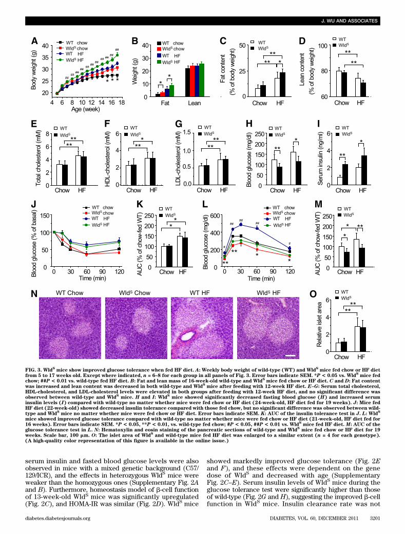
FIG. 3. WldSmice show improved glucose tolerance when fed HF diet. A: Weekly body weight of wild-type (WT) and Wld
Smice fed chow or HF diet
from 5 to 17 weeks old. Except where indicated, n = 6–8 for each group in all panels of Fig. 3. Error bars indicate SEM. *P < 0.05 vs. WldSmice fed
chow; ##P < 0.01 vs. wild-type fed HF diet. B: Fat and lean mass of 16-week-old wild-type and WldSmice fed chow or HF diet. C and D: Fat content
was increased and lean content was decreased in both wild-type and WldSmice after feeding with 12-week HF diet. E–G: Serum total cholesterol,
HDL-cholesterol, and LDL-cholesterol levels were elevated in both groups after feeding with 12-week HF diet, and no significant difference wasobserved between wild-type and Wld
Smice. H and I: Wld
Smice showed significantly decreased fasting blood glucose (H) and increased serum
insulin levels (I) compared with wild-type no matter whether mice were fed chow or HF diet (24-week-old, HF diet fed for 19 weeks). J: Mice fedHF diet (22-week-old) showed decreased insulin tolerance compared with those fed chow, but no significant difference was observed between wild-type and Wld
Smice no matter whether mice were fed chow or HF diet. Error bars indicate SEM. K: AUC of the insulin tolerance test in J. L: Wld
S
mice showed improved glucose tolerance compared with wild-type no matter whether mice were fed chow or HF diet (21-week-old, HF diet fed for16 weeks). Error bars indicate SEM. *P < 0.05, **P < 0.01, vs. wild-type fed chow; #P < 0.05, ##P < 0.01 vs. Wld
Smice fed HF diet. M: AUC of the
glucose tolerance test in L. N: Hematoxylin and eosin staining of the pancreatic sections of wild-type and WldSmice fed chow or HF diet for 19
weeks. Scale bar, 100 mm. O: The islet area of WldSand wild-type mice fed HF diet was enlarged to a similar extent (n = 4 for each genotype).
(A high-quality color representation of this figure is available in the online issue.)
J. WU AND ASSOCIATES
diabetes.diabetesjournals.org DIABETES, VOL. 60, DECEMBER 2011 3201
affected by WldS when monitored by in vivo insulin clear-ance assay and fasting C-peptide-to-insulin molar ratio (Fig.2I and J), which indicated that the elevated serum insulinlevels in WldS mice were not a result of altered insulinclearance. Insulin tolerance test confirmed that WldS andwild-type mice had similar insulin sensitivity as measuredby HOMA-IR (Fig. 2K and L). Insulin-induced phosphory-lation of insulin receptor and AKT in muscle and liver wasalso indistinguishable between WldS and wild-type mice(Fig. 2M-R). In addition, the protein level of SIRT1, an im-portant regulator in hepatic metabolism, and NAD levels inthe liver were similar in wild-type and WldS mice (Supple-mentary Fig. 3A–C). These data show that WldS improvesb-cell function and glucose homeostasis but does not affectinsulin sensitivity.Wld
Smice show improved glucose tolerance when fed
HF diet. The body weight and fat mass were moderatelyincreased in WldS mice compared with wild-type when fedchow, and the differences were more prominent when fedHF diet (Fig. 3A and B). As expected, HF diet caused in-creased fat content and decreased lean content in bothgroups (Fig. 3C and D). Interestingly, when compared withwild-type mice, the fat content was increased in WldS mice fedHF diet (Fig. 3C). Serum total cholesterol, HDL-cholesterol,and LDL-cholesterol levels were elevated in both groupsfed HF diet for 12 weeks, and no differences were observedbetween wild-type and WldS mice (Fig. 3E–G). In WldS
mice fed with either chow or HF diet, fasting blood glucoselevels were decreased, and serum insulin levels but notinsulin sensitivity was remarkably increased, comparedwith corresponding wild-type controls (Fig. 3H–K). Notably,wild-type mice fed HF diet exhibited impaired glucosetolerance, and this was strikingly improved in WldS mice(Fig. 3L and M). The islet area of WldS and wild-typemice fed HF diet was enlarged to a similar extent (Fig.3N and O). These data suggest that WldS alleviates HFdiet-induced glucose intolerance by enhancing insulinproduction.Wld
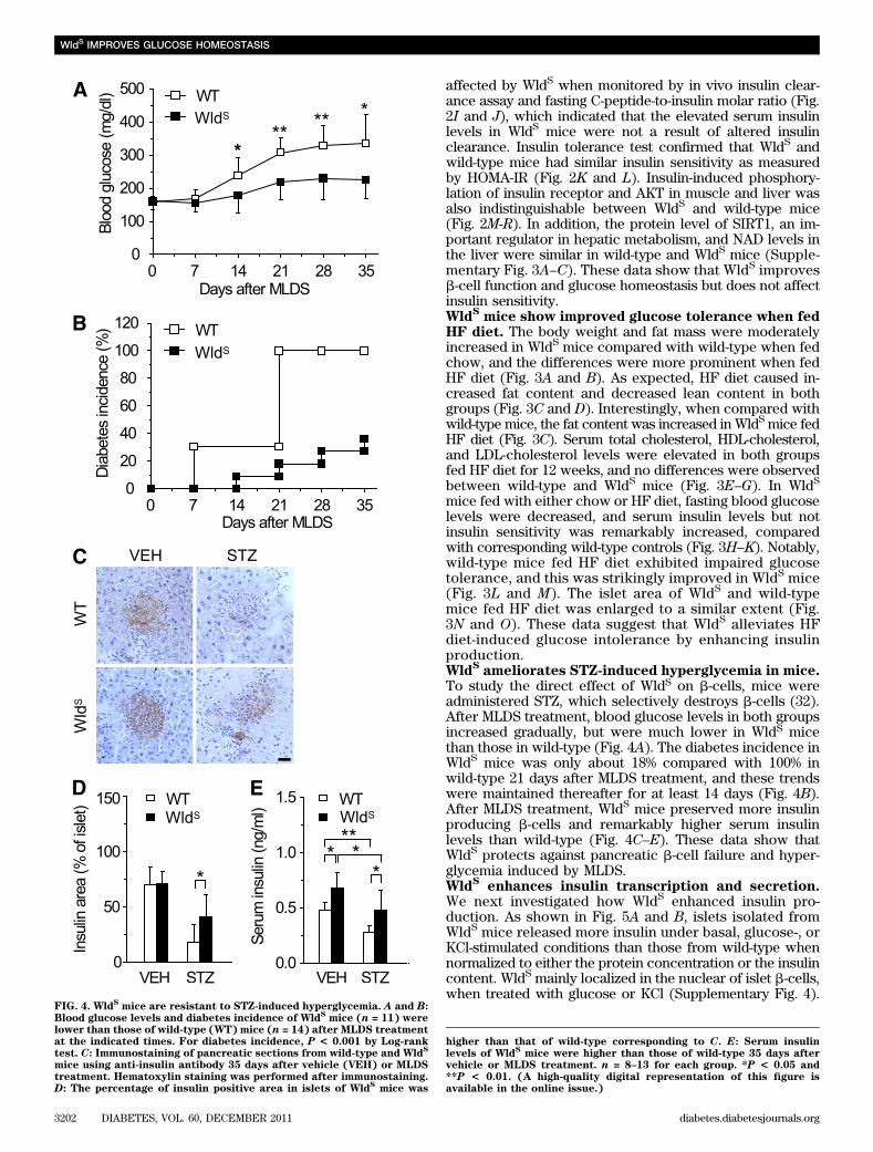
Sameliorates STZ-induced hyperglycemia in mice.
To study the direct effect of WldS on b-cells, mice wereadministered STZ, which selectively destroys b-cells (32).After MLDS treatment, blood glucose levels in both groupsincreased gradually, but were much lower in WldS micethan those in wild-type (Fig. 4A). The diabetes incidence inWldS mice was only about 18% compared with 100% inwild-type 21 days after MLDS treatment, and these trendswere maintained thereafter for at least 14 days (Fig. 4B).After MLDS treatment, WldS mice preserved more insulinproducing b-cells and remarkably higher serum insulinlevels than wild-type (Fig. 4C–E). These data show thatWldS protects against pancreatic b-cell failure and hyper-glycemia induced by MLDS.Wld
Senhances insulin transcription and secretion.
We next investigated how WldS enhanced insulin pro-duction. As shown in Fig. 5A and B, islets isolated fromWldS mice released more insulin under basal, glucose-, orKCl-stimulated conditions than those from wild-type whennormalized to either the protein concentration or the insulincontent. WldS mainly localized in the nuclear of islet b-cells,when treated with glucose or KCl (Supplementary Fig. 4).
FIG. 4. WldSmice are resistant to STZ-induced hyperglycemia. A and B:
Blood glucose levels and diabetes incidence of WldSmice (n = 11) were
lower than those of wild-type (WT) mice (n = 14) after MLDS treatmentat the indicated times. For diabetes incidence, P < 0.001 by Log-ranktest. C: Immunostaining of pancreatic sections from wild-type and Wld
S
mice using anti-insulin antibody 35 days after vehicle (VEH) or MLDStreatment. Hematoxylin staining was performed after immunostaining.D: The percentage of insulin positive area in islets of Wld
Smice was
higher than that of wild-type corresponding to C. E: Serum insulinlevels of Wld
Smice were higher than those of wild-type 35 days after
vehicle or MLDS treatment. n = 8–13 for each group. *P < 0.05 and**P < 0.01. (A high-quality digital representation of this figure isavailable in the online issue.)
WldS IMPROVES GLUCOSE HOMEOSTASIS
3202 DIABETES, VOL. 60, DECEMBER 2011 diabetes.diabetesjournals.org

The effect of WldS on the upregulation of insulin contentin islets was not significant (Fig. 5C), which might bebecause of the very high insulin levels in normal islets.Insulin expression was increased in MIN6 cells stablyexpressing EGFP-fused WldS or WldS with Myc-tag,whereas the increase was attenuated in cells stablyexpressing WldS-H112A or WldS-F116S, in which the NADbiosynthesis activity of WldS was abolished (Fig. 5D-F).The protein levels of WldS and WldS/NMNAT1 protein ratioin the cell lines were even higher than those in the islets of
WldS mice (Supplementary Fig. 5). Moreover, increased NADbiosynthesis activity was observed in the pancreas of WldS
mice (Fig. 5G). In addition, WldS dramatically upregulatedinsulin mRNA levels, which also depended on its enzymeactivity (Fig. 5H and I). Further studies showed WldS acti-vated insulin promoter in a dose-dependent manner, whichalso required its enzyme activity (Fig. 5J and K). NMNAT1,the COOH-terminal of WldS protein, also activated insulinpromoter as WldS, whereas its enzyme-dead mutant NMNAT1-F28S could not (Fig. 5K). These data suggest that the
FIG. 5. WldSenhances insulin transcription and secretion. A and B: Insulin secretion of islets isolated from Wld
Smice was upregulated compared
with wild-type (WT) when treated with the indicated concentration of glucose or KCl (n = 7 for glucose stimulation and n = 4 for KCl stimulation).The protein concentration (A) and the insulin content (B) were measured as internal control, respectively. C: Insulin content in the islets of WldS
mice (n = 4). D: NMNAT enzyme activity in MIN6 cells stably transfected with the indicated plasmids. In this and other panels of this figure, WldS-
H112A stands for EGFP-WldS-H112A. E: Wld
Supregulated insulin expression dependent on its enzyme activity. Insulin expression in MIN6 cells
stably transfected with the indicated plasmids were stained with anti-insulin antibody. In the right panel anti-WldSantibody was added to visualize
the WldSprotein. DAPI was used to stain the nuclei. Scale bar, 5 mm. F: Insulin content in MIN6 cells stably transfected with the indicated
plasmids. G: NMNAT enzyme activity was increased in the pancreas of WldSmice (11-week-old male mice, n = 3 for each genotype). H: Insulin
mRNA levels of islets isolated from WldSmice increased compared with wild-type as determined by real-time PCR (n = 3 for each genotype). I: Wld
S
heightened insulin mRNA levels dependent on its enzyme activity as determined by real-time PCR with MIN6 cells stably transfected withthe indicated plasmids. J and K: Wld
Senhanced insulin promoter activity in a dose-dependent manner (J) and required its enzyme activity (K).
INS-1 cells transfected with pGL3-Insulin-Promoter, and the indicated plasmids were used for luciferase assay to measure insulin promoter ac-tivity. *P < 0.05 and **P < 0.01. (A high-quality digital representation of this figure is available in the online issue.)
J. WU AND ASSOCIATES
diabetes.diabetesjournals.org DIABETES, VOL. 60, DECEMBER 2011 3203

upregulated NMNAT enzyme activity contributes to theenhanced insulin transcription and secretion inducedby WldS.SIRT1 is required for the enhanced transcription,secretion of insulin, and the resistance to STZ-inducedhyperglycemia caused by Wld
S. We next investigated
whether the effect of WldS on insulin production and glu-cose homeostasis depended on SIRT1. Sirtinol, an inhibitorof SIRT1, attenuated WldS-induced activation of insulinpromoter (Fig. 6A). Insulin transcription and secretion inislets and serum insulin levels of SIRT12/2WldS+/+ mice weresignificantly decreased compared with SIRT1+/+ WldS+/+ mice(Fig. 6B–D). The blood glucose levels of SIRT12/2 WldS+/+
mice were similar with SIRT1+/+ WldS+/+ mice no matterwhether mice were fed, fasted, or challenged with glu-cose (Supplementary Fig. 6A–C), which might be as a re-sult of the enhanced insulin sensitivity of SIRT1 null mice(10). The body weight and fat content of SIRT12/2 WldS+/+
mice were decreased compared with SIRT1+/+ WldS+/+
mice (Fig. 6E and F), probably resulting from their in-creased energy expenditure (Supplementary Fig. 6D–H).After MLDS treatment, the blood glucose levels of SIRT12/2
WldS+/+ and SIRT1+/+ WldS2/2 mice increased much fasterthan those of SIRT1+/+ WldS+/+ mice (Fig. 6G). And the di-abetes incidence of SIRT12/2WldS+/+ mice was much higherthan that of SIRT1+/+ WldS+/+ mice (Fig. 6H). Therefore,SIRT1 is necessary for the enhancement of insulin tran-scription, secretion, and the resistance to STZ-inducedhyperglycemia caused by WldS.Wld
Sdownregulates UCP2 expression and upregulates
ATP levels through SIRT1. Next, we explored how WldS
exerts its function through SIRT1. NMNAT1 was reportedto interact with and regulate the activity of SIRT1 inbreast cancer cells (33), suggesting WldS containing thefull-length NMNAT1 has similar effects. As expected, wefound WldS colocalized and coimmunoprecipitated withSIRT1 (Fig. 7A–C). Besides that, both NAD and its pre-cursor NMN were upregulated in the pancreas of WldS mice(Fig. 7D–F), suggesting the activity of SIRT1 was enhanced.SIRT1 has been reported to enhance GSIS by down-regulation of UCP2 and upregulation of ATP (10,11).Similarly, we found WldS downregulated UCP2 promoteractivity like SIRT1 (Fig. 7G) and repressed UCP2 proteinand mRNA levels dependent on its NAD biosynthesisactivity (Fig. 7C and H). The effect of WldS on UCP2 mRNAand protein levels also depended on SIRT1 (Fig. 7I–K).Furthermore, ATP levels were also significantly increased atboth low and high glucose concentrations in islets of WldS
mice compared with wild-type (Fig. 7L), and this functionagain depended on SIRT1 (Fig. 7M).
DISCUSSION
Over the past decade, numerous studies have focused onthe axon protective function of WldS and its potential ap-plication in neuronal diseases (16). The effect of WldS innonneuronal cells would also probably shed light on theunderstanding and treating of other diseases. However,there were few reports concerning this. In this study, wedemonstrate that WldS enhances insulin transcription andsecretion as well as improves glucose homeostasis, andSIRT1 is required in these processes.
It is well established that b-cells show a variety of simi-larities with neuronal cells (34), which implicates that WldS,a protein functional in neuronal cells, may also functionin b-cells. As expected, we found that WldS was highlyexpressed in the pancreas including insulin-producingb-cells (Fig. 1A–C) and enhanced insulin transcription andsecretion (Fig. 5). In addition, WldS mice exhibited in-creased serum insulin levels (Figs. 2A and 3I), even thoughtheir blood glucose levels were normal in the fed state(Fig. 2B). Similarly, SIRT4 knockout mice, for example,also show high insulin and normal fed blood glucose levels(35), which might be as a result of the existence of factorsthat increase blood glucose levels, including glucagon andgluconeogenesis. These factors acted as a counterbalanceto neutralize the glucose-lowering effect of insulin andthus to maintain the blood glucose at a stable level. WldS
mice showed improved glucose tolerance (Fig. 2E). Simi-larly, b-cell–specific SIRT1-overexpressing mice also showimproved glucose tolerance as a result of enhanced GSIS(11). Compatibly, we found that SIRT1 was required forthe function of WldS in insulin secretion (Fig. 6C and D).Increased cellular NAD levels have been shown to enhance
FIG. 6. SIRT1 is required for the enhancement of insulin transcription,secretion, and the resistance to STZ-induced hyperglycemia caused byWld
S. A: Sirtinol attenuated the activation of insulin promoter induced
by WldS. INS-1 cells were transfected with pGL3-Insulin-Promoter and
the indicated plasmids and treated with or without 60 mM Sirtinol for24 h for luciferase assay. B and C: Upregulation of insulin transcriptionand insulin secretion by Wld
Srequired SIRT1. Islets with the indicated
genotypes were used for determination of insulin mRNA levels by real-time PCR (B) and measurement of insulin secretion at the indicatedconcentration of glucose (C; n = 4–8 for each genotype).D: Serum insulinlevels were significantly attenuated in 15-week-old SIRT1
2/2Wld
S+/+mice
compared with SIRT1+/+
WldS+/+
mice (n = 5–9 for each genotype).E and F:Body weight and fat content of 10-week-old SIRT1
2/2Wld
S+/+mice
were decreased compared with SIRT1+/+
WldS+/+
mice (n = 6–11 foreach genotype). G and H: Blood glucose (G) and diabetes incidence(H) of mice with the indicated genotypes after MLDS treatment atthe indicated times (n = 6–9 for each genotype). Error bars indicateSEM. *P< 0.05, **P< 0.01 vs. SIRT1
+/+Wld
S2/2; #P< 0.05, ##P< 0.01 vs.
SIRT1+/+
WldS+/+
. For diabetes incidence, SIRT1+/+
WldS+/+
vs. SIRT1+/+
WldS2/2
, P < 0.05; SIRT1+/+
WldS+/+
vs. SIRT12/2
WldS+/+
, P < 0.05 byLog-rank test. (A high-quality color representation of this figure isavailable in the online issue.)
WldS IMPROVES GLUCOSE HOMEOSTASIS
3204 DIABETES, VOL. 60, DECEMBER 2011 diabetes.diabetesjournals.org

FIG. 7. WldSdownregulates UCP2 expression and upregulates ATP levels through SIRT1. A: Wld
Scolocalized with SIRT1 in MIN6 cells. The stable
cell lines expressing EGFP-WldSwere transiently transfected with pCMV-myc-SIRT1 and stained with anti-Myc antibody and DAPI. Scale bar, 5
mm. B: WldSwas coimmunoprecipitated with SIRT1 from the pancreatic lysates of Wld
Smice. C: Wld
Sand its enzyme-dead mutant Wld
S-H112A
coimmunoprecipitated with SIRT1. The MIN6 cells stably expressing EGFP, EGFP-WldS, or EGFP-Wld
S-H112A were used for immunoprecipitation.
UCP2 protein levels were also detected by Western blot. D and E: Liquid chromatography-tandem mass spectrometry analysis of NMN, NADP,NADPH, NAD, NADH, NA, and NAM extracted from pancreas of 9-week-old wild-type (WT) (D) or Wld
Smice (E). *With significant difference.
F: Quantification of the small molecules corresponding toD and E showed that NAD and NMN levels were upregulated in the pancreas of WldSmice
(n = 4 for each genotype). G: WldSrepressed UCP2 promoter activity like SIRT1. UCP2 promoter activity was measured by luciferase assay in 293T
cells transfected with pGL3-UCP2-Promoter and the indicated plasmids. H: WldSdownregulated UCP2 mRNA levels dependent on its enzyme
activity. UCP2 mRNA level was measured by real-time PCR with MIN6 cell lines stably expressing EGFP, EGFP-WldS, and EGFP-Wld
S-H112A.
I: WldSdownregulated UCP2mRNA levels via SIRT1. UCP2 mRNA levels were determined by real-time PCR using islets isolated from mice with the
indicated genotype (n = 4 for each genotype). J: WldSdownregulated UCP2 protein levels via SIRT1. The protein levels in brown fat tissue with the
indicated genotype were detected with SIRT1, WldS, UCP2, and tubulin antibodies (n = 3 for each genotype). K: Quantification of the UCP2 protein
levels corresponding to J. L: WldSincreased ATP levels in primary cultured islets at the indicated glucose concentration (n = 3).M: Wld
Supregulated
ATP level in islets via SIRT1. ATP levels were measured in islets with indicated genotypes at 2 mmol/L or 20 mmol/L glucose (n = 3). *P < 0.05 and**P < 0.01. (A high-quality digital representation of this figure is available in the online issue.)
J. WU AND ASSOCIATES
diabetes.diabetesjournals.org DIABETES, VOL. 60, DECEMBER 2011 3205
SIRT1 activity (6). Consistently, we found NAD and itsprecursor NMN were upregulated in the pancreas of WldS
mice (Fig. 7D–F), and WldS enhanced insulin transcrip-tion dependent on its NAD biosynthesis activity (Fig. 5). Thesedata suggest that NAD plays a key role in SIRT1-mediatedenhancement of GSIS induced by WldS. It is noteworthythat NMNAT2, an enzyme catalyzing NAD biosynthesis, ishighly expressed in the islets of Langerhans (36), whichalso suggests the importance of NAD biosynthesis in in-sulin secretion. It has been reported that UCP2 knockoutmice show improved GSIS (37), and b-cell–specific SIRT1-overexpressing mice show improved GSIS by decreasingUCP2 expression and elevating ATP levels (11). Analo-gously, we found WldS downregulated UCP2 expressionand upregulated ATP levels via SIRT1 (Fig. 7), whichfurther confirmed that WldS regulates insulin secretionthrough a SIRT1-dependent pathway.
Usually, b-cells will secrete more insulin to overcomethe reduced insulin sensitivity, which is often related withobesity (38). When their compensate mechanisms are im-paired, type 2 diabetes occurs (1). WldS mice show increasedserum insulin levels no matter whether mice were fed chowor HF diet without altering insulin sensitivity (Fig. 3I-K),which indicates enhanced b-cell function in WldS. Further-more, when fed HF diet, the glucose tolerance of WldS micewas strikingly improved (Fig. 3L and M). Similarly, ghrelinknockout mice or GPR40 b-cell–specific transgenic micealso show increased insulin secretary capacity without al-tering insulin sensitivity and improved glucose tolerancewhen fed HF diet (39,40). In addition, WldS promoted in-sulin secretion and downregulated UCP2 via SIRT1 (Figs.6C and 7I–K), which consisted with the studies that bothb-cell–specific SIRT1-overexpressing mice and UCP2 knock-out mice showed enhanced insulin secretion and resistanceto HF diet–induced glucose intolerance (18,41). Takentogether, our findings suggest that enhanced insulin sec-retary capacity by upregulating NAD biosynthesis activityin b-cells would be beneficial to overcome reduced insulinsensitivity induced by HF diet.
Type 1 diabetes is a chronic autoimmune disease, duringwhich b-cells are selectively destroyed (2). MLDS has beenextensively used to generate b-cell destruction to mimictype 1 diabetes (25). In this study, we found that MLDS–induced hyperglycemia was alleviated in WldS mice, whichalso demonstrated increased serum insulin levels (Fig. 4).It is noteworthy that UCP2 knockout mice, which showedenhanced insulin secretory capacity, had accelerated hy-perglycemia after MLDS treatment as a result of strongerinflammation (24). In this scenario, WldS mice were supe-rior to UCP2 knockout mice, probably resulting from somedifferent underlying mechanisms. It has been reportedthat WldS shows protective effects in some neurodegener-ative disease models (12,16). It is likely that ameliorationof MLDS-induced hyperglycemia and attenuation of neu-rodegenerative diseases by WldS could share some com-mon underlying mechanisms. Additionally, the resistanceto MLDS-induced hyperglycemia was abolished in WldS
mice with SIRT1 deficiency (Fig. 6G and H), which isconsistent with the report that intra-arterial targetedislet-specific expression of SIRT1 protects b-cells fromSTZ-induced apoptosis in mice (42). Thereby, the SIRT1-dependent WldS pathway is a potential target not only toenhance insulin secretory capacity, but also to protectagainst b-cell failure.
In this study, our results demonstrate that WldS com-bines an insulinotropic effect with protection against b-cell
failure and suggest that upregulation of the NAD bio-synthesis to increase SIRT1 activity in b-cells will be ben-eficial for diabetes.
ACKNOWLEDGMENTS
This research was supported by grants from the NationalNatural Science Foundation of China (30825009, 30970619,31030022, 81021002, and 30900250), the National Basic Re-search Program of China (973 Program, 2009CB918403and 2007CB914501), the National Science and TechnologySupport Program (2009BAI80B04), the Program of ShanghaiSubject Chief Scientist (11XD1405800), the Director Foun-dation of the Institute for Nutritional Sciences (20090101),SA-SIBS Scholarship Program, the China PostdoctoralScience Foundation (20100480641 and 20080440658), thePostdoctoral Research Program of Shanghai Institutes forBiological Sciences, the Chinese Academy of Sciences(2011KIP511), and the Shanghai Postdoctoral ScientificProgram (11R21417400).
No potential conflicts of interest relevant to this articlewere reported.
J.W. designed the research, performed research, analyzeddata, and wrote the manuscript. F.Z., M.Y., D.W., Q.Y.,Y.Z., and B.Z. performed research. M.W.M. provided theSIRT1+/2 mice and revised the manuscript. Q.Z. designedthe research, analyzed data, and wrote the manuscript.
The authors thank all members of the laboratory forsharing reagents and advice.
REFERENCES
1. Muoio DM, Newgard CB. Mechanisms of disease: molecular and metabolicmechanisms of insulin resistance and beta-cell failure in type 2 diabetes.Nat Rev Mol Cell Biol 2008;9:193–205
2. Lehuen A, Diana J, Zaccone P, Cooke A. Immune cell crosstalk in type 1diabetes. Nat Rev Immunol 2010;10:501–513
3. Kahn SE, Hull RL, Utzschneider KM. Mechanisms linking obesity to insulinresistance and type 2 diabetes. Nature 2006;444:840–846
4. Blander G, Guarente L. The Sir2 family of protein deacetylases. Annu RevBiochem 2004;73:417–435
5. Finkel T, Deng CX, Mostoslavsky R. Recent progress in the biology andphysiology of sirtuins. Nature 2009;460:587–591
6. Haigis MC, Sinclair DA. Mammalian sirtuins: biological insights and dis-ease relevance. Annu Rev Pathol 2010;5:253–295
7. Rodgers JT, Lerin C, Haas W, Gygi SP, Spiegelman BM, Puigserver P.Nutrient control of glucose homeostasis through a complex of PGC-1alphaand SIRT1. Nature 2005;434:113–118
8. Sun C, Zhang F, Ge X, et al. SIRT1 improves insulin sensitivity under insulin-resistant conditions by repressing PTP1B. Cell Metab 2007;6:307–319
9. Kitamura YI, Kitamura T, Kruse JP, et al. FoxO1 protects against pancre-atic beta cell failure through NeuroD and MafA induction. Cell Metab 2005;2:153–163
10. Bordone L, Motta MC, Picard F, et al. Sirt1 regulates insulin secretion byrepressing UCP2 in pancreatic beta cells. PLoS Biol 2006;4:e31
11. Moynihan KA, Grimm AA, Plueger MM, et al. Increased dosage of mam-malian Sir2 in pancreatic beta cells enhances glucose-stimulated insulinsecretion in mice. Cell Metab 2005;2:105–117
12. Coleman M. Axon degeneration mechanisms: commonality amid diversity.Nat Rev Neurosci 2005;6:889–898
13. Coleman MP, Conforti L, Buckmaster EA, et al. An 85-kb tandem tripli-cation in the slow Wallerian degeneration (Wlds) mouse. Proc Natl AcadSci USA 1998;95:9985–9990
14. Conforti L, Tarlton A, Mack TG, et al. A Ufd2/D4Cole1e chimeric proteinand overexpression of Rbp7 in the slow Wallerian degeneration (WldS)mouse. Proc Natl Acad Sci USA 2000;97:11377–11382
15. Mack TG, Reiner M, Beirowski B, et al. Wallerian degeneration of injuredaxons and synapses is delayed by a Ube4b/Nmnat chimeric gene. NatNeurosci 2001;4:1199–1206
16. Coleman MP, Freeman MR. Wallerian degeneration, wld(s), and nmnat.Annu Rev Neurosci 2010;33:245–267
17. Wang J, Zhai Q, Chen Y, et al. A local mechanism mediates NAD-dependentprotection of axon degeneration. J Cell Biol 2005;170:349–355
WldS IMPROVES GLUCOSE HOMEOSTASIS
3206 DIABETES, VOL. 60, DECEMBER 2011 diabetes.diabetesjournals.org
18. Ramsey KM, Mills KF, Satoh A, Imai S. Age-associated loss of Sirt1-mediatedenhancement of glucose-stimulated insulin secretion in beta cell-specificSirt1-overexpressing (BESTO) mice. Aging Cell 2008;7:78–88
19. Revollo JR, Körner A, Mills KF, et al. Nampt/PBEF/Visfatin regulates in-sulin secretion in beta cells as a systemic NAD biosynthetic enzyme. CellMetab 2007;6:363–375
20. McBurney MW, Yang X, Jardine K, et al. The mammalian SIR2alphaprotein has a role in embryogenesis and gametogenesis. Mol Cell Biol2003;23:38–54
21. Mi W, Conforti L, Coleman MP. Genotyping methods to detect a uniqueneuroprotective factor (Wld(s)) for axons. J Neurosci Methods 2002;113:215–218
22. Zehetner J, Danzer C, Collins S, et al. PVHL is a regulator of glucose me-tabolism and insulin secretion in pancreatic beta cells. Genes Dev 2008;22:3135–3146
23. Levy JC, Matthews DR, Hermans MP. Correct homeostasis model assess-ment (HOMA) evaluation uses the computer program. Diabetes Care 1998;21:2191–2192
24. Emre Y, Hurtaud C, Karaca M, Nubel T, Zavala F, Ricquier D. Role ofuncoupling protein UCP2 in cell-mediated immunity: how macrophage-mediated insulitis is accelerated in a model of autoimmune diabetes. ProcNatl Acad Sci USA 2007;104:19085–19090
25. Burkart V, Wang ZQ, Radons J, et al. Mice lacking the poly(ADP-ribose)polymerase gene are resistant to pancreatic beta-cell destruction anddiabetes development induced by streptozocin. Nat Med 1999;5:314–319
26. Jia H, Yan T, Feng Y, Zeng C, Shi X, Zhai Q. Identification of a critical site inWld(s): essential for Nmnat enzyme activity and axon-protective function.Neurosci Lett 2007;413:46–51
27. Jin Q, Zhang F, Yan T, et al. C/EBPalpha regulates SIRT1 expression duringadipogenesis. Cell Res 2010;20:470–479
28. Ge X, Jin Q, Zhang F, Yan T, Zhai Q. PCAF acetylates beta-catenin andimproves its stability. Mol Biol Cell 2009;20:419–427
29. Whelan JA, Russell NB, Whelan MA. A method for the absolute quanti-fication of cDNA using real-time PCR. J Immunol Methods 2003;278:261–269
30. Caruso R, Campolo J, Dellanoce C, Mariele R, Parodi O, Accinni R. Criticalstudy of preanalytical and analytical phases of adenine and pyridine nu-cleotide assay in human whole blood. Anal Biochem 2004;330:43–51
31. Klawitter J, Schmitz V, Klawitter J, Leibfritz D, Christians U. Developmentand validation of an assay for the quantification of 11 nucleotides usingLC/LC-electrospray ionization-MS. Anal Biochem 2007;365:230–239
32. Like AA, Rossini AA. Streptozotocin-induced pancreatic insulitis: newmodel of diabetes mellitus. Science 1976;193:415–417
33. Zhang T, Berrocal JG, Frizzell KM, et al. Enzymes in the NAD+ salvagepathway regulate SIRT1 activity at target gene promoters. J Biol Chem2009;284:20408–20417
34. Corbett J, Serup P, Bonner-Weir S, Nielsen JH. Beta-cell ontogeny: growthand death. Diabetologia 1997;40(Suppl. 3):B27–B32
35. Haigis MC, Mostoslavsky R, Haigis KM, et al. SIRT4 inhibits glutamatedehydrogenase and opposes the effects of calorie restriction in pancreaticbeta cells. Cell 2006;126:941–954
36. Yalowitz JA, Xiao S, Biju MP, et al. Characterization of human brain nic-otinamide 59-mononucleotide adenylyltransferase-2 and expression in hu-man pancreas. Biochem J 2004;377:317–326
37. Zhang CY, Baffy G, Perret P, et al. Uncoupling protein-2 negatively regu-lates insulin secretion and is a major link between obesity, beta cell dys-function, and type 2 diabetes. Cell 2001;105:745–755
38. Biddinger SB, Kahn CR. From mice to men: insights into the insulin re-sistance syndromes. Annu Rev Physiol 2006;68:123–158
39. Dezaki K, Sone H, Koizumi M, et al. Blockade of pancreatic islet-derivedghrelin enhances insulin secretion to prevent high-fat diet-induced glucoseintolerance. Diabetes 2006;55:3486–3493
40. Nagasumi K, Esaki R, Iwachidow K, et al. Overexpression of GPR40 in pan-creatic b-cells augments glucose-stimulated insulin secretion and improvesglucose tolerance in normal and diabetic mice. Diabetes 2009;58:1067–1076
41. Joseph JW, Koshkin V, Zhang CY, et al. Uncoupling protein 2 knockoutmice have enhanced insulin secretory capacity after a high-fat diet. Di-abetes 2002;51:3211–3219
42. Tang MM, Zhu QE, Fan WZ, et al. Intra-arterial targeted islet-specific ex-pression of Sirt1 protects b cells from streptozotocin-induced apoptosis inmice. Mol Ther 2011;19:60–66
J. WU AND ASSOCIATES
diabetes.diabetesjournals.org DIABETES, VOL. 60, DECEMBER 2011 3207