original article effect of octreotide on pancreatic ... · bio, inc, otsu, japan) was used to...
TRANSCRIPT
Int J Clin Exp Pathol 2018;11(10):4784-4794www.ijcep.com /ISSN:1936-2625/IJCEP0079495
Original ArticleEffect of octreotide on pancreatic fibrosis in rats with high-fat diet-induced obesity
Ting Ye1,2, Yan-Hua Chen1,2, Jin-Hang Gao1, Xiao-Xia Wang1,2, Ou Qiang1, Cheng-Wei Tang1,2, Rui Liu1
1Division of Peptides Related with Human Diseases, State Key Laboratory of Biotherapy, West China Hospital, Sichuan University, Chengdu, Sichuan, P. R. China; 2Department of Gastroenterology, West China Hospital, Sichuan University, Chengdu, Sichuan, P. R. China
Received May 11, 2018; Accepted June 22, 2018; Epub October 1, 2018; Published October 15, 2018
Abstract: Background/Aims: To explore the effect of octreotide on pancreatic fibrosis induced by high-fat diet (HFD) and its mechanism of action. Methods: Sprague-Dawley (SD) rats were assigned to control, HFD, or octreotide treat-ment groups. Glucose and insulin tolerance tests (GTT and ITT), fasting plasma glucose (FPG), and fasting insulin (FINS), serum and pancreatic lipid levels, were measured, and the Lee’s index and the homeostatic model assess-ment (HOMA) index were calculated. The expression levels of alpha-smooth muscle actin (α-SMA), desmin, connec-tive tissue growth factor (CTGF), transforming growth factor beta1 (TGF-β1), Smad3, and Smad7 in the pancreas were quantified. The LTC-14 cell line, which has features of primary rat pancreatic stellate cells (PSCs), was used for in vitro studies. Results: The AUC of ipGTT and ipITT, and FPG, FINS, lipid levels, were elevated after HFD feeding; however, they decreased after octreotide administration. The expression of α-SMA, CTGF, TGF-β1, and Smad3 in the HFD group were increased relative to the control group, but Smad7 expression was decreased. After treatment with octreotide, α-SMA, CTGF, TGF-β1, and Smad3 expression decreased, whereas the expression of Smad7 increased. In vitro studies showed that the expression of CTGF, TGF-β1, and Smad3 increased with palmitate treatment (PA), which mimics HFD treatment; and octreotide treatment decreased the expression of these proteins. The α-SMA and Smad7 expression levels remained unchanged among the three groups. Conclusions: Octreotide can ameliorate pancreatic fibrosis and improve pancreatic beta-cell function induced in HFD treated rats, possibly by inhibiting PSC activation and by decreasing pancreatic extracellular matrix (ECM) through the TGF-β1/Smad signaling pathway.
Keywords: Octreotide, high-fat diet, pancreatic stellate cells, pancreatic fibrosis, TGF-β1/Smad signaling pathway
Introduction
Pancreatic fibrosis is a common histopatho- logical characteristic of chronic pancreatitis caused by many varied etiologies [1]. High-fat diet (HFD) can be a cause of inflammatory reac-tions of the pancreas, ultimately leading to pan-creatic fibrosis, which is connected to the acti-vation of pancreatic stellate cells (PSCs) [2-5]. Pancreatic fibrosis damages normal anatomic structures and impairs the exocrine and endo-crine functions of the pancreas [1, 6]. Long-term HFD intake increases the concentration of total triglyceride (TG) in the pancreas. TG is sub-sequently broken down to produce free fatty acids (FFA), which can obstruct the pancreatic capillary network or cause vascular endothelial injury [7, 8]. Currently, therapies for pancreatic fibrosis are limited and their efficacy is poor.
There is therefore an urgent need to study drugs that can alleviate pancreatic tissue fibro-sis [9].
Watari et al [10] observed a type of vitamin A-storing cell located in the rat pancreas through scanning electron microscopy and immunofluorescence analysis in 1982; howev-er, it was not until 1998 that Apte et al [11] first isolated and cultured these vitamin A-storing cells. These cells resemble hepatic stellate cells (HSCs) and were therefore named pancre-atic stellate cells, and they are the major cell type involved in pancreatic fibrosis. The overex-pression of α-smooth muscle actin (SMA) is a marker of activated PSCs [11]. Quiescent pan-creatic stellate cells, which express desmin, represent 4% of the total number of pancreatic cells [12]. In a normal pancreas, only a handful

YE et al: Effect of octreotide on pancreatic fibrosis in HFD rats
4785 Int J Clin Exp Pathol 2018;11(10):4784-4794
of PSCs exist in the activated state for the maintenance of balanced extracellular matrix (ECM) secretion and degradation. When many PSCs are activated, the generation of ECM is upset and excessive amounts of ECM are formed, providing the foundation for develop-ment of pancreatic tissue fibrosis [13].
The transforming growth factor beta (TGF-β) signaling pathway is involved in many cellular processes including cell growth, differentiation, apoptosis, cellular homeostasis and other func-tions [14]. Smads are intracellular proteins that transduce extracellular signals from TGF-β ligands to the nucleus, where they activate downstream gene transcription [15]. There are three classes of Smads: Receptor-regulated Smads (R-Smad), common-mediator Smads (co-Smad), and inhibitory Smads (I-Smad). R-Smads (Smad2, Smad3) are directly phos-phorylated by TGF-β1 receptors, then a co-Smad (Smad4) and Smad3 form a complex that can bind to DNA and modify the expression of several genes related to various cellular activi-ties. I-Smads (Smad6, Smad7) can inhibit the TGF-β1 signaling pathway by blocking the acti-vation of R-Smads and co-Smads [15]. Connective tissue growth factor (CTGF) plays important roles in many biological processes, including cell migration, proliferation, angiogen-esis and tissue wound repair, and is critically involved in fibrotic disease of theliver and pan-creas through its mitogenesis and chemotaxis [16]. Somatostatin is a neuroendocrine hor-mone primarily produced by the hypothalamus, which can suppress the exocrine secretory activity of the pancreas and the release of pan-creatic hormones, and suppress the release of gastrointestinal hormones. Octreotide is an octapeptide that mimics natural somatostatin pharmacologically, though it is a more potent inhibitor of growth hormone, glucagon, and insulin than the natural hormone and has a much longer half-life [17]. Some studies have shown that octreotide can alleviate hepatic fibrosis by inhibiting HSC activation and by inhibiting PSC activation [18, 19].
The effects of octreotide on pancreatic fibrosis in HFD-induced obese rats remain uncertain. In the present study, the authors explored wheth-er octreotide can alleviate pancreatic fibrosis induced by HFD.
Materials and methods
Animals and experimental designs
The Institutional Animal Care and Ethics Com- mittee of Sichuan University (Chengdu, China; approval no. SYXK 2008-119) approved all ani-mal studies. A total of 60 healthy, 21-day-old male Sprague-Dawley rats were purchased from the Animal Center of Sichuan University (Chengdu, China) and were fed at the Animal Center of West China Hospital (Chengdu, China). All rats were housed in individual cages with 12 h light-dark cycles and were given free access to water and chow. Following 7 days of adapted feeding to a standard diet, the rats were divided into a control group (standard diet 320 kcal/100 g, 4.65% calories derived from fat; n=15) and an HFD group (500 kcal/100 g, 60% calories derived from fat; n=45). The body weights and lengths were measured every week for 24 weeks. Lee’s index [body weight (g)1/3 × 1,000/body length (cm)] was calculated to evaluate the degree of obesity. At the 24th
week, 31 eligible obese rats with a body weight at least 1.4 times heavier than the control group were selected from among the HFD rats and were randomly separated into an HFD group (n=15) and an octreotide-treated group (n=16). Both groups of rats continued with an HFD; the octreotide-treated group received a subcutaneous injection of octreotide at a dos-age of 40 μg/kg body weight every 12 h for 8 days.
Rats were fasted for 12 h and anesthetized with 2% sodium pentobarbital administered intraperitoneally at the end of the experiment. The pancreas and abdominal adipose tissue were collected and weighed. Parts of the pan-creatic tissues were put into liquid nitrogen and then stored at -80°C for later use. Remaining pancreatic tissues were fixed in 4% paraformal-dehyde and glutaraldehyde for histopathologi-cal analysis and transmission electron micros-copy (TEM), respectively. Blood samples were centrifuged (100×g, 4°C, 15 min) to separate the plasma/serum then stored at -80°C for later use.
Rat PSC culture and treatment
The immortalized LTC-14 cell line established by Sparmann et al that has been proven to
YE et al: Effect of octreotide on pancreatic fibrosis in HFD rats
4786 Int J Clin Exp Pathol 2018;11(10):4784-4794
maintain essential characteristics of primary PSCs was used for the in vitro studies. The LTC-14 cell line was provided by Professor Robert Jaster from University of Rostock (Rostock, Germany), and cultured in Iscove’s modified Dulbecco’s medium (HyClone; GE Healthcare Life Sciences, Logan, UT, USA) supplemented with 10% fetal bovine serum (Biological Indu- stries, Beit Haemek, Israel), 100 U/ml penicillin and 100 μg/ml streptomycin (HyClone; GE Healthcare Life Sciences). When the cell conflu-ence reached 60%, the cells were starved for 6 h and then divided into three groups with differ-ent treatments: (1) A standard control group (cultured with 0.5% BSA for 24 h); (2) a palmi-tate (PA) treatment group (cultured with 50 μM PA + 0.5% BSA for 24 h); (3) a PA + octreotide treatment group (cultured with 50 μM PA + 0.5% BSA for 24 h and then with 1×10⁻⁹ mmol/l octreotide for 4 h). Radioimmunoprecipitation (Nanjing KeyGen Biotech Co. Ltd, Nanjing, China) assay lysis buffer, mixed with a protease inhibitor and a phosphatase inhibitor, was used to isolate total protein. TRIzol reagent (Takara Bio, Inc, Otsu, Japan) was used to extract total mRNA.
Intraperitoneal glucose tolerance test (ipGTT) and insulin tolerance test (ipITT)
Rats were subjected to the ipGTT and ipITT after fasting for 12 h. For the ipGTT and ipITT assays, rats were given an intraperitoneal injec-tion of glucose at 2.0 g/kg and, separately, of insulin at 7.5 U/kg of body weight. Blood sam-ples were taken from the tail vein at 0, 15, 30, 60 and 120 min after injection. Blood levels were measured with an Accu-Chek active glu-cometer (Roche Diagnostics GmbH, ACCU-CHEK Active, Mannheim, Germany). GraphPad Prism 7 was used to calculate the area under the curve (AUC) of ipGTT and ipITT, and results were expressed as mean ± standard deviation.
FPG, lipids, insulin, FFA and homeostatic mod-el assessment (HOMA) index
The FPG concentration and serum TG and TC levels were determined by enzymatic methods (Changchun Huili Biotech Co. Ltd, Changchun, China). The fasting serum insulin level was measured with an ELISA kit (EZHIASF-14K, EMD Millipore, Billerica, MA, USA). The HOMA index was used to estimate the degree of insu-lin resistance and was calculated as [Fasting
plasma glucose (mmol/l) × fasting serum insu-lin (μU/ml)/22.5]. FFA levels were also mea-sured (Randox, Crumlin, UK).
Pancreas TG and FFA levels
Pancreatic tissue (30 mg per rat) was put in 800 µl of a chloroform/methanol mixed solu-tion (v/v, 2:1), shaken for 20 min, and then cen-trifuged (310×g, 10 min, 4°C). The supernatant was collected and mixed with a 0.9% NaCl solu-tion at ~0.2× the volume of the supernatant. This mixture was centrifuged (550×g, 20 min, 4°C), and the resulting subnatant was collect-ed. The subnatant was dried, and then 0.5 ml of a 2% Triton X-100 solution was used to redis-solve the residue and tissue TG was extracted. The pancreatic TG content was measured by an enzymatic method (Changchun Huili Biotech Co, Ltd). Pancreatic tissue (~50 mg) was put in pH 7.4 PBS to obtain tissue homogenate and then centrifuged (860×g, 20 min, 4°C). The supernatant was collected, and the BCA Protein Assay kit (Pierce; Thermo Fisher Scientific, Inc, Waltham, MA, USA) was used to measure pro-tein concentrations. A FFA Assay kit (Randox, Crumlin, UK) was then used to determine pan-creatic FFA content.
Histopathological and immunohistochemical studies of pancreas
Pancreatic tissues were fixed in formalin, dehy-drated and embedded in paraffin, and then sectioned (thickness of 5 μm) for hematoxylin-eosin and Masson trichrome staining. Glutar- aldehyde-fixed pancreatic specimens were us- ed for TEM analysis (Hitachi Corporation, Tokyo, Japan).
For deparaffinization, sections of pancreatic tissue were subjected to high-pressure antigen retrieval for 10 min in citrate buffer at pH 6.0 and treated with 3% H2O2 for 10 min. After blocking with goat serum, the sections were incubated with anti-desmin (1:100, bs-1026R, BIOSS, Beijing, China) and anti-α-SMA (1:5,000, ab124964, Abcam, Cambridge, UK) antibodies overnight at 4°C. The desmin and α-SMA pro-teins were detected by incubating the sections with biotinylated secondary antibodies (SP-9002, Beijing Zhongshan Golden Bridge Biotechnology, Co, Ltd, Beijing, China) at 37°C for 30 min to form streptavidin-biotin-complex-es. Finally, the immunocomplex was visualized

YE et al: Effect of octreotide on pancreatic fibrosis in HFD rats
4787 Int J Clin Exp Pathol 2018;11(10):4784-4794
using a solution of diaminobenzidine tetrahy-drochloride. All photomicrographs were cap-tured under a microscope (Olympus Corpora- tion, Tokyo, Japan).
RNA isolation and quantitative reverse tran-scription-quantitative polymerase chain reac-tion (RT-qPCR)
An innuPREP RNA Mini kit (Analytik Jena AG, Jena, Germany) was used to extract total RNA from pancreatic tissue. A total of 3 μg RNA was reverse transcribed to cDNA with a Revert Aid First Strand cDNA Synthesis kit (Thermo Fisher Scientific Inc.) following the instructions of the manufacturer. RT-qPCR was performed using 2X SYBR Green Master Mix (Biotool, LLC, Houston, TX, USA). The amplification reactions were run in duplicate and performed at 95°C for 5 min followed by 39 cycles of 95°C for 15 sec and 60°C for 30 sec on a CFX96 PCR Thermocycle Instrument (Bio-Rad Laboratories, Inc, Hercules, CA, USA). The mRNA expression was normalized to β-actin. The relative quanti-tative method was applied to calculate the threshold, and the results were expressed as 2-ΔΔCq [20].
Total protein extraction and western blotting
Total protein was extracted from pancreatic tis-sues or rat PSCs with a KeyGen Whole Cell Assay kit (Nanjing KeyGen Biotech Co. Ltd, Nanjing, China). An enhanced BCA Protein Assay kit was used to measure protein concen-trations. The extracted protein (~40 μg) was electrophoresed on 10% SDS-PAGE and then transferred to polyvinylidene difluoride mem-branes (EMD Millipore). Nonspecific binding sites were blocked with 5% nonfat milk at room temperature for 2 h, and membranes were then incubated with anti-α-SMA rabbit monoclonal antibody (1:20,000, ab124964, Abcam), anti-
Smad3 rabbit monoclonal antibody (1:5,000, ab40854, Abcam), anti-Smad7 rabbit poly-clonal antibody (1:1,000, D160746-0025, BBI Life Sciences, Shanghai, China), anti-TGF-β1 rabbit polyclonal antibody (1:400, D121324-0025, BBI Life Sciences), anti-CTGF rabbit poly-clonal antibody (1:800, D160212-0025, BBI Life Sciences) or anti-β-actin mouse monoclo-nal antibody (1:3,000, mAbcam8226, Epitom- ics, Hangzhou, China) at 4°C overnight. The membranes were washed with 0.1% TBST, then incubated with the appropriate HRP-conjugated secondary antibodies (Beijing Zhongshan Golden Bridge Biotechnology, Co, Ltd, Beijing, China) for 2 h at room temperature. Specific bands were visualized using enhanced chemi-luminescence (Engreen Biosystem, Auckland, New Zealand) detection. The housekeeping protein β-actin was analyzed for normalization. The intensity of the bands was analyzed by Quantity One software (version, 4.6.2; Bio-Rad Laboratories, Inc.).
Statistical analysis
All of the data were analyzed using SPSS soft-ware (version, 20.0; IBM SPSS, Armonk, NY, USA), and the results were expressed as mean ± standard deviation. Comparisons among the three groups were performed by one-way ANOVA, and the Student-Newman-Keuls test was applied to detect significant differences. A test for the homogeneity of variances was also used. P<0.05 was considered a significant difference.
Results
Body weight, Lee’s Index, pancreas weight, abdominal fat and index
The body weight, Lee’s Index, pancreas weight, abdominal fat, and abdominal fat index were calculated at the end of the experiment. The
Table 1. Primers used for qRT-PCRGene Database Forward sequence (5’-3’) Reverse sequence (5’-3’) Product Sizeβ-actin (rat) NM_031144 CGAGTACAACCTTCTTGCAGC CCTTCTGACCCATACCCACC 209 bpTGF-β1 (rat) NM_021578 ATTCCTGGCGTTACCTTGG AGCCCTGTATTCCGTCTCCT 120 bpα-SMA (rat) NM_031004 ACCATCGGGAATGAACGCTT CTGTCAGCAATGCCTGGGTA 199 bpCTGF (rat) NM_022266 CGGGAAATGCTGTGAGGAGT GGCTCGCATCATAGTTGGGT 121 bpSmad3 (rat) NM_013095 AACGGGCAGGAGGAGAAGTG TGGGGATGGTAATGCACTTGG 136 bpSmad7 (rat) NM_030858 TGCAACCCCCATCACCTTAG ATTCGTTCCCCCGGTTTCAT 141 bpThe primers were synthesized by TSINGKE Biological Technology, Ltd. (Chengdu, China).
YE et al: Effect of octreotide on pancreatic fibrosis in HFD rats
4788 Int J Clin Exp Pathol 2018;11(10):4784-4794
Table 3. Data of blood biochemistry, pancreatic TG levels, and HOMA-index in the three groupsControl group (n=15) HFD group (n=15) Octreotide-treated group (n=16)
Plasma glucose (mmol/) 4.60±1.17 6.26±1.55** 4.75±1.60##
Serum insulin (mmol/l) 118.33±37.08 157.68±43.55* 108.85±66.36#
Serum FFA (mmol/l) 225.36±88.20 391.99±105.77** 278.99 ±63.65##
Serum TG (mmol/l) 0.35±0.14 0.70±0.30** 0.49±0.14#
Serum TC (mmol/l) 1.68±0.44 2.54±0.62** 2.31±0.44HOMA-index 23.47±8.86 39.57±13.48** 23.40±16.71##
Pancreas TG (mg/g tissue) 15.40±5.78 31.76±10.53** 23.82±11.99#
Pancreas FFA (mmol/g protein) 104.01±43.20 138.09±62.20* 107.66±37.03#
Values are expressed as mean ± SD, **P<0.01 vs. control group, *P<0.05 vs. control group; ##P<0.01 vs. HFD group, #P<0.05 vs. HFD group.
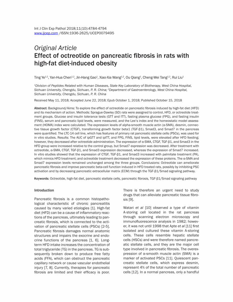
Figure 1. (A and B) Results of ipITT and ipGTT. The glycemic curve in ipITT and ipGTT (C) The AUC of ipITT increased by 32.5% in the HFD group compared to the control group, and decreased by 15.4% after octreotide treatment (C). The AUC of ipGTT in-creased by 28.4% in the HFD group compared to the control group, and decreased by 5% following octreotide treatment (D). Values are expressed as the mean ± stan-dard deviation. *P<0.05 vs. control group. ipITT, insulin tolerance test; ipGTT, intra-peritoneal glucose tolerance test; AUC, area under the curve; HFD, high-fat diet.
(P<0.01). Lee’s Index, an estimate of obesity in adult rats, was also increased by 5.0% in the HFD group when compared with that of the control group (P< 0.01). Abdominal fat in the HFD group was heavier than in the con-trol rats (P<0.01). The abdominal fat index in obese rats was also higher than in the con-trol group (P<0.01). Oct- reotide treatment sig-nificantly reduced these parameters (P<0.01 or P<0.05). Pancreas wei- ght in the HFD group was lighter than in the control group (P<0.01; Table 2).
Plasma glucose, serum lipids, insulin levels, HOMA-index and pan-creas lipids levels
Table 2. Parameters in each groupControl group (n=15) HFD group (n=15) Octreotide-treated group (n=16)
Final body weight (g) 470.07±28.94 600.27±58.38** 512.56±47.90##
Lee’s Index 306.82±4.96 322.30±8.82** 315.32±12.28Pancreas weight (g) 1.39±0.38 0.91±0.23** 0.87±0.25Abdominal fat (g) 10.36±4.72 28.87±8.76** 20.02±4.83##
Abdominal fat Index 2.16±0.88 4.72±1.23** 3.75±0.81#
Values are expressed as mean ± SD, **P<0.01 vs. control group; ##P<0.01 vs. HFD group, #P<0.05 vs. HFD group.
body weight of the HFD group was increased by 27.8% compared with the control group
Compared with the control group, the plasma glucose concentration in obese rats was signifi-
YE et al: Effect of octreotide on pancreatic fibrosis in HFD rats
4789 Int J Clin Exp Pathol 2018;11(10):4784-4794
cantly elevated (P<0.01). Following octreotide administration, plasma glucose levels decre- ased (P<0.01). Obese rats’ serum insulin, FFA, TG, and TC levels were higher than in the con-trol group (P<0.05 or P<0.01). Octreotide inter-vention distinctly decreased these parameters, with the exception of serum TC levels (P>0.05). The HOMA-index, derived from plasma glucose and serum insulin levels, was significantly increased in the HFD group compared with the control group, but was decreased in the octreo-tide-treated group (P<0.01). In HFD-fed rats, the pancreatic TG and FFA levels significantly
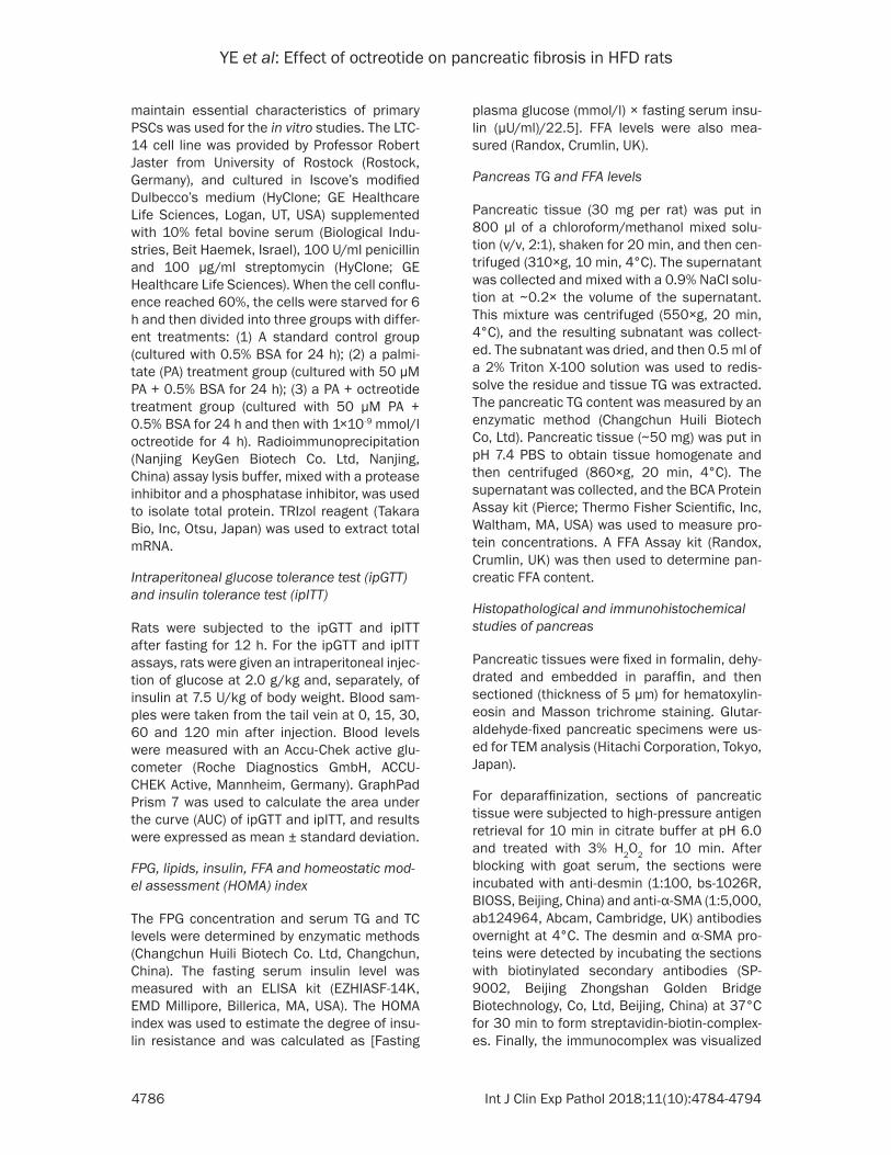
ncreatic calcification was conspicuous. Hema- toxylin and eosin (Figure 2B) and Masson tri-chrome staining (Figure 2C) show histopatho-logical features of the pancreata. In contrast with the control group, the structure of pancre-atic acinar cells was atrophic and accompanied by inflammatory cell infiltration in the HFD group. The pancreatic interlobular fibers in the HFD group were also increased. Octreotide treatment reduced the inflammatory cell infil-tration and alleviated pancreatic tissue fibrosis (Figure 2). Study of pancreatic ultramicrostruc-ture by TEM further showed reduced zymogen
Figure 2. Pancreatic morphology of each group. Pancreatic tissues of the HFD group became harder and smaller, and pancreatic calcification was observed (A, yellow arrow). Hematoxylin and eosin and Masson-Trichrome staining presented pancreatic acinar inflammatory infiltration and atrophy in the HFD group, and pancreatic interlobular fibers also increased in obese rats (B and C, magnifica-tion, ×200). Pancreatic ultramicrostructure was visualized by TEM: red arrow, zymogen granules; white arrow, rough endoplasmic reticula. TEM showed re-duced zymogen granules, dilated rough endoplasmic reticula in the acinar cells in the HFD group, and octreotide treatment increased zymogen granules and the dilation degree of rough endoplasmic reticula was alleviated (D, magnification, ×0.7). HFD, high-fat diet; TEM, transmission electron microscopy.
increased compared with the control group (P<0.05 or P<0.01), and octreotide treatment significantly de- creased their levels (P< 0.05 or P<0.01; Table 3).
Intraperitoneal insulin tolerance test (ipITT) and Intraperitoneal glucose tolerance test (ipGTT)
The blood insulin and glu-cose concentrations of the HFD group at the five time points were higher than in the control group (Figure 1A and 1B). The relative AUC increased by 32.5% or increased by 28.4%, resp- ectively (P<0.05, Figure 1C and 1D). Although the AUC of ipITT and ipGTT in the octreotide-treated gro- up decreased by 15.4% and 5%, respectively, com-pared with the HFD group, these decreases did not meet statistical significan- ce (P>0.05).
Morphology of the pan-creata
It can be seen in Figure 2A that the pancreatic tissues of the HFD group became harder and smaller, pan-creatic weights were light-er than in the control group (P<0.01, Table 2), and pa-

YE et al: Effect of octreotide on pancreatic fibrosis in HFD rats
4790 Int J Clin Exp Pathol 2018;11(10):4784-4794
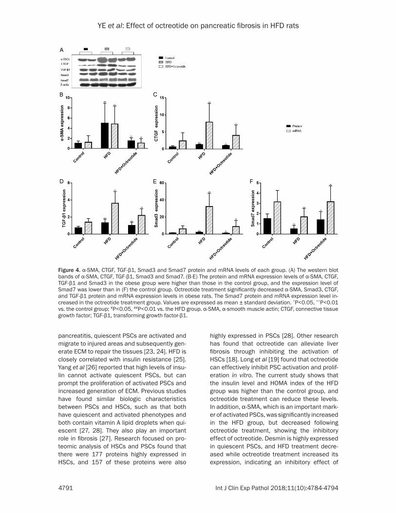
octreotide treatment gro- up (Figure 4, P<0.01).
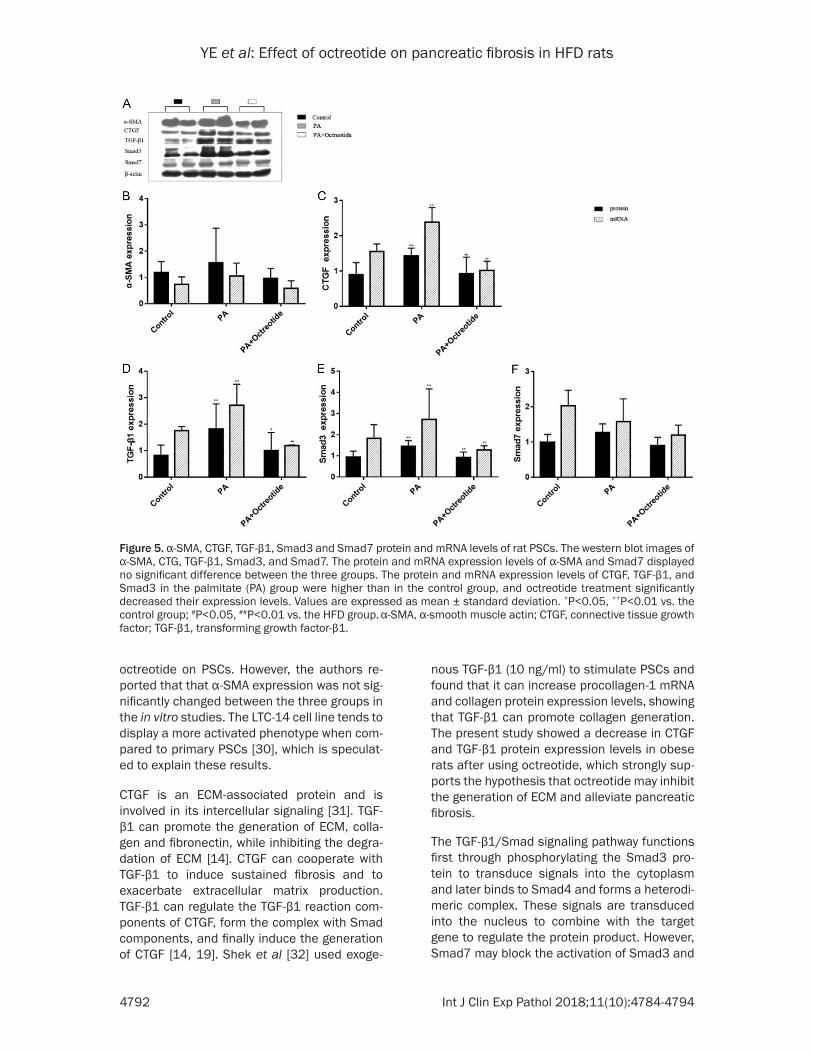
Expression of protein and mRNA for α-SMA, CTGF, TGF-β1, Smad3 and Smad7 of the PSCs
In in vitro studies, the authors found the protein and mRNA expression of α-SMA and Smad7 displ- ayed no significant differ-ences between the three groups (Figure 5). The pro-tein and mRNA expression levels of CTGF, TGF-β1 and Smad3 in the PA group were higher than in the control group (Figure 5, P<0.01 or P<0.05), and
granules, dilated rough endoplasmic reticula in the acinar cells of rats in the HFD group, indi-cating that the secretion function was impaired. In contrast, octreotide treatment increased zymogen granules and the dilation degree of rough endoplasmic reticula was alleviated (Figure 2D).
Immunohistochemical staining for α-SMA and desmin
The increased expression of α-SMA (Figure 3A) and decreased expression of desmin (Figure 3B) were thought to be markers of activated PSCs. Immunohistochemical staining indicated that the expression of α-SMA increased and desmin decreased in the HFD group. The α-SMA protein significantly decreased and the desmin protein increased in response to octreotide treatment.
Expression of pancreatic protein and mRNA for α-SMA, CTGF, TGF-β1, Smad3 and Smad7
The protein and mRNA expression of α-SMA, CTGF, TGF-β1 and Smad3 in the HFD group were higher than in the control group (Figure 4, P<0.01), but the expression level of Smad7 was lower than in the control group (Figure 4F, P<0.01). Octreotide treatment significantly reduced α-SMA, Smad3, CTGF and TGF-β1 pro-tein and mRNA expression in the obese rats (Figure 4, P<0.01 or P< 0.05). The Smad7 pro-tein and mRNA expression increased in the
octreotide treatment significantly decreased their expression (Figure 5, P<0.01 or P<0.05).
Discussion
Long-term HFD-intake leads to energy metabo-lism disequilibrium and increased blood lipid (especially TG) content, which produces an increased burden on pancreatic insulin secre-tion [3]. Excessive TG accumulation in pancre-atic tissue results from the increased serum TG content, and subsequent TG breakdown to FFA are main causes of chronic pancreatic injuries [7, 21]. In the present study, the TG and FFA content increased in HFD rats, subsequently, pancreatic acinar cells were destroyed by inflammatory cell infiltration and pancreatic tis-sue fibrosis formed, indicating the establish-ment of an animal model of chronic pancreati-tis induced by HFD. Interestingly, octreotide can inhibit the activation of PSCs and improve pancreatic endocrine function.
The pancreatic ECM is important for maintain-ing normal pancreatic structure and secretory functions [12]. The generation of ECM primarily occurs by PSCs, and ECM degradation is pri-marily regulated by matrix metalloproteinases and tissue inhibitors of matrix metalloprotein-ases [22]. PSCs are mostly located in the peri-acinar areas and function similarly to fibro-blasts, which can switch between quiescent and activated phenotypes. When stimulated by pathological factors such as acute or chronic
Figure 3. Immunohistochemical staining for α-SMA and desmin. (A) α-SMA and (B) desmin were primarily localized in the periacinar regions of the pancreas. The expression of α-SMA was increased and desmin was decreased in the HFD group. Following octreotide treatment, α-SMA protein significantly decreased and desmin protein increased (magnification, ×400). α-SMA, α-smooth muscle actin; HFD, high-fat diet.
YE et al: Effect of octreotide on pancreatic fibrosis in HFD rats
4791 Int J Clin Exp Pathol 2018;11(10):4784-4794
Figure 4. α-SMA, CTGF, TGF-β1, Smad3 and Smad7 protein and mRNA levels of each group. (A) The western blot bands of α-SMA, CTGF, TGF-β1, Smad3 and Smad7. (B-E) The protein and mRNA expression levels of α-SMA, CTGF, TGF-β1 and Smad3 in the obese group were higher than those in the control group, and the expression level of Smad7 was lower than in (F) the control group. Octreotide treatment significantly decreased α-SMA, Smad3, CTGF, and TGF-β1 protein and mRNA expression levels in obese rats. The Smad7 protein and mRNA expression level in-creased in the octreotide treatment group. Values are expressed as mean ± standard deviation. *P<0.05, **P<0.01 vs. the control group; #P<0.05, ##P<0.01 vs. the HFD group. α-SMA, α-smooth muscle actin; CTGF, connective tissue growth factor; TGF-β1, transforming growth factor-β1.
pancreatitis, quiescent PSCs are activated and migrate to injured areas and subsequently gen-erate ECM to repair the tissues [23, 24]. HFD is closely correlated with insulin resistance [25]. Yang et al [26] reported that high levels of insu-lin cannot activate quiescent PSCs, but can prompt the proliferation of activated PSCs and increased generation of ECM. Previous studies have found similar biologic characteristics between PSCs and HSCs, such as that both have quiescent and activated phenotypes and both contain vitamin A lipid droplets when qui-escent [27, 28]. They also play an important role in fibrosis [27]. Research focused on pro-teomic analysis of HSCs and PSCs found that there were 177 proteins highly expressed in HSCs, and 157 of these proteins were also
highly expressed in PSCs [28]. Other research has found that octreotide can alleviate liver fibrosis through inhibiting the activation of HSCs [18]. Long et al [19] found that octreotide can effectively inhibit PSC activation and prolif-eration in vitro. The current study shows that the insulin level and HOMA index of the HFD group was higher than the control group, and octreotide treatment can reduce these levels. In addition, α-SMA, which is an important mark-er of activated PSCs, was significantly increased in the HFD group, but decreased following octreotide treatment, showing the inhibitory effect of octreotide. Desmin is highly expressed in quiescent PSCs, and HFD treatment decre- ased while octreotide treatment increased its expression, indicating an inhibitory effect of
YE et al: Effect of octreotide on pancreatic fibrosis in HFD rats
4792 Int J Clin Exp Pathol 2018;11(10):4784-4794
octreotide on PSCs. However, the authors re- ported that that α-SMA expression was not sig-nificantly changed between the three groups in the in vitro studies. The LTC-14 cell line tends to display a more activated phenotype when com-pared to primary PSCs [30], which is speculat-ed to explain these results.
CTGF is an ECM-associated protein and is involved in its intercellular signaling [31]. TGF-β1 can promote the generation of ECM, colla-gen and fibronectin, while inhibiting the degra-dation of ECM [14]. CTGF can cooperate with TGF-β1 to induce sustained fibrosis and to exacerbate extracellular matrix production. TGF-β1 can regulate the TGF-β1 reaction com-ponents of CTGF, form the complex with Smad components, and finally induce the generation of CTGF [14, 19]. Shek et al [32] used exoge-
nous TGF-β1 (10 ng/ml) to stimulate PSCs and found that it can increase procollagen-1 mRNA and collagen protein expression levels, showing that TGF-β1 can promote collagen generation. The present study showed a decrease in CTGF and TGF-β1 protein expression levels in obese rats after using octreotide, which strongly sup-ports the hypothesis that octreotide may inhibit the generation of ECM and alleviate pancreatic fibrosis.
The TGF-β1/Smad signaling pathway functions first through phosphorylating the Smad3 pro-tein to transduce signals into the cytoplasm and later binds to Smad4 and forms a heterodi-meric complex. These signals are transduced into the nucleus to combine with the target gene to regulate the protein product. However, Smad7 may block the activation of Smad3 and
Figure 5. α-SMA, CTGF, TGF-β1, Smad3 and Smad7 protein and mRNA levels of rat PSCs. The western blot images of α-SMA, CTG, TGF-β1, Smad3, and Smad7. The protein and mRNA expression levels of α-SMA and Smad7 displayed no significant difference between the three groups. The protein and mRNA expression levels of CTGF, TGF-β1, and Smad3 in the palmitate (PA) group were higher than in the control group, and octreotide treatment significantly decreased their expression levels. Values are expressed as mean ± standard deviation. *P<0.05, **P<0.01 vs. the control group; #P<0.05, ##P<0.01 vs. the HFD group. α-SMA, α-smooth muscle actin; CTGF, connective tissue growth factor; TGF-β1, transforming growth factor-β1.
YE et al: Effect of octreotide on pancreatic fibrosis in HFD rats
4793 Int J Clin Exp Pathol 2018;11(10):4784-4794
the complex formation with Smad4 [14, 15]. The in vivo studies showed that the HFD group’s expression of Smad3 increased and Smad7 decreased compared with the control group. In contrast, following treatment with octreotide, Smad3 decreased and Smad7 reached a nor-mal level. Consistent with these results, Qian et al [33] used exogenous TGF-β1 to stimulate rat primary PSCs and found Smad3 expression lev-els increased along with the concentration of TGF-β1, but Smad7 expression levels were reduced. However, the expression levels of Smad7 remained unchanged among the three groups in the in vitro studies, possibly because the concentration of PA, which mimics HFD to interfere with PSCs, could not reach an effec-tive level to activate the Smad7 protein. Unfortunately, the lipotoxicity of PA increased with the increasing concentration required for effective interference, resulting in the death of PSCs.
As a somatostatin analogue, octreotide is com-monly used to prevent complications following pancreatic surgeries and esophageal variceal hemorrhage in clinical settings [34, 35]. Long et al [19] found that octreotide can also allevi-ate inflammatory infiltration, and slowed the occurrence of insulitis and vasculitis after rat pancreas transplantation. The study found that an HFD can destroy pancreatic acinar cells, causing inflammatory cell infiltration and result-ing in small and tough pancreata. Octreotide reduced pancreatic inflammatory cell infiltra- tion.
In summary, the current study indicated that octreotide can ameliorate pancreatic fibrosis and improve pancreatic beta-cell function in HFD-induced obese rats, possibly by inhibiting PSC activation and decreasing pancreatic extra- cellular matrix through TGF-β1/Smad signal- ing.
Acknowledgements
The project was supported by the National Natural Science Foundation of China (grant no. 30870919).
Disclosure of conflict of interest
None.
Address correspondence to: Dr. Rui Liu, Division of Peptides Related with Human Diseases, State Key
Laboratory of Biotherapy, West China Hospital, Sichuan University, Chengdu 610041, Sichuan, P. R. China. E-mail: [email protected]
References
[1] Majumder S, Chari ST. Chronic pancreatitis. Lancet 2016; 387: 1957-66.
[2] Matsuda A, Makino N, Tozawa T, Shirahata N, Honda T, Ikeda Y, Sato H, Ito M, Kakizaki Y, Akamatsu M, Ueno Y, Kawata S. Pancreatic fat accumulation, fibrosis, and acinar cell injury in the zucker diabetic fatty rat fed a chronic high-fat diet. Pancreas 2014; 43: 735-43.
[3] Castineira-Alvarino M, Lindkvist B, Luaces-Regueira M, Iglesias-García J, Lariño-Noia J, Nieto-García L, Domínguez-Muñoz JE. The role of high fat diet in the development of complica-tions of chronic pancreatitis. Clin Nutr 2013; 32: 830-6.
[4] Lee Y, Lingvay I, Szczepaniak LS, Ravazzola M, Orci L, Unger RH. Pancreatic steatosis: harbin-ger of type 2 diabetes in obese rodents. Int J Obes (Lond) 2010; 34: 396-400.
[5] Yan MX, Li YQ, Meng M, Kou Y. Long-term high-fat diet induces pancreatic injuries via pancre-atic microcirculatory disturbances and oxida-tive stress in rats with hyperlipidemia. Biochem Biophys Res Commun 2006; 347: 192-9.
[6] Enrique Dominguez-Munoz J. Latest advances in chronic pancreatitis. Gastroenterol Hepatol 2016; 39 Suppl 1: 87-92.
[7] Mathur A, Marine M, Lu D, Swartz-Basile DA, Saxena R, Zyromski NJ, Pitt HA. Nonalcoholic fatty pancreas disease. HPB (Oxford) 2007; 9: 312-8.
[8] Miller JP. Serum triglycerides, the liver and the pancreas. Curr Opin Lipidol 2000; 11: 377-82.
[9] Shimizu K. Mechanisms of pancreatic fibrosis and applications to the treatment of chronic pancreatitis. J Gastroenterol 2008; 43: 823-32.
[10] Watari N, Hotta Y, Mabuchi Y. Morphological studies on a vitamin A-storing cell and its com-plex with macrophage observed in mouse pan-creatic tissues following excess vitamin A ad-ministration. Okajimas Folia Anat Jpn 1982; 58: 837-58.
[11] Apte MV, Haber PS, Applegate TL, Norton ID, McCaughan GW, Korsten MA, Pirola RC, Wilson JS. Periacinar stellate shaped cells in rat pan-creas: identification, isolation, and culture. Gut 1998; 43: 128-33.
[12] Omary MB, Lugea A, Lowe AW, Pandol SJ. The pancreatic stellate cell: a star on the rise in pancreatic diseases. J Clin Invest 2007; 117: 50-9.
[13] Erkan M, Adler G, Apte MV, Bachem MG, Buchholz M, Detlefsen S, Esposito I, Friess H,

YE et al: Effect of octreotide on pancreatic fibrosis in HFD rats
4794 Int J Clin Exp Pathol 2018;11(10):4784-4794
Gress TM, Habisch HJ, Hwang RF, Jaster R, Kleeff J, Klöppel G, Kordes C, Logsdon CD, Masamune A, Michalski CW, Oh J, Phillips PA, Pinzani M, Reiser-Erkan C, Tsukamoto H, Wilson J. StellaTUM: current consensus and discussion on pancreatic stellate cell research. Gut 2012; 61: 172-8.
[14] Wrana JL, Cárcamo J, Attisano L, Zentella A, Doody J, Laiho M, Wang XF, Massagué J. TGFβ signals through a heteromeric protein kinase receptor complex. Cell 1992; 71: 1003-14.
[15] Moustakas A. Smad signalling network. J Cell Sci 2002; 115: 3355-6.
[16] Charrier A, Brigstock DR. Regulation of pancre-atic function by connective tissue growth factor (CTGF, CCN2). Cytokine Growth Factor Rev 2013; 24: 59-68.
[17] Sun L, Coy DH. Somatostatin and its analogs. Curr Drug Targets 2016; 17: 529-37.
[18] Klironomos S, Notas G, Sfakianaki O, Kiagia- daki F, Xidakis C, Kouroumalis E. Octreotide modulates the effects on fibrosis of TNF-alpha, TGF-beta and PDGF in activated rat hepatic stellate cells. Regul Pept 2014; 188: 5-12.
[19] Long D, Lu J, Luo L, Guo Y, Li C, Wu W, Shan J, Li L, Li S, Li Y, Lin T, Feng L. Effects of octreo-tide on activated pancreatic stellate cell-in-duced pancreas graft fibrosis in rats. J Surg Res 2012; 176: 248-259.
[20] Livak KJ, Schmittgen TD. Analysis of relative gene expression data using real-time quantita-tive PCR and the 2(-Delta Delta C(T)) method. Methods 2001; 25: 402-8.
[21] Mews P, Phillips P, Fahmy R, Korsten M, Pirola R, Wilson J, Apte M. Pancreatic stellate cells respond to inflammatory cytokines: potential role in chronic pancreatitis. Gut 2002; 50: 535-41.
[22] Michel G, Tonon T, Scornet D, Cock JM, Kloareg B. The cell wall polysaccharide metabolism of the brown alga Ectocarpus siliculosus. Insights into the evolution of extracellular matrix poly-saccharides in Eukaryotes. New Phytol 2010; 188: 82-97.
[23] Jaster R. Molecular regulation of pancreatic stellate cell function. Mol Cancer 2004; 3: 26.
[24] Shimizu K. Pancreatic stellate cells: molecular mechanism of pancreatic fibrosis. J Gastro- enterol Hepatol 2008; 23 Suppl 1: S119-121.
[25] Paniagua JA. Nutrition, insulin resistance and dysfunctional adipose tissue determine the different components of metabolic syndrome. World J Diabetes 2016; 7: 483-514.
[26] Yang J, Waldron RT, Su HY, Moro A, Chang HH, Eibl G, Ferreri K, Kandeel FR, Lugea A, Li L, Pandol SJ. Insulin promotes proliferation and fibrosing responses in activated pancreatic stellate cells. Am J Physiol Gastrointest Liver Physiol 2016; 311: G675-G687.
[27] Kordes C, Sawitza I, Haussinger D. Hepatic and pancreatic stellate cells in focus. Biol Chem 2009; 390: 1003-12.
[28] Wehr AY, Furth EE, Sangar V, Blair IA, Yu KH. Analysis of the human pancreatic stellate cell secreted proteome. Pancreas 2011; 40: 557-66.
[29] Paulo JA, Kadiyala V, Banks PA, Conwell DL, Steen H. Mass spectrometry-based quantita-tive proteomic profiling of human pancreatic and hepatic stellate cell lines. Genomics Proteomics Bioinformatics 2013; 11: 105-13.
[30] Sparmann G, Hohenadl C, Tornoe J, Jaster R, Fitzner B, Koczan D, Thiesen HJ, Glass A, Winder D, Liebe S, Emmrich J. Generation and characterization of immortalized rat pancreat-ic stellate cells. Am J Physiol Gastrointest Liver Physiol 2004; 287: G211-9.
[31] Jun JI, Lau LF. Taking aim at the extracellular matrix: CCN proteins as emerging therapeutic targets. Nat Rev Drug Discov 2011; 10: 945-63.
[32] Shek FW, Benyon RC, Walker FM, McCrudden PR, Pender SL, Williams EJ, Johnson PA, Johnson CD, Bateman AC, Fine DR, Iredale JP. Expression of transforming growth factor-beta 1 by pancreatic stellate cells and its implica-tions for matrix secretion and turnover in chronic pancreatitis. Am J Pathol 2002; 160: 1787-98.
[33] Qian ZY, Peng Q, Zhang ZW, Zhou LA, Miao Y. Roles of Smad3 and Smad7 in rat pancreatic stellate cells activated by transforming growth factor-beta 1. Hepatobiliary Pancreat Dis Int 2010; 9: 531-36.
[34] Gotzsche PC, Hrobjartsson A. Somatostatin analogues for acute bleeding oesophageal varices. Cochrane Database Syst Rev 2008: Cd000193.
[35] Sliwinska-Mosson M, Vesely M, Milnerowicz H. The clinical significance of somatostatin in pancreatic diseases. Ann Endocrinol (Paris) 2014; 75: 232-40.