original article biomarkers and long-term prognosis of ... · biomarkers and long-term prognosis of...
TRANSCRIPT

Int J Clin Exp Pathol 2016;9(2):1005-1013www.ijcep.com /ISSN:1936-2625/IJCEP0019586
Original ArticleBiomarkers and long-term prognosis of colorectal schistosomiasis-associated rectosigmoid cancer: a retrospective study
Wen-Peng Zhang1, Ming-Liang Wang1,3, Ai-Guo Lu3, Jing-Kun Zhao2,3, Shuai Yin2, Rauter Beatrice2, Tobias S Schiergens2, Hao Feng2,3
1Ruijin Hospital, LuWan Branch School of Medicine, Shanghai Jiao Tong University, Shanghai, China; 2Department of General-, Visceral-, Transplantation-, Vascular and Thoracic Surgery, Hospital of University of Munich (LMU), Munich, Germany; 3Ruijin Hospital, School of Medicine, Shanghai Jiao Tong University, Shanghai, China
Received November 11, 2015; Accepted January 12, 2016; Epub February 1, 2016; Published February 15, 2016
Abstract: Aim: This study is to assess the association between the potential long-term effects of previous schis-tosoma infection and rectosigmoid cancer. Methods: From November 2006 to May 2011, 25patients who had rectosigmoid carcinoma combined with colorectal schistosomiasis (SSC), 217patients with non-schistosomiasis rec-tosigmoid carcinoma (NSC), 19 colorectal schistosomiasis (CS) were included. Tumor characteristics, pre-operative investigations, tumor pathological examinations and postoperative courses were evaluated. Results: There was no significant difference in overall survival rate between the SSC and NSC groups. Levels of CEA, AFP and CA19-9 did not differ among three groups; CA19-9 values were significantly larger in SSC group than those in NSC group. Levels of CA-125 were significantly higher (P<0.0001) in CS group compared to the other two groups, but no significant dif-ference could be observed between NSC and SSC groups. Concerning the biopsy staining, CA125 was upregulated in CS and SSC group, significantly stronger staining was observed. Conclusions: For those who travelled from or lived in endemic areas, elevated serum CA19-9 and CA-125 may be signals for those who have colorectal lesions and infested water contact history to go through the circumival precipintin test (COPT), for rectosigmoid cancer patients whose biopsy is CA-125 positive, rectosigmoid carcinoma combined with colorectal schistosomiasis (SSC) should be considered. However, schistosomiasis was not statistically significantly correlated with overall survival in patients suffering rectosigmoid cancer.
Keywords: Schistosomiasis, colorectal carcinoma, biomarker, prognosis, diagnosis
Introduction
Human schistosomiasis was first described by German tropical disease specialist Theodor Bilharz in 1851 [1]. Schistosomiasis could cause acute and long-term clinical syndromes [2], including permanent scarring of the blad-der [3, 4], liver, urogenital system, small bowel [5, 6] and colon [7-9].
Regarding the colonic schistosomiasis, the adult worms of the main intestinal species, Schistosoma mansoni and Schistosoma japon-icum, lay their eggs in the colonic mesenteric vessels. Then these ova penetrate the intesti-nal wall and are shed in the faeces (Figure 1). All segments of the colon may be affected, yet
the rectum, sigmoid, descending colon and the domain of the inferior mesenteric vein, are the main sites of pathology. In chronic disease, the schistosoma ova are calcified and deposited with infiltration of lymphocytes and plasma cells in the submucosa and lamina propria, with giant cell reaction.
It is important to acknowledge that, because of the climate warning [8], schistosomiasis is now also becoming a cause for concern in Europe, especially in south Europe. Schistosomiasis infection in returning travelers is also one of the most common reasons. At present, due to the fact that clinical symptoms [9] and laboratory tests are nonspecific [10, 11] as well as the geographic distribution of the disease, colorec-

Colorectal schistosomiasis with CRC
1006 Int J Clin Exp Pathol 2016;9(2):1005-1013
tal changes as a result of schistosomiasis are generally unsure to clinicians from non-endem-ic areas.
Asian and African reports have proposed the biomarker and a causal relationship between chronic inflammatory changes of colonic muco-sa and colorectal carcinogenesis [12, 13]. It was reported that intestinal schistosomiasis should be considered as a precancerous condi-tion for development of colonic dysplasia and cancer as a consequence of chronic inflamma-tion that altered inflammatory, antioxidant and fulcose status [14]. This study conducted a ret-rospective study, comparing the expression of antioxidant associated biomarkers, endoscopic findings, laboratory tests and prognosis in schistosomal and non-schistosomal rectosig-moid carcinoma.
Patients and methods
Histopathology and diagnoses
For histological examinations, colon tissue samples were fixed in 4% paraformaldehyde, and sections were stained with hematoxylin and eosin (H&E) by the Ruijin Hospital, Department of Pathology. The pathological TNM stages were determined according to the classification established by the American Joint Committee on Cancer. 25 patients were post-operatively histologically diagnosed with recto-sigmoid carcinoma combined with colorectal schistosomiasis (SSC). 217 patients who were diagnosed with rectosigmoid carcinoma only (without colorectal schistosomiasis) were selected as a control group (NSC). Retrospective analysis was also conducted in 19 consecutive
Figure 1. Schematic diagram showed adult schistosoma worms lay their eggs in the colonic mesenteric vessels, notably the inferior mesenteric vein. Then these ova penetrate the intestinal wall and are shed in the faeces. All segments of the colon may be affected, especially the rectum, sigmoid and descending colon. In acute disease, the ova are deposited in the lamina propria. In chronic disease, the schistosoma ova are calcified and deposited with infiltration of lymphocytes and plasma cells in the submucosa and lamina propria. Chronic inflammatory changes, antioxidant and fulcose status make it a precancerous condition for development of colorectal cancer. SMV, supe-rior mesenteric vein; IMV, inferior mesenteric vein; SV, splenic vein; PV, hepatic portal vein.
Colorectal schistosomiasis with CRC
1007 Int J Clin Exp Pathol 2016;9(2):1005-1013
cases in the same period which were histologi-cally diagnosed with colorectal schistosomiasis alone (CS).
Immunohistochemistry (IHC) procedure
IHC was performed on tissues from 83 patients (CS group: 15 cases, SSC group: 12 patients, NSC group: 56 patients). The procedure of IHC was conducted according to the manufacturer instructions (Vectastain Universal anti-mouse IgG/rabbit IgG elite ABC kit, Vector Laboratories, Inc. CA, USA). After baking slides in oven at 65°C overnight, slides were deparaffinized by applying sequential immersion for 5 min in xylene, 95% ethanol, 70% ethanol, and distilled water. Immunohistochemistry was performed on de-paraffinized formalin-fixed sections after antigen retrieval with boiling citrate buffer. IHC staining was conducted using CEA (Anti-Carcino Embryonic Antigen CEA antibody, ab4451, Abcam, England), AFP (Anti-alpha 1 Fetopro- tein antibody, ab46799, Abcam), GFER (growth factor, also known as augmenter of liver regen-eration) rabbit polyclonal antibody (1:600, ab36376, Abcam, England), CA19-9 (Anti-CA19-9 antibody, ab15146, Abcam), CA125 (Anti-MUC16 antibody, ab10033) and Bcl-xL rabbit monoclonal antibody (54H6, #2764, 1:300, Cell Signaling Technology, US). The pri-mary antibodies were detected utilizing a bioti-nylated goat anti-rabbit/mouse antibody and Vectastain Elite ABC kit and DAB substrate
um concentration (CEA), Cancer Antigen 125 (CA-125), Cancer Antigen 19-9 (CA19-9), alpha-fetoprotein (AFP), total colonoscopy, transanal ultrasonography, abdominal magnetic reso-nance imaging (MRI) and thorax and abdominal computed tomography (CT). Between November 2006 and May 2011, all these patients above who underwent laparoscopic resection in the Shanghai Minimally Invasive Surgical Center at Ruijin Hospital, affiliated to Shanghai Jiao-Tong University were prospectively enrolled. Data collection included patient and tumor charac-teristics, pre-operative investigations, tumor pathological examination and postoperative course. Clinical laboratory tests including CEA, CA-125, CA19-9, AFP, hemoglobin collected before treatment (pre-Hb) and pre-treatment total bilirubin (pre-TBil) were performed on admission. The blood samples were collected and analyzed by experienced technicians and doctors from Laboratory Medicine Department.
Ethics statement
Protocol approval for all researches performed was obtained from the medical ethical commit-tee of Shanghai Ruijin Hospital according to the Helsinki Declaration.
Long-term follow-up
Patients who were diagnosed with carcinoma were followed up every 6-month. The examina-
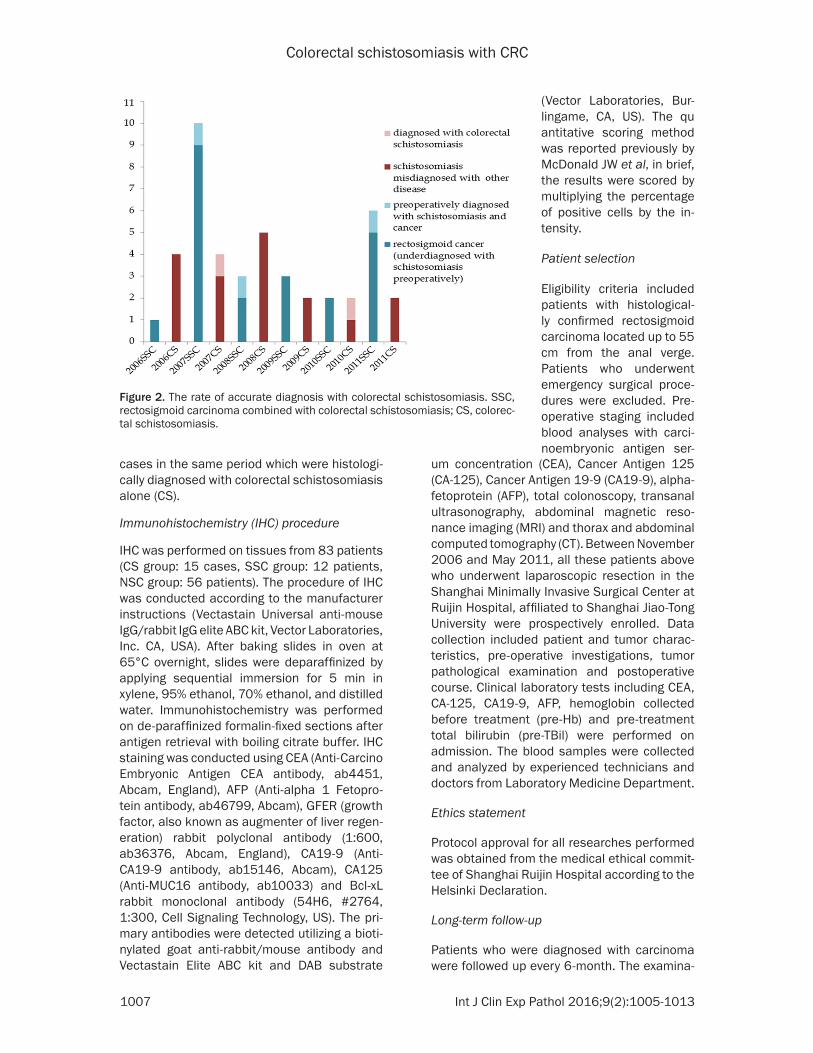
Figure 2. The rate of accurate diagnosis with colorectal schistosomiasis. SSC, rectosigmoid carcinoma combined with colorectal schistosomiasis; CS, colorec-tal schistosomiasis.
(Vector Laboratories, Bur- lingame, CA, US). The qu antitative scoring method was reported previously by McDonald JW et al, in brief, the results were scored by multiplying the percentage of positive cells by the in- tensity.
Patient selection
Eligibility criteria included patients with histological- ly confirmed rectosigmoid carcinoma located up to 55 cm from the anal verge. Patients who underwent emergency surgical proce-dures were excluded. Pre- operative staging included blood analyses with carci-noembryonic antigen ser-

Colorectal schistosomiasis with CRC
1008 Int J Clin Exp Pathol 2016;9(2):1005-1013
tions included colonoscopic examinations, abdominal ultrasound, CT of abdominal and pelvic parts and laboratory examinations (CEA, CA19-9 and CA-125). Overall survival was cal-culated from surgery to death induced by all causes or end of follow up.
Statistical analysis
All measurements were expressed as means ± SD (the bar of Figure 3 was expressed as medi-ans), whereas count data was expressed as numbers (proportions). Differences in propor-tions were tested for statistical significance using the χ2 test, and a Student’s t-test was used to compare the means between groups. Categorical variables were compared using the Fisher’s exact test. Continuous variables were presented as mean ± SD and compared using the Mann-Whitney U test. Kaplan Meier was performed to draw the survival curve. All statis-tical analyses were performed with the sta-
group. In these 25 patients who were diag-nosed with sigmoid or rectal carcinoma com-bined with rectosigmoid schistosomiasis (SSC) by postoperative pathology, 21 were preopera-tive diagnosed with rectosigmoid carcinoma alone, only 3 patients got the accurate diagno-ses (Figure 2).
Comparison of serum biomarkers in rectosig-moid carcinoma complicated with schistoso-miasis
Levels of CEA, AFP and CA19-9 did not differ among three groups (CEA: P=0.4833, AFP: P=0.1108, CA19-9: P=0.2719). In contrast, lev-els of CA-125 were significantly higher (P<0.0001) in CS group, however, no statistical difference was found between SSC group and NSC group. When comparing the pre-Hb and pre-TBil, we found that patients in CS group pro-duced significantly lower Hb levels (P<0.0001), they also produced higher TBil levels, although
Figure 3. Laboratory examinations among three groups of patients. SSC, rec-tosigmoid carcinoma combined with colorectal schistosomiasis; CS, colorectal schistosomiasis. NSC, rectosigmoid carcinoma (without colorectal schistoso-miasis).
tistical software Stat View 5.0 for Windows (SAS Institute Inc., Cary, NC, USA), and a P value <0.05 was considered statistical-ly significant.
Results
Colorectal schistosomiasis was easily underdiagnosed in patients with or without rectosigmoid carcinoma
In these 19 patients who had colorectal schistoso-miasis (CS), 5 patients were underdiagnosed with ulcerative colitis, 3 patients were diagnosed with Crohn disease, 1 was diagnosed with intestinal tuberculosis, 6 patients were diagnosed with colonic polyps, 2 patients were diagnosed with rectosigmoid carcino-ma. Only 2 patients (11%) were diagnosed with colo- rectal schistosomiasis be- fore histologically confirma-tion, high-grade intraepi-thelial neoplasia was con-firmed in 3 patients in this
Colorectal schistosomiasis with CRC
1009 Int J Clin Exp Pathol 2016;9(2):1005-1013
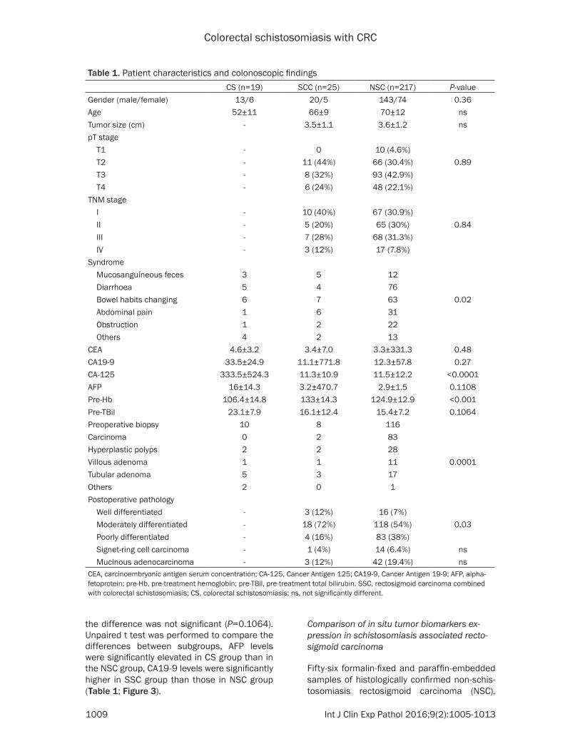
Table 1. Patient characteristics and colonoscopic findingsCS (n=19) SCC (n=25) NSC (n=217) P-value
Gender (male/female) 13/6 20/5 143/74 0.36Age 52±11 66±9 70±12 nsTumor size (cm) - 3.5±1.1 3.6±1.2 nspT stage T1 - 0 10 (4.6%) T2 - 11 (44%) 66 (30.4%) 0.89 T3 - 8 (32%) 93 (42.9%) T4 - 6 (24%) 48 (22.1%)TNM stage I - 10 (40%) 67 (30.9%) II - 5 (20%) 65 (30%) 0.84 III - 7 (28%) 68 (31.3%) IV - 3 (12%) 17 (7.8%)Syndrome Mucosanguineous feces 3 5 12 Diarrhoea 5 4 76 Bowel habits changing 6 7 63 0.02 Abdominal pain 1 6 31 Obstruction 1 2 22 Others 4 2 13CEA 4.6±3.2 3.4±7.0 3.3±331.3 0.48CA19-9 33.5±24.9 11.1±771.8 12.3±57.8 0.27CA-125 333.5±524.3 11.3±10.9 11.5±12.2 <0.0001AFP 16±14.3 3.2±470.7 2.9±1.5 0.1108Pre-Hb 106.4±14.8 133±14.3 124.9±12.9 <0.001Pre-TBil 23.1±7.9 16.1±12.4 15.4±7.2 0.1064Preoperative biopsy 10 8 116Carcinoma 0 2 83Hyperplastic polyps 2 2 28Villous adenoma 1 1 11 0.0001Tubular adenoma 5 3 17Others 2 0 1Postoperative pathology Well differentiated - 3 (12%) 16 (7%) Moderately differentiated - 18 (72%) 118 (54%) 0.03 Poorly differentiated - 4 (16%) 83 (38%) Signet-ring cell carcinoma - 1 (4%) 14 (6.4%) ns Mucinous adenocarcinoma - 3 (12%) 42 (19.4%) nsCEA, carcinoembryonic antigen serum concentration; CA-125, Cancer Antigen 125; CA19-9, Cancer Antigen 19-9; AFP, alpha-fetoprotein; pre-Hb, pre-treatment hemoglobin; pre-TBil, pre-treatment total bilirubin. SSC, rectosigmoid carcinoma combined with colorectal schistosomiasis; CS, colorectal schistosomiasis; ns, not significantly different.
the difference was not significant (P=0.1064). Unpaired t test was performed to compare the differences between subgroups, AFP levels were significantly elevated in CS group than in the NSC group, CA19-9 levels were significantly higher in SSC group than those in NSC group (Table 1; Figure 3).
Comparison of in situ tumor biomarkers ex-pression in schistosomiasis associated recto-sigmoid carcinoma
Fifty-six formalin-fixed and paraffin-embedded samples of histologically confirmed non-schis-tosomiasis rectosigmoid carcinoma (NSC),

Colorectal schistosomiasis with CRC
1010 Int J Clin Exp Pathol 2016;9(2):1005-1013
twelve schistosomiasis-associated rectosig-moid carcinoma (SSC) and fifteen rectosigmoid schistosomiasis (CS) samples were assessed with CEA, CA19-9, CA125, AFP, Bcl-xL and GFER (ALR) protein expression. Immunoreactivity for
CEA was present in forty samples in NSC group (71.4%), nine in SSC group (75%) and nine in CS group (60%). The positive rate and levels of AFP protein expression in the CS group (34%) were higher than those in other groups (NSC 19%,
Figure 4. Representative immunohistochemistry staining of CEA, AFP, CA19-9 and CA125 in paraffin-embedded samples of histologically confirmed schistosomiasis-associated rectosigmoid carcinoma or non-schistosomiasis rectosigmoid carcinoma. (A-C) showed CEA expression in the tissues collected from rectosigmoid schistosomiasis (A); non-schistosomiasis rectosigmoid carcinoma (B); schistosomiasis-associated rectosigmoid carcinoma (C). (D-F) showed AFP expression in the tissues collected from rectosigmoid schistosomiasis (D); non-schistosomiasis rec-tosigmoid carcinoma (E); schistosomiasis-associated rectosigmoid carcinoma (F). (G-I) showed the expression of CA19-9 in those three groups, respectively; (J-L) showed the expression of CA125 in those three groups, respectively (400×).
Colorectal schistosomiasis with CRC
1011 Int J Clin Exp Pathol 2016;9(2):1005-1013
SSC 26%), but there was no significant differ-ence. The positive rate of CA19-9 protein was higher in SSC and NSC groups (83.3% vs 77.5%). CA125 was upregulated in CS and SSC group, significantly stronger staining was observed. 7/12 SSC tissues and 36/56 NSC tissues showed GFER cytoplasm immunoreac-tivity. In addition, Bcl-xL has significantly elevat-ed expression level in SSC group (Figure 4).
The prognosis of rectosigmoid carcinoma com-plicated with schistosomiasis
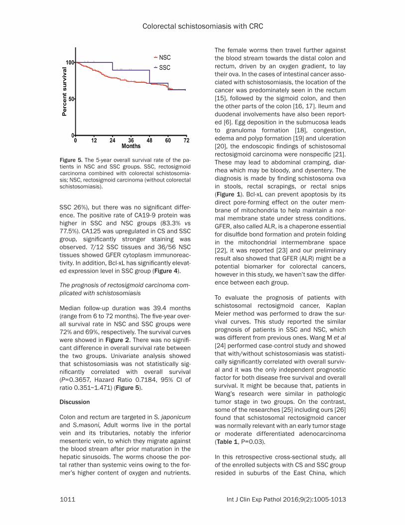
Median follow-up duration was 39.4 months (range from 6 to 72 months). The five-year over-all survival rate in NSC and SSC groups were 72% and 69%, respectively. The survival curves were showed in Figure 2. There was no signifi-cant difference in overall survival rate between the two groups. Univariate analysis showed that schistosomiasis was not statistically sig-nificantly correlated with overall survival (P=0.3657, Hazard Ratio 0.7184, 95% CI of ratio 0.351~1.471) (Figure 5).
Discussion
Colon and rectum are targeted in S. japonicum and S.masoni, Adult worms live in the portal vein and its tributaries, notably the inferior mesenteric vein, to which they migrate against the blood stream after prior maturation in the hepatic sinusoids. The worms choose the por-tal rather than systemic veins owing to the for-mer’s higher content of oxygen and nutrients.
The female worms then travel further against the blood stream towards the distal colon and rectum, driven by an oxygen gradient, to lay their ova. In the cases of intestinal cancer asso-ciated with schistosomiasis, the location of the cancer was predominately seen in the rectum [15], followed by the sigmoid colon, and then the other parts of the colon [16, 17]. Ileum and duodenal involvements have also been report-ed [6]. Egg deposition in the submucosa leads to granuloma formation [18], congestion, edema and polyp formation [19] and ulceration [20], the endoscopic findings of schistosomal rectosigmoid carcinoma were nonspecific [21]. These may lead to abdominal cramping, diar-rhea which may be bloody, and dysentery. The diagnosis is made by finding schistosoma ova in stools, rectal scrapings, or rectal snips (Figure 1). Bcl-xL can prevent apoptosis by its direct pore-forming effect on the outer mem-brane of mitochondria to help maintain a nor-mal membrane state under stress conditions. GFER, also called ALR, is a chaperone essential for disulfide bond formation and protein folding in the mitochondrial intermembrane space [22], it was reported [23] and our preliminary result also showed that GFER (ALR) might be a potential biomarker for colorectal cancers, however in this study, we haven’t saw the differ-ence between each group.
To evaluate the prognosis of patients with schistosomal rectosigmoid cancer, Kaplan Meier method was performed to draw the sur-vival curves. This study reported the similar prognosis of patients in SSC and NSC, which was different from previous ones. Wang M et al [24] performed case-control study and showed that with/without schistosomiasis was statisti-cally significantly correlated with overall surviv-al and it was the only independent prognostic factor for both disease free survival and overall survival. It might be because that, patients in Wang’s research were similar in pathologic tumor stage in two groups. On the contrast, some of the researches [25] including ours [26] found that schistosomal rectosigmoid cancer was normally relevant with an early tumor stage or moderate differentiated adenocarcinoma (Table 1, P=0.03).
In this retrospective cross-sectional study, all of the enrolled subjects with CS and SSC group resided in suburbs of the East China, which
Figure 5. The 5-year overall survival rate of the pa-tients in NSC and SSC groups. SSC, rectosigmoid carcinoma combined with colorectal schistosomia-sis; NSC, rectosigmoid carcinoma (without colorectal schistosomiasis).
Colorectal schistosomiasis with CRC
1012 Int J Clin Exp Pathol 2016;9(2):1005-1013
used to be heavily endemic for S. japonicum and men worked more in the infested water [27]. That may be the reason why in this study the CS and SSC group contained high propor-tions of men.
In this study, TBil and AFP were significantly elevated while Hb had a significantly less level in CS group, which may be explained as follows: though most of the advanced schistosomiasis patients have received schistosomicides, but before the development of praziquantel, patients who received other schistosomicides might not complete the whole therapeutic pro-cess because of side effects. Thus, some of the advanced schistosomiasis patients still have live schistosomes in their portal veins. Meanwhile, there are many reports about rela-tionship between the clinical liver fibrosis diag-nosis and histopathology in advanced schisto-somiasis. In addition, the tumor biochemical markers CA19-9 and CA-125 [28], which were routinely tested in gastrointestinal or ovarian cancers, were significantly higher in schistoso-miasis patients (SSC and CS group respective-ly) than in NSC group. Therefore, elevated CA19-9 and CA-125 levels, as well as reduced Hb may be signals for those who have colorec-tal lesions, especially for those who have infest-ed water contact history and gastrointestinal syndromes, to go through the circumival precip-intin test (COPT).
Conclusions
Colorectal schistosomiasis is still easily under-diagnosed in patients with or without rectosig-moid carcinoma. For those who travelled from or lived in endemic areas, elevated serum CA19-9 and CA-125 may be signals for those who have colorectal lesions and infested water contact history to go through the circumival precipintin test (COPT), for rectosigmoid cancer patients whose biopsy is CA-125 positive, rec-tosigmoid carcinoma combined with colorectal schistosomiasis (SSC) should be considered. This study reported the similar 5-year overall survival rate of patients in SSC and NSC grou- ps, which may be because that schistosomal rectosigmoid cancers are normally relevant with early tumor stages and moderate di- fferentiation.
Disclosure of conflict of interest
None.
Address correspondence to: Dr. Hao Feng, Depart- ment of General, Visceral, Transplantation, Vascular and Thoracic Surgery, Hospital of University of Munich, Ludwig-Maximilian-University (LMU), Marchioninistr.15, 5H-02-428, Munich 81377, Germany. Tel: +49-89-440073438; Fax: +49-89-440076433; E-mail: [email protected]; Ming-Liang Wang, Ruijin Hospital, LuWan Branch School of Medicine, Shanghai Jiao Tong University, Shanghai, China; Ruijin Hospital, School of Medi- cine, Shanghai Jiao Tong University, Shanghai, China. E-mail: [email protected]
References
[1] Schaderwaldt H. The first report and drawings of Schistosoma haematobium and monsoni by Theodor Bilharz. Z Tropenmed Parasitol 1953; 4: 410-4.
[2] Coltart C, Whitty CJ. Schistosomiasis in non-endemic countries. Clin Med (Lond) 2015; 15: 67-9.
[3] Eissa S, Habib H, Ali E, Kotb Y. Evaluation of urinary miRNA-96 as a potential biomarker for bladder cancer diagnosis. Med Oncol 2015; 32: 413.
[4] Eissa S, Matboli M, Mansour A, Mohamed S, Awad N, Kotb YM. Evaluation of urinary HURP mRNA as a marker for detection of bladder cancer: relation to bilharziasis. Med Oncol 2014; 31: 804.
[5] Xiang JJ, Cheng BJ, Tian F, Li M, Jiang XF, Zhao HC, Hu XM, Xiao BL, Xie JP, Shrestha A. Perforation of small bowel caused by Schistosoma japonicum: A case report. World J Gastroenterol 2015; 21: 2862-4.
[6] Contractor QQ, Benson L, Schulz TB, Contractor TQ, Kasturi N. Duodenal involvement in Schistosoma mansoni infection. Gut 1988; 29: 1011-1012.
[7] El-Garem AA. Schistosomiasis. Digestion 1998; 59: 589-605.
[8] McCreesh N, Nikulin G, Booth M. Predicting the effects of climate change on Schistosoma mansoni transmission in eastern Africa. Parasit Vectors 2015; 8: 4.
[9] Wu TS, Chen TC, Chen RJ, Chiang PC, Leu HS. Schistosoma japonicum infection presenting with colon perforation: case report. Changgeng Yi Xue Za Zhi 1999; 22: 676-81.
[10] Titi S, Kosik-Warzyńska R, Sycz K, Chosia M. Intestinal schistosomiasis--a case report. Pol J Pathol 2003; 54: 283-5.
[11] Hosho K, Ikebuchi Y, Ueki M, Nakamura K, Yashima K, Maeda N, Koda M, Murawaki Y, Suou T, Inoue M. Schistosomiasis japonica identified by laparoscopic and colonoscopic ex-amination. Dig Endosc 2010; 22: 133-6.
Colorectal schistosomiasis with CRC
1013 Int J Clin Exp Pathol 2016;9(2):1005-1013
[12] Liu W, Zeng HZ, Wang QM, Yi H, Mou Y, Wu CC, Hu B, Tang CW. Schistosomiasis combined with colorectal carcinoma diagnosed based on endoscopic findings and clinicopathological characteristics: a report on 32 cases. Asian Pac J Cancer Prev 2013; 14: 4839-42.
[13] Zhang R, Takahashi S, Orita S, Yoshida A, Maruyama H, Shirai T, Ohta N. p53 gene muta-tions in rectal cancer associated with schisto-somiasis japonica in Chinese patients. Cancer Lett 1998; 131: 215-21.
[14] Soliman NA, Keshk WA, Shoheib ZS, Ashour DS, Shamloula MM. Inflammation, oxidative stress and L-fucose as indispensable partici-pants in schistosomiasis-associated colonic dysplasia. Asian Pac J Cancer Prev 2014; 15: 1125-31.
[15] Anonymus. Schistosomes, liver flukes and Helicobacter pylori. IARC Monogr Eval Carcinog Risks Hum 1994; 3: 1-241.
[16] Lin CT, Chen CY, Jao SW, Wu CC. An unusual pedunculated polyp of the transverse colon as-sociated with schistosomiasis. Endoscopy 2010; 42 Suppl 2: E160.
[17] Abe T, Yunokizaki H, Iijima H, Tamura K, Lee ZL, Higashi D, Ebara R, Tanabe J, Tsujimoto M, Tsujii M. Schistosoma japonicum showing flat colon polyp appearance. Gastrointest Endosc 2011; 73: 820-2.
[18] Meybehm M, Dworák O, Gugler R. Granulo- matous colitis with pseudopolyp in schistoso-miasis. Dtsch Med Wochenschr 1989; 114: 19-22.
[19] Osman M, Aftimos G. Schistosomiasis mani-festing as a colon polyp: a case report. J Med Case Rep 2014; 8: 331.
[20] Elbaz T, Esmat G. Hepatic and Intestinal Schis-tosomiasis: Review. J Adv Res 2013; 4: 445-452.
[21] Cao J, Liu WJ, Xu XY, Zou XP. Endoscopic find-ings and clinicopathologic characteristics of colonic schistosomiasis: a report of 46 cases. World J Gastroenterol 2010; 16: 723-727.
[22] Sankar U, Means AR. Gfer is a critical regulator of HSC proliferation. Cell Cycle 2011; 10: 2263-2268.
[23] Gatzidou E, Mantzourani M, Giaginis C, Giagini A, Patsouris E, Kouraklis G, Theocharis S. Aug-menter of liver regeneration gene expression in human colon cancer cell lines and clinical tissue samples. J BUON 2015; 20: 84-91.
[24] Wang M, Zhang YC, Yang XY, Wang ZQ. Prog-nostic analysis of schistosomal rectal cancer. Asian Pac J Cancer Prev 2014; 15: 9271-5.
[25] Probst A, Schaller T, Ebigbo A, Messmann H. Colonic schistosomiasis and early rectal can-cer: coincidence or causal relationship? En-doscopy 2014; 46 Suppl 1 UCTN: E671.
[26] Feng H, Lu AG, Zhao XW, Han DP, Zhao JK, Shi L, Tobias S Schiergens, Serene ML Lee, Zhang WP, Wolfgang E Thasler. Comparison of non-schistosomal rectosigmoid cancer and schisto-somal rectosigmoid cancer. World J Gastroen-terol 2015; 21: 7225-7232.
[27] Ross AG, Sleigh AC, Li Y, Davis GM, Williams GM, Jiang Z, Feng Z, McManus DP. McManus. Schistosomiasis in the People’s Republic of China: Prospects and Challenges for the 21st Century. Clin Microbiol Rev 2001; 14: 270-295.
[28] Herszényi L, Farinati F, Cardin R, István G, Mol-nár LD, Hritz I, De Paoli M, Plebani M, Tulassay Z. Tumor marker utility and prognostic rele-vance of cathepsin B, cathepsin L, urokinase-type plasminogen activator, plasminogen acti-vator inhibitor type-1, CEA and CA 19-9 in colorectal cancer. BMC Cancer 2008; 8: 194.