original article analyze two cases of encapsulated ... · analyze two cases of encapsulated...
TRANSCRIPT
Int J Clin Exp Pathol 2016;9(7):7493-7497www.ijcep.com /ISSN:1936-2625/IJCEP0026329
Original ArticleAnalyze two cases of encapsulated papillary oncocytic neoplasms (EPONs) of the thyroid
Li Ding, Xianjin Zou, Wan Yang, Junhua Xie
Department of Pathology, The First People’s Hospital of Jingmen, Xiangshan Road No. 168, Jingmen 448000, Hubei Province, China
Received February 21, 2016; Accepted May 21, 2016; Epub July 1, 2016; Published July 15, 2016
Abstract: Aim: Analyzing the pathological features of encapsulated papillary oncocytic neoplasms (EPONs) of the thyroid based on 2 cases in our department. Methods: Medical history and morphological characteristics of the two cases were reviewed. Immunohistochemical staining was applied to detect the expression of CK19, CD56, CD31, galectin-3 and so on with paraffin-embedded tissues. Sanger sequencing was performed to detect the genotype of codons V600 in BRAF and codons GG12/13 in KRAS. Results: The first patient has been diagnosed as Hurthle cell adenoma 8 years ago; the second patient discovered that the mass grew up gradually for 3 months. Oncocytic cells of EPONs mainly arranged and formed to papillary architecture but did not exhibit the nuclear feature of papillary thyroid carcinoma under microscope. Invasion of capsule and vascular vessels could be observed in case 1. The two cases of EPONs displayed CK19(-), CD56(+), galectin-3(+), Wide Genotype of BRAF and KRAS. In the follow-up, the two patients remain alive within 5 and 3 years, respectively. Conclusions: EPONs might occurring through the malignant transformation from Hurthle cell adenoma; however, this process was different from classical Hurthel cell adenocarcinoma. They were malignant tumor which didn’t diagnosed on infiltrating of vessels or capsule.
Keywords: EPONs, oxyphilic thyrocytes, papillary, malignant transformation
Introduction
EPONs (Encapsulated Papillary Oncocytic Neo- plasms) of the Thyroid were rare tumors which defined as encapsulated thyroid tumors com-posed of oncocytic thyrocytes with at leastfocal papillary architecture. Cases demonstrating readily apparent nuclear features of papillary thyroid carcinomas (PTCs) were excluded. EPONs were very rare and their relationship with other oncocytic neoplasms of the thyroid remained unclear. In some instances, EPONs were often misdiagnosed as other neoplasms with a similar pattern. In this study, the clinico-pathologic and molecular features of two cases of EPONs were analyzed.
Materials and methods
Two cases of EPONs in our department were considered. All of the tissues were fixed in 10% neutral formalin, desiccated, and embedded in paraffin. A two-step immunohistochemical staining technique (EnVision) was employed to
detect TG, CK19, galectin-3, CD56, calcitonin, CD31, CD34, and Ki-67. Sanger sequencing was applied to detect the gene phenotype of codons V600 in BRAF and codons GG12/13 in KRAS for the two samples.
Results
Introduction of illness and clinical characteris-tics
Case 1: In 2010, a 58-year-old female patient manifested a mass in the isthmic portion of the thyroid but did not complain of hoarseness, bucking, and dyspnea. A pathologist found a pinkish-gray nodule with a dimension of 4.5× 4×4 cm in the isthmic portion of the thyroid with an integrated capsule. In a follow-up, the patient remains alive and does not suffer from recur-rence after the whole thyroid was resected. Past history: In 2002, the patient was diag-nosed with Hurthle cell adenoma. The sections in 2002 were reviewed which displayed a typi-cal image of Hurthle cell adenoma without pap-
EPONs of the Thyroid
7494 Int J Clin Exp Pathol 2016;9(7):7493-7497
EPONs of the Thyroid
7495 Int J Clin Exp Pathol 2016;9(7):7493-7497
illary structure and did not invade the vascular vessels or the capsule.
Case 2: A 47-year-old female patient was detected with a chicken-egg-sized neck mass 3 month ago before see a doctor in 2012. The mass grew to a size of a duck egg within a month. The patient did not suffer from heart palpitations, fever, and pain. Conservative treatment was not applicable. Color Doppler ultrasound: A regularly sized mass was detect-ed in the right lobe of the thyroid. The mass exhibited a slightly lower echo but a non-uni-form echo. A 6×5.5×4.2 cm nodule of the thy-roid tissue was examined, and the pinkish-gray cross section was found. The thyroid was resected completely.
Microscopic characteristics
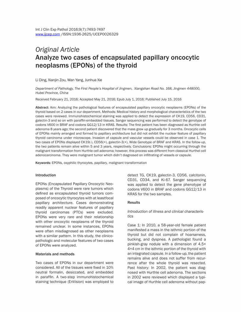
The tumor cells were columnar with a granular eosinophilic cytoplasm. Most of the tumor cells were arranged in the papillary form, and the subsection was arranged in a trabecular pat-tern, with a solid and alveolar structure. The cells were arranged in single layer; the nucleus was partly found in the center of the cell and partly located in the top of the adenoid (the reverse phenomenon). The nuclei were round or oval with prominent nucleoli. The typical nuclear features of PTCs, such as large and crowded and translucent nuclei with nuclear groove and intranuclear pseudoinclusion, were not observed in the two cases. Small vessel invasion was found in the local area of the fiber within the capsule in Case 1 (Figure 1A-D).
Molecular characteristics
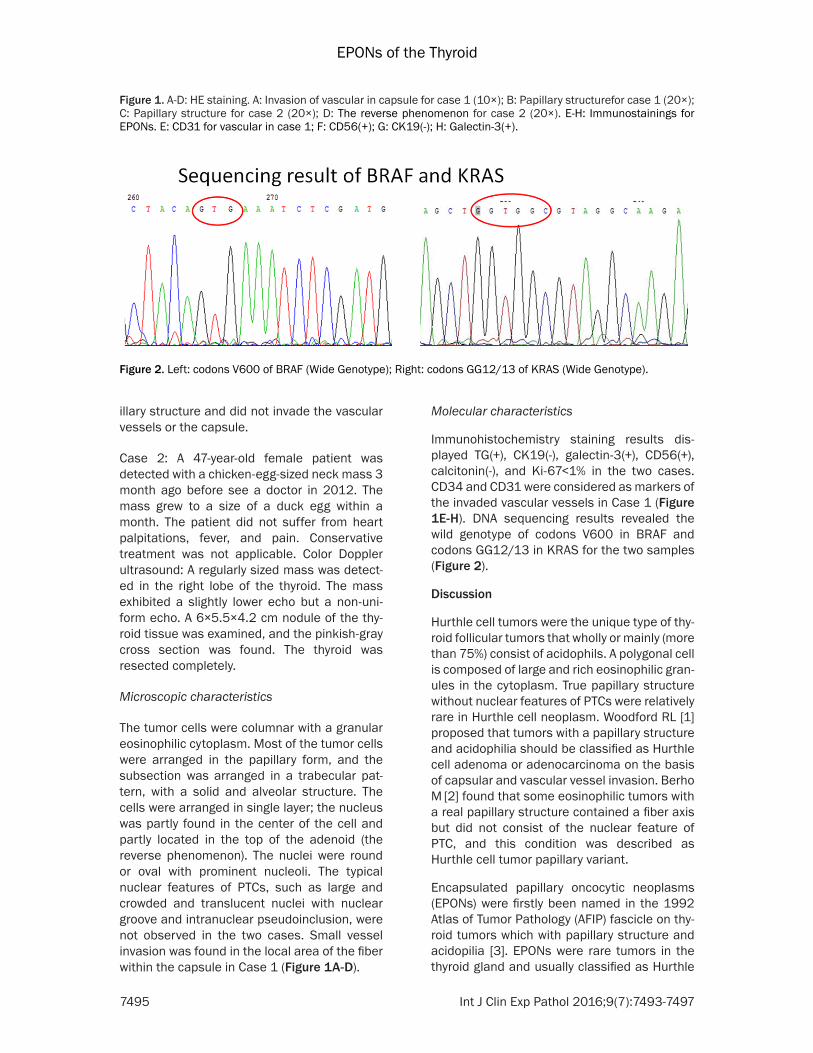
Immunohistochemistry staining results dis-played TG(+), CK19(-), galectin-3(+), CD56(+), calcitonin(-), and Ki-67<1% in the two cases. CD34 and CD31 were considered as markers of the invaded vascular vessels in Case 1 (Figure 1E-H). DNA sequencing results revealed the wild genotype of codons V600 in BRAF and codons GG12/13 in KRAS for the two samples (Figure 2).
Discussion
Hurthle cell tumors were the unique type of thy-roid follicular tumors that wholly or mainly (more than 75%) consist of acidophils. A polygonal cell is composed of large and rich eosinophilic gran-ules in the cytoplasm. True papillary structure without nuclear features of PTCs were relatively rare in Hurthle cell neoplasm. Woodford RL [1] proposed that tumors with a papillary structure and acidophilia should be classified as Hurthle cell adenoma or adenocarcinoma on the basis of capsular and vascular vessel invasion. Berho M [2] found that some eosinophilic tumors with a real papillary structure contained a fiber axis but did not consist of the nuclear feature of PTC, and this condition was described as Hurthle cell tumor papillary variant.
Encapsulated papillary oncocytic neoplasms (EPONs) were firstly been named in the 1992 Atlas of Tumor Pathology (AFIP) fascicle on thy-roid tumors which with papillary structure and acidopilia [3]. EPONs were rare tumors in the thyroid gland and usually classified as Hurthle
Figure 1. A-D: HE staining. A: Invasion of vascular in capsule for case 1 (10×); B: Papillary structurefor case 1 (20×); C: Papillary structure for case 2 (20×); D: The reverse phenomenon for case 2 (20×). E-H: Immunostainings for EPONs. E: CD31 for vascular in case 1; F: CD56(+); G: CK19(-); H: Galectin-3(+).
Figure 2. Left: codons V600 of BRAF (Wide Genotype); Right: codons GG12/13 of KRAS (Wide Genotype).
EPONs of the Thyroid
7496 Int J Clin Exp Pathol 2016;9(7):7493-7497
cell adenoma/adenocarcinoma or papillary car-cinoma inaccurately.
We analyzed two cases of EPONs that rich in eosinophilic granules and arranged into fibro-vascular papillae. The first patient suffered from typical Hurthle cell adenoma 8 years ago and the recurrent tumor displayed papillary structure and invaded the vascular vessel in the capsule. This result indicated that EPONs were related to Hurthle cell adenoma in the ori-gin. The second patient displayed a tumor that increased in size significantly within a month. The recent increasing in tumor size was evi-dently manifestation of malignant transforma-tion. We proposed that EPONs were the special results from Hurthle cell adenomas.
Hurthle cell adenocarcinoma referred to Hur- thle cell adenoma with vascular vessel or cap-sular invasion. Carcangiu ML [4] proposed that EPONs should be evaluated on the basis of the histological criteria of follicular tumors, includ-ing vascular or capsular invasion and distant metastasis. In case 2, all the mass was select-ed for dehydration and embedding but no inva-sion were discovered under microscope. We proposed on biological behavior that EPONs were different from Hurthle cell adenocarci- noma regardless of vascular or capsular inva- sion.
Galectin-3 has been implicated in the regula-tion of cellular growth, differentiation and ma- lignant transformation in thyroid gland. Diffuse and strong immunohistochemistry staining for galectin-3 might potentially serve as a marker in difficult differential diagnosis cases involving Hurthle cell adenomas and Hurthle cell carcino-mas [5]. Galectin-3 immunostaining in Hurthle cell carcinomas was significantly higher than in Hurthle cell adenomas [6]. The two cases all di- splayed Galectin-3 positive with immunostain-ing which indicated that EPONs were malignant neoplasms independent of tumor invasion.
KRAS mutationsand heteroploid were detected more often in malignant follicular neoplasm than benign [7]. All the two cases showed codons GG12/13 of KRAS wide genotype with Sanger sequencing. We speculated that the 2 cases could not represent the whole phenom-enons of KRAS. Moreover, the tissues were saved too long and DNA fragment might have been fractured in same degree. In addition,
mutated tumors less than 20% and exon 3/4 mutation could not been detected in our study with Sanger sequencing.
EPONs were different from PTCs because of two features: morphological characteristics and molecular biomarkers. Despite of the actu-al papillary structure, EPONs didn’t possess the characteristics of the nucleus similar to that of PTCs. PTCs usually displayed CK19(+), HBME-1(+), and CD56(-). Polymerase chain reaction test results revealed that 69% of PTCs display-ing gene mutation in BRAF [8]. Bellevicine C [9] subjected EPONs to immunostaining and found CK19(-), HBME-1(-), and CD56(+). Woodford RL [1] detected nine cases of EPONs but did not find BRAF gene mutation. These results were not consistent with that observed in PTCs. Our immunostaining results also showed that the two cases were CK19(-)/CD56(+). DNA sequ- encing results demonstrated that codons V600 in BRAF were wild genotype within the two cases.
Differential diagnosis mainly includes the fol-lowing: (1) Hurthle cell adenomas: EPONs showed cytological characteristics similar to Hurthle cell adenoma, but EPONs were arr- anged at least focally to typical papilla with true fiber axis. Hurthle cell adenomas does not invade the envelope and vascular vessels. (2) Hurthle cell adenocarcinomas: Large tumor cells contained a large nucleus and exhibited heteromorphism. Large acreage of the papilla was missing. Vascular vessel/capsular invasion or distant metastases were the diagnostic mor-phology features for Hurthle cell adenocarcino-mas but didn’t appropriate for EPONs. (3) Subtype of PTCs with an acidophil type: This conditions exhibited the typical characteristic of the nucleus. PTCs were characterized by glass-like nucleus, with thick nuclear mem-brane and intranuclear pseudoinclusions. Molecular biomarker revealed CK19(+), HBME-1(+), CD56(-) and mutation of BRAF gene. (4) Other tumors: Other tumor types, such as PTC with a high cell type, medullary carcinoma with an acidophilic cell type, and metastatic tumors.
Summary
We considered that EPONs were included in Hurthle cell neoplasms. They might the unique results of malignant transformation from Hur-
EPONs of the Thyroid
7497 Int J Clin Exp Pathol 2016;9(7):7493-7497
thle cell adenomas and different from classical Hurthle cell adenocarcinomas. Diagnosis of EPONs mainly based on the pathological mor-phology independent of tumor invasion. Immunostaining results displayed CK19(-), CD56(+), and galectin-3(+). The BRAF gene always showed wild genotype in EPONs. EPONs should be treated with the same strategies as those used to cure invasive tumors and closely following up was necessary [10].
Acknowledgements
Thanks to all the colleagues in our department for their assistance.
Disclosure of conflict of interest
None.
Authors’ contribution
Ding Li: put in order of data and write the arti-cle, sequencing for BRAF and KRAS. Zou Xianjin: pathological diagnosis for EPONs and guide the work. Yang Wan: immunohistochemi-cal staining. Xie Junhua: HE staining.
Address correspondence to: Xianjin Zou, Depart- ment of Pathology, The First People’s Hospital of Jingmen, Xiangshan Road No. 168, Jingmen 448- 000, Hubei Province, China. E-mail: [email protected]
References
[1] Woodford RL, Nikiforov YE, Hunt JL, Bellizzi AM, Zhang X, Mills SE, Stelow EB. Encapsulated papillary oncocytic neoplasms of the thyroid: morphologic, immunohistochemical, and mo-lecular analysis of 18 cases. Am J Surg Pathol 2010; 34: 1582-90.
[2] Berho M, Suster S. The oncocytic variant of papillary carcinoma ofthe thyroid: a clinico-pathologic study of 15 cases. Hum Pathol 1997; 28: 47-53.
[3] Rosai J, Carcangiu ML, DeLellis RA. Atlas of Tu-mor Pathology. Third Series, Fascicle Five. Tu-mors of the Thyroid Gland. Washington, DC: Armed Forces Institute of Pathology; 1992.
[4] Carcangiu ML, Bianchi S, Savino D, Voynick IM, Rosai J. Follicular Hurthle celltumors of the thy-roid gland. Cancer 1991; 68: 1944-53.
[5] Sumana BS, Shashidhar S, Shivarudrappa AS. Galectin-3 Immunohistochemical Expression in Thyroid Neoplasms. J Clin Diagn Res 2015; 9: EC07-11.
[6] Nascimento MC, Bisi H, Alves VA, Longatto-Fil-ho A, Kanamura CT, Medeiros-Neto G. Differ-ential reactivity for galectin-3 in Hürthle cell adenomas and carcinomas. Endocr Pathol 2001; 12: 275-9.
[7] Kondo T, Ezzat S, Asa SL. Pathogenetic mecha-nisms in thyroid follicular-cell neoplasia. Nat Rev Cancer 2006; 6: 292-306.
[8] Cohen Y, Xing M, Mambo E, Guo Z, Wu G, Trink B, Beller U, Westra WH, Ladenson PW, Sidran-sky D. BRAF mutation in papillary thyroid carci-noma. J Natl Cancer Inst 2003; 95: 625-7.
[9] Bellevicine C, Malapelle U, Docimo G, Ciancia G, Mossetti G, Pettinato G, Troncone G. Multi-centric encapsulated papillary oncocytic neo-plasm of the thyroid: A case diagnosed by a combined cytological, histological, immunohis-tochemical, and molecular approach. Diagn Cytopathol 2012; 40: 450-4.
[10] Gundry SR, Burney RE, Thompson NW, Lloyd R. Total thyroidectomyfor Hurthle cell neoplasm of the thyroid. Arch Surg 1983; 118: 529-532.