organoid models of human and mouse ductal pancreatic cancer · patients with pancreatic cancer are...
TRANSCRIPT

Resource
Organoid Models of Human and MouseDuctal Pancreatic CancerSylvia F. Boj,1,2,14 Chang-Il Hwang,3,4,14 Lindsey A. Baker,3,4,14 Iok In Christine Chio,3,4,14 Dannielle D. Engle,3,4,14
Vincenzo Corbo,3,4,14 Myrthe Jager,1,14 Mariano Ponz-Sarvise,3,4 Herve Tiriac,3,4 Mona S. Spector,3,4 Ana Gracanin,1,2
Tobiloba Oni,3,4,5 Kenneth H. Yu,3,4,6,7 Ruben van Boxtel,1 Meritxell Huch,1,15 Keith D. Rivera,3 John P. Wilson,3
Michael E. Feigin,3,4 Daniel Ohlund,3,4 Abram Handly-Santana,4,8 Christine M. Ardito-Abraham,3,4 Michael Ludwig,3,4
Ela Elyada,3,4 Brinda Alagesan,3,4,9 Giulia Biffi,3,4 Georgi N. Yordanov,4,8 Bethany Delcuze,3,4 Brianna Creighton,3,4
KevinWright,3,4 Youngkyu Park,3,4 Folkert H.M. Morsink,10 I. QuintusMolenaar,11 Inne H. Borel Rinkes,11 Edwin Cuppen,1
Yuan Hao,3 Ying Jin,3 Isaac J. Nijman,1 Christine Iacobuzio-Donahue,6 Steven D. Leach,6 Darryl J. Pappin,3
Molly Hammell,3 David S. Klimstra,12 Olca Basturk,12 Ralph H. Hruban,13 George Johan Offerhaus,10 Robert G.J. Vries,1,2
Hans Clevers,1,* and David A. Tuveson3,4,6,*1Hubrecht Institute, Royal Netherlands Academy of Arts and Sciences (KNAW), University Medical Centre Utrecht and CancerGenomics.nl,
3584 CT Utrecht, the Netherlands2foundation Hubrecht Organoid Technology (HUB), 3584 CT Utrecht, the Netherlands3Cold Spring Harbor Laboratory, Cold Spring Harbor, NY 11724, USA4Lustgarten Foundation Pancreatic Cancer Research Laboratory, Cold Spring Harbor, NY 11724, USA5Graduate Program in Molecular and Cellular Biology, Stony Brook University, Stony Brook, NY 11794, USA6Rubenstein Center for Pancreatic Cancer Research, Memorial Sloan Kettering Cancer Center, New York, NY 10065, USA7Weill Medical College at Cornell University, New York, NY 10065, USA8Watson School of Biological Sciences, Cold Spring Harbor Laboratory, Cold Spring Harbor, NY 11724, USA9Graduate Program in Genetics, Stony Brook University, Stony Brook, NY 11794, USA10Department of Pathology, University Medical Centre Utrecht, 3584 CX Utrecht, the Netherlands11Department of Surgery, University Medical Center Utrecht, 3584 CX Utrecht, the Netherlands12Department of Pathology, Memorial Sloan Kettering Cancer Center, New York, NY 10065, USA13The Sol Goldman Pancreatic Cancer Research Center, Johns Hopkins University School of Medicine, Baltimore, MD 21231, USA14Co-first author15Present address: Gurdon Institute-University of Cambridge, Tennis Court Road, Cambridge CB2 1QN, UK
*Correspondence: [email protected] (H.C.), [email protected] (D.A.T.)http://dx.doi.org/10.1016/j.cell.2014.12.021
SUMMARY
Pancreatic cancer is one of the most lethal malig-nancies due to its late diagnosis and limitedresponse to treatment. Tractable methods to iden-tify and interrogate pathways involved in pancreatictumorigenesis are urgently needed. We establishedorganoid models from normal and neoplastic mu-rine and human pancreas tissues. Pancreatic orga-noids can be rapidly generated from resectedtumors and biopsies, survive cryopreservation,and exhibit ductal- and disease-stage-specific char-acteristics. Orthotopically transplanted neoplasticorganoids recapitulate the full spectrum of tumordevelopment by forming early-grade neoplasmsthat progress to locally invasive and metastaticcarcinomas. Due to their ability to be geneticallymanipulated, organoids are a platform to probegenetic cooperation. Comprehensive transcriptionaland proteomic analyses of murine pancreaticorganoids revealed genes and pathways alteredduring disease progression. The confirmation ofmany of these protein changes in human tissuesdemonstrates that organoids are a facile model
324 Cell 160, 324–338, January 15, 2015 ª2015 Elsevier Inc.
system to discover characteristics of this deadlymalignancy.
INTRODUCTION
Mortality due to pancreatic cancer is projected to surpass that of
breast and colorectal cancer by 2030 in the United States (Rahib
et al., 2014; Siegel et al., 2013). This dire scenario reflects an ag-
ing population, the improvement of outcomes for breast and
colorectal cancer patients, the advanced stage at which most
patients with pancreatic cancer are diagnosed, and the lack of
durable treatment responses in pancreatic cancer patients.
Indeed, effective therapeutic strategies for patients with pancre-
atic ductal adenocarcinoma (PDA) have been difficult to identify
(Abbruzzese and Hess, 2014).
The therapeutic resistance of PDA has been explored in a
variety of cell culture and animal model systems, with clinically
actionable findings encountered only occasionally (Villarroel
et al., 2011). Patient-derived xenografts (PDXs) have yielded in-
sights into PDA, but their generation requires a large amount of
tissue, and they take multiple months to establish (Kim et al.,
2009; Rubio-Viqueira et al., 2006). Genetically engineered
mouse models (GEMMs) of PDA have also been generated as
a parallel system for fundamental biological investigation and
preclinical studies (Perez-Mancera et al., 2012). These GEMMs

accuratelymimic the pathophysiological features of humanPDA,
including disease initiation from preinvasive pancreatic intraepi-
thelial neoplasms (PanINs) (Hingorani et al., 2003; Perez-Man-
cera et al., 2012) and were used to discover that PDA possesses
a deficient vasculature that impairs drug delivery (Erkan et al.,
2009; Jacobetz et al., 2013; Koong et al., 2000; Olive et al.,
2009; Provenzano et al., 2012). Although GEMMs have informed
PDA therapeutic development (Beatty et al., 2011; Frese et al.,
2012; Neesse et al., 2014), they are expensive and time
consuming (Perez-Mancera et al., 2012). In addition, both human
PDA and GEMMs exhibit an extensive stromal component that
decreases the neoplastic cellularity, making it difficult to isolate
and characterize the epithelium-derived malignant cells in
pancreatic neoplastic tissues.
To study neoplastic cells, dissociated human tumors are often
grown in two-dimensional (2D) culture conditions (Sharma et al.,
2010), which do not support growth of untransformed, nonneo-
plastic pancreatic cells. Three-dimensional (3D) culture strate-
gies have been developed to study normal, untransformed cells
but so far have only allowed minimal propagation (Agbunag and
Bar-Sagi, 2004; Lee et al., 2013; Means et al., 2005; Rovira et al.,
2010; Seaberg et al., 2004). A comprehensive 3D cell culture
model of murine and human PDA progression would facilitate
investigation of genetic drivers, therapeutic targets, and diag-
nostics for PDA.
To address this deficiency, we sought to generate normal
and neoplastic pancreatic organoids by modifying approaches
we previously pioneered to culture intestinal (Sato et al., 2009),
gastric (Barker et al., 2010), colon carcinoma (Sato et al.,
2011), hepatic (Huch et al., 2013b), pancreatic (Huch et al.,
2013a), and prostatic organoids (Gao et al., 2014; Karthaus
et al., 2014). We developed 3D organoids from normal and ma-
lignant murine pancreatic tissues and used this model system
to investigate PDA pathogenesis. Pancreatic organoids derived
from wild-type mice and PDA GEMMs accurately recapitulate
physiologically relevant aspects of disease progression in vitro.
Following orthotopic transplantation, organoids from wild-type
mouse normal pancreata are capable of regenerating normal
ductal architecture, unlike other 3D model systems. We further
developed methods to generate pancreatic organoids from
normal and diseased human tissues, as well as from endoscopic
needle biopsies. Following transplantation, organoids derived
from murine and human PDA generate lesions reminiscent of
PanIN and progress to invasive PDA. Finally, we demonstrate
the utility of organoids to identify molecular pathways that corre-
late with disease progression and that represent therapeutic and
diagnostic opportunities.
RESULTS
Murine Pancreatic Ductal Organoids ExpressingOncogenic Kras Recapitulate Features of PanINsRecently, we derived continuously proliferating, normal pancre-
atic organoids from adult murine ductal cells (Huch et al., 2013a).
We optimized this approach to generatemodels of PDA progres-
sion. We manually isolated small intralobular ducts and estab-
lished organoid cultures from C57Bl/6 mouse normal pancreata
and pancreatic tissues that contained low-grade murine PanIN
(mPanIN-1a/b) from Kras+/LSL-G12D; Pdx1-Cre (‘‘KC’’) mice (Fig-
ure 1A). KC mice develop a spectrum of preinvasive ductal le-
sions that mirror human PanINs and, upon aging, stochastically
develop primary and metastatic PDA (Hingorani et al., 2003).
Ducts from KC pancreata were often larger and exhibited higher
grades of dysplasia compared to those fromwild-typemice (Fig-
ure 1A). After 1–3 days in culture, organoid growth was observed
from isolated ducts (Figure 1A). We created a collection of 10
murine normal (mN) and 9 PanIN (mP) organoid cultures that
we have continuously propagated for over 20 passages and suc-
cessfully cryopreserved (Table S1A available online). mP orga-
noids exhibited recombination of the conditional KrasLSL-G12D
allele and higher levels of Kras-GTPwhen compared tomNorga-
noids (Figure 1B).
To determine the contribution of different pancreatic lineages
to the organoids, we evaluated the expression of pancreatic line-
age markers in these cultures. Genes associated with the ductal
lineage (Ck19 and Sox9) (Cleveland et al., 2012) were enriched in
the mN and mP organoids compared to total pancreatic tissues,
which contain relatively few ductal cells (Figure 1C). In addition,
the mP organoids upregulated genes indicative of a PanIN dis-
ease state (Muc5ac, Muc6, and Tff1) relative to mN, with no dif-
ference in Klf4 (Figure 1D) (Prasad et al., 2005). GFP-transduced
mN andmP organoids were orthotopically transplanted into syn-
geneic C57Bl/6 or Nu/Nu mice. mN organoids quickly formed
ductal structures comprised of simple cuboidal cells that per-
sisted for up to 1 month (n = 9/27 transplants) but were not
observed after 2 months (n = 0/13 transplants) (Figure 1E and
Table S1B). In comparison, mP organoids formed small cysts
lined with a single layer of simple cuboidal ductal cells
interspersed with mucin-containing columnar epithelial cells.
Although we could not demonstrate that the mP transplants
were contiguous with the native ductal system, they resembled
preinvasive mPanIN (Figure S1C). These dysplastic epithelial
cells persisted for 2 months or longer (n = 16/18 transplants),
were GFP and Ck19 positive, expressed themPanIN-associated
mucin Muc5ac, and stained prominently with Alcian blue (Fig-
ure 1E and Table S1C). In addition, when compared to mN trans-
plants, mP transplants had increased proliferation and a robust
stromal response, which are characteristics of autochthonous
mPanIN tissue (Figures S1A–S1C). The ability of transplanted
mP organoids to form lesions with many of the features of
mPanINs demonstrates the utility of this system as a model for
early pancreatic neoplasia.
Multiple cellular origins have been proposed for the develop-
ment of PDA, with the pancreatic acinar cell hypothesized to be
amajor contributor to PDA initiation (De La O et al., 2008; Gidekel
Friedlander et al., 2009; Guerra et al., 2003; Habbe et al., 2008;
Kopp et al., 2012; Morris et al., 2010; Sawey et al., 2007).
However, recent studies have suggested that transformation of
pancreatic ductal cells can also give rise to PDA (Pylayeva-Gupta
et al., 2012; Ray et al., 2011; von Figura et al., 2014). Acinar cells
isolated fromwild-type pancreata are unable to formorganoids in
our conditions (Huch et al., 2013a). Therefore, our pancreatic
ductal organoid system offers a unique opportunity to determine
whether ductal cells can give rise to mPanIN. To assess whether
expression of oncogenic Kras in pancreatic ductal organoids
is sufficient to induce mPanIN formation in vivo, we derived
Cell 160, 324–338, January 15, 2015 ª2015 Elsevier Inc. 325

Figure 1. Oncogenic KrasG12D Expression in Pancreatic Ductal Organoids Is Sufficient to Induce Preinvasive Neoplasms
(A) Hematoxylin and eosin (H&E) staining of murine pancreatic tissue used to prepare organoids (top). Arrows indicate mouse normal or PanIN ductal structures.
Ducts embedded in Matrigel immediately following isolation (middle) and organoids 3 days postisolation (bottom). Arrowheads mark isolated ducts and growing
organoids. Scale bars, 50 mm.
(B) Immunoblots for Kras, pan Ras, Kras-GTP by RBD-GST pull-down, and Tubulin in mN and mPanIN (mP) organoids. PCR confirmation of Cre-mediated
recombination of the KrasLSL-G12D allele (bottom).
(C) qRT-PCR of ductal (Pdx1, Ck19, Sox9, and Hnf6), acinar (Ptf1a, Cpa1, and Amy), and endocrine (Ngn3, Chga, and Ins2) lineage markers in mN and mP
organoids. Means of three biological replicates are shown. Error bars indicate SEMs. Values were normalized to mouse normal pancreas.
(D) qRT-PCR of genes indicative of PanIN lesions (Muc5ac,Muc6, Tff1, and Klf4) in mN and mP organoids. Values were normalized to mN organoids. Means of
three biological replicates are shown. Error bars indicate SEMs. **p < 0.01 by two-tailed Student’s t test.
(E) H&E, Alcian blue staining, and immunohistochemistry (IHC) of orthotopic, syngeneic transplants of GFP-transduced mN and mP organoids. Scale bars,
200 mm.
(F) Immunoblots for Kras, pan Ras, Kras-GTP by RBD-GST pull-down, and tubulin in Kras+/LSL-G12D organoids transduced with adenoviral-Cre (Ad-Cre) or
adenoviral-blank (Ad-Bl). PCR confirmation of Cre-mediated recombination of the KrasLSL-G12D allele (bottom).
(G) H&E, Alcian blue staining, and IHC of orthotopic syngeneic transplants of organoids transduced with Ad-Bl (Kras+/LSL-G12D; R26LSL-YFP) and Ad-Cre
(Kras+/G12D; R26YFP) 2 weeks posttransplant. Scale bars, 200 mm.
See also Figure S1 and Table S1.
326 Cell 160, 324–338, January 15, 2015 ª2015 Elsevier Inc.

organoids from ducts harboring the conditional KrasLSL-G12D
allele (Hingorani et al., 2003). Following activation of Kras
by adenoviral-Cre (Ad-Cre) infection, KrasG12D organoids main-
tained expression of genes specific to ductal cells and not acinar
or endocrine lineages (Figures S1D and S1E). Recombination of
the KrasLSL-G12D allele was confirmed by PCR, and levels of
GTP-bound Kras were increased relative to control-infected
organoids (Figure 1F). In addition, expression ofKrasG12D resulted
in the upregulation of genes associated with human PanIN
(Figure S1F). The KrasG12D-expressing organoids demonstrated
increased proliferation relative to control organoids (Figure S1G).
Finally, KrasG12D organoids formed mPanIN-like structures with
columnar cell morphology when implanted orthotopically into
syngeneic mice (Figure 1G). This morphology contrasted with
thenormal-appearingductal architecture formedby transplanting
Kras+/LSL-G12D organoids or wild-type mN (Figures 1E and 1G).
The ability of mPanIN-like structures to develop from KrasG12D-
expressing ductal organoids following transplantation demon-
strates that ductal cells are also competent to form mPanINs.
Tumor-Derived Organoids Provide a Model for MurinePDA ProgressionWe prepared pancreatic ductal organoids from multiple murine
primary tumors (mT) and metastases (mM) from KC and
Kras+/LSL-G12D; Trp53+/LSL-R172H; Pdx1-Cre (‘‘KPC’’) mice, which
develop mPDAmore rapidly than KCmice (Figures 2A and Table
S2A) (Hingorani et al., 2005). mT and mM organoids exhibited
recombination of the Kras LSL-G12D allele, as well as increased
levels of Kras-GTP and Kras protein (Figure 2B). mT and mM
organoids had increased levels of S6 phosphorylation, but not
of Erk or Akt phosphorylation (Figure 2B).
Orthotopic transplantation of mT organoids initially generated
low- and high-grade lesions that resembled mPanIN (Figure 2C
and Table S2B). Over longer periods of time (1–6 months), trans-
plants developed into invasive primary and metastatic mPDA
(Figure 2C and Table S2B). mT organoids engraftedwith a similar
efficiency upon orthotopic transplantation in Nu/Nu mice
(91.7%) compared to C57Bl/6 mice (85%), but disease progres-
sion was accelerated inNu/Nu hosts (Table S2B). Although most
mT organoid transplants required several months to progress
from early mPanIN-like lesions to invasive andmetastatic cancer
(Figure 2C and Table S2B), mM organoids rapidly formed inva-
sive mPDA within 1 month (Table S2C). The ability of organoid
transplants to reproduce the discrete stages of disease progres-
sion contrasts with the rapid formation of advanced mPDA
following transplantation of 2D cell lines (Figures S2A–S2C)
(Olive et al., 2009).
Tumors derived from transplanted mT and mM organoids ex-
hibited prominent stromal responses and resembled autochtho-
nous tumors from KPCmice (Figure S2A) (Olive et al., 2009). This
stromal response is often absent in tumors formed from 2D
cell lines (Figure S2A) (Olive et al., 2009). Low vascular density
and high vessel-to-tumor distance were also observed, demon-
strating the close resemblance of the organoid transplantation
models to autochthonous mPDA, in contrast to transplanted
2D cell lines (Figures S2A–S2C) (Olive et al., 2009).
Loss of heterozygosity (LOH) for Trp53 has been reported as a
common feature of mPDA based on studies of 2D cell lines (Hin-
gorani et al., 2005). Therefore, we assayed for Trp53 LOH in our
murine 3D organoids. All mT organoids prepared from KPC tu-
mors maintained expression of p16, did not exhibit Trp53 LOH,
andmaintained a stable karyotype, whereasmostmMorganoids
lost the wild-type Trp53 allele and were aneuploid (Figures 2D,
2E, and S2D). We generated 2D cell lines frommT andmM orga-
noids but found that mN andmP organoids were unable to prop-
agate in 2D. mT1 was derived from a KC mouse PDA, lacks the
mutant Trp53 allele, and was also unable to propagate in 2D. All
mT-derived 2D cell lines exhibited Trp53 LOH and were aneu-
ploid (Figures 2E and S2D).
To determinewhether organoids are suitable for genetic coop-
eration experiments, shRNAs targeting p53 and p16/p19 were
introduced intomP organoids (Figure S2E). Although the prolifer-
ation of mP organoids increased upon knockdown of either
p53 or p16/p19 (Figure S2G), only p53 knockdown enabled
2D growth and colony formation (Figure S2F; data not shown).
Also, only p53 knockdown promoted progression of mP
organoid transplants to invasive carcinoma within 3 months (Fig-
ure S2H). This contrasts with a previous report that Kras muta-
tion and biallelic loss of p16/p19 promoted mPDA (Aguirre
et al., 2003; Bardeesy et al., 2006) and may reflect differences
in the genetic system or the initiating cellular compartment.
Nevertheless, the cooperation between p53 depletion and onco-
genic Kras demonstrates that organoids are a facile system to
evaluate genetic mediators of PDA progression.
Human Pancreatic Organoids Model PanIN to PDAProgressionWe modified our culture conditions to support the propagation
of human normal and malignant pancreatic tissues. Isolation of
ductal fragments was not always feasible because some normal
pancreatic tissue samples were predigested in preparation for
islet transplantation. Therefore, we directly embedded digested
material into Matrigel. This approach achieved an isolation effi-
ciency of 75%–80% for human normal (hN) organoids (Figures
3A and S3 and Table S3). hN organoids require transforming
growth factor b (TGF-b) pathway inhibitors (A83-01 and Noggin),
R-Spondin1 and Wnt3a-conditioned media, EGF, and PGE2 for
propagation (Figures 3B and 3C). Unlike mN organoids, which
have unlimited propagation in culture, hN organoids ceased
proliferating after 20 passages or �6 months but could be
cryopreserved.
We adapted the methods described above to accommodate
the extensive desmoplastic reaction in freshly resected PDA
specimens and generated human tumor-derived organoids
(hT) (Figures 3A and S3 and Table S3). hT organoids could be
passaged indefinitely and cryopreserved (Figure 3C). The estab-
lishment of hT organoids had efficiencies of 75% (n = 3/4) and
83% (n = 5/6) in the Netherlands and USA, respectively (Table
S3). The first specimen that failed to generate an organoid cul-
ture was obtained from a patient that had undergone neo-adju-
vant chemotherapy, and histologic examination of this specimen
revealed extensive necrosis. The second specimen that did not
generate an organoid culture was predominantly composed of
stromal cells, without sufficient viable tumor cells to establish
a culture. Although the hN organoids had a simple, cuboidal
morphology, the hT organoids had differing degrees of
Cell 160, 324–338, January 15, 2015 ª2015 Elsevier Inc. 327
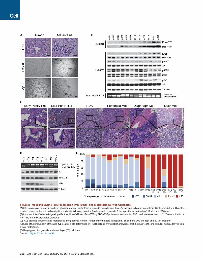
Figure 2. Modeling Murine PDA Progression with Tumor- and Metastasis-Derived Organoids(A) H&E staining of murine tissue from which tumor and metastasis organoids were derived (top). Arrowhead indicates metastasis. Scale bars, 50 mm. Digested
murine tissues embedded in Matrigel immediately following isolation (middle) and organoids 3 days postisolation (bottom). Scale bars, 200 mm.
(B) Immunoblots of selected signaling effectors, Kras-GTP and Ras-GTP by RBD-GST pull-down, and tubulin. PCR confirmation of KrasLSL-G12D recombination in
mP, mT, and mM organoids (bottom).
(C) H&E staining of tumors and metastases (Met) derived from mT organoid orthotopic transplants. Scale bars, 200 mm (top) and 50 mm (bottom).
(D) Loss of heterozygosity of thewild-type Trp53 allele determined by PCR (top) and immunoblot analysis of Trp53, Smad4, p16, and Tubulin. mM3L, derived from
a liver metastasis.
(E) Karyotypes of organoids and monolayer (2D) cell lines.
See also Figure S2 and Table S2.
328 Cell 160, 324–338, January 15, 2015 ª2015 Elsevier Inc.

Figure 3. Human Pancreatic Ductal Organoids Recapitulate Features of Normal and Neoplastic Ducts
(A) Representative images (top) and H&E staining (middle) of human organoid cultures established from normal tissues (hN1-2), resected primary tumors (hT1-2),
a resected metastatic lung lesion (hM1), and a fine-needle aspiration biopsy of a metastatic lesion (hFNA2). H&E staining of the resected tissues from which the
organoids were derived (bottom). Scale bars, 500 mm (top), 250 mm (middle), and 500 mm (bottom).
(B) Representative images of hN and hT organoids cultured for 2 weeks (1 passage) in human complete media or in human complete media lacking the indicated
factors. Scale bars, 500 mm.
(C) Number of passages hN and hT organoids could be propagated in the absence of the indicated factors.
(D) Targeted sequencing analysis of human organoids. Genes altered in more than one sample and/or known to be mutated in PDA are shown. If multiple
mutations were found in a gene, only onemutation per gene is shown. Color key for the type of genetic alterations is shown.Met indicates organoids derived from
metastatic samples.
See also Figure S3 and Tables S3 and S4.
Cell 160, 324–338, January 15, 2015 ª2015 Elsevier Inc. 329

dysplastic tall columnar cells, resembling low-grade PanINs (Fig-
ures 3A). hT organoids tolerated the withdrawal of certain growth
factors from the media (Figures 3B and 3C).
85% of pancreatic cancer patients are ineligible for surgical
resection of their tumors (Ryan et al., 2014). Therefore, we deter-
mined whether hT organoids could be generated from the limited
amount of cellular material provided by endoscopic biopsies
using fine needle aspirations (FNA). Initial attempts to generate
organoids from FNA biopsies were hampered by loss of cellular
material during digestion. Upon optimization of these conditions,
human FNA biopsy organoids (hFNA) were generated from two
specimens that were not dissociated prior to suspension in Ma-
trigel (Figures 3A and S3 and Table S3). This approach is broadly
applicable to PDA patients and enables serial sampling.
Targeted sequencing of 2,000 cancer-associated genes was
performed on hN and hT organoids. As expected, no mutations
were detected in the hN organoid cultures. These analyses
identified oncogenic KRAS mutations in the majority of tumor-
derived samples (n = 8), as well as mutations in TP53 (n = 7),
SMAD4 (n = 5), and CDKN2A (n = 4) (Figure 3D and Table S4).
We also noted amplification of known oncogenes, such as
MYC (n = 4), and loss of tumor suppressors, including TGFBR2
(n = 3) and DCC (n = 5). Importantly, the same KRAS mutations
observed in several hT organoids were confirmed in the primary
PDA from which they were derived (Table S4). The allele fre-
quency of oncogenic KRAS variants in hT1–hT5 and hFNA2
ranged from �50–100%. In contrast, the KRASG12V allele fre-
quency in hFNA1 was only 1% (Table S4), which may result
from coexistence of wild-type ductal cells. Although KRAS
mutations were not detected in hT8 (Figure 3D and Table S4),
the presence of mutations in known PDA genes (ARID1A and
MLL3) suggests that hT8 contains malignant cells (Table S4).
To further characterize the cell types present in primary PDA
organoids, we evaluated the expression of pancreatic lineage
markers. hN and hT organoids expressed markers of ductal
cells, but not other pancreatic lineages (Figure 4A). The karyo-
types of hT organoids were highly aneuploid, whereas the hN
organoids were predominantly and stably diploid (Figure 4B).
The PDA-associated biomarker CA19-9 (Makovitzky, 1986)
was also elevated in hT relative to hN organoids (Figure 4C).
The hN and hT organoids are therefore reflective of normal and
neoplastic human pancreatic ductal cells and offer a model sys-
tem to explore pancreatic cancer biology in the more genetically
complex background of human cancer.
Following orthotopic transplantation into Nu/Nu mice, hN
organoids produced normal ductal structures at low efficiency
(n = 2/23), whereas hT organoids efficiently generated a spec-
trum of low- and high-grade, extraductal PanIN-like lesions
within 1 month (n = 9/12) (Figures 4D and S4A and Table S4D).
The hT-derived transplants initially formed well-defined hollow
lesions lined by a single layer of columnar epithelial cells with
apical mucin and basally located, relatively uniform nuclei. The
nuclei were small and lacked the pleomorphism and hyperchro-
masia often seen in invasive PDA. These lesions progressed over
several months to infiltrative carcinoma comprised of poorly
defined and invasive glands (Figures 4D and S4A and Table
S4). A prominent desmoplastic reaction was present in hT-
derived PanIN-like structures and PDA, including the deposition
330 Cell 160, 324–338, January 15, 2015 ª2015 Elsevier Inc.
of a collagen-rich stroma and the recruitment of aSMA-positive
cells (Figure S4B). The mutation or loss of TP53 or SMAD4 in
hT1 and hT2 was also detected by IHC in these tumors (Fig-
ure S4C and Table S4). Overall, hT organoids represent a trans-
plantable model of human pancreatic cancer progression.
Gene Expression Analysis of Murine Pancreatic DuctalOrganoids Implicates Candidate Genes in PDAProgressionThe mouse organoids were prepared from syngeneic mice, of-
fering the ability to discern gene expression changes in organo-
ids and determine whether these changes correlate with PDA
progression. We harvested RNA from mN (n = 7), mP (n = 6),
and mT (n = 6) organoids and generated strand-specific RNA-
sequencing (RNA-seq) libraries. Sequences were mapped to
the mm9 version of the mouse genome, and relative transcript
abundances (transcripts per million) of 29,777 mouse genes
were determined (Table S5). Principal component analysis
revealed that mN organoids were distinct frommP andmT orga-
noids (Figure 5A and Table S5).
Genes whose levels differed significantly among mN, mP, and
mT organoids were identified. 772 genes were found downregu-
lated and 863 genes upregulated in mP relative to mN organoids
(Figure 5B and Table S5). WhenmT organoids were compared to
mNorganoids, 2,721 genes were downregulated and 2,695were
upregulated. In addition, 823geneswere downregulated and 640
genes were upregulated in mT relative to mP organoids. Distinct
patterns of gene expression were found in the data set (Fig-
ure 5C). Themajority of genes differentially expressed inmP rela-
tive tomNorganoids changed in a similarmanner inmT relative to
mN organoids (Figure 5D). However, a much larger cohort of
genes changed in expression inmT relative tomN than inmP rela-
tive to mN organoids (Figure 5D), suggesting that mP organoids
represent an intermediate state between mN and mT organoids.
The glycosyltransferase Gcnt3 and putative protein disulfide
isomerase Agr2 were among the most upregulated genes in
both mP and mT organoids and have been demonstrated to be
elevated in human PDA (Figure 5E) (Dumartin et al., 2011; Zhao
et al., 2014). The most upregulated gene in both mP and mT
relative to mN organoids was the acyl-CoA synthetase Acsm3
(Figure 5E). RNA-seq results were confirmed by qRT-PCR for
35 out of 40 genes (Table S5), including the upregulation of
Agr2, Acsm3, Gcnt1, Gcnt3, and Ugdh and the downregulation
of Ptprd in mP and mT organoids (Figure 5F and Table S5).
Among the genes upregulated in mP and mT relative to mN
organoids, Gcnt1, Gcnt3, Acsm3, Agr2, Syt16, Nt5e, and Ugdh
were upregulated following the Ad-Cre-induced expression of
oncogenic KrasG12D, suggesting that these genes are activated
downstream of mutant KrasG12D (Figure S5A). To determine
whether organoid RNA-seq profiles resembled gene expression
patterns in vivo, we compared our organoid RNA-seq data to
a published transcription profile of murine pancreatic tumors
uponKrasG12D inactivation (Ying et al., 2012). Genes differentially
expressed upon inactivation of oncogenic Kras overlapped
significantly with those up or downregulated in mP or mT relative
to mN organoids (Figure S5B). These analyses demonstrate the
ability of the organoid system to identify molecular alterations
associated with PDA progression.
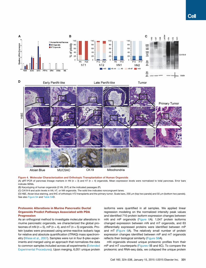
Figure 4. Molecular Characterization and Orthotopic Transplantation of Human Organoids
(A) qRT-PCR of pancreas lineage markers in hN (n = 3) and hT (n = 4) organoids. Mean expression levels were normalized to total pancreas. Error bars
indicate SEMs.
(B) Karyotyping of human organoids (2 hN, 2hT) at the indicated passages (P).
(C) CA19-9 and actin levels in hN, hT, or hM organoids. The solid line indicates noncongruent lanes.
(D) H&E, Alcian blue staining, and IHC of orthotopic hT2 transplants and the primary tumor. Scale bars, 200 mm (top two panels) and 50 mm (bottom two panels).
See also Figure S4 and Table S4D.
Proteomic Alterations in Murine Pancreatic DuctalOrganoids Predict Pathways Associated with PDAProgressionAs an orthogonal method to investigate molecular alterations in
murine pancreatic organoids, we characterized the global pro-
teomes of mN (n = 5), mP (n = 4), and mT (n = 5) organoids. Pro-
tein lysates were processed using amine-reactive isobaric tags
for relative and absolute quantification (iTRAQ) mass spectrom-
etry (Wiese et al., 2007). Samples were run in four 8-plex exper-
iments and merged using an approach that normalizes the data
to common samples included across all experiments (Extended
Experimental Procedures). Upon merging, 6,051 unique protein
isoforms were quantified in all samples. We applied linear
regression modeling on the normalized intensity peak values
and identified 710 protein isoform expression changes between
mN and mP organoids (Figure 6A). 1,047 protein isoforms
changed expression between mN and mT organoids, and 63
differentially expressed proteins were identified between mP
and mT (Figure 6A). The relatively small number of protein
expression changes identified between mP and mT organoids
reflects their biological similarity (Figure S6A).
mN organoids showed unique proteomic profiles from their
mP and mT counterparts (Figures 6B and 6C). To compare the
proteomic and RNA-seq data, we collapsed the unique protein
Cell 160, 324–338, January 15, 2015 ª2015 Elsevier Inc. 331

Figure 5. Gene Expression Analysis of Murine Organoids Reveals Genetic Changes Correlated with Pancreatic Cancer Progression
(A) Principal component analysis of gene expression data for mN, mP, and mT organoids.
(B) The number of genes differentially expressed (DESeq adjusted p value < 0.05) among mN (n = 7), mP (n = 6), and mT (n = 6) organoids.
(C) Heatmap showing relative expression levels using Z score normalization among mN, mP, and mT organoids. Color key of Z score is shown.
(D) Venn diagrams show overlap of genes significantly differentially expressed in mP andmT relative tomN organoids. The p values for overlaps were determined
by two-tailed Fisher’s exact test.
(E) Genes with the largest fold changes in mP or mT relative to mN organoids.
(F) qRT-PCR validation of mN,mP andmT organoid gene expression changes. Values were normalized tomean levels inmN organoids. n = 8mN, 7mP, and 8mT
organoid cultures. Error bars indicate SEMs. *p < 0.05, **p < 0.01, ***p < 0.001, and ns, not significant by two-tailed Student’s t test.
See also Figure S5 and Table S5.
isoforms into their corresponding 4,155 genes. Some protein
expression changes (e.g., 123/150 for downregulated and 96/
151 for upregulated mP proteins) did not reflect corresponding
transcriptional changes, indicating that protein stability may
play a role in cancer progression, particularly in mP organoids
(Figure 6D). Nonetheless, the proteomic data validated many
of the expression changes identified by RNA-seq (Figure 6D),
including upregulation of Gcnt3, Agr2, and Ugdh (Table S6).
Additionally, of the 1,599 genes whose expression levels
changed in mT relative to mN organoids that were measured
by mass spectrometry, 301 (19%) showed corresponding
protein changes (Figure 6D).
Gene Set Enrichment Analysis (GSEA) on the RNA-seq and
proteomic data (Subramanian et al., 2005) revealed elevated
332 Cell 160, 324–338, January 15, 2015 ª2015 Elsevier Inc.
expression of genes and proteins involved in glutathione meta-
bolism and biological oxidations in mP relative to mN organoids
(Figures 6E, S6B, and S6C and Table S7), which is consistent
with elevations in reactive oxygen species metabolism previ-
ously reported in KrasG12D cells (DeNicola et al., 2011; Ying
et al., 2012). Enrichment of proteins involved in glutathionemeta-
bolism was also found in mT relative to mN organoids (Table S7).
Additionally, we identified a significant positive enrichment of
proteins involved in the steroid biosynthesis, cholesterol biosyn-
thesis, one carbon pool by folate, and pyrimidine metabolism
pathways (Figures 6E, S6B, and S6C and Table S7), which is
consistent with an earlier report (Ying et al., 2012). Similar
pathways were enriched in mP relative to mN organoids (choles-
terol biosynthesis, one carbon pool by folate, and pyrimidine

Figure 6. Proteomic Profiling of Murine Organoids Uncovers Molecular Pathways Linked to Pancreatic Cancer Progression
(A) Protein expression changes by iTRAQ proteomic analysis of murine organoids. Both unique protein isoforms and protein isoforms encoded by the same gene
are included (adjusted p value < 0.1 by linear regression analysis).
(B) Heatmap of unique protein isoforms that differ (adjusted p value < 0.05) among mN, mP, and mT organoids. Color key of the Z score is shown.
(C) Venn diagrams showing overlaps between proteins differentially expressed (p < 0.05) in mP and mT relative to mN organoids. p values for overlaps were
determined by two-tailed Fisher’s exact test.
(D) Venn diagrams showing overlaps between genes and proteins found differentially expressed byRNA-seq and proteomic analyses (adjusted p < 0.05). p values
for the overlaps were determined by two-tailed Fisher’s exact test.
(E) Molecular pathways found enriched by GSEA analysis of RNA-seq and proteomic data. Normalized enrichment scores (NESs), p and q values are shown.
(F) Heatmap showing relative gene expression levels of nucleoporins in mN, mP, and mT organoids determined by RNA-seq. Color key of the Z score is shown.
See also Figure S6 and Tables S6 and S7.
metabolism) (Figures S6B and S6C and Table S7), whereas fatty
acid metabolism and TCA cycle/respiratory electron transport
pathways were downregulated (Figure S6C and Table S7).
The increase in anabolic and decrease in catabolic pathways
suggest that complex alterations in fatty acid and nucleotide
metabolism occur during PDA progression.
Interestingly, we also found broad upregulation of the nucleo-
porin family at both the RNA and protein levels in the mT relative
tomNorganoids (Figures 6E and 6F and Table S6). The individual
nucleoporins NUP214, NUP153, and NUPL1 were previously
identified in shRNA dropout screens in PDA cell lines (Cheung
et al., 2011; Shain et al., 2013). Furthermore, amplification of
NUP153 was detected in one human PDA cancer cell line,
and elevation of NUP88 was detected in human primary PDA
(Cheung et al., 2011; Gould et al., 2000; Shain et al., 2013).
This systematic analysis of molecular alterations in pancreatic
organoids implicates nuclear transport as a pathway correlated
with pancreatic cancer progression.
In Vivo Mouse and Human Validation of CandidatesAssociated with PDA Progression in OrganoidsTo demonstrate that the mouse organoid culture system
represents a biological resource for the accurate discovery of
genes associated with PDA progression, we selected 16 genes
Cell 160, 324–338, January 15, 2015 ª2015 Elsevier Inc. 333

Figure 7. Increased Levels of ACSM3, NT5E, and GCNT3 Correlate with Mouse and Human PDA Progression
(A) IHC analysis of 14 candidate genes in mouse adjacent normal ducts, mPanlN and mPDA. Differential expression is indicated as � (negative), + (weak), ++
(moderate), or +++ (strong). Only the ductal component of the normal pancreas was scored.
(B) IHC analysis of Acsm3, Nt5e, and Gcnt3 in mouse normal ducts, mPanlN and mPDA tissues. Arrow indicates adjacent normal ducts in mPanlN tissues.
Arrowhead indicates mPanlN or mPDA. Scale bars, 50 mm.
(C) IHC analysis of seven candidate genes in human normal pancreas, hT orthotopic transplants, and PDA tissues. Differential expression is indicated as �(negative), + (weak), ++ (moderate), or +++ (strong). Only the ductal component of the normal pancreas was scored.
(D) IHC analysis of ACSM3, NT5E, and GCNT3 in human normal pancreas and PDA tissues. Arrow indicates normal ducts, and arrowhead indicates PDA. Scale
bars, 50 mm.
See also Figure S7.
upregulated in mT organoids for validation in primary tissue
specimens by IHC and immunofluorescence (IF) (Figure 7A).
These 16 genes included enzymes, membrane proteins, struc-
tural proteins, and secreted ligands, which could represent
candidate biomarkers and therapeutic targets. Of the 14 anti-
bodies that generated a detectable signal on murine pancreatic
tissue sections, 13 antibodies confirmed the increased expres-
334 Cell 160, 324–338, January 15, 2015 ª2015 Elsevier Inc.
sion of the candidate protein in mPanIN and mPDA lesions in
concordance with the RNA-seq and proteomic data (Figures
7A, 7B, and S7A). 11 of the 13 candidate antibodies were
compatible for evaluation in human tissues, and 7 of these can-
didates were upregulated in human PDA when compared to
normal pancreatic ductal tissues (Figures 7C, 7D, and S7B).
The high expression of many of these markers was recapitulated

in orthotopic transplants of hT organoids into Nu/Nu mice (Fig-
ure 7C). These results indicate that the organoid culture system
accurately models PDA progression and can serve as a resource
for the discovery and genetic dissection of pathways driving
human pancreatic tumorigenesis.
DISCUSSION
We have established pancreatic organoids as a tractable and
transplantable system to probe the molecular and cellular prop-
erties of neoplastic progression in mice and humans. In contrast
to prior reports (Agbunag andBar-Sagi, 2004; Rovira et al., 2010;
Seaberg et al., 2004), our culture conditions prevent the rapid
exhaustion of normal ductal cells in vitro and generate a normal
ductal architecture following orthotopic transplantation. Impor-
tantly, the ability to passage and transplant both normal and
neoplastic ductal cells enables a detailed analysis of molecular
pathways and cellular biology that is not possible when neonatal
pancreatic fragments are propagated in air-liquid interfaces or
when induced pluripotent cells are employed (Agbunag and
Bar-Sagi, 2004; Kim et al., 2013; Li et al., 2014). Our finding
that nucleoporins are broadly upregulated in the neoplastic mu-
rine organoids, coupled with the known associations of nucleo-
porins to cell proliferation and cell transformation, presents a
class of proteins to investigate in pancreatic cancer progression
(Gould et al., 2000; Kohler and Hurt, 2010). Furthermore, the
ability to systematically characterize human pancreatic cancer
organoids that lack KRAS mutations, such as hT8, will reveal
driver genes for PDA. Finally, because organoids can be readily
established from small patient biopsies, they should hasten the
development of personalized approaches for pancreatic cancer
patients.
EXPERIMENTAL PROCEDURES
Animals
Trp53+/LSL-R172H, Kras+/LSL-G12D, and Pdx1-Cre strains in C57Bl/6 back-
ground were interbred to obtain Pdx1-Cre; Kras+/LSL-G12D (KC) and Pdx1-
Cre; Kras+/LSL-G12D; Trp53+/LSL-R172H (KPC) mice (Hingorani et al., 2005).
The R26LSL-YFP strain was interbred to get the desired genotype. C57Bl/6
and athymic Nu/Nu mice were purchased from Charles River Laboratory
and Jackson Laboratory. All animal experiments were conducted in accor-
dance with procedures approved by the IACUC at Cold Spring Harbor
Laboratory (CSHL).
Murine Pancreatic Ductal Organoid Culture
Detailed procedures to isolate normal pancreatic ducts have been described
previously (Huch et al., 2013a). In brief, normal and preneoplastic pancreatic
ducts were manually picked after enzymatic digestion of pancreas with
0.012% (w/v) collagenase XI (Sigma) and 0.012% (w/v) dispase (GIBCO) in
DMEM media containing 1% FBS (GIBCO) and were seeded in growth-fac-
tor-reduced (GFR) Matrigel (BD). For tumors and metastases, bulk tissues
were minced and digested overnight with collagenase XI and dispase and
embedded in GFR Matrigel.
Human Specimens
Pancreatic cancer tissues and adjacent normal pancreas were obtained from
patients undergoing surgical resection at the University Medical Centre
Utrecht Hospital, Memorial Sloan-Kettering Cancer Center (MSKCC), MD An-
derson Cancer Center (MDACC), and Weill Cornell Medical College (WCMC).
Normal pancreatic tissue was also obtained from islet transplant programs at
the University of Illinois at Chicago and University of Miami Miller School of
Medicine. All human experiments were approved by the ethical committees
of the University Medical Centre Utrecht or the IRBs of MSKCC, MDACC,
WCMC, and CSHL. Written informed consent from the donors for research
use of tissue in this study was obtained prior to acquisition of the specimen.
Samples were confirmed to be tumor or normal based on pathological
assessment.
Human Pancreatic Tumor and Normal Organoid Culture
Tumor tissue was minced and digested with collagenase II (5 mg/ml,
GIBCO) in human complete medium (see below) at 37�C for a maximum
of 16 hr. The material was further digested with TrypLE (GIBCO) for
15 min at 37�C, embedded in GFR Matrigel, and cultured in human com-
plete medium (AdDMEM/F12 medium supplemented with HEPES [13, Invi-
trogen], Glutamax [13, Invitrogen], penicillin/streptomycin [13, Invitrogen],
B27 [13, Invitrogen], Primocin [1 mg/ml, InvivoGen], N-acetyl-L-cysteine
[1 mM, Sigma], Wnt3a-conditioned medium [50% v/v], RSPO1-conditioned
medium [10% v/v, Calvin Kuo], Noggin-conditioned medium [10% v/v] or
recombinant protein [0.1 mg/ml, Peprotech], epidermal growth factor [EGF,
50 ng/ml, Peprotech], Gastrin [10 nM, Sigma], fibroblast growth factor 10
[FGF10, 100 ng/ml, Preprotech], Nicotinamide [10 mM, Sigma], and A83-
01 [0.5 mM, Tocris]).
Normal samples were processed as above, except that the collagenase
digestion was done for a maximum of 2 hr in the presence of soybean
trypsin inhibitor (1 mg/ml, Sigma). Following digestion, cells were embedded
in GFR Matrigel and cultured in human complete medium with the addition
of PGE2 (1 mM, Tocris).
Additional experimental details and methods can be found in the Extended
Experimental Procedures.
ACCESSION NUMBERS
All RNA-seq data are available at Gene Expression Omnibus (GEO) under
accession number GSE63348. The proteomic raw data are available at
PeptideAtlas under accession number PASS00625. The targeted DNA-
sequencing data are available at EMBL European Nucleotide Archive under
the accession number ERP006373.
SUPPLEMENTAL INFORMATION
Supplemental Information includes Extended Experimental Procedures, seven
figures, and seven tables and can be found with this article online at http://dx.
doi.org/10.1016/j.cell.2014.12.021.
AUTHOR CONTRIBUTIONS
S.F.B. initiated the project, developed themethods for isolatingmouse and hu-
man organoids, and characterized human organoids (Figures 1A, 3, 4B, 4D,
and S4C and Tables S3 and S4). C.-I.H. developed transplantation models
for organoids and performed shRNA knockdown and histological and karyo-
typic analyses (Figures 1A, 1E, 1G, 2A, 2C–2E, 4D, 7, S1A–S1C, S2A–S2F,
S2H, S4A, S4B, S7A, and S7B and Tables S1, S2, and S4). L.A.B. performed
RNA-seq on mouse organoids and analyzed RNA-seq and proteomic data
(Figures 5, 6, S5B, and S6 and Tables S5, S6, and S7). I.I.C.C. conducted pro-
teomic evaluation of mouse organoids and analyzed proteomic data (Figures 6
and S6C). D.D.E. developed mouse organoid methods and evaluated CA19-9
levels in human organoids (Figures 1A, 2A, 4C, S3, and S6 and Table S6). V.C.
developed human organoid methods, performed molecular analyses of orga-
noids, and prepared material for DNA-sequencing and sequencing of Kras
(Figures 1C, 1D, 3A, 3D, 4A, S1D–S1F, and S5A and Tables S3, S4, and S5).
M.J. performed and analyzed the DNA sequencing of human organoids (Fig-
ure 3D and Table S4). Mouse and human organoid preparation and character-
ization was performed byM.P.-S., H.T., M.S.S., T.O., D.O., A.H.-S., C.M.A.-A.,
M.L., E.E., B.A., M.E.F., G.N.Y., G.B., B.D., B.C., K.W., K.H.Y., Y.P., M. Huch,
A.G., F.H.M.M., and S.D.L. Sequencing analyses were performed by Y.H.,
Y.J., M. Hammell, I.J.N., E.C., and R.v.B. Pathological analyses were
Cell 160, 324–338, January 15, 2015 ª2015 Elsevier Inc. 335

performed by G.J.O., R.H.H., D.S.K., O.B., and C.I.-D. Surgical resections and
tissue dissection were performed by I.Q.M. and I.H.B.R. Proteomic develop-
ment was performed by D.J.P., K.D.R., and J.P.W. Overall study management
was conducted by D.A.T., H.C., and R.G.J.V. S.F.B., D.D.E., L.A.B., M.E.F.,
C.H., H.T., V.C., M.P.S., R.G.J.V., H.C., I.I.C.C., and D.A.T. contributed to
manuscript writing.
ACKNOWLEDGMENTS
We thank Peter Kapitein and Jan Schuurman from Inspire 2 Live for helping to
establish the collaboration between D.A.T. and H.C. We also thank H. Begthel
andJ.Korving for technical assistance.Thisworkwasperformedwithassistance
from the CSHL Proteomic, Histology, DNA Sequencing, Antibody, and Bioinfor-
matics Shared Resources, which are supported by the Cancer Center Support
Grant 5P30CA045508. D.A.T. is a distinguished scholar of the Lustgarten Foun-
dation and Director of the Lustgarten Foundation-designated Laboratory of
PancreaticCancerResearch.D.A.T. is also supportedby theColdSpringHarbor
Laboratory Association, the Carcinoid Foundation, PCUK, and the David Rubin-
stein Center for Pancreatic Cancer Research at MSKCC. In addition, we are
grateful for support from the following: Stand Up to Cancer/KWF (H.C.), the
STARR foundation (I7-A718 for D.A.T.), DOD (W81XWH-13-PRCRP-IA for
D.A.T.), theSolGoldmanPancreaticCancerResearchCenter (R.H.H.), the Italian
Ministry of Health (FIRB - RBAP10AHJ for V.C.), Sociedad Espanola deOncolo-
gıaMedica (SEOMforM.P.S.), LouisMorinCharitableTrust (M.E.F.), theSwedish
ResearchCouncil (537-2013-7277 forD.O.),TheKempeFoundations (JCK-1301
for D.O.) and the Swedish Society of Medicine (SLS-326921, SLS-250831 for
D.O.), the Damon Runyon Cancer Research Foundation (DRG-2165-13 for
I.I.C.C.), the Human Frontiers Science Program (LT000403/2014 for E.E.), the
Weizmann Institute of Science Women in Science Award (E.E.), the American
Cancer Society (PF-13-317-01-CSM for C.M.A.A.), the Hearst Foundation
(A.H.S.), and the NIH (5P30CA45508-26, 5P50CA101955-07, 1U10CA180944-
01, 5U01CA168409-3, and 1R01CA190092-01 for D.A.T.; CA62924 for
R.H.H.; CA134292 for S.D.L.; 5T32CA148056 for L.A.B. and D.D.E.; and
CA101955 UAB/UMN SPORE for L.A.B.). In addition, S.F.B. and M.H. are
supported by KWF/PF-HUBR 2007-3956, A.G is supported by EU/232814-
StemCellMark, and R.G.J.V. is supported by GenomiCs.nl (CGC). M.J., R.B.,
and E.C. are supported by the CancerGenomics.nl (NWOGravitation) program.
Ralph Hruban receives royalty payments fromMyriad Genetics for the PalB2 in-
ventions.HansClevers andMeritxell Huch havepatents pending and granted on
the organoid technology.
Received: August 1, 2014
Revised: November 24, 2014
Accepted: December 10, 2014
Published: December 31, 2014
REFERENCES
Abbruzzese, J.L., andHess, K.R. (2014). New option for the initial management
of metastatic pancreatic cancer? J. Clin. Oncol. 32, 2405–2407.
Agbunag, C., and Bar-Sagi, D. (2004). Oncogenic K-ras drives cell cycle
progression and phenotypic conversion of primary pancreatic duct epithelial
cells. Cancer Res. 64, 5659–5663.
Aguirre, A.J., Bardeesy, N., Sinha, M., Lopez, L., Tuveson, D.A., Horner, J.,
Redston, M.S., and DePinho, R.A. (2003). Activated Kras and Ink4a/Arf defi-
ciency cooperate to produce metastatic pancreatic ductal adenocarcinoma.
Genes Dev. 17, 3112–3126.
Bardeesy, N., Aguirre, A.J., Chu, G.C., Cheng, K.H., Lopez, L.V., Hezel, A.F.,
Feng, B., Brennan, C., Weissleder, R., Mahmood, U., et al. (2006). Both
p16(Ink4a) and the p19(Arf)-p53 pathway constrain progression of pancreatic
adenocarcinoma in the mouse. Proc. Natl. Acad. Sci. USA 103, 5947–5952.
Barker, N., Huch, M., Kujala, P., van de Wetering, M., Snippert, H.J., van Es,
J.H., Sato, T., Stange, D.E., Begthel, H., van den Born, M., et al. (2010).
Lgr5(+ve) stem cells drive self-renewal in the stomach and build long-lived
gastric units in vitro. Cell Stem Cell 6, 25–36.
336 Cell 160, 324–338, January 15, 2015 ª2015 Elsevier Inc.
Beatty, G.L., Chiorean, E.G., Fishman, M.P., Saboury, B., Teitelbaum, U.R.,
Sun, W., Huhn, R.D., Song, W., Li, D., Sharp, L.L., et al. (2011). CD40 agonists
alter tumor stroma and show efficacy against pancreatic carcinoma in mice
and humans. Science 331, 1612–1616.
Cheung, H.W., Cowley, G.S., Weir, B.A., Boehm, J.S., Rusin, S., Scott, J.A.,
East, A., Ali, L.D., Lizotte, P.H., Wong, T.C., et al. (2011). Systematic investiga-
tion of genetic vulnerabilities across cancer cell lines reveals lineage-specific
dependencies in ovarian cancer. Proc. Natl. Acad. Sci. USA 108, 12372–
12377.
Cleveland, M.H., Sawyer, J.M., Afelik, S., Jensen, J., and Leach, S.D. (2012).
Exocrine ontogenies: on the development of pancreatic acinar, ductal and
centroacinar cells. Semin. Cell Dev. Biol. 23, 711–719.
De La O, J.P., Emerson, L.L., Goodman, J.L., Froebe, S.C., Illum, B.E., Curtis,
A.B., and Murtaugh, L.C. (2008). Notch and Kras reprogram pancreatic acinar
cells to ductal intraepithelial neoplasia. Proc. Natl. Acad. Sci. USA 105, 18907–
18912.
DeNicola, G.M., Karreth, F.A., Humpton, T.J., Gopinathan, A., Wei, C., Frese,
K., Mangal, D., Yu, K.H., Yeo, C.J., Calhoun, E.S., et al. (2011). Oncogene-
induced Nrf2 transcription promotes ROS detoxification and tumorigenesis.
Nature 475, 106–109.
Dumartin, L., Whiteman, H.J., Weeks, M.E., Hariharan, D., Dmitrovic, B., Iaco-
buzio-Donahue, C.A., Brentnall, T.A., Bronner, M.P., Feakins, R.M., Timms,
J.F., et al. (2011). AGR2 is a novel surface antigen that promotes the dissem-
ination of pancreatic cancer cells through regulation of cathepsins B and D.
Cancer Res. 71, 7091–7102.
Erkan, M., Reiser-Erkan, C., Michalski, C.W., Deucker, S., Sauliunaite, D.,
Streit, S., Esposito, I., Friess, H., and Kleeff, J. (2009). Cancer-stellate cell
interactions perpetuate the hypoxia-fibrosis cycle in pancreatic ductal adeno-
carcinoma. Neoplasia 11, 497–508.
Frese, K.K., Neesse, A., Cook, N., Bapiro, T.E., Lolkema, M.P., Jodrell, D.I.,
and Tuveson, D.A. (2012). nab-Paclitaxel potentiates gemcitabine activity by
reducing cytidine deaminase levels in a mouse model of pancreatic cancer.
Cancer Discov. 2, 260–269.
Gao, D., Vela, I., Sboner, A., Iaquinta, P.J., Karthaus, W.R., Gopalan, A., Dow-
ling, C., Wanjala, J.N., Undvall, E.A., Arora, V.K., et al. (2014). Organoid cul-
tures derived from patients with advanced prostate cancer. Cell 159, 176–187.
Gidekel Friedlander, S.Y., Chu, G.C., Snyder, E.L., Girnius, N., Dibelius, G.,
Crowley, D., Vasile, E., DePinho, R.A., and Jacks, T. (2009). Context-depen-
dent transformation of adult pancreatic cells by oncogenic K-Ras. Cancer
Cell 16, 379–389.
Gould, V.E., Martinez, N., Orucevic, A., Schneider, J., and Alonso, A. (2000). A
novel, nuclear pore-associated, widely distributed molecule overexpressed
in oncogenesis and development. Am. J. Pathol. 157, 1605–1613.
Guerra, C., Mijimolle, N., Dhawahir, A., Dubus, P., Barradas, M., Serrano, M.,
Campuzano, V., and Barbacid, M. (2003). Tumor induction by an endogenous
K-ras oncogene is highly dependent on cellular context. Cancer Cell 4,
111–120.
Habbe, N., Shi, G., Meguid, R.A., Fendrich, V., Esni, F., Chen, H., Feldmann,
G., Stoffers, D.A., Konieczny, S.F., Leach, S.D., and Maitra, A. (2008). Sponta-
neous induction of murine pancreatic intraepithelial neoplasia (mPanIN) by
acinar cell targeting of oncogenic Kras in adult mice. Proc. Natl. Acad. Sci.
USA 105, 18913–18918.
Hingorani, S.R., Petricoin, E.F., Maitra, A., Rajapakse, V., King, C., Jacobetz,
M.A., Ross, S., Conrads, T.P., Veenstra, T.D., Hitt, B.A., et al. (2003). Preinva-
sive and invasive ductal pancreatic cancer and its early detection in themouse.
Cancer Cell 4, 437–450.
Hingorani, S.R., Wang, L., Multani, A.S., Combs, C., Deramaudt, T.B., Hruban,
R.H., Rustgi, A.K., Chang, S., and Tuveson, D.A. (2005). Trp53R172H and
KrasG12D cooperate to promote chromosomal instability and widely meta-
static pancreatic ductal adenocarcinoma in mice. Cancer Cell 7, 469–483.
Huch, M., Bonfanti, P., Boj, S.F., Sato, T., Loomans, C.J., van deWetering, M.,
Sojoodi, M., Li, V.S., Schuijers, J., Gracanin, A., et al. (2013a). Unlimited in vitro

expansion of adult bi-potent pancreas progenitors through the Lgr5/R-spon-
din axis. EMBO J. 32, 2708–2721.
Huch,M., Dorrell, C., Boj, S.F., van Es, J.H., Li, V.S., van deWetering, M., Sato,
T., Hamer, K., Sasaki, N., Finegold, M.J., et al. (2013b). In vitro expansion of
single Lgr5+ liver stem cells induced by Wnt-driven regeneration. Nature
494, 247–250.
Jacobetz, M.A., Chan, D.S., Neesse, A., Bapiro, T.E., Cook, N., Frese, K.K.,
Feig, C., Nakagawa, T., Caldwell, M.E., Zecchini, H.I., et al. (2013). Hyaluronan
impairs vascular function and drug delivery in a mouse model of pancreatic
cancer. Gut 62, 112–120.
Karthaus, W.R., Iaquinta, P.J., Drost, J., Gracanin, A., van Boxtel, R.,
Wongvipat, J., Dowling, C.M., Gao, D., Begthel, H., Sachs, N., et al. (2014).
Identification of multipotent luminal progenitor cells in human prostate orga-
noid cultures. Cell 159, 163–175.
Kim, M.P., Evans, D.B.,Wang, H., Abbruzzese, J.L., Fleming, J.B., and Gallick,
G.E. (2009). Generation of orthotopic and heterotopic human pancreatic
cancer xenografts in immunodeficient mice. Nat. Protoc. 4, 1670–1680.
Kim, J., Hoffman, J.P., Alpaugh, R.K., Rhim, A.D., Reichert, M., Stanger, B.Z.,
Furth, E.E., Sepulveda, A.R., Yuan, C.X., Won, K.J., et al. (2013). An iPSC line
from human pancreatic ductal adenocarcinoma undergoes early to invasive
stages of pancreatic cancer progression. Cell Rep. 3, 2088–2099.
Kohler, A., and Hurt, E. (2010). Gene regulation by nucleoporins and links to
cancer. Mol. Cell 38, 6–15.
Koong, A.C., Mehta, V.K., Le, Q.T., Fisher, G.A., Terris, D.J., Brown, J.M.,
Bastidas, A.J., and Vierra, M. (2000). Pancreatic tumors show high levels of
hypoxia. Int. J. Radiat. Oncol. Biol. Phys. 48, 919–922.
Kopp, J.L., von Figura, G., Mayes, E., Liu, F.F., Dubois, C.L., Morris, J.P.,
4th, Pan, F.C., Akiyama, H., Wright, C.V., Jensen, K., et al. (2012). Identifica-
tion of Sox9-dependent acinar-to-ductal reprogramming as the principal
mechanism for initiation of pancreatic ductal adenocarcinoma. Cancer Cell
22, 737–750.
Lee, J., Sugiyama, T., Liu, Y., Wang, J., Gu, X., Lei, J., Markmann, J.F.,
Miyazaki, S., Miyazaki, J., Szot, G.L., et al. (2013). Expansion and conversion
of human pancreatic ductal cells into insulin-secreting endocrine cells. eLife 2,
e00940.
Li, X., Nadauld, L., Ootani, A., Corney, D.C., Pai, R.K., Gevaert, O., Cantrell,
M.A., Rack, P.G., Neal, J.T., Chan, C.W., et al. (2014). Oncogenic transforma-
tion of diverse gastrointestinal tissues in primary organoid culture. Nat. Med.
20, 769–777.
Makovitzky, J. (1986). The distribution and localization of the monoclonal anti-
body-defined antigen 19-9 (CA19-9) in chronic pancreatitis and pancreatic
carcinoma. An immunohistochemical study. Virchows Arch. B Cell Pathol.
Incl. Mol. Pathol. 51, 535–544.
Means, A.L., Meszoely, I.M., Suzuki, K., Miyamoto, Y., Rustgi, A.K., Coffey,
R.J., Jr., Wright, C.V.E., Stoffers, D.A., and Leach, S.D. (2005). Pancreatic
epithelial plasticity mediated by acinar cell transdifferentiation and generation
of nestin-positive intermediates. Development 132, 3767–3776.
Morris, J.P., 4th, Cano, D.A., Sekine, S., Wang, S.C., and Hebrok, M. (2010).
Beta-catenin blocks Kras-dependent reprogramming of acini into pancreatic
cancer precursor lesions in mice. J. Clin. Invest. 120, 508–520.
Neesse, A., Frese, K.K., Chan, D.S., Bapiro, T.E., Howat, W.J., Richards, F.M.,
Ellenrieder, V., Jodrell, D.I., and Tuveson, D.A. (2014). SPARC independent
drug delivery and antitumour effects of nab-paclitaxel in genetically engi-
neered mice. Gut 63, 974–983.
Olive, K.P., Jacobetz,M.A., Davidson, C.J., Gopinathan, A., McIntyre, D., Hon-
ess, D., Madhu, B., Goldgraben, M.A., Caldwell, M.E., Allard, D., et al. (2009).
Inhibition of Hedgehog signaling enhances delivery of chemotherapy in a
mouse model of pancreatic cancer. Science 324, 1457–1461.
Perez-Mancera, P.A., Guerra, C., Barbacid, M., and Tuveson, D.A. (2012).
What we have learned about pancreatic cancer from mouse models. Gastro-
enterology 142, 1079–1092.
Prasad, N.B., Biankin, A.V., Fukushima, N., Maitra, A., Dhara, S., Elkahloun,
A.G., Hruban, R.H., Goggins, M., and Leach, S.D. (2005). Gene expression
profiles in pancreatic intraepithelial neoplasia reflect the effects of Hedge-
hog signaling on pancreatic ductal epithelial cells. Cancer Res. 65, 1619–
1626.
Provenzano, P.P., Cuevas, C., Chang, A.E., Goel, V.K., Von Hoff, D.D., and
Hingorani, S.R. (2012). Enzymatic targeting of the stroma ablates physical bar-
riers to treatment of pancreatic ductal adenocarcinoma. Cancer Cell 21,
418–429.
Pylayeva-Gupta, Y., Lee, K.E., Hajdu, C.H., Miller, G., and Bar-Sagi, D. (2012).
Oncogenic Kras-induced GM-CSF production promotes the development of
pancreatic neoplasia. Cancer Cell 21, 836–847.
Rahib, L., Smith, B.D., Aizenberg, R., Rosenzweig, A.B., Fleshman, J.M., and
Matrisian, L.M. (2014). Projecting cancer incidence and deaths to 2030: the un-
expected burden of thyroid, liver, and pancreas cancers in the United States.
Cancer Res. 74, 2913–2921.
Ray, K.C., Bell, K.M., Yan, J., Gu, G., Chung, C.H., Washington, M.K., and
Means, A.L. (2011). Epithelial tissues have varying degrees of susceptibility
to Kras(G12D)-initiated tumorigenesis in a mouse model. PLoS ONE 6,
e16786.
Rovira, M., Scott, S.G., Liss, A.S., Jensen, J., Thayer, S.P., and Leach, S.D.
(2010). Isolation and characterization of centroacinar/terminal ductal progeni-
tor cells in adult mouse pancreas. Proc. Natl. Acad. Sci. USA 107, 75–80.
Rubio-Viqueira, B., Jimeno, A., Cusatis, G., Zhang, X., Iacobuzio-Donahue, C.,
Karikari, C., Shi, C., Danenberg, K., Danenberg, P.V., Kuramochi, H., et al.
(2006). An in vivo platform for translational drug development in pancreatic
cancer. Clin. Cancer Res. 12, 4652–4661.
Ryan, D.P., Hong, T.S., and Bardeesy, N. (2014). Pancreatic adenocarcinoma.
N. Engl. J. Med. 371, 1039–1049.
Sato, T., Vries, R.G., Snippert, H.J., van de Wetering, M., Barker, N., Stange,
D.E., van Es, J.H., Abo, A., Kujala, P., Peters, P.J., and Clevers, H. (2009). Sin-
gle Lgr5 stem cells build crypt-villus structures in vitro without a mesenchymal
niche. Nature 459, 262–265.
Sato, T., Stange, D.E., Ferrante, M., Vries, R.G., Van Es, J.H., Van den Brink,
S., Van Houdt, W.J., Pronk, A., Van Gorp, J., Siersema, P.D., and Clevers,
H. (2011). Long-term expansion of epithelial organoids from human colon,
adenoma, adenocarcinoma, and Barrett’s epithelium. Gastroenterology 141,
1762–1772.
Sawey, E.T., Johnson, J.A., and Crawford, H.C. (2007). Matrix metallopro-
teinase 7 controls pancreatic acinar cell transdifferentiation by activating
the Notch signaling pathway. Proc. Natl. Acad. Sci. USA 104, 19327–
19332.
Seaberg, R.M., Smukler, S.R., Kieffer, T.J., Enikolopov, G., Asghar, Z.,
Wheeler, M.B., Korbutt, G., and van der Kooy, D. (2004). Clonal identification
of multipotent precursors from adult mouse pancreas that generate neural
and pancreatic lineages. Nat. Biotechnol. 22, 1115–1124.
Shain, A.H., Salari, K., Giacomini, C.P., and Pollack, J.R. (2013). Integrative
genomic and functional profiling of the pancreatic cancer genome. BMC
Genomics 14, 624.
Sharma, S.V., Haber, D.A., and Settleman, J. (2010). Cell line-based platforms
to evaluate the therapeutic efficacy of candidate anticancer agents. Nat. Rev.
Cancer 10, 241–253.
Siegel, R., Naishadham, D., and Jemal, A. (2013). Cancer statistics, 2013. CA
Cancer J. Clin. 63, 11–30.
Subramanian, A., Tamayo, P., Mootha, V.K., Mukherjee, S., Ebert, B.L.,
Gillette, M.A., Paulovich, A., Pomeroy, S.L., Golub, T.R., Lander, E.S., andMe-
sirov, J.P. (2005). Gene set enrichment analysis: a knowledge-based approach
for interpreting genome-wide expression profiles. Proc. Natl. Acad. Sci. USA
102, 15545–15550.
Villarroel, M.C., Rajeshkumar, N.V., Garrido-Laguna, I., De Jesus-Acosta, A.,
Jones, S., Maitra, A., Hruban, R.H., Eshleman, J.R., Klein, A., Laheru, D.,
et al. (2011). Personalizing cancer treatment in the age of global genomic
analyses: PALB2 gene mutations and the response to DNA damaging agents
in pancreatic cancer. Mol. Cancer Ther. 10, 3–8.
Cell 160, 324–338, January 15, 2015 ª2015 Elsevier Inc. 337

von Figura, G., Fukuda, A., Roy, N., Liku, M.E., Morris Iv, J.P., Kim, G.E., Russ,
H.A., Firpo, M.A., Mulvihill, S.J., Dawson, D.W., et al. (2014). The chromatin
regulator Brg1 suppresses formation of intraductal papillary mucinous
neoplasm and pancreatic ductal adenocarcinoma. Nat. Cell Biol. 16, 255–267.
Wiese, S., Reidegeld, K.A., Meyer, H.E., and Warscheid, B. (2007). Protein
labeling by iTRAQ: a new tool for quantitative mass spectrometry in proteome
research. Proteomics 7, 340–350.
338 Cell 160, 324–338, January 15, 2015 ª2015 Elsevier Inc.
Ying, H., Kimmelman, A.C., Lyssiotis, C.A., Hua, S., Chu, G.C., Fletcher-San-
anikone, E., Locasale, J.W., Son, J., Zhang, H., Coloff, J.L., et al. (2012). Onco-
genic Krasmaintains pancreatic tumors through regulation of anabolic glucose
metabolism. Cell 149, 656–670.
Zhao, L.L., Zhang, T., Liu, B.R., Liu, T.F., Tao, N., and Zhuang, L.W. (2014).
Construction of pancreatic cancer double-factor regulatory network based
on chip data on the transcriptional level. Mol. Biol. Rep. 41, 2875–2883.