organic constituents of the marine sponge dvsidea …
TRANSCRIPT

Organic constituents of the marine sponge Dysidea etheria, the nudibranch Hypselodoris zebra, and thegorgonian soft coral Briareum polyanthesby Stephen Howard Grode
A thesis submitted in partial fulfillment of the requirements for the degree of Doctor of Philosophy inChemistryMontana State University© Copyright by Stephen Howard Grode (1983)
Abstract:This thesis comprises a report of research into the chemical constituents of the marine sponge Dysideaetheria, the gorgonian soft coral Briareum polyanthes, and the nudibranch Hypselodoris zebra. A totalof eleven compounds were characterized, six of which were new molecules.
The compounds isolated and characterized from D. etheria were the sesquiterpenes furodysinin, 27, andthe heretofore unreported furodysinin lactone, 56, and the ceramides α-hydroxy N-acylsphingo-sines,60, and N-acylsphingosines, 61. The extraction and subsequent fractionation of H. zebra yielded thefuranosesquiterpenes furodysinin, euryfuran, 34, 5-acetoxy nakafuran—8, 24, and5-hydroxynakafuran8, 59. Briareum polyanthes was the source of a series of novel, highlyfunctionalized diterpenes which possessed the briaran carbon skeleton. These molecules were given thetrivial names, brianthein X, 69, Y, 68, and Z, 67.
Characterization of all isolates was accomplished, predominantly, by analysis of NMR, MS, IR, andUV data on the purified compounds and their chemical derivatives. The structure of brianthein Y wasconfirmed by an X—ray diffraction analysis.

ORGANIC CONSTITUENTS OF THE MARINE SPONGE Dvsidea etheria. THE
NUDIBRANCE Hypselodoris zebra, AND THE GORGONIAN
SOFT CORAL Briarepm uolvanthes
by
Stephen Howard Grode
A thesis submitted in partial fulfillment of the requirements for the degree
of
Doctor of Philosophy
in
Chemistry
MONTANA STATE UNIVERSITY Bozeman, Montana
March 1983

D 3 T %
APPROVAL
ii
of a thesis submitted by
Stephen Howard Grode
This thesis has been read by each member of the thesis committee and has been found to be satisfactory regarding content, English usage, format, citations, bibliographic style, and consistency, and is ready for submission to the College of Graduate Studies.
f da /yg-3Date airperson, Graduate Committee
Approved for the Major Department
[ n nDate Head, Major Department
Approved for the College of Graduate Studies
"3 ~ F 'IDate Graduate Dean

STATEMENT OF PERMISSION TO USE
In presenting this thesis in partial fulfillment of the require
ments for a doctoral degree at Montana State University, I agree that
the Library shall make it available to borrowers under rules of the
Library. I further agree that copying of this thesis is allowable
only for scholarly purposes, consistent with "fair use" as pre
scribed in the U.S. Copyright Law. Requests for extensive copying or
reproduction of this thesis should be referred to University Micro
films International, 300 North Zeeb Road, Ann Arbor, Michigan 48106,
to whom I have granted the "exclusive right to reproduce and distri
bute copies of the dissertation in and from microfilm and the right
to reproduce and distribute by abstract in any format."
iii
Signatur e
Date // /9/S’

iv
To my grandparents, whose foresight and fortitude brought them to theshores of this great nation.
To my parents, who allowed me the freedom to find my own way.
To my wife.

V
ACKNOWLEDGMENT
Acknowledging all those responsible for my professional develop
ment at Montana State University would not be possible. I would,
however, like to thank Thomas R. James, Jr., for the isolation of the
hrianthein series. Thanks are also due to Dr. Robert T. Orth for
helpful discussions on mass spectrometry and Richard Taylor and Dr.
Mark Waddington for helpful instruction on the NMR spectrometer. The
assistance of William Grey in setting up the plant pathogenic antimi
crobial assays is also gratefully acknowledged. Dr. Bradford P.
Mundy, Dr. P.W. Jennings, and their respective research groups also
deserve to be thanked for allowing me the occasional use of their
chemicals and glassware.
The most important acknowledgement is due my advisor. Dr. John
E. Cardellina II, whose wit and good cheer were a source of constant
encouragement during the trials and tribulations encountered during
the course of this research project.
List of Tables ........................................viii
List of Figures................................... .. ix
List of Schemes.............................................. ..
Abstract..................... xii
Introduction .................................... . . . . . . I
Statement of Problem . . . . . . ................... . . . . 17
Results and Discussion . . . . . . . . . . . . . .......... 18
Sesquiterpenes from Dvsidea etheria. . . ............... 18
Furanosesquiterpenes from Evnselodoris zebra ........... 39
Pharmacological Activity of Furodysinin and Furodysinin Lactone..................... 40
Ceramides from Dvsidea etheria . . . ........ . . . . . 41
Pharmacological Activity of the Ceramides.............. .57
Diterpenes from Briareum noIvanthes.......... 58
Pharmacological and Insecticidal Activity of the Briantheins. ..................... 80
Conclusion . . . . . . . . .................................. 83
Experimental . . . . . . . . ........ . ................... 86
Physical Measurements . . . . . . . . ................. 86
Pharmacological and Insecticidal Screening ............. 87
Collection and Extraction of Dvsidea etheria ........... 90
Crude Separation of 1979 Collection.................... 91
Isolation of Furodysinin . ................. 91
Isolation of Furodysinin Lactone ............ . . . . . 92
viTABLE OF CONTENTS
Page
(continued)

TABLE OF CONTENTS (continued)Page
Oxidation of Furodysinin..................... .. . . . . 92
Photooxidation of Furodysinin in Methanol. . . . . . . . 93
Photooxidation of Furodysinin in Tetrahydrofuran . . . . 93LIS Study of Furodysinin Lactone ....................... 94
LIS Study of Semisynthetic Furodysinin Lactone . . . . . 95
Collection and Extraction of Evnselodoris zebra........ 95
Isolation of Furanosesquiterpenes. . . . . ............. 95
Isolation of Ceramides . . . . . . . . . . . . . . . . . 96
Acetylation of a—Hydroxy-N—acylsphingosines. . . . . . . 97
Acetylation of N-Acylsphingosine . . . . . . . . . . . . 98
Hydrolysis of a—Hydroxy—N-acylsphingosines ...............98
Hydrolysis of N-Acylsphingosines . . . . . . . . . . . . 99
Acetylation of Sphingosine . . . . . . . . . . . . . . . 99
Collection and Extraction of Briareum nolvanthes . . . .100
Partitioning and Fractionation of Crude Extract. . . . .100
Isolation of Briantheins Z and Y .......... .. .101
Isolation of Brianthein X. . . ............. .. .101
Characterization of Briantheins X, Y, and Z. . . . . . .102
Acetylation of Brianthein X........ . . . . . . . . . .102
References Cited..................... 105
vii
viii
Table Page
1. ^K-NMR Assignments for Furodysinin Lactone . . . . . . . . . .24132. C-NMR Assignments, Furodysinin Lactone andFurodysinin . . . . . . . . . . . . . . . 3 1
3. Slopes from the Plot of AS vs.[Eu(fod)g]/[Substrate] ........ . . . . . . . . . . . . . .34
4. Agreement Factors for Furodysinin Lactone and its C-4Epimer . . . . . . . . . . . . . . . . . . . . . . . . . . .37
5. ^H-NMR Assignments for the Ceramide Acetates . . . . . . . .47
6. ^H-NMR Assignments for the Ceramides . . . . . . . . . . . .52
7. Optical Rotations, Esters of 2-Hydroxyacids. . . . . . . . .56
8. ^H-NMR Assignments for Briantheins X, T, and Z . . . . . . .66
9. ^C-NMR Assignments for Briantheins X, Y, and Z............ 75
10. Chromatography Packing Material. . . . . . . . . . . . . . . .87
11. Test Organisms for Antimicrobial Assays. ................... 89
LIST OF TABLES

LIST OF FIGURES
Figure Page131. C-NMR Spectrum of Furodysinin Lactone 21
2. Mass Spectrum of Furodysinin Lactone.................22
3. ^H-NMR Spectrum of Furodysinin Lactone. . . . . . . . 23
4. NMR Spectrum of Furodysinin. . . . . . . . . . . . 27
5. Mass Spectrum of Furodysinin. . . . . . . . . . . . . 28
6. Inverse Gated ^C-NMR Spectrum of Furodysinin . . . . 29
7. ^C-NMR Spectra of the Highfield Region of Furodysinin . . . . . . . . . . . . . . . . . . . . . 32
8. Plot of Change in Chemical Shift vs. Equivalents Eu(fod)g. . . . . . . . . . . . . . . . . . . . . . . 35
9. Plot of R Values vs. Europium—Oxygen Bond Distance. . 38
10. ^H-NMR Spectrum of Ceramide 60. . . . . . . . 42
11. ^H-NMR Spectrum of Geramide 61. . . . . . . . . . . . 43
12. ^H-NMR Spectrum of Ceramide Diacetate 60a . . 45
13. ^H-NMR Spectrum of Ceramide Triacetate 61a . . . . . 46
14. ^C-NMR Spectrum of Ceramide 60 . . . . . . . . . . . 49
15. Inverse Gated "^C-NMR Spectrum of Ceramide 61 » . . .501 316. Off Resonance Decoupled C-NMR Spectrum of
Ceramide 6 1 ................................. .. . . . 51
17. o-Hydroxylation of Fatty Acids. . . . . . . . . . . . 57
18. ^^C-NMR Spectrum of Brianthein Z. . . . . . . . . . . 63
19. ^H-NMR Spectrum of Brianthein Z . . . . . . . . . . . 64
20. Mass Spectrum of Brianthein Z . . . . . . . . . . . . 65(continued)
ix

LIST OF FIGURES (continued)
21. ^H-NMR Spectrum of Brianthein Y . . . . . . . . . . . 70
22. ^G-NMR Spectrum of Brianthein Y. . . . ............. 71
23. Mass Spectrum of Briathein X. . . . . . . . . . . . . 72
24. ^H-NMR Spectrum of Brianthein X . . . . . . . . . . . 73
25. ^C-NMR Spectrum of Brianthein X. . . . . . . . . . . 74
26. Computer Generated Perspective Drawing of1 1 1 X ♦ e e e e e e e e e e o e e e e « e e e o 7 8
27. Mass Spectrum of the Glycerol—KI Matrix . . . . . . .103
X

Scheme Page
1. Proposed Biosynthetic Pathways from Geraniolin the Marine and Terrestrial Environments. . . . . . . 2
2. Proposed Biosynthetic Pathway of Disidein ............ 11
xiLIST OF SCHEMES
I I

Xii
ABSTRACT
This thesis comprises a report of research into the chemical constituents of the marine sponge Dysidea etheria. the gorgonian soft coral Briareum polyanthes, and the nudibranch Evnselodoris zebra. A total of eleven compounds were characterized, six of which were new molecules.
The compounds isolated and characterized from D. etheria were the sesquiterpenes furodysinin, 27, and the heretofore unreported furodysinin lactone, 56, and the ceramides a-hydroxy N-acylsphingo- sines, 60, and N-acylsphingosines, 61. The extraction and subsequent fractionation of E; zebra yielded the furanosesquiterpenes furodysinin, euryfuran, 34, 5-acetoxy nakafuran—8, 24, and 5-hydroxy nakafuranS, 59. Briareum polyanthes was the source of a series of novel, highly functionalized diterpenes which possessed the briaran carbon skeleton. These molecules were given the trivial names, brianthein X, 69, T, 68, and Z, 67.
Characterization of all isolates was accomplished, predominantly, by analysis of NMR, MS, IR, and UV data on the purified compounds and their chemical derivatives. The structure of brianthein Y was confirmed by an 5—ray diffraction analysis.
IINTRODUCTION
The natural products chemist is primarily motivated by the
search for new and unique compounds. From a biological perspective,
the uniqueness might be inherent in biological activity (e.g.,
antibiotic, antimicrobial, antiviral, or ant!neoplastic), a role in
chemical communication, or use as a taxonomic marker. From a
chemical perspective, the uniqueness would be inherent in novel structure.
It is quite reasonable to suspect that the high ionic content
of the ocean would be conducive to formation of unique compounds;
that is, it would be possible to form biogenetic intermediates in
the marine environment that would be unstable or highly unlikely in
the terrestrial environment. An example of the differing
intermediates formed in the marine and terrestrial environments, as
presented in Scheme I, is the biosynthesis of cis and trans—3,3
dime thy l-A*,a-cyclohexaneacet aldehyde, I, a sex pheromone from the
male boll weevil Anthonomis grandis (I), and ochtodene, 2, isolated
from tropical red seaweeds of the family Rhizophyllidaceae (2).
Starting from the same precursor, geraniol, 3, the first step in the
terrestrial environment is proton induced cyclization, whereas the
first step in the marine environment is presumed to be bromonium ion
induced cyclization. The next step in both is proton loss to form
the alkene. Ochtodene is then further halogenated.
The intense interest in marine natural products began about
fifteen years ago and was spurred by the aforementioned search for

X=Br
Scheme I. Proposed Biosynthetic Pathways from Geraniol in the Marine and Terrestrial Environments.

3
novel structures. Systematic studies have heen directed toward a
particular phylum, such as sponges (3), toward a particular class of
compounds (4-8), or toward a class of compounds in a phylum (9-13).
Since many of these novel structures demonstrated biological
activity, three major pharmaceutical firms, Hoffman La Roche in
Australia, Roussel in Brazil, and Suntory in Japan, as well as
independent researchers (14,15), have undertaken systematic
screening of marine organisms for biological activity.
One of the most intensely studied invertebrate phyla is
Porifera, with a steady stream of publications dating back to the
late eighteen hundreds (16). Sponges are among the lowest and
simplest forms of animal life. They are primitive multicellular
animals that are more complicated than the protozoa and less
complicated than the typical coelenterate. Some of the more
interesting species, from a chemical standpoint, belong to the genus
Dysidea. There are three classes in the phylum Porifera; Dvsidea
belongs to the class Demospongiae, order Dictyoceratida and family
Disideidae. About one hundred different species of the class
Demospongiae have been examined, yielding about two hundred new
molecules, many of them novel and not observed in the terrestrial
environment (12).
Most natural products chemists concentrate their search on
secondary metabolites (e.g., terpenes, sterols, carotenoids,
tyrosine derived bromo compounds, and bromopyrrole derivatives)
leaving the primary metabolites (amino acids, carbohydrates, and

4
proteins) to the biochemists. Consequently, the following review of natural products will concentrate solely on secondary metabolites.
The secondary metabolites reported from species of Dysidea can
be separated into three main classes: phenolic compounds,
polychlorinated amino acid derived metabolites, and the terpenes.
Avarol, 4, is a sesquiterpenoid hydroquinone isolated from Dvsidea avara
and has been shown to inhibit cell division in sea urchin eggs (17).
The (+) enantiomer of chromazonarol, 5, was isolated from D.
pallescens (18). Chromazonarol was originally isolated from
Dictvonteris undulata. a brown seaweed (19).
Several polychlorinated amino acid derived metabolites were all
isolated from Dysidea herbacea: however, they may actually be products
of symbiotic blue-green algae or bacteria (20). The structure of
dysidin (21), 6, was' determined by single crystal X-ray analysis,
while the diketopiperazine (22), 7, and dysidenin (23), 8, were
identified by NMR, IR, UV, and MS studies.
Terpenes are the most abundant secondary metabolites to be
isolated from sponges. Most of these have been isolated from the
order Dictyoceratida, to which Dvsidea belongs. Sesquiterpenes are
the most abundant terpenoids to be isolated from Dvsidea species and
all but one have a furan or oxidized furan ring.
A series of sesquiterpenes have been isolated from D.
pallescens. Identification was made by chemical degradations.
spectral data, biogenetic considerations, and establishment of the

CH3 CONHCHCH3
Oa

6
interrelationship amongst them. Pallescensin-1, 9, -2, 10, and -3,
11, are of the known monocyclofarnesane type (24) while pallescensin
A, 12, B, 13, C, 14, D, 15, E, 16, F, 17, and G, 18, are of
previously unknown skeletons (25,26). Isolates from D. fragilis
include, microcionin-2, 19, and -4, 20, (27) (originally isolated
from Microciona toxvstila (28)), nakafuran-8, 21, and -9, 22, (29)

7
and upial, 23, (30). All were identified by analysis of spectral
data and chemical degradations. The 5-acetoxy nakafuran-8, 24, was
isolated from D. etheria and was also identified by spectral data
and chemical degradations (31-33). Another series of sesquiterpenes
has been characterized from D. herbacea (20). They are furodysin,
25, thiolfurodysin, 26, fnrodysinin, 27, and thiolfurodysinin, 28,
(previously reported from an unidentified Dysidea species (34)),
(4aS ,7R ,8aR )-6,9,9, trimethyl-4,4 a, 7,8,8a,9-hexahydronaptho [2,3-
b] furan-7-ol, 29, (4aR ,6R ,8aS )-4,4,7 trimethyl-4,4a,5,6,8a,9-
hexahydronaphtho[2,3-b]furan-6-ol, 30, (4aR*,7R*,8aS*)-4,4,7-
trim ethyl-4,4 a, 5,6,8a, 9-hexahydr onaphtho [2,3-b] fur an-6-ol, 31, 4,4-
dimethyl-7-methylene-cis-4,4a,5,6,7,8,8a,9-octahydronaptho[2,3-
b]furan, 32, (2Z,4E,6E)-3-(4,8-dime thylnona-2,4,6-tr ienyl) fur an, 33,
and euryfuran, 34. All were identified using spectral data only.
The probable precursor of furodysin and fnrodysinin is spirodysin,

835, also isolated from D. herbacea (35). Furodysin and furodysinin were obtained by exposure of spirodysin to boron trifluoride etherate or heat (35).
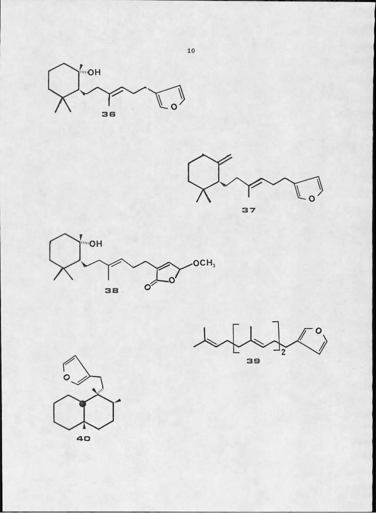
Until a few years ago, diterpenes isolated from the marine
environment were quite rare. Recently, however, compounds have been
identified from seaweeds (36—38), coelentcrates of the orders
Alcyonacea (39-43) and Gorgonacea (44-50) and from a few sponges
(51) . A series of diterpenes have been isolated from Dysidea amblia
(52) . Ambliol A, 36, dehydroambliol A, 37, ambliolide, 38,
ambliofuran, 39, and ambliol B, 40, were identified by spectral data
and chemical degradation. ' The sponge was steeped in methanol for
two weeks at O0C, homogenized, and then Soxhlet extracted with fresh
methanol for two days. Ambliolide, therefore, was probably formed
by air oxidation of ambliol A in the methanolic solution.
Sesterterpenes are very rare in terrestrial fauna and flora but
are more commom in the marine environment, especially in sponges.
Disidein, 41, isolated from D. pallescens is a pentacyclic
sesterterp.ene, whose identification was accomplished primarily by
the mass spectral framentation pattern of the diester degradation
product (53). Its biogenesis, as presented in Scheme II, is
probably the proton catalyzed cyclization of the linear phenolic
sesterterpene, 42. Furospongolide, 43, is a twenty one carbon
terpene isolated from D. herbacea collected in the Red Sea (54).
Tetranorsesterterpenes are among the most unusual terpeniod

2 5 H H a B SAC H a a H /SOH
H OCOCH
a-7 H Ha a SAc H s o H OH3 1 H « 0 H
3 2
34
10
OH
OCH3
40

Scheme II. Proposed Biosynthetic Pathway of Disidein.
OH
MH

12compounds isolated from marine sponges and are thought to arise from oxidative degradation of sesterterpenes.
Nudibranchs, gastropod mollusks of the order Nudibranchia
(subclass Opisthobranchia) are often observed feeding upon marine
sponges. Their often vibrant coloring and lack of external shell
would, seemingly, make them easy targets for predation, yet they are
not sought after by fish, other mollusks, and crab predators;
carnivorous fish have been observed to reject nudibranchs. The
suborder Doridaceae has been the most intensely studied; two
chemical defense mechanisms have been observed, secretion of a
strong acid (55,56) or secretion of a neutral toxic or noxious
substance (57,58). The compounds responsible for the latter
mechanism are presumed to be of dietary origin.
Recently, dorid nudibranchs of the genus Evnselodoris have been
the subject of interest. The secondary metabolites isolated are
predominantly furanosesquiterpenes. Faulkner's group at Scripps
Institution of Oceanography examined four nudibranchs of the genus
Hvnselodoris and isolated a total of nine furanoterpenes (59).
Agassizin, 44, was found in H. aeassizi collected at Cruz de
Juanacaxtle, Nayarit, Mexico, and nakafuran-9, 22, the butenolide of
nakafuran-9, 45, dendrolasin, 46, and the furanoditerpene
ghiselinin, 47, were isolated from H. ghiselini collected at Isla
Danzante, Gulf of California. Specimens of E. californiensis
collected at Isla San Jose, Gulf of California yielded dendrolasin
and nakafuran-8, 21, while those collected at Point Loma, California
contained furodysinin, 27, euryfuran, 34, and pallescensin A, 12.

13
Six specimens of H. porterae found feeding on Dvsidea amblia, also
collected at Point Loma, California, contained furodysinin and
euryfuran. The marine sponge Eurvspongia so., collected at Casa
Cove, La Jolla, California, was found to contain euryfuran and is
thought to be the source of that isolated from Evuselodoris species.
IL godeffrovana. observed feeding on Dvsidea fragilis in Kaneohe
Bay, Oahu, contained nakafuran-8 and nakafuran-9. Dt fragilis was
subsequently shown to be the source of the metabolites (29). H.
daniellae, collected at the Ala Wai Canal, Honolulu, posessed
spiniferin—2, 48, (60) first isolated from the sponge PlerauIvsilla suinifera (61).
It is presumed all furanoterpenes isolated from Hvuselodoris
species have antifeedant properties (59), however, it has been
demonstrated only in nakafuran-8, nakafuran-9, furodysinin, and
pallescensin A (29,59,60).
Soft corals are sessile, soft bodied invertebrates usually
found in tropical waters. They belong to the phylum Cnidaria, class
Anthozoa, and subclass Octocorallia. There are three orders,
Stolonifera, Alcyonacea, and Gorgonacea. Recently much interest has
been devoted to the gorgonian soft corals due to the isolation of
prostaglandins (62-64) and unique terpenoid compounds therefrom (9).
One of the most examined genera from Gorgonacea is Briareum.
Briareum asbestinum, heretofore the only species investigated,
has yielded, a series of highly functionalized diterpenes.
Specimens collected at St. Thomas, Virgin Islands, and at Grand

14
as
a y

15Bahama yielded the not fully characterized briareins C and D (47), and from a Jamaican collection, briarein A, 49,(48). From a
collection off the coast of Belize, briarein B and asbestinins —I,
50, -2, 51, —3, 52, —4, 53, and —5, 54, have been isolated (49). A
collection from Roatan Island, Honduras produced asbestinin epoxide,
55, and asbestinin-5 acetate, 56, (50). The structure of briarein A
and the diol obtained by hydrolysis of asbestinin-1 were elucidated
by single crystal X-ray. The structure of briarein B, briarein A
with a butyrate ester replacing one of the acetate groups, is not
fully solved. Characterization of the remaining asbestinins was
accomplished by analysis of spectral data and chemical
interconversion. Asbestinin—I and its hydrolysis product, and
asbestinin-5 were shown to antagonize the effects of acetylcholine on guinea pig ileum preparations (50).

16OAc
4 3
0
S I H CH31 a 6’7 5 3 - 0 = C H 25 4 ...OH = C H 25 B ...OAc = C H 2
5 0 Ac 5 2 H
S B

17STATEMENT OF THE PROBLEM
The central problem confronting the natural products chemist is
structure elucidation, thus the primary objective of the research
described herein is the characterization of novel compounds. The
criterion in choosing which marine organism to examine was the
uniqueness of compounds isolated from related organisms and the lack
of literature pertaining to the particular organism. The marine
sponge Dysidea etheria and the soft coral Briareum poIvanthes met
both criteria. In addition, the weakly antibiotic activity of
Dysidea etheria, the taxonomic identification of Briareum
polyanthes, and the role of chemical communication in the isolates
of Hypselodoris zebra, prompted this investigation. Antimicrobial
activity was to be assessed for all compounds isolated.

18RESULTS AND DISCUSSION
Sesquiterpenes from Dvsidea etheria
Dvsidea etheria was first described as a new species by M. W.
deLaubenfels in 1936 (65). The description of the beauty of a
tropical reef and his encounter with D. etheria is as follows:
As one walks about on the bottom of the sea near the Dry Tortugas, using a diving apparatus, at depths of less than 10 meters... Gaudy fish swim through the forests of purple and yellow bush-like coelenterates (corals and gorgonians). Not the least striking nor least beautiful are the sponges in this habitat. Big dark Snheciosnongia vesnarias stand up as big as barrels, and shaped somewhat like them. Graceful vase-shaped Hircinia camnanas abound. There are rich red bushes of Haliclona rubens. chrome-yellow bushes of Verongia fulva. two or three Haliclonas of different shades of green, and dark purple bushes of Iotrochota birotulata. There are small flaming orange sponges such as Mvriastra kallitetilla and such azure blue ones as Dysidea etheria... Comparisons to a 'fairyland' are appropriate.
Literature accounts of D. etheria are sparse. Jakowska and Nigrelli
(66) reported extracts of D. etheria demonstrated weak antimicrobial
activity against Escherichia coli and poikilotherm and bovine forms
of tubercle bacilli and, after this research was completed, a brief
reference to the appearance of furodysinin in D. etheria was
reported (67).
D. etheria was collected from a variety of shallow water
habitats in Bermuda, primarily in Harrington Sound, and stored in
acetone at —5°C prior to extraction. The sponge was homogenized and
then extracted with acetone, followed by dichloromethane.
19
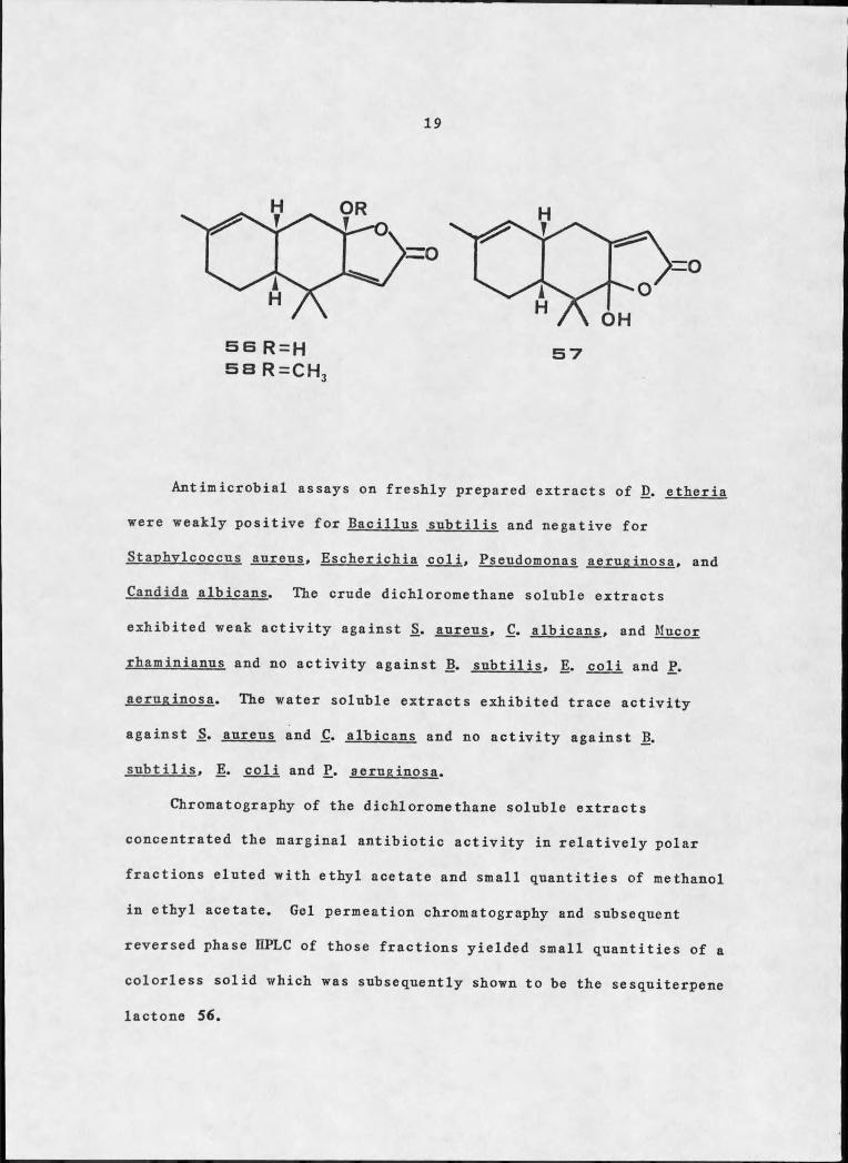
SB R = HSB R=CH
Antimicrobial assays on freshly prepared extracts of D. etheria
were weakly positive for Bacillus subtilis and negative for
Staphylcoccus aureus, Escherichia coli. Pseudomonas aeruginosa. and
Candida albicans. The crude dichloromethane soluble extracts
exhibited weak activity against S. aureus. C. albicans, and Mucor
rhaminianus and no activity against B. subtilis. E. coli and P.
aeruginosa. The water soluble extracts exhibited trace activity
against _S. aureus and C. albicans and no activity against B.
subtilis, E. coli and P. aeruginosa.
Chromatography of the dichloromethane soluble extracts
concentrated the marginal antibiotic activity in relatively polar
fractions eluted with ethyl acetate and small quantities of methanol
in ethyl acetate. Gel permeation chromatography and subsequent
reversed phase HPLC of those fractions yielded small quantities of a
colorless solid which was subsequently shown to be the sesquiterpene lactone 56.

20Inspection of the 13C-NMR spectral data (see Figure I) for 56
revealed the presence of one carbonyl, two olefinic linkages, a 3
quaternary sp carbon substituted by two heteroatoms (8104.91), one
other quaternary carbon, two methines, three methylenes, and three
methyl groups. The total of fifteen carbons, including three methyl
groups, indicated a probable sesquiterpene. Accurate mass spectral
measurements (see Figure 2) delineated a molecular formula of
C15E20°3* ^he carbonyl and two carbon-carbon double bonds left three sites of unsaturation, which were attributed to a tricyclic skeleton.
-IFrom the H-NMR data (see Figure 3), the two olefinic protons,
a singlet at 85.67 and a broad doublet at 85.36, had to be on
separate trisubstituted double bonds. A broad D2O exchangeable
singlet at 83.5 was assigned to a hydroxyl group (3580, 3330 cm-1).
An unsaturated y-lactone, 1745 cm-1 and Xfflax 221 nm (6=8700),
accounted for one ring and the other ^oxygens, meaning that the
quaternary carbon at 8104.91 bore both the hydroxyl group and the
lactone oxygen, as illustrated in 56a.
Careful integration in the 1E-NMR between 81.50 and 1.75
indicated six protons instead of the apparent five protons. The LIS
experiments revealed the hidden proton and extrapolation to zero
equivalents indicated its chemical shift to be 81.63. Spin-spin
decoupling of the normal spectrum and that recorded after addition
of 0.552 equivalents Eu(fod)g suggested part structure 56b.
Examination of coupling constants (see Table I) indicated a cis ring
juncture (1-2.5 Hz between Hg and H11) of the two carbocyclic rings.

5 TBO 140 120 IOO SO
13Figure I. C-NMR Spectrum of Furodysinin Lactone.

Figure
100.0-1112.1
2 3 0 .1
215.1140.0
5 0 .0 -
202.1
187.1119.0
159.1
Mass Spectrum of Furodysinin Lactone

Figure 3. H NMR Spectrum of Furodysinin Lactone.

24Table I. ^E-NMR Assignments for Fnrodysinin Lactone
ChemicalHydrogen Shift(S) Multiplicity Coupled to (J, Hz)
5.67 sOH 3.5 hr s
H5a 1.57 dti E5b (14), H6 (13)
H5b 2.28 dd H5a (14), H6 (3.8)
H6 2.80 m H5a (13), H7 (5.7)H5b (3.8), HlObH11 (2.5)**
H7 5.36 hr dd Hg (5.7), Hpa,b (1.4)
H9a,b 1.96 overlapping m HiQa (9'5), H7 (1.4)
E10 a 1.12 hr ddd HlOb (12.6) H9a.b (9-5,
H n (12.6)unresolved, small)
H1 Ob 1.70 br ddd Hioa (12.6) hH (3.1)
, H6 (3.7)*
“9a,b (tmresoIved, small)
E11 1.63** m E10a (12.6) % (2.5)**
» Hiob (3.D
E13 1.38 S
H14 1.21 S
H15 1.61 br s
*W coupling
**Not resolved in normal spectrum; chemical shift obtained by extrapolation to zero equivalents Eu(fod)q; after addition of 0.554 eq., an apparent dt (J = 13.3, 2.5, 2.5).
Z

25
OH
Somewhat perplexing at first glance was the coupling between Hg and
H1Ob' bnt construction of a Dreiding model illustrated that when the dihedral angle between Hg and Hn was minimized, Hg and were
aligned quite properly for W coupling. The two methyl singlets
(61.38 and 1.21), IR absorptions at 1371 and 1348 cm™1, and a fully
substituted sp carbon (638.47) were ample evidence for a germinal
dimethyl group and accounted for the remaining elements in the molecule.
Assembling the substructures gave 56 or 57. Originally, 57 was
favored because it followed the isoprene rule. Two subsequent
experiments; however, rigorously established 56 as the proper structure.
The H-NMR of the lactone bore a resemblance to the 1E-NMR of a
major non polar component isolated from the first fraction of the
aforementioned Florisil chromatography. Purification could not be
accomplished by gel permeation (Sephadex LE-20 and Bio-Beads S-X8),
silica gel chromatography (flash and gravity), argentation
chromatography (15% silver nitrate impregnated SiO2) and HPLC

26
7 H 5
io H14 13
(Ultrasphere-Cyano, ODS, and Si). Thus, all spectral work was done
on nearly pure material. The analysis of the 1E-N1MR (see Figure 4),
which indicated a 2 ,3-disubstituted furan (57.21 and 6.22) and a
resemblance between the signals at 51.96, 1.70, and 1.61 of 56 and
52.03, 1.71, and 1.66 of the furanoid compound suggested molecules
with a similar skeleton. Mass spectral analysis (see Figure 5)
indicated a molecular formula of C-15H20® and the fragmentation pattern along with the inverse gated 12C-NMR (see Figure 6)
unequivocally established the compound as the non-isoprenoid furodysinin 27 (20).
In order to confirm the apparent relationship of furodysinin and the
newly discovered lactone, the oxidation of 27 was undertaken.
Photoinduced oxygenation (63) of 27 in methanol gave, exclusively, a
ketal-lactone shown to be 58 by 1B-NMR (53.12, 3H, s). The ketal
could not be hydrolyzed to the hydroxy lactone under a variety of
acidic conditions: 4% BCl at room temperature for 8 hours, 4% BCl
at 50 C for 2.5 hours, and 4% HCl at reflux for 2.25 hours.

L . I .. I . . . I . I I . . . . . . . . . I . . . . . . . . . I5 7 6 5 4Figure 4. NMR Spectrum of Furodysinin.
23

M 02 3
Figure 5. Mass Spectrum of Furodysinin.

8130 30
Figure 6. Inverse Gated 13C-NMR Spectrum of Furodysinin.
K>NO
-LU
50 2 0

30
Repetition of this oxidation, using tetrahydrofBran-water in
place of the methanol, gave mostly starting material. The
conversion was finally achieved by a two step oxidation with meta—
chloroperbenzoic acid, followed by Jones reagent (29). The sole
product of this sequence gave a ^H-NMR spectrum superimposable with
that of the natural lactone, indicating that one of the
diastereomers of 56 (epimers at C-4) was the structure of both the
natural product and the semisynthetic lactone.
The 13C-NMR assignments for 56 and 27 are presented in Table 2
The assignments for furodysinin lactone followed from an off-
resonance decoupled and three single frequency off-resonance
decoupled experiments (SFORD) irradiating at 52.80, 2.28, and 1.12.
The assignments for furodysinin were not as simple to ascertain.
The six sp^ carbons were assigned with the aid of an off—resonance
decoupled experiment, but the upfield region, due to the congestion
of the four signals between 533.2 and 31.2, required additional
experiments for full assignment. The off—resonance decoupled
spectrum provided the multiplicity of five sp^ carbons (Figure 7,
spectrum b), a spin—echo experiment (0.5/J) identified the
quaternary carbon (533.13) (Figure 7, spectrum d), and a second
spin-echo experiment (1/J) established the remaining CH2 (531.69)
and indicated that the resonances at 531.22 and 32.85 were due
either to methine or methyl groups (Figure 7, spectrum c). The
information desired from a J-modulated spin-echo experiment is
dependent upon the time between the 90° pulse and the 180° pulse
(the echo delay). An echo delay of 0.5/J (J is the 13C-H coupling

31Table 2 13C-NMR Assignments, Fnrodysinin Lactone and Furodysinin
Fnrodysinin Lactone Fnrodysinin
J-Modnlated Spin EchoCarbon # 8 ORD 6 ORD 0.5/J 1/J
I 174.76 S 140.51 d2 115.27 d 108.20 d3 169.82 S 124.71 S
4 104.91 S 147.48 S
5 41.17 t 27.56 t PO S
6 30.37 d 31.22 negc7 123.54 d 126.18 d8 134.49 S 133.62 S
9 30.96 t 31.69 po S
10 18.65 t 19.26 t pos
11 47.33 d 44.60 d neg12 38.47 S 33.13 S pos
13 25.28* Q 32.85% negd14 26.84* I 26.21% q neg15 22.98 . Q 23.12 q neg
^assignments interchangeable
^assignments interchangeable
ca me thine (see text)
^a methyl (see text)

32
j lJV ------------------— u— I U v u - ^ — A uXjk~ ju^ juu-~w jN w
. i t . . ......... I I I . ,<5 40 3 O p> n
Figure 7. I3C-NMR Spectra of the Highfield Region of Furodysinin

33
constant) will give a spectrum exhibiting only quatenary carbons (defined as the positive phase)# while a 1/J echo delay will show
quatenary and methylene carbons as the positive and methine and
methyl carbons as the negative phase (69). Correlation of the off-
resonance decoupled and spin-echo data indicated that the signal at
831.22 was the methine and that at 832.85 the methyl. An SFOED
(irradiating 81.61) identified the allylic methyl (823.12) and
another SFORD (irradiating at 82.65) confirmed the resonance at831.22 as the methine.
A lanthanide induced chemical shift study (LIS) was undertaken to establish the full relative configuration of 56. A full
equivalent of Eu(fod)g was added in increments to a CDClg solution
of the lactone and a ^H-NMR spectrum recorded after each addition.
Table 3 contains the slopes, obtained by linear least squares
method, along with correlation coefficients, of a plot of induced
chemical shift versus equivalents of shift reagent added and Figure
8 is a graphical interpretation of the same.
The assignment of relative configuration of polycyclic systems
from LIS data is rarely straightforward. In this case, analysis was
rendered particularly difficult because the hydroxyl group did not
appear to be the site of chelation, as might be expected. This
assumption was based on the observation that the hydroxyl proton did
not undergo the exceedingly large shifts attributed to pseudocontact with the lanthanide.
In order to determine first the site of chelation and then the
relative configuration of the three chiral centers in 56, the PDIGM

34
Table 3. Slopes from the Plot of AS vs. [Eu(fod)3]/[Substrate]
SemisyntheticFnrodysinin Lactone Fnrodysinin Lactone
Proton(S) Slope
CorrelationCoefficient Slope
CorrelationCoefficient
5.67 2.02 .986 2.11 . .9985.36 .35 .993 .35 .9983.50 2.01 .992 2.12 .9912.80 .65 .994 .70 .9962.28 1.16 .990 1.22 .9981.96 .20 .990 .21 .9991.70 .26 .996 .25 .9991.63 .54 .986 .50 .9881.57 1.42 .985 1.50 .9961.61 .09 .995 .10 .9991.38 .47 .999 .50 .9981.21 .28 .983 .31 .9981.12 .89 .999 .89 .999

35
eq. Eulfodl
Figure 8. Plot of Change in Chemical Shift vs. Equivalents Eu(fod)3

36
program of Willcott and Davis (70) was utilized. The program calculates the expected induced shift for a given set of proton
coordinates and compares them to the experimental induced shifts.
The europium position is varied to obtain the lowest agreement factor, R.
Since the cis ring juncture between the two cyclohexane rings
was clearly defined from the chemical correlation with 27, only the
two diastereomers epimeric at the hemiketal carbon had to be
compared. Dreiding models of the C—4 epimers of 56 were constructed
and Cartesian coordinates were generated by projection onto the XY
plane; Z coordinates were measured directly. Any of the three
oxygens could have served as the site of chelation of the lanthanide
with a lone pair of electrons. Each of these possibilities was
evaluated and the best fit for the LIS data indicated complexation
of the europium with the carbonyl oxygen at a distance of 2.4 X in the isomer with the hydroxyl cis to the ring juncture protons as
shown in 56; the minimum R value was 7.98%. The C-4 epimer gave a minimum R value of 16.83%. No other combination of relative
configuration and site of complexation resulted in minimum R values under 28.48% (see Table 4).
The R factor ratio test is often used to determine the
confidence level with which one isomer may be favored over another
R = [

37Table 4. Agreement Factors for Furodysinin Lactone and its C-4
Epimer.
Minimum R(%) for Minimum R(%) for the Site of Complexation 56 C-4 Epimer of 56
OH 63.70 61.76
0 28.48 30.98
CO 7.98 16.83
(71). The thirteen protons used in the experiment leave nine
degrees of freedom. Thus an R1/R2 of 2.11 (16.83/7.98) indicates we
have a greater than 99.5% confidence level that 56 is the correct
isomer. Figure 9 illustrates the graphical relationship of the R
values obtained for 56 with the distance between the europium and
carbonyl oxygen atom.
Consideration of steric effects provides a suitable explanation
for the failure of the Eu(fod)g to chelate with the hydroxyl group.
The molecular model suggested the likelihood of intramolecular
hydrogen bonding of the hydroxyl with a non-bonding electron pair on
the lactone oxygen. This idea was supported by the sharpness and
constancy of chemical shift (no effect of concentration on 5 value)
exhibited by the hydroxyl hydrogen in the ^H-NMR. Such a
configuration would preclude access of the lanthanide to the lone
pairs of the hydroxyl oxygen and to one pair on the lactone oxygen.
The net result would be that the carbonyl oxygen should become the
primary complexation site.

38
R value
B.O-
2.8 3.02.2
Distance (Al
Figure 9. Plot of R Values vs. Europium-Oxygen Bond Distance
The Eu(fod)g shift reagent induced identical chemical shift changes in the lactone prepared by oxidation of furodysinin (see
Table 3), confirming that a single diastereomer, identical to the
natural product, was produced in the course of the oxidation.

39
sa R =OAc53 = O H2 1 = H
Fnranosesquiterpenes from Evnselodoris zebra
The nudibranch Hy-pselodoris zebra was frequently observed
grazing on Dysidea etheria in Bermudian waters. When a single
specimen of Hypselodoris zebra was dissected and offered in separate
portions to Abudefduf saxatilis, an omnivorous scavenger fish common
on Bermuda's reefs, only the gonads were consumed. The digestive
tract and outer skin were categorically rejected by the fish.
Extraction of several nudibranchs gave very limited quantities of
extract; the extract, however, contained a significant percentage (36%) of sesquiterpenes.
Gel permeation chromatography of this extract through Bio-Beads
S-X8 with cyclohexane-dichloromethane (2:3) yielded a mixture of
furodysinin, 27, euryfuran, 34, and 5-acetoxy nakafuran-8, 24, as
major constituents and 5-hydroxy nakafuran-8, 59, as a minor
constituent of the extract. The identity of furodysinin and the two
nakafuran—8 derivatives (31—33) were secured by direct comparison

40
with the fnranosesqniterpenes isolated earlier from the sponge. Euryfnran was identified by comparison of its mass spectral and Aff-
NMR data with those reported in the literature (20,59,72). An
apparent typographical error in reference 59 made the initial
comparison to literature data confusing. We found chemical shifts
of 50.90 and 0.93 for two of the methyl groups in 61, consistent
with the data reported in reference 20 and 72 while reference 59
reports those signals at 80.90 and 0.99.
Furodysinin (67,75) and nakafuran-8, 21, and -9, 22, (29) have
previously been identified as feeding deterrents. Thus, it is
likely that furodysinin serves the same function in Hypselodoris
zebra. The large amount of 5-acetoxy nakafuran—8 and euryfuran
concentrated in H. zebra suggest they also may be antifeedants. The
structural similarity of nakafuran—8 to 5-acetoxy nakafuran-8
reinforces this view.
Euryfuran is conspicuously absent from D. etheria. Attempts
will be made in the future to identify the dietary source of
euryfuran in H. zebra.
Pharmacological Activity of Furodysinin and Furodysinin Lactone
Metabolites that are biological deterrents in nature often
demonstrate in vitro biological activity. The biological activity
of furodysinin and furodysinin lactone was assessed against a series
of common plant pathogenic microbes. Both 56 and 27 were mildly
active against Curvularia lunata and Rhodotorula glutinis and
demonstrated no activity against Fusarium solani. Corvnebactarium

41michiganese, and Psendomonas svringae. In addition, furodysinin lactone proved negative in tests against bacteria deficient in DNA
repair capacity.
Ceramides from Dvsidea etkeria
The fractions which contained furodysinin lactone from the initial separation of D. etheria on Florisil, also contained, as
revealed by subsequent purification via gel permeation
chromatography, a white solid which ultimately proved to be a
mixture of ceramides.
Ceramides are common isolates of many terrestrial fauna (74-
78), fungi (79—82), and bacteria (83,84), and have recently been
found in a variety of marine organisms, including the first
isolation of such compounds from plants, red (85) and green (86,87)
alga, as well as the electric organ of the ray Torpedo marmorata
(88), the rectal gland of the spiny dogfish (89), the starfish
Patiria pectinifera. (90), octopus (91), and the shellfish Pinctada
martensii (92), but they had previously not been reported from
sponges.
Florisil chromatography of the dichloromethane soluble extracts
of Dysidea etheria gave three polar fractions which, when further
separated by gel permeation chromatography, yielded fractions (one
each) which were appreciably soluble in tetrahydrofuran and pyridine
only. Purification by flash chromatography yielded two fractions
comprised of an amorphous white solid. The ^H-NMR spectra (see
Figures 10 and 11), recorded in deuterated tetrahydrpfuran,

XX
A uw<-
< 5 B
Figure 10. 1H-NMR Spectrum of Ceramide 60.

J--- -—

44
suggested moderately functionalized molecules with a long aliphatic
chain. The minor fraction, Rf 0.35 in silica gel TLC, appeared to
contain three exchangeable protons, while the major fraction, Rf
0.26, had four such protons. However, acetylation of the minor
fraction gave only a diacetate (see Figure 12), while the major
fraction yielded a triacetate (see Figure 13). Absorption in the IR
spectrum (KBr pellet) at 1623 cm-1 for the minor fraction and 1624
cm 1 for the major fraction suggested an amide functionality. Thus,
in order to account for the number of deuterium exchangeable protons
in each molecule, a secondary amide, along with a diol in the minor
fraction and a triol in the major fraction, must be present.
The 1H-NMR spectra of the two molecules, and their acetylated
derivatives, had the typical paraffinic resonances at 50.88 (broad
triplet), 1.28 (broad singlet), and 1.45-1.70 (multiplet);
additional straightforward information was precluded by the overlap
of numerous midfield signals. 1H-NMR decoupling experiments on the
acetylated derivatives established part structure 60a for the minor
fraction and 61a for the major fraction. The broad deuterium
exchangeable resonance at 55.58 was readily assigned to the amide
proton in 60a (see Table 5 for a complete list of 1H-NMR assignments
and coupling constants). It was coupled to a one proton multiplet
at 54.40 which in turn was vicinally coupled to three one proton
doublet of doublets. Two of the doublets of doublets, 64.29 and
4.02, demonstrated geminal coupling while the remaining doublet of
doublets, 55.26, was vicinally coupled to an olefinic proton at
55.37. A coupling of 14.9 Hz to the remaining olefinic proton.

Figure 12. IH-NMR Spectrum of Ceramide Diacetate 60a.

5 "7 B 5 4
Figure 13. 1H-NMR Spectrum of Ceramide Triacetate 61a.
I3

47Table 5. IE-NMR Assignments for the Ceramide Acetates.
Chemical Shift (5) Multiplicity Coupling Constants (Hz)
H 60aa 61ab 60a 61ala 4.29 4.21 dd 12.3,6.2 11.6,7.9lb 4.02 4.07 dd 12.3,3.9 11.6,4.72 4.40 4.35 m3 5.26 5.31 dd 7.0,6.5 6.7,6.54 5.37 5.46 . dd 14.9,6.5 15.3,6.55 5.77 5.78 dt 14.9,6.2 15.3,6.56(2H) 2.00 1.97° dt 6.2,6.8 6.5,
unresolved2 ' 2.15(2E,t) 4.95
(IH.br t)7.1 6.7
3' 1.72(2E,m)
COCH3 2.05,2.07 2.13c, 2.02c, 2.OOc
S
NE 5.58 7.13 brd 8.6 8.5
CE2d 1.45-1.65 1.45-1.65 m
CH2d 1.27 1.28 brs
CH3(GE) 0.84 0.83 brt 7.4 7.6
aCDCl3 was used as solvent and internal standard.^Dg_acetone was nsed as solvent and internal standard (see following note).
cThe acetone impurity in dg—acetone necessitated measurement in CDCl3.
^The methylene protons of the alkyl chains. An indeterminant number due to the presence of a mixture of chain lengths.

4885.77, suggested trans geometry. A two proton doublet of triplets
at 52.00 coupled to the olefinic proton at 85.77 indicated an
allylic methylene. Chemical shift considerations placed the two
acetates on C-I and C—3. ^H-NMR assignments for 61a followed from
60a. The broad one proton triplet at 84.95 necessitated placement
of the third acetate on C-2% The proton on 0-2' was coupled to the
two proton multiplet at 81.72 which, in turn, was coupled to the
methylene envelope, 81.28.13The C-NMR spectra of 60 and 61 indicated one carbonyl, two
olefinic carbons and three heteroatom bearing carbons (two doublets
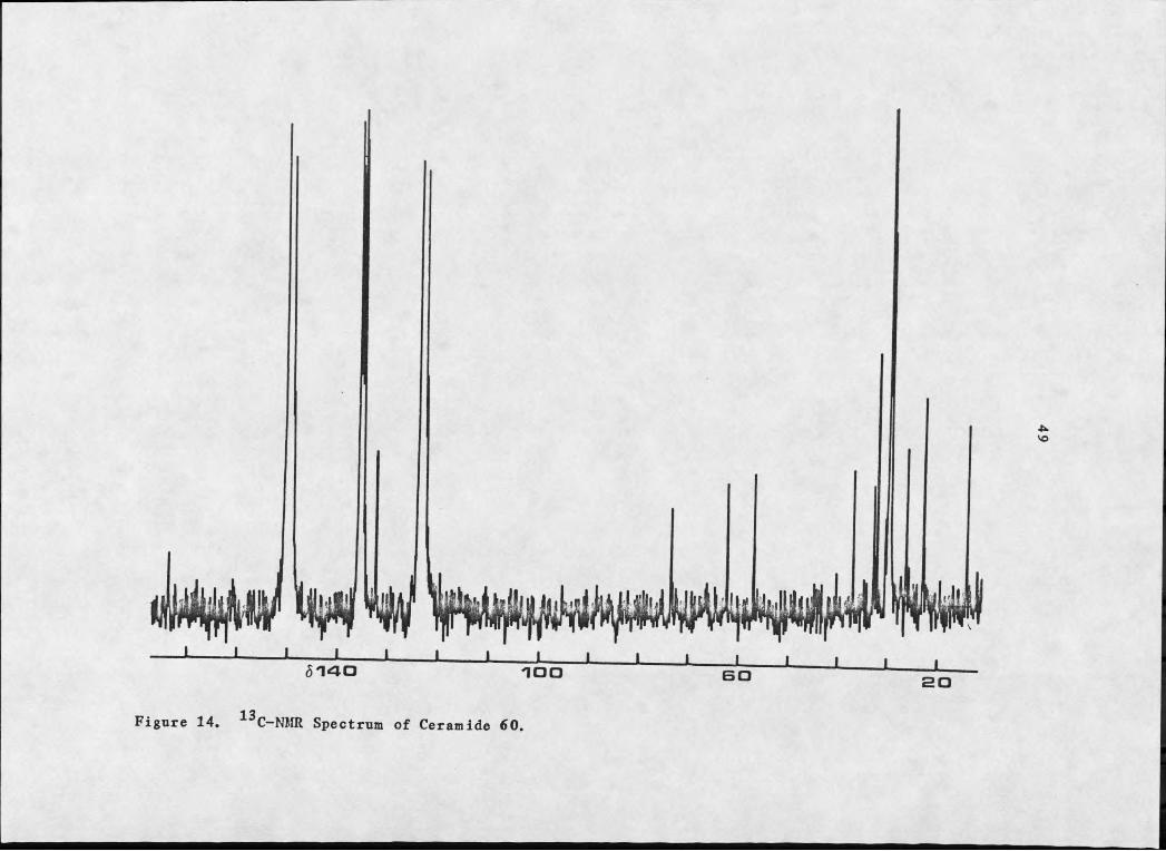
and a triplet), for 60 (see Figure 14), and one carbonyl, two
olefinic carbons and four heteroatom bearing carbons (three doublets
and a triplet) for 61 (see Figure 15), which led to the 1,3-
diacetoxy, 2 amido groupings, establishing both fractions as
sphingosine type compounds. The upfield region of inverse gated
(see Figure 15) and off resonance decoupled (see Figure 16) ^^C-NMR
spectra of 61 in pyridine indicated approximately thirty—two
methylenes (all but 7 between 829.66 and 29.30) and two methyls.
After the structures of 60a and 61a were ascertained with
certainty and the ^H-NMR resonances were assigned it was possible,
through spin— spin decoupling, to assign the resonances of 60 and 61
(see Table 6 for a list of ^E-NMR assignments). Chemical shift
considerations led to the assignment of the broad deuterium
exchangeable proton at 56.87 in 60, which was coupled to the
multiplet between 83.78 and 3.90, to the secondary amide (N-E). The
integration (2 protons), as well as the coupling to 53.73, 3.53, and
iii WL i l l i
6140 __I____ L l l i iIOO BO I I20
Figure 14. 13C-NMR Spectrum of Ceramide 60.

L I I I I I I I I I I
/I
UiO
Wwtwi8 1 4 0 IOO I I I I
Figure 15. Inverse Gated 13C-NMR Spectrum of Ceramide 61.

I_____ L I6 1 4 0
UL W Li.I_____ L I
IOOJ_____ I J--------L______L I
Figure 16. Off-Resonance Decoupled 13C-NMR Spectrum of Ceramide 61.

52
Table 6. ^H-NMR Assignments for the Ceramides.
Chemical Shift (8) Multiplicity Coupling Constants (Hz)
H 60a 61a 60 61la 3.73(ddd) 3.77-3.90(m) 14.9,4.7,4.4lb 3.53 3.58 m2 3.78-3.90 3.77-3.90 m3 4.08 4.13 ddd 6.2,5.3,5.3 6.4,5.2,54 5.50 5.49 dd 15.5,6.2 15.5,6.35 5.65 5.70 dt 15.5,6.5 15.5,6.36 (2E) 2.20 2.04 m2' 2.13(2H,t) 3.97(lH,m) 6.83' 1.95(2H,m)OH(C-I) 3.78-3.90 3.77-3.90 m
0H(C-3) 4.30 4.43 brd 5.3 5.20H(C-2') 4.79(d) 4.8NE 6.87 7.39 brd 8.1 8.9
CH2b 1.45-1.65 1.45-1.65 m
CH2b 1.30 1.30 brsCH3 (6H) 00 00 CO00 brt 8.1 8.1
aD8-THF was used as solvent and TMS as internal standard.^The methylene protons of the alkyl chains. An indeterminantnumber due to the presence of a mixture of chain lengths.

53
OAc
AcO
B T a R = alky!, X = OAc
OH
G O X = H , n =12,13,14,15,19,20,21
B T X = O H 1 n = 19,20,21

544.08 suggested the multiplet (83.78—3.90) constituted the primary
alcohol proton and Hg. When THF-dg was used as solvent, all the alcoholic protons demonstrated coupling to protons on neighboring
carbons. This is primarily due to the decrease in the rate of
exchange of the hydroxylic protons upon the use of a more polar
solvent. The doublet of doublet of doublets at 84.08 was coupled to
the olefinic proton at 85.50 (a doublet of doublets) and the
hydroxyl proton at 84.30 (a broad doublet). A coupling constant of
14.9 Hz between 83.73 and 3.53 indicated geminal coupling, and one
of 15.5 Hz between the olefinic protons at 85.50 and 5.65 indicated
trans geometry. The olefinic resonance at 85.65 was coupled to the
allylic methylene at 82.02 and a two proton triplet at 82.13
necessitated placement next to the carbonyl. Once again IE-NMR
assignments for 61 followed from 60. The additional alcoholic
proton appeared as a broad doublet at 84.79 coupled to a multiplet
at 83.97, which in turn was coupled to the methylene at 81.95.
Hydrolysis of both 60 and 61 gave sphingosine and a mixture of fatty
acid methyl esters. The ^H-NMR spectra of the sphingosine from both
fractions were identical in all respects to that reported previously
(85). The sphingosine was converted to its triacetate and
identified by comparison with literature values {[a]j)» ^H-NMR
(85,93)}. Fast atom bombardment mass spectral studies indicated an
M+H ion at m/z 426 and diagnostic fragments at 366 (loss of O2CCH3)
and 352 (loss of CHg(^CCHg), to verify the presence of sphingosine triacetate. The fatty acid methyl esters of 61 were shown to be a-
hydroxylated by the IH-NMR (84.17, IR, dd, J = 7.8, 3.6) and MS (M-

55
59, loss of CO2CH3) spectra and to be composed predominately of n-
C22 acids with minor amounts of H-C23 and H-C24 acids. The fatty acid methyl esters of 60 were shown by mass spectrometry to be
composed mainly of n—C22, H-C23, and n—C24 acids, with smaller
amounts of H-C^5, n-C^g, n-C^y and n-C^g acids also present. The
aliphatic chains of the sphingosinetriacetate and the fatty acid
methyl esters of 60 and 61 were proven to be normal by virtue
of the low mass fragmentation pattern of their mass spectra (m/z 29, 43, 57, 71, 85, etc.).
In spite of the ubiquitous nature of a—hydroxy fatty acids,
relatively few stereochemical assignments have been made. This is
due to the difficulty in assigning absolute configuration to
compounds of weak optical activity (94-96). However, some
configurations have been assigned and the subject has been reviewed
(97). On the basis of comparison with literature data (see Table
7), the optical rotation [aJd + 19.5 of the hydroxyacid methyl
esters suggested an JS configuration at C-2.
This isolation of sphingosine derivatives is the first reported
occurrence of ceramides in marine sponges and is important for a number of reasons.
The biological function of free ceramides is not known (102).
The a-hydroxy and non-hydroxy ceramides serve as precursors to
cerebrosides (l-Ji-galactosyl ceramide) and sulfatides (sulfate in
position 3 of the galactose) (103), both are abundant in mammalian
nervous tissue (104). These sphingolipids are components of the

56
Table 7. Optical Rotations, Esters of 2—Hydrozyacids.
Compounda [ci]d Solvent Reference
S—methyl valerate +16.6® CHCl3 98_S—methyl octanoate +11* CHCl3 99R-methyI tetradecanoate -3.6® CHCl3 100
R-methyl hexadecanoate -3.6® CHCl3 101
R—methyl octadecanoate -2.1® CHCl3 101
aAll esters have hydroxyl substituents a t C—2.
surface membrane, the hydrogen bonding capabilities of the polarendgroups ensure cellular integrity. The function of the a-hydroxyI
is unknown, however, it has been suggested that it serves to alter
permeability and stability by creating a system of laterally
oriented hydrogen bonds (105).
All known a-hydroxy fatty acids from ceramide sources are
optically active (106). The stereochemistry about C—2 of a—hydroxy^-
n—tetracosanoic acid formed by the brain a—hydroxylating enzyme has
always been the R configuration (i.e. D-hydroxyacids) (107-110). A
synthetic mechanism for a-hydroxylation of ceramides from a cell
free preparation of rat brain has been proposed to explain the
reason for this preference (107). Hydroxylation of the fatty acid
occurs by direct substitution of the pro R hydrogen (see Figure 17),
with retention of stereochemistry. The fatty acid is subsequently
incorporated into the ceramide (108). A similar explanation is

57obtained from studies on the a-oxidation of fatty acids in plants (101,111—113). Fatty acid degradation is accomplished by successive
a—oxidation. Studies on the o—oxidation system of pea leaf and
germinating peanut seed demonstrated that only the D a-hydroxy fatty
acid is formed and suggested that it must go through a D a-
hydroperoxy intermediate (113).HO. .Hcv ..Mg
R COOH
X / ' 'R COOH
Figure 17. a—Hydroxylation of Fatty Acids
The .S enantiomers found for the ceramide derived a—hydroxy
fatty acids suggest an alternate pathway for the production or
accumulation of the a—hydroxy fatty acids in Dvsidea etheria.
The fatty acids, both hydroxylated and saturated, represented
in the ceramides of Dvsidea etheria were predominately C-22 or
larger, in keeping with recently described profiles of fatty acids
(114,115) and their derivatives (116) in the Porifera.
Sponges harbor and support considerable microbial communities;
it is possible, therefore, that the ceramides could be of bacterial
or fungal origin. However, the consistent quantities of these
compounds found in several collections of D. etheria from numerous
sites suggest that they are, in fact, produced by the sponge.
Pharmacological Activity of the Ceramides
Antimicrobial activity of 60 and 61 was assessed against the
same series of plant pathogens as were furodysinin and furodysinin

58
lactone. The ceramide, 60, demonstrated mild activity against C.
michiganese and no activity against F. solani. C. lunata, R.
Rlntinis, and P. svringae. The a—hydroxy ceramide, 61, was negative
against all microbes tested.
Diterpenes from Briareum nolvanthes
Along with the marine sponges, the soft corals are among the most
primitive of animals. They are carnivorous invertebrates whose diet
consists essentially of plankton. Their month, which is surrounded
by eight pinulated tentacles, opens to an internal cavity that has
no anus. The soft corals are widely distributed. The order
Alcyonacea is especially prevalent in the South Pacific and Indian
Oceans, and the order Gorgonacea is found in great abundance in the
tropical western Atlantic.
While on a collecting expedition to the Bermuda Biological
Station in 1979, Dr. John H. Cardellina II found a moderately large
community of a soft coral from the genus Briareum at the eastern end
of the Bermudian archipelago. Briareum species had not previously
been reported from Bermudian waters and subsequent identification
indicated a taxonomically confused species recently renamed B.
nolvanthes (117). Analysis of the organic metabolites revealed a
series of highly functionalized diterpenes possessing the briaran
carbon skeleton.
Prior to this work, compounds possessing the briaran ring
skeleton have been found in one species of Briarenm (B. asbestinum)
and three different genera of the distantly related sea pens.

59
Briarein A, 49, B, C, and D (see Introduction) have been isolated
from B. asbestinin (47,48). Stylatulide, 62, from Stvlatula s£.,
was identified as the major component of a series of five
metabolites by single crystal X-ray analysis (118). The structure
of ptilosarcone, 63 from Ptilosarcus eurnevi. was identified by
chemical degradations, comparison with NMR spectra of briarein A,
and spin-spin decoupling experiments (119). A series of three
diterpenes have been identified from Scvtalium tentaculatum (120)
and have been assigned structures 64, 65, and 66. Structure
elucidation was accomplished by high resolution mass spectrometry, 13C- and ^B-NMR, and chemical degradations.
Our collections on Briareum nolvanthes were extracted with
acetone, followed by dichloromethane. The dichloromethane extract
was then equilibrated with the aqueous residue remaining after
concentration of the acetone extract. The resulting organic phase
was concentrated and then separated according to a partition scheme
popularized by Kupchan (121). This scheme successively partitions
the organic solubles against progressively more polar organic
phases. Four organic phases were used; hexane, carbon
tetrachloride, chloroform, and ethylacetate, which were partitioned
against 10% aqueous methanol, 25% aqeous methanol, 35% aqueous
methanol, and water respectively. The carbon tetrachloride and

60
65 6 6

61
chloroform soluble extracts provided 1C-NMR suggesting significant quantities of terpenoid components.
Crude separation of the chloroform residue was accomplished by
step gradient gel permeation chromatography (122). This technique
capitalizes upon the adsorption and partition effects of Sephadex
LH-20 as well as the size separation which is expected of gel
permeation. The gel was eluted successively with hexane-
dichloromethane (1:4), acetone-dichloromethane (2:3), and acetone-
dichlorcmethane (4:1). The first phase gave five fractions. 1H-NMR
analysis revealed the terpenoid components to be concentrated in
fractions 2 and 4. Subsequent separation and purification was
accomplished by gel permeation chromatography through Sephadex LH-
60, then Biobeads S-X8 followed by HPLC on a nitrile-bonded phase
column. Three diterpenes were obtained, briantheins Z, 67, and Y,
68, from fraction 2 and brianthein X, 69, from fraction 4.
9 % 15 Q ^ 3y B 7 R 1= R 2 = R 3 = Ac
S B R1= R 2 = A c
BB R1 = R2- Ac R3= H
O

62
The C NMR (see Figure 18) of 67 showed resonances for four
carbonyls, four olefinic carbons (two doublets, one triplet, and one
singlet), eight heteroatom bearing carbons (one singlet and seven
doublets) and ten saturated carbons (six quartets, three doublets,
and one singlet). An intense absorption at 1739 cm in the IR
spectrum, along with three singlets (each SE) in the 1E-NMR (see
Figure 19) at 82.15, 2.06, and 2.04, indicated the presence of three
acetate groups and accounted for three of the four carbonyls, three
of the eight heteroatom bearing carbons, and three of the ten
saturated carbons. Another intense absorption in the IR at 1790
cm-1 suggested a y-lactone and an absorption at 3540 cm-1 required a
hydroxyl group. A positive Beilstein test then left two heteroatom
bearing carbons, in addition to the seven saturated carbons, to be assigned.
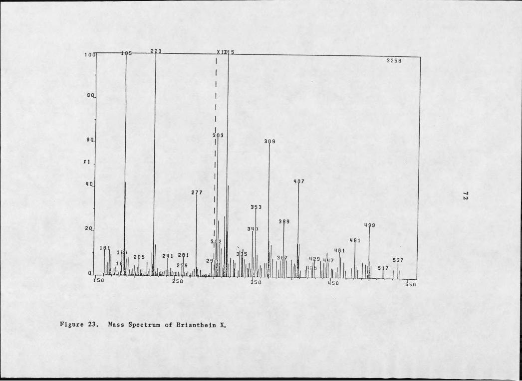
A molecular ion was not obtained using electron impact or
chemical ionization mass spectral techniques. Eowever, fast atom
bombardment in a glycerol-potassium iodide matrix gave an (M+K)+ at
m/z 579 and an (M+K+2)+ at m/z 581 for 67 whose isotopic abundance
indicated the presence of chlorine (see Figure 20). Fragments at
m/z 519 (loss of BOAC) and m/z 561 (loss of E2O) verified the acetyl
and hydroxyl moieties respectively. A molecular weight of 540/542
is obtained upon subtraction of potassium from M+K. Addition of the
number of carbons (obtained from the 1^C-NMR), hydrogens (obtained
from 1^C- and 1E-NMR data), oxygens mandated by the acetate,
lactone, and hydroxyl moieties, and the single chlorine leave 16


6 7 G 5Figure 19. 1H-NMR Spectrum of Brianthein Z.
Ia
ON4x
JjU aJJLJ'3 3 I

150jy.
281 ^lhllIkUAlIl1L u
250
353 385
Figure 20. Mass Spectrum of Brianthein Z.

66
a.m.u. to account for. The inclusion of one more oxygen gave the
molecular formula C26H33C1O10*
1H-NMR decoupling experiments (see Table 8 for a list of 1H-NMR
assignments and coupling constants for brianthein X, Y, and Z) on 67
revealed three isolated systems 67a—c» assigning all resonances
except a methyl singlet (81.09) and a one proton singlet at 83.2
(OH). Irradiation of the one proton doublet of doublets at 83.55
collapsed the sharp doublet at 82.95 (IR) as well as eliminating the
5.8 Hz coupling of the doublet of doublets at 84.68 (1H). The
resonance at 84.68 was coupled to the complex of signals between
82.15 and 2.26 (2H) which in turn was coupled to the one proton
doublet at 85.22 and the methyl doublet at 81.05. The 1,3-
disubstituted butadiene of the second isolated system, 67b, was
formulated by consideration of the four sp2 carbon resonances in the 13 iC-NMR as well as H coupling constants. Irradiation of the
olefinic doublet of doublets at 85.61 collapsed the doublet at 86.22
(IH) as well as the broad 11.9 Hz doublet at 85.93 (IE). The one
proton broad doublets at 86.30 and 5.99, upon irradiation, sharpened
the signal at 85.93 and each partially collapsed the one proton
doublet of doublet of doublets at 85.25. The one proton doublet at
84.90 was then shown to be coupled to the signal at 85.25. The
final isolated system, 67c, consisted simply of a methyl doublet
(81.15) coupled to a one proton quartet at 82.29.
The unusual downfield chemical shift of many of the sp3 protons
in 67a and 67b suggested likely sites of heteroatom substitution.
The resemblance of isolated spin system 67b to briarein A prompted

Table 8. IH-NMR Assignments for Briantheins X, Y, and Z
Multi-Chemical Shift (8) plicity Coupling Constants (Hz)
Proton X I Z X I ZC8-OH 3.47 3.18 3.20 SH2 5.23 6.22 6.22 da 9.1 8.8H3 5.21 5.60 5.61 dda 11.8,9.1 11.9,8.8H4 5.83 5.91 5.93 brd 8,1 11.8,1 11.9,1H6 5.25 5.25 5.25 ddd 3.3,3,2.7 3.3,2.7,2.3. 3,2.4,2H7 4.93 4.90 4.90 d 3 3.3 3H9 5.19 5.22 5.22 brd 8.2 8.5 8.5H10 2.18-2.27 2.15-2.26 2.15-2.26 mH11 2.18-2.27 2.15-2.26 2.15-2.26 mH12 4.73 4.68 4.68 dd 6.1,2.? 5.5,3.2 5.8,3H13 3.61 3.55 3.55 dd 6.1,3.1 5.5,3.3 5.8,3.2H14 3.14 2.92 2.95 d 3.1 3.3 3.2H15 1.04 1.07 1.09 SH16a 5.59 5.99 5.99 brd 2.4 2.3 2H16b 5.90 6.32 6.30 brd 3.0 2.7 2.4H17 2.32 2.29 2.29 9 7.7 7.0 6.5H18 1.15 1.15 1.15 d 7.7 7.0 6.5H20 1.04 1.04 1.05 d 7.2 7.2 7.6R1 (3H) 2.15% 2.14b 2.15b SR9 (3H) 2.07^ . 2.05b 2.05b SR3 2.18(lH,s) 2.29(2H,t) 2.05(3H,s) 6.7R3 (2H) 1.64 sextet 6.7R3 (3H) 0.92 t 6.7^Overlapping multiplets in X.^May be interchanged within each column.

68
assemblage of the three substructures to give 67d, which left just
the heteroatom substitution about the cyclohexane ring to be
assigned. The chemical shift of the proton residing on C-12 (54.68)
necessitated placement of the remaining acetate. The two untendered
heteroatom bearing carbons from the 13C-NMR, the chemical shift of
^i3 and and the, as yet, unaccounted for oxygen from the
molecular formula, suggested C-13 and C-14 as the site of an epoxide
linkage, yielding structure 67 for brianthein Z.
55.61 55.93
56.22
55.22
X — Clor OR
Brianthein Y, 68, the most abundant diterpene showed a
molecular weight 28 a.m.u. (M+K, m/z 607/609) greater than 67.

69
Comparison of the 1H- and 13C-NMR (see Figures 21 and 22) of 67 and 68 were identical except for the highfield regions. Missing from
the 1H-NMR of 68 was an acetate methyl near 52.0. The apparent
replacement, a methyl triplet at 50.92, a multiplet at 51.64, andJ
additional integral strength in the complex pattern of overlapping
signals from 52.18-2.27, suggested a butyrate ester. The upfield
shift of a carbonyl from 5172.48 to 169.75 and the presence of two
additional methylene carbons in the sp3 region of the 13C-NMR of 68
substantiated this conclusion.
Brianthein X, 69, the most polar diterpene, showed a molecular
weight 42 a.m.u. (M+K, m/z 537/539) less than 67 (see Figure 23).
Their 1H-NMR (see Figure 24) differed in that 69 showed only two
acetate methyls near 52.0, additional complexity and intensity
between 55.18 and 5.27, loss of the resonance at 56.22, and an
additional one proton singlet at 52.18. The shift of the one proton
resonance from 56.22 to 5.23 and the appearance of the resonance at
2.18 suggested substitution of a hydroxyl for an acetate. The loss
of a carbonyl and the shift of the heteroatom bearing carbon, C-2,
from 575.66 and 75.34 in brianthein Z and Y, respectively, to 672.16 in 69 in the 13C-NMR spectra (see Figure 25) verified this finding.
The relationship of the two molecules was confirmed by acetylation
of 69 in good yield to obtain a product identical to 67 (by 1E-NMR
and m.p.).
A series of 13C-NMR single frequency off resonance decoupled
experiments of 68 were untertaken to assist in the assignment of the

_ 187 J______ I_______ LS 5 4
Figure 21. 1H-NMR Spectrum of Brianthein Y.
O
3 I

13Figure 22. C-NMR Spectrum of Brianthein Y.
3256
Figure 23. Mass Spectrum of Brianthein X.

H-NMR Spectrum of Brianthein X.

Figure 25. C-NMR Spectrum of Brianthein X.

75
numerous heteroatom bearing carbons as well as to verify the
position of the epoxide (see Table 9 for a list of 13C-NMR
assignments for brianthein X, T, and Z). The chemical shift of sp3
carbons roughly follows substituent electronegativity, as such, the
five most deshielded heteroatom bearing carbons bore the three
ester, one hydroxyl, and one lactone functionalities and the
Table 9 13C-NER Assignments for Briantheins X, Y, and Z.
Carbon # X Y Z Multiplicity
I 41.54 40.78 40.81 S2 72.16 75.34J 75.66 d3 135.63 131.09 131.07 d4 126.12 127.86 127.92 d5 138.00 136.75 136.80 S6 62.50 62.38 62.42 d7 69.39 68.99J 69.18 d8 84.61 84.58 84.64 S9 77.66 77.41 77.10 d
10 33.24h 32.69° 32.86° d11 36.45*1 36.71% 36.78° d12 69.84 69.3 8*i 69.45 d13 53.24 52.64-3 52.70 d14 62.36 61.80J 61.85 d15 6.22 6.01 6.04 q16 117,91 118.88 118.94 t17 44.63 44.75 44.78 d18 15.28 15.81 15.81 q19 174.42 174.40 174.27 S20 12.55 12.35 12.41 q% 20.35t 20.30° 20.29f qRi 21.701 21.82° 21.78f qR3 — 36.13 — tR3 — 18.26 — trS, — 13.45 — qR3(CO) — 172.48 169.75 SR2(CO) 170.13s 170.18* 170.07d SR1(CO) 169.98s 170.16* 169.98d S
a,b, c, d, e, f, g,h, i_^ss;.giunents are £nterc]iangeaj3 e ^Assignments verified by SFORD

76
relatively highfield resonance at 562.38 bore the chlorine.
Paralleling the chemical shift of cyclopropanes in respect to their
acyclic equivalents, epoxides are notable for the relatively
highf ield chemical shift of their ^ C -NMR resonances. Irradiation
of signals at 52.92 and 3.55, as expected, collapsed the remaining
two upfield doublets at 661.80 and 52.64 respectively.
Left to be assigned were the configuration about the twelve
chiral centers of the briantheins and the location of the butyrate
ester in 68. The conformation of brianthein Y will be discussed,
however, it is assumed the conclusions will be the same for the
entire brianthein series. Relative stereochemistry was suggested by
an analysis of the ^H-NMR coupling constants and by comparison with
briarein A, whose absolute stereochemistry was determined by X-ray
diffraction studies (46). The most striking characteristic of the
substituents about the cyclohexane ring of 49 was the axial
distribution of the two methyl groups and the acetate groups on C-14
and C-12. Assuming a similar conformation for the acetate and two
methyl groups of the cyclohexane ring of 68, steric considerations
and coupling constants would then suggest that the epoxide bond
between C-13 and C-14 would be o. The 1H-NMR coupling constants of
E13' E12' anc* would then fit nicely for an axial arrangement of the C—12 acetate and the C—11 methyl. Construction of Dreiding
models, examination of the dihedral angle between and Hg, and
comparison with their coupling constants indicated a
pseudoequatorial arrangement (0) for the acetate on C-2. C-9 of

77briarein A contains an axially disposed acetoxyl group and a proton with a coupling constant less than 0.5 Hz. The equivalent proton in
68 has a coupling constant of 8.5 Hz, suggesting opposite
stereochemistry. A 3.3 Hz coupling constant for Hg and Hy mandated
an axial disposition .(a) for the chlorine atom and examination of
Dreiding models suggested that the hydroxyl and C-17 methyl should
also be axial (a).
The location of the butyrate ester in brianthein Y remained to
be assigned. Comparison with ptilosarcone, 63, suggested a position
on the cyclohexane ring, in this case on C-12. The absence of a
brianthein with two hydroxyls, one acetate, and one butyrate
(brianthein X, substituting a butyrate for an acetate) argued
against that proposal and suggested instead the butyric acid ester
resided on C—2. Brianthein X could then serve as a precursor to 67
and 68. In either case, spectral evidence was insufficient for
assignment.
An X-ray diffraction analysis of brianthein Y was undertaken
which proved the location of the butyrate to be C-2 and determined
the absolute stereochemistry of the brianthein series to be C-I(S),
C-2(S), C-6(S), C-7(R), C-S(R), C-P(S), C-IO(S), C-Il(R), C-12(S),
C—13(R), C—14(R), and C—17(R). A computer generated perspective
drawing of 68 is presented in Figure 26.
The most notable observation concerning the crystallographic
data is that the configuration about C-9 is not opposite of that in
briarein A, 49, as suggested by the ^E-NMR decoupling experiments.
The difference in coupling constants is readily explained by the

Figure 26. Computer Generated Perspective Drawing of Brianthein Y.

79
difference in the C-9 - C-IO torsional angle between the two
molecules, 45.1*. The ten membered ring in 68 has adopted a much
more open conformation due to the absence of an acetoxyl substituent
on C-Il. In 49 the transannular interaction of the C - H substituent
(a) with the oxygen of the hydroxyl and the exOmethylene is greater
than in 68. Indeed, the internuclear distance of C-16 - 0-30 and C-
20 - 0-31 are respectively 3.35 and 3.35 A in 49 and 3.82 and >4.0 A
in 68. Also, as a result of a more relaxed cyclodecane ring, the
inclination of the olefinic bonds decrease from 68.9* (planar being
0°) in briarein A to 48.7® in brianthein Y.
The absolute stereochemistry about C-9 of briarein A and brian
thein Y are the same, j}. The absolute stereochemistry of stylatu-
lide, 62, was not determined, however, the relative stereochemistry
was proven to be identical to that of briarein A (118). Of the
diterpenes with the briaran ring skeleton only ptilosarcone, 63, has
been assigned C-9 stereochemistry opposite to that of 49 (119).
Their assignment was based on the observation that the 1H-NMR coup
ling constant at H9 in 63 was 5.5 Hz while that of 49 was <0.5 Hz.
Briarein A, brianthein Y, and ptilosarcone^ all possess the same
array of substituents about the cyclodecane ring, their only
difference being the substituent array about the cyclohexane ring.
As discussed previously, the small coupling constant in 49 is due to
the steric congestion of the C-Il acetyl with the C-8 hydroxyl which
consequently effects the torsional angle between C-9 and C-10. The
absence of an acetoxyl substituent on C - H in 63 provides for a less
constrained 10 membered ring which, as in 68, changes the C-9 - C-10

80
torsional angle and necessarily increases the H-NMR coupling
constant of Eg. This suggests the stereochemistry of C-9 in ptilosarcone is identical to that of briarein A and brianthein Y and
not of opposite stereochemistry.as previously assigned.
The chemotaxonomic significance of the characterization of the
brianthein series lies in the confusion surrounding the identity of
the producing organism. Dr. Frederick M. Bayer of the Smithsonian
Institution attests to B . noIvanthes having been variously described
as Ammothea nolvanthes Duchassaing and Michelotti (1860),
Erythronodium nolvanthes Deichmann (1936) and synonymous with
Briareum asbestinium Bayer (1961). The absence of briarein A and
the presence of large amounts of brianthein X, Y, and Z, diterpenes
possessing the briaran ring skeleton, but with a different array of
functionalities, lends credence to the current classification of
Briareum nolvanthes as a distinct species.
Pharmacological and Insecticidal Activity of the Briantheins
The pharmacological activity of diterpenes possessing the
briaran carbon skeleton is well documented. Ptilosarcone, 63,
inhibited acetylcholinesterase and serum cholinesterase and was just
mildly toxic to mice, LDgQ 7.4 mg/kg (119), whereas stylatulide, 66,
showed toxicity (LD^q q) to larvae of the marine copepod Tishbe furcata iohnsonii at concentrations of 0.5 ppm (118). One of the
three diterpenes from Scvtalium tentaculatum. 65, was measured for
its effect on the cardiac output of anaesthetized cats.
Interperitonal injection (150 mg/kg) produced increased blood
81
pressure and heart rate for 30-40 minutes followed by a decrease to levels below normal (120).
Field tests on freshly prepared extracts of B. nolvanthes.
using the same microbes as described previously for D„ etheria. were
negative. Brianthein X, T, and Z were also tested for activity
against C. Iunataj R. glutinis. F. solani. P. svrinsae. and C.
michiganense. Each brianthein showed no activity against any of the
plant pathogenic microbes. In addition 68 exhibited no mutagenicity
in Salmonella strains TA 97, TA 98, and TA 100. The onset of
toxicity to the bacteria occured at concentrations of 7 pg/ml;
however, mutagenicity was not observed. Also, 68 had no effect in
tests for selective toxicity to bacteria deficient in DNA repair
capacity.
The lack of activity against any of the microbes tested
suggested the briaran diterpenes had a more sophisticated mode of
action.
The grasshopper Melanonlus bivittatus is a major source of
agricultural crop loss in the plain and plateau states. Brianthein
Y, because it was the most abundant diterpene, was chosen to
determine if the briantheins have insecticidal or insect repellant
activity by observing its effect on the feeding behavior of the
grasshopper.
Approximately 3 mg of 68 were coated, in an ethanol—water
solution, onto freshly cut squares (2x2 cm) from leaves of sunflower
plants. Three sets of grasshoppers were maintained. One set was
82
given the treated squares, one set squares treated with just an ethanol-water solution, and the final set of controls untreated
sunflower leaves. All grasshoppers were given water regularly. The
insects exposed to 68 expired within five days, while all but one
(of eight) controls survived. Before death it was observed that the
exposed insects did not respond to stimuli as the controls did (i.e.
knocking or shaking of their beaker). They appeared to be in a
stupor with concomitant loss of equilibrium and coordination,
suggesting perhaps, the mode of action of brianthein Y is through
the central nervous system. In support of this view is the
inhibitory activity demonstrated by ptilosarcone on
acetylcholinesterase and serum cholinesterase, enzymes which control
the breakdown and biosynthesis of the well known neurotransmitter
acetylcholine (123).

83CONCLUSION
The isolation and characterization of the novel diterpenes,
brianthein X, 69, T, 68, and Z, 67, from the gorgonian soft coral
Briarenm -polyanthes is significant for two reasons. From a taxonomic
perspective, their presence supports the new classification of B.
polyanthes as a taxonomically distinct species. From a biochemical
perspective, the absence of inhibitory activity demonstrated by the
antimicrobial and DNA repair assays in conjunction with the
demonstrated insecticidal activity against Melanoplus bivittatus
suggested a mode of action involving the central nervous system. The
inhibitory action of ptilosarcone on acetylcholinesterase and serum
cholinesterase supports this view.
The characterization of these compounds utilized, by necessity,
a relatively novel mass spectral technique. Fast atom bombardment of
a glycerol matrix containing dissolved potassium iodide and one of
the briantheins gave (M+K)+ and (M+K+2)+ ions from which the
molecular formulas could be obtained. ^H-NMR decoupling experiments
and ^C-NMR allowed structure elucidation (except for the placement
of the butyrate in 68). The correct relative stereochemical
assignments of all but one chiral center were afforded by examination
of ^H-NMR coupling constants and comparison with briarein A. An X—
ray diffraction analysis of brianthein Y placed the butyrate and
assigned the absolute configuration in the series. The
stereochemistry of ptilosarcone 63 and 68 were discussed which led to
the suggested reassignment of the C-9 configuration in 63.

84The characterization and presence of the discussed isolates of
Dy_s„idea etheria were important for predominantly one reason. A
source for the reported antibiotic activity of D. etheria (66) was
suggested by the weak activity demonstrated by ceramide, 60,
furodysinin, 27, and furodysinin lactone, 56, against some plant pathogenic microbes.
The characterization of 56 was accomplished by spin-spin
decoupling experiments, by comparison with 27, and by a lanthanide
induced shift experiment, which utilized the calculations of the
PDIGM program to determine the best fit. The unusual preference of
the carbonyl oxygen over the hydroxyl oxygen as site of europium-
oxygen chelation was discussed and explained. The assignment of the 13C-NMR resonances of 27 was realized by a series of heteronuclear J-
modulated spin echo experiments which differentiated a group of four
overlapping highfield signals (in the off-resonance decoupled spectrum).
Analysis of the nudibranch Hypselodoris zebra, found feeding on
D. etheria, revealed high concentrations of furanosesquiterpenes.
Furodysinin and 5-acetoxy nakafuran-8, 24, were identified by direct
comparison with those previously isolated from D. etheria (1E-NMR and
MS), 5-hydroxy nakafuran-8, 59, by direct comparison with the
hydrolysis product of 24 (1E-NMR and MS), and euryfuran, 34, by
comparison with the physical constants reported in the literature
(1E-NMR and MS). The antifeedant nature of furodysinin was discussed
(67,73) and a similar deterrent role was suggested for the remaining
furanosesquiterpenes.

85
Structure elucidation of the ceramides was afforded by spin-spin decoupling experiments on the acetylated derivatives and by analysis
of the products obtained by acid hydrolysis. The sphingosine
hydrolyzate was converted to its triacetate and identification
accomplished by comparison with literature reported physical data.
The methyl esters of the fatty acids obtained by hydrolysis were
analyzed by MS and revealed a profile typical of fatty acids isolated
from marine sponges (114,115). The optical rotation of the a-hydroxy
fatty acid methyl esters suggested an S. configuration about C-2. The
importance of this finding, which suggests a different biosynthetic
mechanism for the production of a-hydroxy fatty acids in D. etheria.
was discussed.
86
EXPERIMENTAL
Physical Measurements
NMR spectra were obtained on a Brucker WM-250 (250 MHz)
multinuclear Fourier Transform Spectrometer, using CDCl3, dg-acetone,
ds-pyridine, pyridine, or dg-tetrahydrofuran as solvent and internal standard. Chemical shifts are reported in ppm (8 units) relative to
tetramethylsilane (8=0). Mass spectra were obtained at Montana State
Unversity on a VG MM—16F operating at 70 eV in the electron impact
mode or chemical ionization mode or on a VG 7070HE operating in the
fast atom bombardment mode (FAB) or at the Midwest Center for Mass
Spectrometry on a Kratos MS—50 high resolution mass spectrometer. IR
spectra were determined on a Beckman IR-20 spectrophotometer, UV
spectra on a Varian G34 spectrophotometer, and optical rotations with
a Carl Zeiss Circle Polarimeter. GC separations were obtained on a
Varian 3700 Gas Chromatograph with an injection port temperature of
230°C, utilizing a 5% SE-30 packed column (6'x 1/8") whose
temperature was programed to rise from 150 to 220°C at a rate of
IO0CZmin. The carrer gas was helium and the flow rate was maintained
at 30 mL/min; detection was accomplished by the MM-16F mass
spectrometer with a source temperature of 2OO0C. Column
chromatography was accomplished with the packings listed in Table 10
and eluants were monitored on an ISCO UA-5 UV monitor operating at
254 and 280^mn. The Beckman-Altex component system, consisting of
the model IlOA pump and the model 153 UV monitor (operating at 254
nm), was used for separations requiring high performance liquid
87chromatography. Melting points were determined on a Fisher-Johns apparatus and are uncorrected.
Table 10. Chromatography Packing Material.
Material Mesh Manufacturer
Florisil 60-500 Floridin Corp.
Silica gel 70-230 EM Reagents
Silica gel 37—53 pm Whatman Inc.
Sephadex LH-60 25-100 Pharmacia Fine ChemicalsSephadex LE-60 40-120 Pharmacia Fine Chemicals
Biobeads S-X8 200-400 Bio—Rad Laboratories
Pharmacological and Insecticidal Screening
Assays for mutagenicity and selective toxicity to bacteria
deficient in DNA repair capacity were conducted by Dr. Samuel J.
Rogers of this department.
Qualitative antimicrobial assays were conducted on freshly
collected organisms and on their purified isolates. Each freshly
collected organism was chopped and extracted briefly with acetone.
Sterile paper discs were dipped in the extract and placed on agar
plates which were subsequently streaked with a test microbe. The
test microbes were Bacillus subtilis. Stanhvlcoccus aureus.
Escherichia coli. Pseudomonas aeruginosa, and Candida albicans
The purified isolates were assessed for activity against a
series of plant pathogens. Table 11 lists the microbes used, their

88appropriate growth media, and the diseases they inflict. The isolated metabolites were each dissolved in an appropriate solvent;
tetrahydrofnran for the ceramides, dichloromethane for furodysinin,
fnrodysinin lactone, and the briantheins. A sterile paper disc was
dipped into the solution and placed onto an agar plate. The test
microbes were prepared four to six days in advance by transferring
and streaking onto fresh growth media. Transfer of the test microbe
was accomplished by immersion in distilled water, vigorous scraping
and shaking, pouring into a glass jar, and finally spraying onto the
agar plates containing the test compounds.
The insecticidal activity of brianthein Y was tested in the
following way. Approximately 20 grasshoppers. Melanoolus bivittatus.
were collected on the day the test began. A total of eight insects
were isolated in separate 500 mL beakers enclosed with a sheet of
aluminum foil that had been punctured with 0.5 cm holes to allow for
respiration. Three of the isolated grasshoppers were fed untreated
sunflower leaves to ensure their eating habits had not changed in the
new environment. All demonstrated hearty appetites. Brianthein Y
was dissolved in an ethanol-water solution and applied to a sunflower
leaf such that 3 mg of compound resided on each cut 2x2 cm square.
The treated squares were placed with three of the isolated
grasshoppers and two squares treated with just the ethanol-water
solvent system, were placed with the remaining grasshoppers. All the
insects were fed water regularly. Two of the treated grasshoppers

89
Table 11. Test Organisms for Antimicrobial Assays.8
Microbe Growth medium Disease Name
Pseudomonassvrineae
Kings medium Bacterial canker of stone fruits. Citrus blast. Lilac blight.
Corvnebacteriummichieanese
Nutrient agar^ Bacterial canker of tomato.
Fusariumsolani
Potato dextrose agar*1 Fusarium wilt of chili pepper.
Curvularialunata
Potato dextrose agar^ Gladiolus flower blight and leaf spot, Curvularia disease.
Rhodotorula aIutinis
Potato dextrose agar**. Contaminating organism found in microbiological laboratories, no economic significance.
aInformation on these plant pathogenic microbes was obtained from references 124 and 125.
^Obtained from Difco Laboratories, Detroit, Michigan.

90
died within 36 hours, the third after being fed an additional square treated with 3 mg of 72 on the third day, died on the fifth day.
After 48 hours one of the controls being fed untreated sunflower
leaves expired.
Collection and Extraction of Dvsidea etheria
Dysidea etheria was collected from a variety of shallow water
(2—8 m deep) habitats in Bermuda, primarily in Earringon Sound, in
October, 1979, and August, 1982, and stored in acetone prior to
extraction.
The 1979 collection (dry weight 210.7 g) was homogenized in a
Waring Blender and extracted with acetone (twice, 24 hours each),
then with dichloromethane (thrice, 24 hours each). The aqueous
suspension remaining after evaporation of the acetone extract was
equilibrated with the dichloromethane extracts. The organic phase
was concentrated to yield 17.5 g of a brown oil (8.3% of dried
weight).
The acetone extracts from the 1982 collection (dry wt. 350 g)
were decanted and filtered. The sponges were then ground in a Waring
blender and extracted with acetone (twice, 24 hours each), then with
dichloromethane (thrice, 24 hours each). The combined acetone
extracts were reduced, .in vacuo, to an aqueous suspension. The
dichloromethane extracts were then equilibrated with the aqueous
suspension which gave, upon subsequent evaporation of the

91
dichloromethane phases, 17.4 g of crude extract (4.9% of dried weight).
Crude Separation of 1979 Collection
Initial separation was accomplished hy chromatography of 7.3 g
of the crude extract on 275 g of Florisil with a hexane-ethyl
acetate—methanol gradient which gave fifteen fractions. Fraction I, eluted with hexane and hexane-ethyl acetate (49:1), was a nearly
colorless oil with a pleasant aroma (912 mg) and fraction 10, eluted
with ethyl acetate-methanol (97:3) was a brown colored gum (272 mg).
Isolation of Furodysinin
A portion of Florisil fraction I (210 mg) was further purified
on silica gel (30g, 70-230 mesh). Elution with hexane gave 146 mg of
a colorless oil. Additional separation was accomplished by gel
permeation through Bio-Beads S-X8 (2 x 125 cm). Elution with
dichloromethane—cyclohexane (3:2) yielded one major fraction (118
mg), which was then submitted to flash chromatography (100 g silica
gel, 37-53|im). Hexane eluted one major fraction (109 mg),whose "*"H-
NMR and ^C-NMR (see Table 2) spectra were identical with those
reported (32) for furodysinin, 27; Ymax^ 2861,2838,1632,1377,1361 cm-1; X^tOH 223 nm (e =8300); 1E-NMR (CDCl3): 57.21 (IE, br s), 6.22(IE, d J=1.8), 5.62 (IE, dd 1=4.7,1.3), 2.70 (IE, dd 1=19.5,6.5),
2.65 (IE, m), 2.28 (IE, dd 1=19.5,13), 2.03 (2H, m), 1.71 (IE, m),
1.66 (3H, br s), 1.55 (IE, ddd 1=10.3,3.7,1.6), 1.28 (IE, m), 1.21
(3H, s), 1.20(3H, s); MS: m/z (rel int) 216(33), 201(15), 122(100),
107(11), refer to Figure 5 for additonal fragments; ERMS: m/z

92
216.1510 (M , requires 216.1506), 122.0742 (CgE-^gO requires122.0751).
Isolation of Furodysinin Lactone
Florisil fraction 10 was permeated through Sephadex LH-20 (2 x
125 cm) with dichloromethane-methanol (1:1) to give six fractions.
The sixth fraction was further separated by HPLC on an Ultrasphere-
ODS column. Elution with acetonitrile-water (3:2) yielded 12 mg of
furodysinin lactone, 56: ^ a x H 221 nm (e=8700); Ymaxl33 5 80,33 3 0,2925,2 8 90,2 8 57,1745,16 4 0 cm-1; MS: m/z (rel int) 248 (M+,
6), 230(95), 215(66), 202(30), 187(25), 159(20), 140(63), 112(97),
93(100), 79(66), 67(71); HRMS: m/z 248.1404 (M+, C15H20O3 requires
248.1412), 93.0715 (C^Hg requires 93.0705).
Oxidation of Furodysinin
To a stirring mixture of 113 mg furodysinin, 27, in 2.5 mL
dichloromethane, 120 mg m-chloroperbenzoic acid were added over ten
minutes at room temperature. The mixture was then heated to 35°C for
thirty minutes. The dichloromethane was evaporated and the residue
was taken up in 2 mL acetone. Jones reagent (1.5 mL) was then added
over twenty five minutes at O0C. After the mixture was heated to
SO0C and stirred for an additional half hour, the reaction was
quenched with isopropanol. The mixture was then filtered twice
through Hyflo-Super Cel and evaporated. The residue was suspended in
dichloromethane, washed with 5% NaECOg, dried over MgSO^, and concentrated again to yield 39 mg of a light brown gum, which was

93
purified by gel permeation chromatography through Bio-Beads S-X8 with dichloromethane-cyclohexane (3:2) to yield 27 mg of furodysinin
lactone, 56 (21% yield), identical in all respects with the natural
product.
Photooxidation of Furodysinin in Methanol
In a test tube, 188 mg of furodysinin, 27 were dissolved in 2.5
mL methanol and 3.5 mL dichlorome thane to which was added a catalytic
amount of Rose bengal. Oxygen was bubbled through the mixture which
was immersed in cold water during illumination by a Sylvania 300 watt
bulb for 4 hours (68). The mixture was then reduced JLn vacuo,
dissolved in dichloromethane and washed with distilled water. The
organic phase was evaporated and permeated through Sephadex LH-20
with dichloromethane-methanol (1:1) to yield 148 mg of the 4-0-
methyl-furodysinin lactone, 59; ^H-NMR (CDClg): § 5.78 (IE, s), 5.30 (IE, br dd, J=5.7,1.5), 3.12 (3B, s), 2.70 (IE, m), 2.3 0 (IE, dd,
1=13.8,4), 1.96 (2E, m), 1.69 (IE, br ddd, 1=12.6,3.7,3.1), 1.59 (SB,
br s), 1.55 (IB, dd, 1=14,14), 1.31 (SE, s), 1.20 (SB s) 1.10 (IB, br
ddd, 1=12.6,12,9.5).
Photooxidation of Furodysinin in Tetrahydrofuran
In a test tube immersed in cold water, 121 mg of 27 was
dissolved in 3 mL TBF to which was added a catalytic amount of Rose
bengal. Distilled water was added dropwise (0.4 mL) over ten minutes
as oxygen was bubbled through the mixture during illumination by a
Sylvania 300 watt bulb for 1.5 hours (68). The mixture was then
reduced .in vacuo, leaving an aqueous residue which was diluted and

94
then extracted three times with dichloromethane. The dichloromethane
extract was evaporated and analysis by ^H-NMR revealed mostly
starting material.
LIS Study of Furodysinin Lactone
Furodysinin lactone, 56 (3 mg) was dissolved in 0.5 mL CDCl3and
transferred to a 5 mm NMR tube. One equivalent of Eu(fod)g (12.4 mg) was added to the solution in approximately 0.1 equivalent increments.
E-NMR spectra were recorded after the addition of each equivalent.
The change in chemical shift for each distinguishable proton was then
determined for each recorded spectrum.
Linear least squares slopes of the change in chemical shift
versus equivalents Eu(fod)3 added were obtained on a Texas Instrument
TI-55 hand calculator by consideration of the points between 0.3 and
0.8 equivalents. Coordinate files were obtained by projecting a
Dreiding model- of 56 and its C—4 epimer onto graph paper containing I
mm grids. The Dreiding models were secured to the graph paper by
tape. X and Y coordinates were then obtained by projection onto the
graph paper and Z coordinates were obtained directly by a centimeter
ruler. All coodinates were then converted to angstrom units.
The coordinates thus obtained were entered into the PDIGM
program (70) along with the slopes obtained from the change in proton
chemical shift versus equivalents Eu(fod)3 added. The agreement
factors (R values) and europium-oxygen distances were then
calculated. The R values versus sites of chelation for 56 and its C-
4 epimer were tabulated and are presented in Table 4.

95
LIS Study of Semisynthetic Furodysinin Lactone
Semisysnthetic furodysinin lactone (5.2 mg) was dissolved in 0.5
mL CDCl3 and transferred to a 5 mm NMR tube. One equivalent (21.8
mg) of En(fod)g was added in approximately 0.1 equivalent intervals.
The 1E-NMR spectra were recorded, changes in proton chemical shift
calculated, slopes of change in proton chemical shift versus
equivalents Eu(fod)3 added were determined, and R values obtained, in the exact manner as previously described. The only site of europium
chelation considered for the PDIGM calculations was the carbonyl
oxygen.
Collection and Extraction of Eypselodoris zebra
Five specimens of Eypselodoris zebra were collected in Shark's
Hole and Church Bay, Harrington Sound, and under the airport causeway
in Castle Harbour, Bermuda. The nudibranchs were stored in acetone
prior to extraction. The animals (dry weight 1.5 g) were extracted
with acetone (twice, 24 hours), then with dichloromethane (thrice, 24
hrs). The aqueous suspension remaining after evaporation of the
acetone extracts was equilibrated with the dichloromethane extracts..
The organic phase was then concentrated to yield 192 mg of a pleasant smelling orange oil.
Isolation of Furanosesquiterpenes
The organic extract (192 mg) was initially separated by gel
permeation chromatogrphy on Bio-Beads S-X8 (125 x 2 cm). Elution
with cyclohexane-dichloromethane (2:3) yielded seven fractions. The

96
fifth fraction (71 mg) was further purified by permeation through Sephadex LH-20 (125 x 2 cm). Elution with dicbloromethane-methanol
(1:1) yielded five fractions. The third fraction's (54 mg) ^E-NMR
spectrum indicated a mixture of furodysinin, 27, euryfuran, 34, and
5-acetoxy-nakafuran-8, 24. Analysis by GC-MS indicated that the
ratio of furodysinin (m/z 216, 122 base peak) to euryfuran (m/z 218,
201 base peak) to 5-acetoxy-nakafnran-8 (m/z 274, 232, 43 basepeak)
was 2:1:1. The sixth fraction (16 mg) from the initial Bio-Beads S-
X8 separation had an NMR spectrum identical to that of a mixture of
furodysinin, 5-hydroxy-nakafuran-8, 59, and euryfuran. Analysis, by
GC-MS indicated that the ratio of euryfuran to furodysinin to 5—
hydroxy—nakafuran—8 (m/z 232, 136 base peak) was 3:2:0.I.
Isolation of Ceramides
The dicbloromethane soluble extract (17.4 g) from the 1982
collection was chromatographed on Florisil (350 g). Elution with
combinations of hexane, ethyl acetate, and methanol of gradually
increasing polarity gave 13 fractions. ETOAc-MeOH (97:3), ETOAc-MeOH
(95:5) and ETOAc-MeOH (90:10) eluted three fractions (302 mg, 276 mg,
and 51 mg, respectively), each of which contained the sphingosine
derivatives as detected by TLC (SiO2 , THF-hexane, 1:1) Rf 0.26 and 0.35. Detection was best achieved with phosphomolybdic acid. The
crude fractions were individually submitted to gel permeation on
Sephadex LH-20 (125 x 2 cm), using CEaCl2-MeOE (1:1), to give 64 mg,
160 mg, and 23 mg fractions, respectively, each highly enriched in
sphingosine derivatives (as indicated by TLC). These three fractions

97
were combined, washed with cold methanol and then purified by flash
chromatography (100 g, Si02, 37—53pm), using hexane—tetrahydrofuran
(1:1) to obtain two fractions. The minor fraction (32 mg, Rf 0.35) contained a mixture of N-acylspingosines whose spectral data were similar to those previously reported (87): ^max
3300,2912,2845,1623,1525,1453 cm-1; 13C-NMR (pyridine-dg); 5
173.54(s), 132.38(d), 132.33(d), 73.36(d), 62.17(t), 56.84(d),
36.82(t), 32.64(t), 32.03(2C, t), 30.37-29.50(XC, t), 26.33(t),
22.83(2C, t), 14.14(2C, q). The major fraction (44 mg, Rf 0.26) contained a mixture of u—hydroxy—N-acylsphingosines,- 61,
3330,2912,2842,1624,1517,1447 cm-1; 13C-NMR (pyridine): 175.00(s),
132.15(d), 131.58(d), 72.50(d), 71.94(d), 61.32(t), 55.46(d),
3 5.18(t), 32.16(t), 31.53(2C, t), 29.41-29.03 (XC, t), 25.26(f),
22.32(2C, t), 13.66(2C, q). There was an indeterminant number of
carbons between 530.37-29.50 in 60 and 29.41-29.03 in 61 due to the
presence of a mixture of ceramides. ^H-NMR data has been tabulated
and reported for 60 and 61 in Table 6.
Acetylation of a-Hydroxy-N-acylsphingosines
A mixture of a-hydroxy-N-acylsphingosines (11 mg) was added to
0.5 mL pyridine, 2 mg 4—N,N-dimethylaminopyridine and 0.5 mL acetic
anhydride. The flask was flushed with nitrogen, heated to 35°C for
1.5 hours and left at room temperature overnight. The organic
solvents were evaporated and the residue separated on Sephadex LH-20
(125 x 2 cm) by elution with CI^C^-MeOH (1:1) to yield 11 mg of the
triacetylated N—acy lsphingo sine, 61a; Ymax^ 3409,2887,2839,1723,

98
1668,1347 cm 1. 13C-NME (CDCl3): 6171.45(g), 169.96(g), 169.91 (g),169.77(g), 130.77(d), 124.01(d), 74.16(d), 73.74(d), 62.13 (t),
50.98(d), 32.26(t), 31.88(2C, t), 29.66-29.3 0(XC, t), 28.82 (t),
24.79(t), 22.63(2C, t), 21.12(g), 21.10(g), 20.77(g), 13.99 (2C, g).
There wag an indeterminant nnmber of carbon regonanceg between 629.66
and 29.30 due to the pregence of a mixture of ceramideg. The 1E-NMR data wag tabulated and preeented in Table 5.
Acetylation of N-Acylgphingogine
A mixture of N-acylgphingogine (4 mg) was added to 0.5 mL
pyridine, I mg 4—N,N-d ime thy I am inopy r i d ine and 0.5 mL acetic
anhydride. The flagk wag flughed with nitrogen heated to 35°C for I
hour and left at room temperature overnight. The organic eolventg
were evaporated and the reeidue separated on Sephadex LH-20 (125 x 2
cm) by elution with CE2Clg-MeOE (1:1) to yield 3 mg of the
diacetylated N—acylsphingosine derivative, 60a. The 1E-NMR data has
been tabulated and recorded in Table 5.
Hydrolysis of a—Eydroxy-N-acylsphingosines
A portion of the mixture of a-hydroxy-N-acylsphingosines (28
mg), in 7 mL of TBF, was hydrolyzed with 7 mL 1.2 M EgSO^ in 85% MeOE at gentle reflux for 4 hours (85). The organic solvents were then
evaporated, yielding an acidic agueous suspension which was extracted
twice with hexanes to remove the a-hydroxy-fatty acid methyl esters
(16 mg). The agueous phase was then basified with 2 M KOE and
extracted twice with hexane-CE^Clg (1:1) to remove the sphingosine (8

99mg). The fatty acid esters were separated by gel permation on
Sephadex LH—20 with CE^C^-tleOH (1:1) to yield one major fraction (14
mg) which contained U-C22 (MS: m/z 370, 311), n-C23 (MS: m/z 384,
325) and n—C2^ (MS: m/z 398, 339) acid methyl esters in ca. 9:1.5:1ratio, [<z]p+19.5 (CHCl3, c 0.61).
Hydrolysis of N—Acylsphingosine
A portion of the mixture of N—acylsphingosines (27 mg) was
hydrolyzed exactly as above. The fatty acid methyl esters (10 mg)
were separated on Sephadex LH-20 by CH2Cl2-MeOH (1:1) to yield three
major fractions. The first two (2 mg and 4 mg, respectively)
contained n-C22 (MS: m/z 354, 311), n-C23 (MS: m/z 368, 325), n-C24
(MS: m/ z 382, 229) in an overall ratio of ca. 8:1.5:1. The third
fraction (I mg) contained pentadecanoic (MS: m/z 256, 213),
hexadecanoic (MS: m/z 270, 227), heptadecanoic (MS: m/z 284, 241),
and octadecanoic (MS: m/z 298, 255) acid methyl esters in ca.
1:11:5:3 ratio.
Acetylation of Sphingosine
Sphingosine (8 mg) was added to 0.5 mL pyridine, 0.5 mL acetic
anhydride and I mg 4-N,N dimethylpyridine. The mixture was sealed
under nitrogen and then heated to 40°C for two hours. After cooling
overnight, the solvent and excess reagent were evaporated and the
residue separated by gel permeation on Sephadex LH-20 with CH2Cl2-
MeOH (1:1) to yield one major fraction, 6 mg of sphingosine
triacetate, [a]g-8.3 (CHCl3, c 0.58) [lit. 93 [alp-ll.? (CHCl3, c
1.0)], 1H-NMR (CDCl3): 65.76 (1H dt, J= 15.3,6.9), 5.63 (1H, br d

100
J=9.0)„ 5.37 (IE, dd, 1=15.3,6.5) 5.25 (IE, dd, J=6.5,6.9), 4.32 (IE
m), 4.27 (IB, dd, J=Il.7,5.9), 4.02 (IE, dd, J=Il.7,3.7)., 2.05 (3B,
s), 2.04 (3E, s), 1.96 (SE, s), 1.32 (2E, m), 1.24 (22E, br s)> 0.86
(3B, br t, J=7.2)» MS m/z (rel int): 426(3)* 370(4), 369(4), 366(2),
352(2), 338(7), 277(30), 75(100), 57(100).
Collection and Extraction of Briarenm nolvanthes
Briarenm nolvanthes was collected in October, 1979, and
September, 1981, at depths of 4 to 7 meters in the channel between
Governor's Island and Smith's Island, The specimens were chopped and
stored in acetone at -5°C prior to extraction. The acetone was
decanted and the gorgonian pieces were homogenized in a Waring
blender with fresh acetone. The combined filtered extracts were then
reduced to an aqueous suspension. The soft coral marc was extracted
twice with dichloromethane, after which the dichlor©methane extracts
and the aqueous suspension were equilibrated. Evaporation of the
dichloromethane phase, in vacuo, gave 25.4 g of a thick brown oil
(from 482.3 g dry weight).
Partitioning and Fractionation of Crude Extract
The crude extract was distributed between hexane and 10%
aqueous methanol. The polar phase was increased to 25% water and
then extracted with carbon tetrachloride. Finally, the upper phase
was increased to 35% water and extracted with cholorform. The
methanol was then evaporated from the aqueous phase and the remaining
aqueous solution was then extracted with ethyl acetate. Evaporation

101
of the hexane phase gave 18.62 g extract, the carbon tetrachloride 3.75 g, the chloroform 2.27 g, and the ethyl acetate 0.13 g.
The chloroform soluble extract was applied to a 2.2 x 195 cm
column of Sephadex LH-20 and elution with hexane—dichloromethane
(1:4) gave five fractions. Subsequent elution with acetone-
dichloromethane (2:3), then (4:1), yielded three fractions with each
solvent combination.
Isolation of Briantheins Z and T
Fraction 2 from the step gradient gel permeation chromatography
(565 mg) was permeated through Sephadex LE-60 (column 2 x 168 cm)
with dichloromethane—acetonitrile (3:2). Two fractions were
obtained. The first, 492 mg, was further separated by Bio-Beads S-X8
(column 1.7 x 126 cm) with cyclohexane-dichloromethane (2:3) to give
five fractions. The third fraction, 451 mg, was subjected to HPLC on
an Ultrasphere-Cyano column (0.9 x 25 cm). Seven fractions were
obtained by elution with hexane-isopropanol (2:1). The fourth was
brianthein T, 68, 129 mg, and the fifth was brianthein Z, 69, 30 mg.
Isolation of Brianthein X
Fraction 4 from the step gradient gel permeation chromatography
(528 mg) was permeated through Sephadex LE-20 (column 2 x 122 cm)
with dichloromethane—methanol (1:1). The second of three fractions,
310 mg, was filtered next through Sephadex LE-60 with
dichloromethane—acetonitrile (3:2). The second of three fractions,
241 mg, was submitted to HPLC on an Ultrasphere—Cyano column (0.9 x

102
25 cm). Elution with hexane-isopropanol (2:1) gave brianthein X, 69, 105 mg, as the second of three fractions.
Characterization of Briantheins X, Y, and Z
Brianthein X (69): mp 230-243°C (dec); 231 nm (e=6900);
'!mix13 3566,2963,1788,1739,1363 cm""1 5 MS (rel int) (M+K)+ 537(.7), (M+K+2)+ 539(.4), 519(.4), 502(.6), 477(.4). For additional fragments, see Figure 24.
Brianthein Y(68): mp 233-235«C (dec); XCH^Cl2 229 nm (8=6500);
Ymaxl3 3575,2964,1788,1739,1364 cm-1; MS (rel int) (M+K)+ 607(2.7),
(M+K+2)+(1.2), 587(2.1), 567(3.9), 566(2.3), 565(3.9), 549(.7), 519(.9), 517(2.7).
Brianthein Z(67): mp 240-2420C (dec); X ^ C l 2 230 ^ (e=670o);
3 5 40,2930,17 90,1739,13 6 5 cm-1; MS (rel int) (M+K)+ 579 (.4),
(M+K+2)+ 58K.2), 56K.3), 537(.3), 519(.4). For additional
fragments see Figure 21.
An MS of the glycerol-KI matrix is provided (Figure 27) for
comparison with Figures 21 and 24. The 1H- and 13C-NMR data are
tabulated and presented in Tables 8 and 9.
Acetylation of Brianthein X
Brianthein X, 5 mg, was dissolved in 0.5 mL dry pyridine.
Acetic anhydride (0.5 mL) was added and the mixture was allowed to
stand in a sealed flask at 50°C for one hour. Solvent and excess
reagent were removed, jn vacuo, and the residue was permeated through
Bio-Beads S-X8 with dichloromethane-cyclohexane (3:2) to give, in
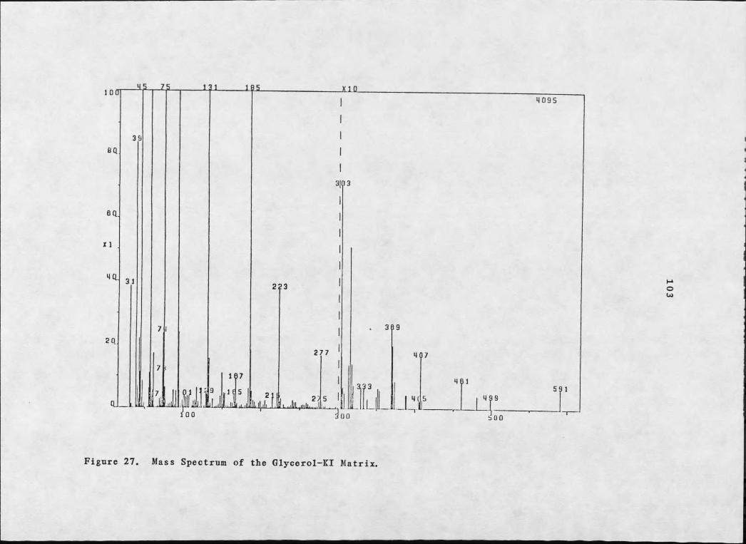
Figure 27. Mass Spectrum of the Glycerol-KI Matrix.
103

104
quantitative yield, a crystalline solid, mp 243-246°C, whose 1II-NMR spectrum was superimposable on that of brianthein Z, 71.

105
REFERENCES CITED
1. J. H. Turn I inson, D. D. Hardee, R. C, Gue I dne r, A. G. Thompson,P. A. He din, and J. P. Minyard, Science. 166 1010 (1969).
2. D. J. Faulkner, Tetrahedron. 33 I (1978).
3. W. Bergman and M. F. Stempien, Ir., J. Ore. Chem.. 22 1575 (1957) .
4. C. Djerassi. N. Theobald, W. C. M. C. Kokke, C. S. Pak, and R.M. K. Carlson, Pure Ann I. Chem.. 51 1815 (1979).
5. W. F'enical, "Marine Natural Products, Chemical and Biological Perspectives”, vol. 2, P.J. Scheuer, ed., Academic Press, Inc., New York, 1978, 173.
6. L. Chevolot, "Marine Natural Products, Chemical and Biological Perspectives", vol. 4, P.J. Scheuer, ed., Academic Press, Inc., New York, 1981, 54.
7. T. Higa, ibid., 93.
8. S. Liaaen-Jensen, "Marine Natural Products, Chemical and Biological Perspectives", vol. 2, P.J. Scheuer, ed., Academic Press, Inc., New York, 1978, 2.
9. B. Tursch, J. C. Braekman, D. Daloze, and M. Kaisin, ibid., 247.
10. R. E. Moore, "Marine Natural Products, Chemical and Biological Perspectives", P.J. Scheuer, ed., vol. I, Academic Press, Inc., New York, 1978, 44.
11. J. D. Martin and J. Darias, ibid., 124.
12. L. Minale, ibid., 175.
13. A. J. Weinheimer, F. J. Schmitz, and L. S. Ciereszko, "Drugs from the Sea", H. D. Freudenthal, ed., Marine Technology Society, Washington, D.C., 1968, 135.
14. K. L. Rinehart, Jr., P. D. Shaw, L. S. Shield, J. B. Gloer, G.C. Harbour, M. E. S. Koker, D. Samain, R. E. Schwartz, A. A. Tymiak, D. L. Weller, G. T. Carter, M. H. G. Munro, R. G. Hughes, Jr., H. E. Renis, E. B. Swynenberg, D. A. Stringfellow, J. J. Vavru, J. H. Coats, G. E. ZuGrenko, S. L. Kuentzel, L. H. Li, G. J. Bakus, R. C. Brusca, L. L. Craft, D. N. Young, and J. L. Connor, Pure Ann I. Chem., 53. 795 (19 81).

106
15. Y. Shimizu, "Marine Natural Products, Chemical and Biological Perspectives", vol. I, P. J. Scheuer, ed„ Academic Press, Inc., New York, 1978, 32.
16. P. R. Berquist, "Biologie de s Spongiaires", C. Levi and N. Boury-Esnault, eds., Colloques Internationaux du C.N.R.S.,Paris, 1978, 3 83.
17. S. DeRosa, L. Minale, R. Riccio, and G. Sodano, J. Chem. Soc.. Perkin Trans. JT 1408 (1976).
18. G. Cimino, S. De Stefano, and L. Minale, Exnerientia. 31 1117(1975).
19. W. Fenical and 0. McConnell, Exnerientia. 31 1001 (1975).
20. R. W. Dunlop, R. Kazlauskas, G. March, P. T. Murphy, and R. J. Wells, Aust. I. Chem., 35 95 (1982).
21. W. Kofheinz and W. E. Oberhansl i, Helv. Chim. Acta. 60 660 (1977).
22. R. Kazlauskas, P. T. Murphy, and R. I. Wells, Tetrahedron Lett.. 4945 (1978).
23. R. Kazlauskas, R. 0. Lidgard, R. I. Wells, and W. Vetter, Tetrahedron Lett.. 3183 (1975).
24. G. Cimino, S. De Stefano, A. Guerriero, and L. Minale, Tetrahedron Lett.. 1417 (1975).
25. G. Cimino, S. De Stefano, A. Guerriero, and L. Minale, Tetrahedron Lett.. 1421 (1975).
26. G. Cimino, S. De Stefano, A. Guerriero, and L. Minale, Tetrahedron Lett.. 1425 (1975).
27. P. I. Scheuer, "Marine Natural Products, Chemical and Biological Perspectives" P. J. Scheuer, ed., vol. 4, Academic Press, Inc., New York, 1981, 1 87.
28. G. Cimino, S. De Stefano, A. Guerriero, and L. Minale, Tetrahedron Lett.. 3723 (1975).
29. G. Schulte, P. I. Scheuer, and 0. McConnell, Helv. Chim. Acta.63 2519 (1980).
30. G. Schulte and P. I. Scheuer, J. Ore. Chem., 45 552 (1980).
31. I. H. Cardellina II, Abstracts, 41st Meeting, Montana Academy of Sciences, April, 1981.

107
32. J. E. Cardellina II, Abstracts, 36th Northwest Regional Meeting, American Chemical Society, June, 1981.
33. J. H. Cardellina II, Abstracts, 16th Meeting, Association of Island Marine Laboratories of the Caribbean, September, 1981.
34. R. Kazlauskas, P. T. Murphy, R. J. Wells, J. J. Daly, and P. Schonholzer, Tetrahedron Lett.. 4951 (1978).
35. R. Kazlauskas, P. T. Murphy, and R. J. Wells, Tetrahedron Lett.. 4949 (1978).
36. A. J. Blackman and R. J. Wells, Tetrahedron Lett.. 2729 (1976).
37. V. Amico, G. Oriente, M. Piattelli, C. Tringali, E. Fattorusso,S. Magno, and L. Mayol, J. Chem. Soc., Chem. Commun.. 1024(1976) .
38. H. H. Sun, S. M. Waraszkiewicz, K. L. Erickson, J. Finer, and J. Clardy, J. Am. Chem. Soc.. 99 3516 (1977).
39. D. J. Vanderah, P. A. Steadier, L. S. Ciereszko, F. J. Schmitz,J. D. Ekstrand, and D. van der Helm, J. Am. Chem. Soc.. 99 5780(1977) . ~
40. F. J. Schmitz, D. J. Vanderah, and L. S. Ciereszko, J. Chem.Soc., Chem. Commun.. 407 (1974).
41. B. Tursch, Pure Appl. Chem., 48 I (1976).
42. A. J. Weinheimer, R. E. Middlebrook, J. 0. Bledsoe, W. E.Marsico, and T. K. B. Earns, Chem. Commun.. 384 (1968).
43. T. Kashman, E. Zadock, and I. Neeman, Tetrahedron. 30 3615 (1974).
44. S. H. Grode, T. R. James, Jr., J. H. Cardellina II, TetrahedronLett.. 691 (1983). ~
45. 0. Kennard, D. G. Watson, L. R. Di Sanseverino, B. Tursch, R. Bosmans, and C. Djerassi, Tetrahedron Lett.. 2879 (1968).
46. Y. Kashman and A. Groweiss, Tetrahedron Lett.. 1159 (1977).
47. C. Bartholome', Ph.D. thesis, Univ. of Brussels, 1974; B. Tursch, J. C. Brackman, D. Daloze, and M. Kaisin, "Marine Natural Products, Chemical and Biological Perspectives", vol. 2, P. J. Scheuer, ed., Academic Press, Inc., New York, 1978, 278.

10848. J. E. Burks, D. van der Helm, C. Y. Chang, and L. S. Ciereszko,
Acta Crystallogr., Sect. B, 33. 704 (1977).
49. D. B. Stierle, B. Carte, D. J. Fanlkner, B. Tagle, and J.Clardy, J. Am. Chem. Soc.. 102 5088 (1980).
50. S. J. Selover, P. Crews, B. Tagle, and J. Clardy, J. Ore. Chem., 46 964 (1981).
51. R. Kazlauskas, P. T. Murphy, R. J. Wells, K. Noack, W. E. Oherhansli, and P. Schonholzer, Aust. J., Chem.. 32 867 (1979).
52. R. P. Walker and D. J. Faulkner, J. Ore. Chem., 46 1098 (1981).
53. G. Cimino, P. DeLuca, S. De Stefano, and L. Minale, Tetrahedron. 31 271 (1975).
54. Y. Kashman, Exnerientia. 36 1279 (1980).
55. T. E. Thompson, J. Mar. Biol. Assoc. U. K., 39. 115 (1960).
56. T. E. Thompson, Aust. J. Zool.. 17 755 (1969).
57. T. E. Thompson, J. Mar. Biol. Assoc. U. K., 39. 123 (1960).
58. H. P. K. Agersberg, Am. Nat., 55. 222 (1921).
59. J. E. Hochlowski, R. P. Walker, C. Ireland, and D. J. Faulkner,J. 0r£. Chem.. 47 88 (1982).
60. G. R. Schulte and P. J; Scheuer, Tetrahedron. 38 1857 (1982).
61. G. Cimino, S. De Stefano, L. Minale, and E. Trivellone, Tetrahedron Lett.. 3727 (1975).
62. J. C. Coll, B. F. Bowden, and S. J. Mitchell, Chem. Aust., 47.259 (1980).
63. J. N. Dominguez, D. R. Adams, and J. Flamerich, Rev. Latinam. Quim., 11 56 (1980); Chem. Abstr. 94:2209%.
64. W. P. Schneider, R. A. Merge, and B. E. Henson, J. Am. Chem. Soc.. 99 6062 (1977).
65. M. W. de Laubenfels, ''A Discussion of the Sponge Fauna of The Dry Torugas in Particular and the West Indies in General, with Material for a Revsion of the Families and Orders of the Porifera, Carnegie Institution of Washington, Washington, D.C., 1936.

109
66. S. Jakowska and R. F. Nigrelli, Ann. N.T. Acad. Sci., 913 (1960).
61. J. E. Thompson, R. P. Walker, S. J. Wratten, and D. J. Faulkner, Tetrahedron. 38 1865 (1982); footnote 40.
68. C. S. Foote, M. T. Wuesthoff, S. Wexler, I. G. Burstain, R. Denny, G. 0. Schenck, and E-H. Schulte-Elte, Tetrahedron. 23 2583 (1967).
69. D. J. Cookson and B. E. Smith, Ore. Magn. Reson.. 16 111 (1981),
70. M. R. Willcott, III, R. E. Davis, and R. W. Holder, J. Org.Chem.. 40 1952 (1975).
71. W. C. Hamilton, Acta Crvstallogr.. 18 502 (1965).
72. J. S. Henderson, R. McCrindle, and D. McMaster, Can. J. Chem.,51 1346 (1973).
73. J. Hellou, R. J. Andersen, and J. E. Thompson, Tetrahedron. 38 1875 (1982).
74. M. G. Gray and R. J. White, J. Invest. Dermatol.. 70 336 (1978).
75. T. Ariga, A. Susumu, A. Takahashi, and T. Miyatake, Biochim.Biophvs. Acta. 618 480 (1980).
76. S. Sato, N. Kawamura, and T. Taketomi, Jpn. J. Exp. Med.. 49 139 (1979).
77. I. Singh and T. Kishimoto, Brain ResJr, 232 500 (1982).
78. U. H. Do, P. T. Pei, and R. D. Minard, Lipids. 16. 855 (1981).
79. M. K. Wassef and J. W. Hendrix, Biochim. Biophvs. Acta. 486 172 (1977).
80. V. Ondrusek and M. Prostenik, Exp. Mycol.. 2. 156 (1978).
81. A. Kazutaka and Y. Tometaka, Mokuzai Gakkaishi. 26 819 (1980); Chem. Abstr. 94:117774n.
82. A. Kazutaka and M. Sawada, Mokuzai Gakkaishi» 25 749 (1979);Chem. Abstr. 92:90877t.
83. E. Miyagawa, R. Azuma, T. Suto, and I. Yano, J. Biochem.(Tokyo), 86 311 (1979); Chem. Abstr. 91:153149n.
84. D. Andrews and D. L. Nelson, Biochim. Biophvs. Acta. 550 174 (1979).

H O
85. J. H. Cardellina II and R. E. Moore, Pkvtochemistrv. 17 554(1978). —
86. M. Mahendran, S. Somasundaram, and R. E. Thompson, Phytochemistry. 18 1885 (1979).
^ Nielsen, T, S. Carle, and C. Christophersen. Phytochemistry. 21 1643 (1982).
88. G. C. Hanson, E. EeiTbronn, K.-A. Karlsson, and B. E. Samuelson, J. Lipid Res.. 20 509 (1979).
89. K.-A. Karlsson, B. E. SamueIsson, and T. 0. Steen, Biochim- Biophys. Acta. 337 356 (1974).
90. G. P. Smirnova and N. K. Kochetkov, Biochim. Bionhvs. Acta. 618486 (1980). ---- ---
91. A. J. DeKoning, I. Sci. Food Agr., 23. 1471 (1972).
92. M. Sugita, I. Arakawa, T. Eori, and Y. Sawada, Seikasaku. 40 158 1968; Chem. Abstr. 70:35428t.
93. H. E. Carter, W. P. Norris, F. J. Click, G. E. Phillips, and R. Harris, J. Biol. Chem.. 170 269 (1947).
94. D. H. S. Horn and Y. Y. Pretorius, J. Chem. Soc.. 670 (1966).
95. E. Baer and V. Mahadevan, J. Am. Chem. Spc., 81.2494 (1959).
96. R. Kleiman, R. W. Miller, F. R. Earl, and I. A. Wolff, Lipids. 2473 (1967). --- -
97. C. R. Smith, Jr., "Topics in Lipid Chemistry", vol. I, F. D. Gunstone, ed., Logos Press, Ltd., London, 1970, 338.
98. R. U. Lemieuz and J. Giguere, Can. J. Chem.. 29 678 (1951).
99. D. H. S. Horn, and Y. Y. Pretorius, J. Chem. Spc., 1460 (1954).
100 D. H. S. Horn, F. W. Hougen, E. von Rudloff, and D. A. Sutton,J. Chem. Soc.. 177 (1954).
101. C. R. Smith, Jr., and I. A. Wolff, Lipids. 4 9 (1969).
102. A. Ease, Arch. Biochem. Biophvs.. 210 280 (1981).
103. I. Singh and Y. Kishimoto, J. Biol. Chem.. 254 7698 (1979).
104. Y. Kishmoto and N. S. Radin, Ji Lipid Res.. I 79 (1959).

Ill
105. K.-A. Karlsson, "Structure of Biological Membranes", S. Abrahamsson and I. Pascher, eds., Plenum Press, Inc., New York, 1977, 245.
106. C. Hitchcock, L. J. Morris, and A. T. James, European J. Biochem.. 3 473 (1968),
107. M. Hoshi and Y. Kishmoto, J. Biol. Chem.. 248 4123 (1973).
108. K. Tatsumi, S. Murad, and Y. Kishimoto, J. Biol. Chem.. 252 5206 (1977).
109. K. Tatsumi, S. Murad, and Y. Kishimoto, Arch. Biochem. Biouhvs.. 171 87 (1975).
H O . K. Tatsumi, Y. Kishimoto, C. Hignite, Arch. Biochem. Biophvs.. 165 656 (1974).
111. C. Hitchcock, L. J. Morris, and A. T. James, European J. Biochem.. 3. 419 (1968).
112. C. Hitchcock, L. J. Morris, and A. T. James, European J, Biochem.. 3. 473 (1968).
113. W. E. Shine and P. K. Stumpf, Arch. Biochem. Biophvs.. 162 147 (1974).
114. E. Jefferts, R. W. Morales, and C. Litchfield, Lip ids. 9.244 (1974).
115. R. W. Morales and C. Litchfield, Biochem. Biophvs. Acta, 431 206 (1976).
116. J. H. Cardellina II, C. J. Graden, B. Greer, and J. E. Kern, Lipids, in press (1983).
117. Personal Communication from Dr. F. M. Bayer, Smithonian Institution.
118. S. J. Wratten, D. J. Faulkner, K. Hirotsu, and J. Clardy, J. Am. Chem. Soc.. 99 2824 (1977).
119. S. J. Wratten, W. Fenical, D. J. Faulkner, and J. C. WekelI, Tetrahedron Lett., 1559 (1977).
120. B. N. Ravi, J. F. Marwood, and R. J. Wells, Aust. J. Chem.. 33 2307 (1980).
121. S. M. Kupchan, R. W. Britton, M. F. Ziegler, and C. W. Sigel, J. Ore. Chem., 38 17 8 (1973).

122. J. E. Cardellina II, J« Nat. Prod., in press.123. D. Nachmansohn, "Protein Metabolism of the Nervous System", A.
Laj tha, ed., Plenum Press, Inc., New York, 1970, 313.
124. C. Westcott, "Plant Disease Handbook", Van Nostrand Reinhold Co., New York, 1971, 843.
125. C. J. Alexopoulos and C.W. Mims, "Introductory Mycology", John Wiley and Sons, Inc., New York, 1979, 613.
112

M O N T A N A S T A T E U N I V E R S I T Y L I B R A R I E Sstks D378.G892@Theses RL
3 1762 00185546 7
337*$ S'? a.a a p-A.