oral mucous membranes-2 /certified fixed orthodontic courses by indian dental academy
TRANSCRIPT

www.indiandentalacademy.com

Objective- At the end of the lecture student should know-
Details of non-keratinised epithelium
Details of different types of oral mucosa.
www.indiandentalacademy.com

NON-KERATINIZED EPITHELIUM
They do not produce cornified surface layer.
There are four layers of non- keratinized epithelium.
1. Basal ( Stratum basale)
2. Prickle cell
3. Intermediate (Stratum intermedium)
4. Superficial (Stratum superficiale)
www.indiandentalacademy.com

Cell Layers Features
Stratum Basale •A layer of cubiodal or columnar cells adjacent to basal lamina containing separate tonofilamentsand other cell organelles •Site of most cell divisions
Prickle Cell •Larger ovoid cells containing dispersed tonofilament•Membrane coating granules appear in upper part of layer, filaments become numerous .
StratumIntermedium
• Slightly flattened cells containing many dispersed tonofilaments and glycogen.
StratumSuperficiale
•Slightly flattened cells with dispersed filaments and glycogen •Fewer organells are present , but nuclie persist.
www.indiandentalacademy.com

Principal structure of oral epithelium in successive layers
A. Orthokeratinized B. Non- keratinized
oral epithelium oral epithelium
www.indiandentalacademy.com

Histologic section
A. Orthokeratinization B. Parakeratinization C. Non- keratinization
www.indiandentalacademy.com

LAMINA PROPRIA The connective tissue
supporting the oral epithelium is termed LAMINA PROPRIA.
It can be divided into two layers.
- Superficial papillary layer
- Deeper reticular layer
www.indiandentalacademy.com

In papillary layer, collagen fibers are thin and loosely arranged and many capillary loops are present.
In reticular layer, collagen fibers are arranged in thick bundles and lie parallel to surface plane.
The lamina propria may be attached to the periosteumof the alveolar bone.
www.indiandentalacademy.com

SUBMUCOSA
The submucosa consists of connective tissue of varying thickness and density. It attaches the mucous membrane to the underlying structures.
www.indiandentalacademy.com

Glands, blood vessels, nerves, lymph vessels and adipose tissue are present in this layer.
It is in the submucosa that larger arteries divide into smaller branches which then enter the lamina propria.
www.indiandentalacademy.com

SUBDIVISIONS OF ORAL MUCOSAFor descriptive purposes , the oral mucosa can be divided into following areas :
1. Keratinized Areas
- Masticatory mucosa ( gingiva and hard palate)
- Vermillion border of lip
2. Non-Keratinized Areas
- Lining or reflecting mucosa
3. Specialised or sensory mucosa
www.indiandentalacademy.com

KERATINIZED AREASMASTICATORY MUCOSA
1. Hard palate2. Gingiva
HARD PALATE
The palate functionsanatomically as the roof ofthe oral cavity and the floorof the nasal cavity.
It is divided in two regions,anterior hard palate andposterior soft palate.
www.indiandentalacademy.com

Both are distinguished from one another by colorand palpation.
The hard palate is light pink while soft palate isred.
The hard palate is firm and less movable than softpalate because the mucous membrane of hardpalate is tightly fixed to underlying periosteum.
www.indiandentalacademy.com

The hard palate is further divided into three regions –
1. The raphe
2. The fatty zone
3. The glandular zone
The raphe divided the hard palate in two lateral halves. The halves are subdivided microscopically into two areas, the fatty anterolateral zone and the glandular posterolateral zones.
www.indiandentalacademy.com

The epithelium is uniform with well keratinized surface.
The lamina propria is thicker in anterior region than posterior parts of palate.
Various regions in the Hard palate differ because of varying structure of the submucosal layer.1. Gingival region adjacent to the teeth2. Palatine raphe also known as median area3. Anterolateral area or fatty zone4. Posterolateral area or glandular zone
www.indiandentalacademy.com
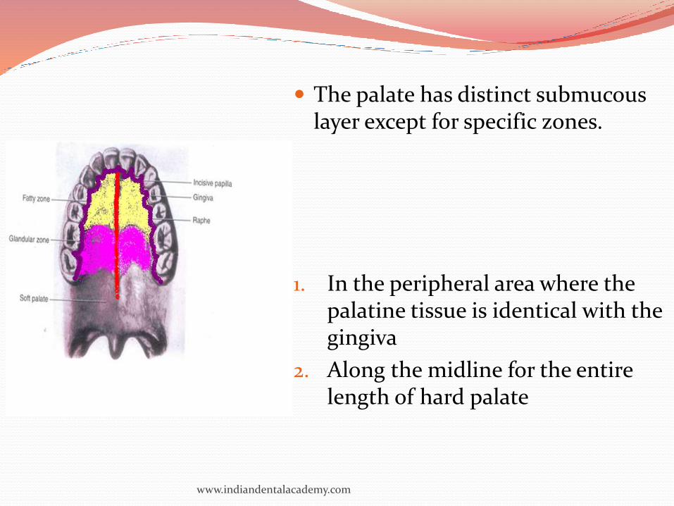
The palate has distinct submucous layer except for specific zones.
1. In the peripheral area where the palatine tissue is identical with the gingiva
2. Along the midline for the entire length of hard palate
www.indiandentalacademy.com

Incisive papilla The oral incisive papilla is
formed of dense connectivetissue.
It contains the oral part ofthe vestigial Nasopalatineducts.
They are, the blind ducts ofvarying length lined bysimple or pseudostratifiedcolumnar epithelium rich ingoblet cells.
www.indiandentalacademy.com

Jacobson's organ/vomeronasal organ
Considered as auxiliary olfactory sense organ alongwith Nasopalatine ducts
It is a small ellipsoid (cigar shaped) structurelined with olfactory epithelium that extends fromthe nose to the oral cavity.
Is apparent in the twelth to fifteenth fetal week,after which it undergoes involution
www.indiandentalacademy.com

Palatine rugae (Transverse palatine ridges)
Irregular and often asymmetric in humans are ridges of mucous membrane extending laterally from the incisive papilla and the anterior part of the raphe
Made up of dense connective tissue layer with the interwoven fibers.
www.indiandentalacademy.com

Epithelial pearls
In the midline especially in the region of theincisive papilla, epithelial pearls may be found inthe lamina propria.
They consist of concentrically arranged epithelialcells that are frequently keratinized.
They are remnants of the epithelium formed in theline of fusion between the palatine processes.
www.indiandentalacademy.com

www.indiandentalacademy.com

GINGIVA The gingiva is that portion of
the oral mucosa that surrounds and is attached to the teeth and the alveolar bone.
The gingiva extends from the dentogingival junction to the alveolar mucosa
It is subject to friction and pressure of mastication
www.indiandentalacademy.com

GINGIVA Color of gingiva – Normally pink
but sometimes have blackish tint depending upon keratinization & pigmention.
Epithelium :- Orthokeratinized (15%) or parakeratinized(75%) and non keratinized (10%).
Stratified squamous epithelium often showing stippled surface
www.indiandentalacademy.com

Lamina propria :- Long and narrow papillae; dense collagenous connective tissue, not highly vascular but has long capillary loops with numerous anastomoses
Submucosa :- No distinct layer; mucosa firmly attached by collagen fibers to periosteum of alveolar process.(mucoperiosteum)
www.indiandentalacademy.com

Gingiva can be divided into
i) Free gingiva
ii) Attached gingiva
iii) Interdental gingiva.
Free gingival groove separates the free gingiva from attached gingiva, which runs parallel to margin of the gingiva at distance of 0.5 to 1.5 mm.
Mucogingival junction separates the attached gingivafrom alveolar mucosa.
www.indiandentalacademy.com

www.indiandentalacademy.com

www.indiandentalacademy.com

A. Different types of epitheliumB. Histologic section showing tissues
www.indiandentalacademy.com

Gingiva and alveolar mucosa are separated by themucogingival junction.
The alveolar mucosa is thin and loosely attachedto the periosteum by a well-defined submucouslayer of loose connective tissue , and it maycontain small mixed glands.
www.indiandentalacademy.com

The epithelium is thin and nonkeratinized, and the epithelial ridges and papillae are low and often entirely missing. These differences cause the variation in color between the pale pink gingiva and the red lining mucosa.
The gingival surface appears stippled .Portions at the epithelium appear to be elevated and between the elevations there are shallow depressions, the net result of which is stippling.
www.indiandentalacademy.com

The depression corresponds to the center of heavier epithelial ridges
Functional adaptations to mechanical impacts
Disappearance of stippling- an indication of edema.
The degree of stippling and the texture of collagenous fibers vary with the different individuals, according to age and sex.
www.indiandentalacademy.com

Interdental papilla
Part of gingiva that fills the space between two adjacent teeth
The interdental papilla when viewed from the oral or vestibular aspect, the surface is triangular.
In a 3 dimensional view the interdental papilla of the post teeth is tent shaped, where as pyramidal between the anterior teeth.
www.indiandentalacademy.com

Col: It is the central concave area which fits below the contact point seen in the tent shaped interdental papilla of the posterior teeth
It is covered by non-keratinized epithelium and is more vulnerable to periodontal diseases.
www.indiandentalacademy.com

www.indiandentalacademy.com

Gingival ligament
1. Dentogingival fibres :- Extend from cervical cementuminto lamina propria of the gingiva. Most numerous
2. Alveologingival fibres : The fibers arise from the alveolar crest and extend into the lamina propria
3. Circular fibres : This group of fibers encircle the tooth
4. Dentoperiosteal fibres : They run from the cementuminto the periosteum of the alveolar crest
5. Transseptal fibres : They are accessory fibers that extend interproximally between adjacent teeth
www.indiandentalacademy.com

BLOOD SUPPLY It is chiefly from the branches of the alveolar
arteries
The lymph vessels of gingiva lead to submentaland submandibular lymph nodes
It is also well innervated by various nerve endings
www.indiandentalacademy.com

Summary- Student should understand-
Details of non-keratinised epithelium
Details of different types of oral mucosa.
www.indiandentalacademy.com

Thank you
www.indiandentalacademy.com