optom-234; no.of pages10 article in press · please cite this article in press as: huang j, et al....
TRANSCRIPT

ARTICLE IN PRESS+ModelOPTOM-234; No. of Pages 10
Journal of Optometry (2017) xxx, xxx---xxx
www.journalofoptometry.org
ORIGINAL ARTICLE
Comparison of manual & automated analysis methodsfor corneal endothelial cell density measurements byspecular microscopy
Jianyan Huanga,b, Jyotsna Marama,b, Tudor C. Tepelusa,b, Cristina Modaka,Ken Mariona, SriniVas R. Saddaa,b, Vikas Chopraa,b, Olivia L. Leea,b,∗
a Doheny Eye Institute, Los Angeles, CA 90033, United Statesb Department of Ophthalmology, David Geffen Medical School at UCLA, Los Angeles, CA 90095, United States
Received 10 April 2017; accepted 26 June 2017
KEYWORDSCorneal endothelialcell density;Automated method;Center Method;Flex-Center Method;Specular microscopy
AbstractPurpose: To determine the reliability of corneal endothelial cell density (ECD) obtained by auto-mated specular microscopy versus that of validated manual methods and factors that predictsuch reliability.Methods: Sharp central images from 94 control and 106 glaucomatous eyes were captured withKonan specular microscope NSP-9900. All images were analyzed by trained graders using KonanCellChek Software, employing the fully- and semi-automated methods as well as Center Method.Images with low cell count (input cells number <100) and/or guttata were compared with theCenter and Flex-Center Methods. ECDs were compared and absolute error was used to assessvariation. The effect on ECD of age, cell count, cell size, and cell size variation was evaluated.Results: No significant difference was observed between the Center and Flex-Center Methodsin corneas with guttata (p = 0.48) or low ECD (p = 0.11). No difference (p = 0.32) was observedin ECD of normal controls <40 yrs old between the fully-automated method and manual CenterMethod. However, in older controls and glaucomatous eyes, ECD was overestimated by the fully-automated method (p = 0.034) and semi-automated method (p = 0.025) as compared to manualmethod.Conclusion: Our findings show that automated analysis significantly overestimates ECD in theeyes with high polymegathism and/or large cell size, compared to the manual method. There-fore, we discourage reliance upon the fully-automated method alone to perform specularmicroscopy analysis, particularly if an accurate ECD value is imperative.
r Espana, S.L.U. on behalf of Spanish General Council of Optometry.cle under the CC BY-NC-ND license (http://creativecommons.org/
© 2017 Published by ElsevieThis is an open access artilicenses/by-nc-nd/4.0/).
Please cite this article in press as: Huang J, et al. Comparison of manual & automated analysismethods for corneal endothelial cell density measurements by specular microscopy. J Optom. (2017),http://dx.doi.org/10.1016/j.optom.2017.06.001
∗ Corresponding author at: 800S Fairmount Ave Suite 215, Pasadena, CA 91105, United States.E-mail address: [email protected] (O.L. Lee).
http://dx.doi.org/10.1016/j.optom.2017.06.0011888-4296/© 2017 Published by Elsevier Espana, S.L.U. on behalf of Spanish General Council of Optometry. This is an open access articleunder the CC BY-NC-ND license (http://creativecommons.org/licenses/by-nc-nd/4.0/).

ARTICLE IN PRESS+ModelOPTOM-234; No. of Pages 10
2 J. Huang et al.
PALABRAS CLAVEDensidad celularendotelial corneal;Métodoautomatizado;Método de centrado;Método de centradoflexible;Microscopio especular
Comparación de los métodos de análisis manual y automatizado para medir ladensidad celular endotelial corneal mediante microscopio especular
ResumenObjetivo: Determinar la fiabilidad de la densidad celular endotelial corneal (ECD) obtenidamediante microscopio especular automático frente a métodos manuales validados y factorespredictivos de la fiabilidad.Métodos: Se capturaron imágenes nítidas de 94 controles y 106 ojos glaucomatosos con unmicroscopio especular Konan NSP-9900. Todas las imágenes fueron analizadas por examinadoresexpertos mediante el software Konan CellChek, utilizando los métodos automatizado total,semiautomático y de centrado. Se compararon las imágenes con bajo recuento celular (númerode células <100) y/o córnea guttata con el método de centrado y centrado flexible. Se com-pararon las ECD, utilizándose el error absoluto para valorar la variación. Se evaluó el efecto dela ECD sobre la edad, el recuento celular, el tamano celular y la variación del tamano celular.Resultados: No se observó diferencia significativa entre los métodos de centrado y centradoflexible en las córneas con guttata (p = 0,48) o baja ECD (p = 0,11). No se observó diferencia(p = 0,32) en cuanto a ECD en los controles normales < 40 anos entre el método totalmenteautomatizado y el método de centrado manual. Sin embargo, en los controles mayores y enlos ojos glaucomatosos, la ECD fue sobreestimada por el método totalmente automatizado(p = 0,034) y el método semiautomático (p = 0,025), en comparación al método manual.
Conclusión: Nuestros hallazgos muestran que los análisis automatizados sobreestiman consid-erablemente la ECD en los ojos con alto polimegatismo y/o gran tamano celular, en comparaciónal método manual. Por tanto, no recomendamos confiar en el método totalmente automatizadopor sí solo para realizar estudios mediante microscopio especular, particularmente en casos enque la precisión del valor de ECD sea imperativo.© 2017 Publicado por Elsevier Espana, S.L.U. en nombre de Spanish General Council of Optom-etry. Este es un artıculo Open Access bajo la licencia CC BY-NC-ND (http://creativecommons.org/licenses/by-nc-nd/4.0/).
I
Eietc
9rfiaa
aEaosdatCMtcbM
ott
mope
M
P
NcottuCeMt
ntroduction
ndothelial cell density (ECD) values are routinely usedn clinical practice to evaluate the status of the cornealndothelium to make treatment and surgical decisions ando gauge the safety of new drugs, devices and surgical pro-esses during clinical trials.
Non-contact specular microscopes such as the Konan NSP-900 capture sharp images with sufficient magnification foreliable ECD determination or morphometric analysis. Thexed-frame method for determine ECD allows quantitativenalysis of cell structure, including ECD, coefficient of vari-tion (CV), and percentage of hexagonal cells (HEX).1---3
The Konan CellChek software uses several differentpproaches, varying in speed and complexity, to obtainCDs, from fully- and semi-automated to manual.4---8 Theutomated analysis method, or Auto-Trace, automaticallyutlines endothelial cells and calculates cell density, cellize and hexagonality. In the fully-automated method, theefault cell size S pattern is used, while in the semi-utomated method, the cell size is manually selected from So XL. The most commonly manual analysis methods are theenter Method and the Flex-Center Method. In the Centerethod, the user marks the center of each cell in a con-
Please cite this article in press as: Huang J, et
methods for corneal endothelial cell density measurehttp://dx.doi.org/10.1016/j.optom.2017.06.001
iguous group, and the software then counts the number ofells by determining cell area from a polygon digitizationy locating cell border intersections.9 In the Flex-Centerethod, based on the Center Method, the outer boundary
CtaH
f all visible cells is outlined by clicking the intersection ofhree cells. The use of Flex-Center Method is suggested byhe manufacturer when fewer contiguous cells are visible.
We compared automated and manual methods of ECDeasurements to determine the optimal methods in terms
f accuracy under different conditions and the factors thatredict reliability of ECD values in normal and glaucomatousyes.
ethods
atients and endothelial photography
inety-nine normal control eyes and 112 open angle glau-omatous eyes with or without uveitis spanning a wide rangef cell densities were recruited from the Doheny Eye Cen-er between May 2013 and May 2014, and were included inhe study. None of the eyes had any history of prior intraoc-lar surgery, ocular trauma, keratitis or contact lens wear.entral images of corneal endothelium were captured forach eye with a Konan NSP-9900 specular microscope (Konanedical USA Inc., Irvine, CA). This study was approved by
he Institutional Review Board of University of Southernalifornia (at that time the affiliation of Doheny Eye Insti-
al. Comparison of manual & automated analysisments by specular microscopy. J Optom. (2017),
ute). Informed consent was obtained from all participants,nd the study followed the tenets of the Declaration ofelsinki.
IN PRESS+Model
rneal ECD 3
3400
3200
3000
2800
2600
2400
EC
D (
cells
/mm
2 )
2200
2000Control
MANUAL SEMI AUTO
n=94
p=0.001∗
p<0.001∗
p=0.77 p=0.89
p=0.025∗ p<0.001∗
n=106
Glaucoma
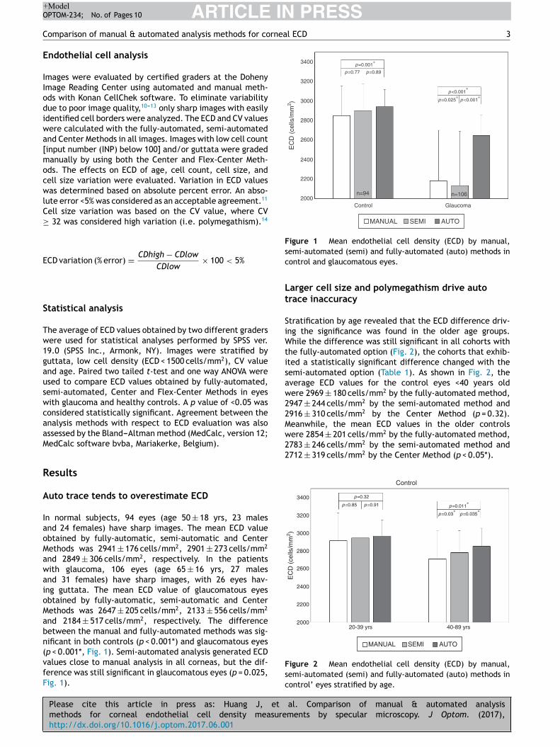
Figure 1 Mean endothelial cell density (ECD) by manual,sc
Lt
SiWtisaw22Mwere 2854 ± 201 cells/mm2 by the fully-automated method,2783 ± 246 cells/mm2 by the semi-automated method and2712 ± 319 cells/mm2 by the Center Method (p < 0.05*).
3400 p=0.32
p=0.011∗
p=0.03∗
p=0.035∗
p=0.85 p=0.91
Control
3200
3000
2800
2600
2400
EC
D (
cells
/mm
2 )
2200
200020-39 yrs 40-89 yrs
MANUAL SEMI AUTO
ARTICLEOPTOM-234; No. of Pages 10
Comparison of manual & automated analysis methods for co
Endothelial cell analysis
Images were evaluated by certified graders at the DohenyImage Reading Center using automated and manual meth-ods with Konan CellChek software. To eliminate variabilitydue to poor image quality,10---13 only sharp images with easilyidentified cell borders were analyzed. The ECD and CV valueswere calculated with the fully-automated, semi-automatedand Center Methods in all images. Images with low cell count[input number (INP) below 100] and/or guttata were gradedmanually by using both the Center and Flex-Center Meth-ods. The effects on ECD of age, cell count, cell size, andcell size variation were evaluated. Variation in ECD valueswas determined based on absolute percent error. An abso-lute error <5% was considered as an acceptable agreement.11
Cell size variation was based on the CV value, where CV≥ 32 was considered high variation (i.e. polymegathism).14
ECD variation (% error) = CDhigh − CDlow
CDlow× 100 < 5%
Statistical analysis
The average of ECD values obtained by two different graderswere used for statistical analyses performed by SPSS ver.19.0 (SPSS Inc., Armonk, NY). Images were stratified byguttata, low cell density (ECD < 1500 cells/mm2), CV valueand age. Paired two tailed t-test and one way ANOVA wereused to compare ECD values obtained by fully-automated,semi-automated, Center and Flex-Center Methods in eyeswith glaucoma and healthy controls. A p value of <0.05 wasconsidered statistically significant. Agreement between theanalysis methods with respect to ECD evaluation was alsoassessed by the Bland---Altman method (MedCalc, version 12;MedCalc software bvba, Mariakerke, Belgium).
Results
Auto trace tends to overestimate ECD
In normal subjects, 94 eyes (age 50 ± 18 yrs, 23 malesand 24 females) have sharp images. The mean ECD valueobtained by fully-automatic, semi-automatic and CenterMethods was 2941 ± 176 cells/mm2, 2901 ± 273 cells/mm2
and 2849 ± 306 cells/mm2, respectively. In the patientswith glaucoma, 106 eyes (age 65 ± 16 yrs, 27 malesand 31 females) have sharp images, with 26 eyes hav-ing guttata. The mean ECD value of glaucomatous eyesobtained by fully-automatic, semi-automatic and CenterMethods was 2647 ± 205 cells/mm2, 2133 ± 556 cells/mm2
and 2184 ± 517 cells/mm2, respectively. The differencebetween the manual and fully-automated methods was sig-nificant in both controls (p < 0.001*) and glaucomatous eyes
Please cite this article in press as: Huang J, et
methods for corneal endothelial cell density measurehttp://dx.doi.org/10.1016/j.optom.2017.06.001
(p < 0.001*, Fig. 1). Semi-automated analysis generated ECDvalues close to manual analysis in all corneas, but the dif-ference was still significant in glaucomatous eyes (p = 0.025,Fig. 1).
Fsc
emi-automated (semi) and fully-automated (auto) methods inontrol and glaucomatous eyes.
arger cell size and polymegathism drive autorace inaccuracy
tratification by age revealed that the ECD difference driv-ng the significance was found in the older age groups.
hile the difference was still significant in all cohorts withhe fully-automated option (Fig. 2), the cohorts that exhib-ted a statistically significant difference changed with theemi-automated option (Table 1). As shown in Fig. 2, theverage ECD values for the control eyes <40 years oldere 2969 ± 180 cells/mm2 by the fully-automated method,947 ± 244 cells/mm2 by the semi-automated method and916 ± 310 cells/mm2 by the Center Method (p = 0.32).eanwhile, the mean ECD values in the older controls
al. Comparison of manual & automated analysisments by specular microscopy. J Optom. (2017),
igure 2 Mean endothelial cell density (ECD) by manual,emi-automated (semi) and fully-automated (auto) methods inontrol’ eyes stratified by age.
ARTICLE IN PRESS+ModelOPTOM-234; No. of Pages 10
4 J. Huang et al.
Table 1 Stratification by cell size and polymegathism.
Subjects Cell Info ECD (cells/mm2) mean ± SD
Cohort Eyesn Size CV Manual Semi Auto
Control eyesn = 94
29 S <32 2943 ± 294 2980 ± 191 2980 ± 19147 ≥32 2868 ± 263 2909 ± 223(*) 2928 ± 263(*)18 M-XL Any 2384 ± 293 2372 ± 257 2729 ± 120(*)
Glaucomatous eyewithout guttatan = 80
15 S <32 2684 ± 172 2883 ± 138(*) 2883 ± 138(*)27 ≥32 2598 ± 445 2789 ± 298(*) 2841 ± 256(*)38 M-XL any 1348 ± 713 1355 ± 688 2571 ± 172(*)
cient
acitiW
o(t
FaCwaba(+
ECD = endothelial cell density; SD = standard deviation; CV = coeffi
Further stratification revealed that fully-automatednalysis can yield comparable ECD values in normalorneas with uniformly small cell size, 2980 ± 191 cells/mm2
2
Please cite this article in press as: Huang J, et
methods for corneal endothelial cell density measurehttp://dx.doi.org/10.1016/j.optom.2017.06.001
n the fully-automated method, 2980 ± 191 cells/mm inhe semi-automated method, and 2943 ± 294 cells/mm2
n the Center Method (p = 0.12) (Table 1, S + CV < 32).hen the cells size pattern is M-XL, the ECD values
(sdi
UNGRADED
UNGRADED
AUTOMATED GRADING
AUTOMATED
A
B
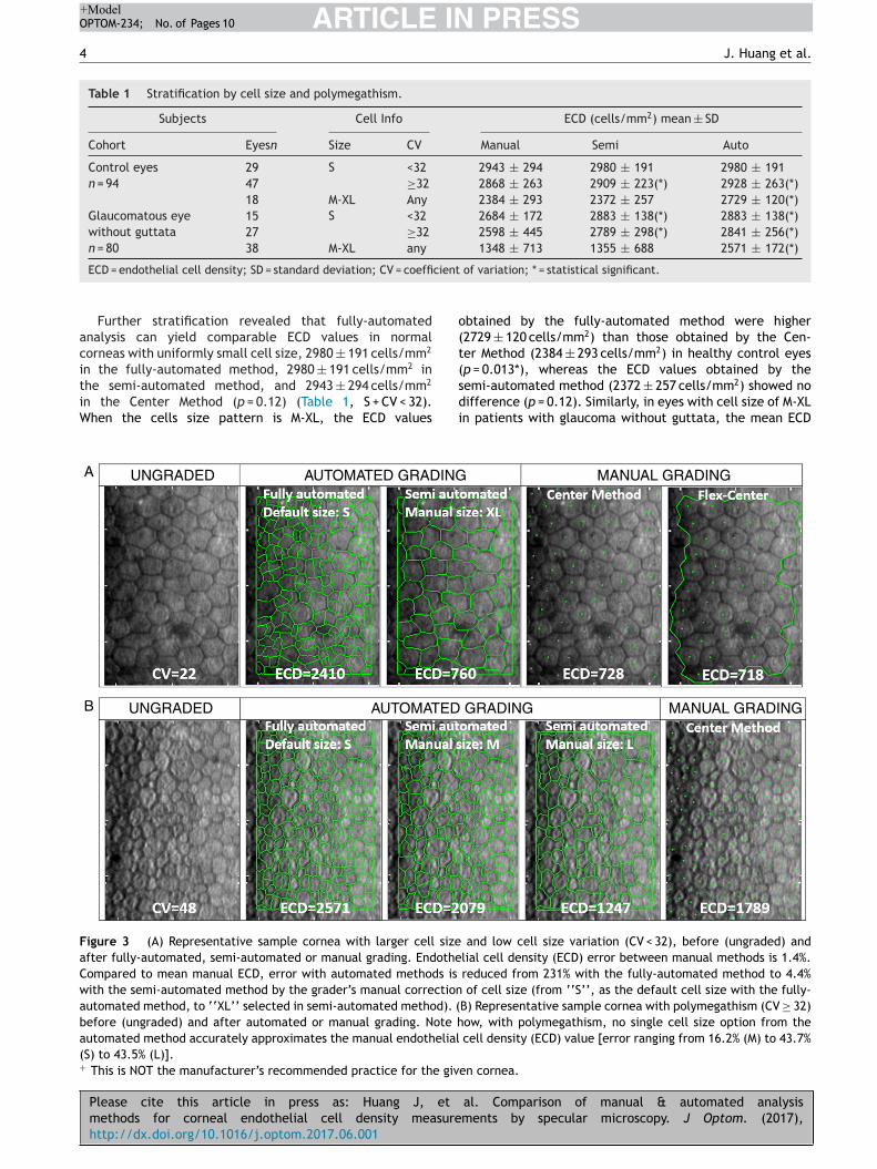
igure 3 (A) Representative sample cornea with larger cell sizefter fully-automated, semi-automated or manual grading. Endotheompared to mean manual ECD, error with automated methods is
ith the semi-automated method by the grader’s manual correctionutomated method, to ‘‘XL’’ selected in semi-automated method). (efore (ungraded) and after automated or manual grading. Note hutomated method accurately approximates the manual endothelialS) to 43.5% (L)].This is NOT the manufacturer’s recommended practice for the giv
of variation; * = statistical significant.
btained by the fully-automated method were higher2729 ± 120 cells/mm2) than those obtained by the Cen-er Method (2384 ± 293 cells/mm2) in healthy control eyes
al. Comparison of manual & automated analysisments by specular microscopy. J Optom. (2017),
p = 0.013*), whereas the ECD values obtained by theemi-automated method (2372 ± 257 cells/mm2) showed noifference (p = 0.12). Similarly, in eyes with cell size of M-XLn patients with glaucoma without guttata, the mean ECD
GRADING
MANUAL GRADING
MANUAL GRADING
and low cell size variation (CV < 32), before (ungraded) andlial cell density (ECD) error between manual methods is 1.4%.reduced from 231% with the fully-automated method to 4.4%
of cell size (from ‘‘S’’, as the default cell size with the fully-B) Representative sample cornea with polymegathism (CV ≥ 32)ow, with polymegathism, no single cell size option from the
cell density (ECD) value [error ranging from 16.2% (M) to 43.7%
en cornea.

ARTICLE IN PRESS+ModelOPTOM-234; No. of Pages 10
Comparison of manual & automated analysis methods for corneal ECD 5
UNGRADED MANUAL GRADING
guttanual
a1rv2Falwoa8
B
Tadwcmmhu
Figure 4 Representative sample cornea with severe guttata (low number of gradable cells (INP < 100) graded by different ma
values was 2571 ± 172 cells/mm2 by the fully-automatedmethod (p < 0.001*), 1355 ± 688 cells/mm2 by the semi-automated method (p = 0.24), and 1348 ± 713 cells/mm2 bythe Center Method.
While the semi-automated method can further correctECD values in both normal and diseased eyes (Table 1,M-XL; Fig. 3A), it fails in the presence of polymegath-ism (Table 1, S + CV ≥ 32); Fig. 3B). In terms of high cellsize variation (CV ≥ 32), the mean ECD value obtainedby the fully-automated method (2928 ± 263 cells/mm2)was higher than that obtained by the Center Method(2868 ± 263 cells/mm2, p = 0.034*). There was no signifi-cant difference from that obtained by the semi-automatedmethod (2909 ± 223 cells/mm2, p = 0.21) in control eyes<40 yrs of age, because most of the cells are of Spattern. Likewise, in the eyes of patients with glau-coma but without guttata, the mean ECD value obtainedwith the fully-automated method (2841 ± 256 cells/mm2)was higher than that obtained with the Center Method(2598 ± 445 cells/mm2, p = 0.034*) and there was no dif-ference when compared to the semi-automated method(2789 ± 298 cells/mm2, p = 0.37).
Center Method offers comparable ECD even at low
Please cite this article in press as: Huang J, et
methods for corneal endothelial cell density measurehttp://dx.doi.org/10.1016/j.optom.2017.06.001
cell count
No significant difference was observed between the resultsobtained by manual Center and Flex-Center Methods in
hIat
Table 2 Mean endothelial cell density by Center and Flex-Center
Subjects Center values
Cohort Eyesn
Age (yrs) INP CV
Guttata 26 30---85 16---73 29 ± 4
Low ECD 23 50---94 36---96 34 ± 4
ECD = endothelial cell density; SD: standard deviation; INP = input numb
e covering >40% of the viewable field) resulting in a drastically methods.
ll analyzed corneas with an INP value of less than00. In the corneas with presence of guttata, whicheduced the INP value to 16---73 cells, the mean ECDalue was 2022 ± 569 cells/mm2 by Center Method and069 ± 745 cells/mm2 by Flex-Center Method (p = 0.48).ig. 4 showed a sample image obtained in both Centernd Flex-Center Methods in severe guttata, resulting iness than 40 cells countable. Even in corneas with low ECD,hen only 36---96 cells were countable, the mean ECD valuebtained by the Center Method was 856 ± 343 cells/mm2
nd that obtained by the Flex-Center Method was79 ± 352 cells/mm2 (p = 0.11). (Table 2; Fig. 3A).
land---Altman analysis
he Bland---Altman analysis showed differences in ECD evalu-tion by using different analysis methods in all corneas. Theifference in ECD values between different analysis methodsas low in normal controls, but high in glaucomatous eyes. Inontrol eyes, ECD values determined by the fully-automatedethod tended to be 39 cells/mm2 higher than those deter-ined by the semi-automated method and 91 cells/mm2
igher than those obtained by the Center Method; ECD val-es obtained by semi-automated method were 52 cells/mm2
al. Comparison of manual & automated analysisments by specular microscopy. J Optom. (2017),
igher than those obtained by the Center Method (Fig. 5).n glaucomatous eyes, the ECD values determined by fully-utomated method tended to be 514 cells/mm2 higherhan those determined by the semi-automated method and
Methods in eyes with low cell count.
ECD (cells/mm2) mean ± SD
Center Flex-Center Error p
2022 ± 569 2069 ± 745 2.3% 0.48856 ± 343 879 ± 352 2.7% 0.11
er; CV = coefficient of variation.

Please cite this article in press as: Huang J, et al. Comparison of manual & automated analysismethods for corneal endothelial cell density measurements by specular microscopy. J Optom. (2017),http://dx.doi.org/10.1016/j.optom.2017.06.001
ARTICLE IN PRESS+ModelOPTOM-234; No. of Pages 10
6 J. Huang et al.
700
Control
Control
Control
+1.96 SD
307.3
Mean
39.2
–228.9
–1.96 SD
+1.96 SD
365.6
Mean
91.1
–183.5
–1.96 SD
+1.96 SD
313.9
Mean
51.9
–210.1
–1.96 SD
600
500
400
300
200
100
0
–100
–200
–300
600
500
400
300
200
100
–100
–200
–300
400
300
200
100
–100
–200
–300
0
0
2400
Diff
eren
ce in
EC
D v
alue
sF
ully
- vs
sem
i-aut
omat
ed m
etho
dD
iffer
ence
in E
CD
val
ues
Ful
ly-
auto
mat
ed m
etho
d vs
. Cen
ter
met
hod
Diff
eren
ce in
EC
D v
alue
sS
emi-a
utom
ated
met
hod
vs. C
ente
r m
etho
d
2600 2800 3000 3200
Mean of ECD values (cell/mm2)
3400 3600
2400
2200 2400 2600 2800 3000 3200 3400 3600 3800
2600 2800 3000 3200
Mean of ECD values (cell/mm2)
Mean of ECD values (cell/mm2)
3400 3600 3800
Figure 5 The Bland---Altman plots illustrate the level of agreement between the analysis methods for the evaluation of ECD incontrol eyes. The solid line represents the mean difference, and the dashed lines show the 95% limits of agreement.

ARTICLE IN PRESS+ModelOPTOM-234; No. of Pages 10
Comparison of manual & automated analysis methods for corneal ECD 7
2500
Glaucoma
Glaucoma
Mean of ECD values (cells/mm2)
Glaucoma
2000
1500
1000
500
–500
–1000
1000
Diff
eren
ce in
EC
D v
alue
sF
ully
vs.
Sem
i-aut
omat
ed m
etho
dD
iffer
ence
in E
CD
val
ues
Ful
ly-a
utom
ated
met
hod
vs. C
ente
r m
etho
dD
iffer
ence
in E
CD
val
ues
Sem
i-aut
omat
ed m
etho
d vs
. Cen
ter
met
hod
1500 2000 2500 3000 3500
Mean of ECD values (cells/mm2)
1000 1500 2000 2500 3000 3500
Mean of ECD values (cells/mm2)
1000500 1500 2000 2500 3000 3500
0
2500
2000
1500
1000
500
–500
–1000
400
300
200
100
–100
–200
–300
–400
–500
0
0
+1.96 SD
1474.3
Mean
513.9
–446.5
–1.96 SD
+1.96 SD
+1.96 SD
295.5
Mean
–50.9
–1.96 SD
–397.3
1388.0
Mean
463.1
–461.9
–1.96 SD
emece, a
Figure 6 The Bland---Altman plots illustrate the level of agreglaucomatous eyes. The solid line represents the mean differen
2
Please cite this article in press as: Huang J, et
methods for corneal endothelial cell density measurehttp://dx.doi.org/10.1016/j.optom.2017.06.001
463 cells/mm higher than those obtained by the CenterMethod; ECD values obtained by semi-automated methodwere 51 cells/mm2 lower than those obtained by the Cen-ter Method (Fig. 6). The mean difference of ECD values
dbw(
nt between the analysis methods for the evaluation of ECD innd the dashed lines show the 95% limits of agreement.
al. Comparison of manual & automated analysisments by specular microscopy. J Optom. (2017),
erived by Center Method was lower than those obtainedy the Flex-Center Method, 47 cells/mm2 lower in corneasith guttata and 23 cells/mm2 lower in corneas with low INP
Fig. 7).
ARTICLE IN PRESS+ModelOPTOM-234; No. of Pages 10
8 J. Huang et al.
Guttata150
100
50
0
–50
–100
–150
–200
20
10
–10
–20
–30
–40
–50
–60
–70
–80
0
Diff
eren
ce in
EC
D v
alue
sC
ente
r m
etho
d vs
. Fle
x-C
ente
r m
etho
dD
iffer
ence
in E
CD
val
ues
Cen
ter
met
hod
vs. F
lex-
Cen
ter
met
hod
500
400 600 800 1000 1200 1400
1000 1500
Mean of ECD values (cells/mm2)
Mean of ECD values (cells/mm2)
2000
Low INP
2500 3000 3500
+1.96 SD
64.1
Mean
–47.0
–158.2
–1.96 SD
+1.96 SD
13.8
Mean
–22.8
–59.4
–1.96 SD
Figure 7 The Bland---Altman plots illustrate the level of agreement between the analysis methods for the evaluation of ECDi he ma
D
Ecaem
EfMctabctaa
n
mwsmafwMasaaaswsw
n corneas with guttata or low INP. The solid line represents tgreement.
iscussion
ndothelial cell density assessment is essential for accuratelinical assessment of endothelial health, following patientsfter keratoplasty, as well as for clinical trials to documentndothelial safety. It is important to know which analysisethod is appropriate and reliable for use.Clinical trials typically use Konan CellChek software for
CD analysis. Herein we studied 4 methods, comparing theully-automatic, semi-automatic, Center and Flex-Centerethods in normal and glaucomatous eyes. We found thatomparable ECD values can be obtained with either the Cen-er or Flex-Center Method when fewer than 100 cells arenalyzed. Automated analysis tends to overestimate ECD inoth control and diseased eyes. The semi-automated optionan compensate for this in eyes with larger cell size. Ofhe parameters tested, larger cell size and polymegathism
Please cite this article in press as: Huang J, et
methods for corneal endothelial cell density measurehttp://dx.doi.org/10.1016/j.optom.2017.06.001
re the major factors limiting ECD accuracy in automatednalysis.
Based on our findings, the fully-automated method isot reliable because of its poor agreement with other
aPcu
ean difference, and the dashed lines show the 95% limits of
ethods, with the exception of younger normal corneasith uniformly small cell size. The Bland---Altman analysis
howed the ECD obtained by fully-automated method wasuch higher than those obtained by both semi-automated
nd Center Methods in glaucomatous eyes, therefore theully-automated method cannot be used interchangeablyith semi-automated and Center Method in disease eyes.any studies have shown that the results of automatednalysis programs did not match those of manual andemi-automated analysis methods.7,15---18 With automatednalysis, the software independently recognizes cell bordersnd then makes calculations based on the identified cellsnd their areas. The fully-automated method has a defaultetting of S cell size, which is not appropriate in corneasith lower ECDs and therefore larger cells. When the cell
ize matched the M- and L-pattern, the default settingould misidentify the cells as being smaller than they were
al. Comparison of manual & automated analysisments by specular microscopy. J Optom. (2017),
nd overestimate the ECD. Similar results were reported byrice et al.6 when the manual and automated endothelialell density analysis in normal eyes and eyes that hadndergone Descemet’s Stripping Endothelial Keratoplasty

IN+Model
rnea
inoaceFbw
D
Tifa
C
T
R
ARTICLEOPTOM-234; No. of Pages 10
Comparison of manual & automated analysis methods for co
was compared with using the Konan Noncon Robo Sp-8800specular microscope.
The semi-automated method allows the grader to use hisor her judgment in selecting the pattern that matches themajority of the cells, which helps the computer to appro-priately identify the endothelial cell borders and generatea more reliable ECD value than the fully-automated methoddoes. However, in those eyes with high polymegathism, itis difficult to determine what size to select, because cellssizes within the same frame could vary from S to XL. Insuch cases, the Center Method should be used to identifythe cell centers regardless of their cell size. This methodhas been approved by the FDA for use in clinical trials togenerate reliable ECD data. Price et al.19 also used theKonan Center Method for ECD measurement to assess 5-yeargraft survival after Descemet’s Stripping Endothelial Kerato-plasty and to determine endothelial cell loss in the survivinggrafts. The Flex-Center Method allows the gradable areato be defined by selecting cell borders and maximizing theNUM value by passing the Center Method’s requirement ofcounting cells within a box-shaped contiguous area. How-ever, the Flex-Center Method is more time-consuming, andthere is a potential extra source of error in defining borders.Patel et al. reported the Flex-Center Method could be usedinterchangeably with the Center Method when measuringECD in 10 normal corneas and 10 corneas after penetrat-ing keratoplasty.20 They suggested the Flex-Center Methodis good for assessing corneas with high endothelial cell lossover time, such as transplanted corneas. Villalba et al.,21
used Flex-Center Method versus Center Method for endothe-lial corneal evaluation in eye banking. In the 67 corneasthey studied, they found there were differences in NUM val-ues between the Center and Flex-Center Methods. However,there were no differences in the ECD, CV and HEX betweenthe two methods. Similarly, in our study, the Bland---Altmananalysis showed lower difference in ECD values betweenCenter and Flex-Center Methods, suggesting that the twomethods can be used interchangeably in corneas with gut-tata or low INP. For assessment of corneas with guttata, onlythe local ECD was assessed, not the effective ECD. The localECD was calculated by the number of contiguous clear cellsdivided by the counting area of the cells. In Center Method,the area occupied by each cell is derived based on the dis-tance between the center of a cell and the center of allits each neighbors. This means, the outer-most border ofcells dotted by the grader is not counted toward the ‘‘NUMvalue (number of cells analyzed)’’, which the computer usesto calculate the cell density. In the Flex-Center Method theouter border of the grading area must be drawn first, so allthe cell counted are intact. Without using partial profiles ofcells to estimate the ECD, the error was eliminated. On thecontrary, the error may increase by estimating the local celldensity of the image with guttae if the partial cells wereincluded.22,23
One potential limitation of this study was that only 23cases had an INP of less than 100. Further study is needed todetermine the minimum INP that requires use of the Flex-Center Method. A second limitation was that the inter- and
Please cite this article in press as: Huang J, et
methods for corneal endothelial cell density measurehttp://dx.doi.org/10.1016/j.optom.2017.06.001
intra-grader reproducibility of each method was not com-pared. Nevertheless, the Konan Center Method has beenreported to be a reliable way to analyze ECD and has beenused as gold standard for clinical trials.24
PRESSl ECD 9
In summary, the use of the fully-automated method alones discouraged. With the exception of corneas from youngormal controls, the automated specular analysis yieldsverestimated ECD values. For improved accuracy, the semi-utomated method with manual cell size selection may beonsidered in corneas with uniformly larger cell size. How-ver, manual determination of ECD by either Center orlex-Center Method represent the most accurate analysisy specular microscopy and is highly recommended for eyesith polymegathism and guttata.
eclaration of interests
he authors alone are responsible for the content and writ-ng of the paper. No commercial relationship existed in theorm of financial support or personal financial interest forny author.
onflicts of interest
he authors report no conflicts of interest.
eferences
1. Siertsema JV, Landesz M, van den Brom H, van Rij G. Auto-mated video image morphometry of the corneal endothelium.Doc Ophthalmol. 1993;85:35---44.
2. Laing RA. Image processing of corneal endothelial images.In: Cavanagh HD, ed. The Cornea: Transactions of theWorld Congress on the Cornea, III. New York: Raven Press;1988:259---265.
3. Benetz BA, Diaconu E, Bowlin SJ, Oak SS, Laing RA, Lass JH.Comparison of corneal endothelial image analysis by KonanSP8000 noncontact and Bio-Optics Bambi systems. Cornea.1999;18:67---72.
4. Gain P, Thuret G, Kodjikian L, et al. Automated tri-imageanalysis of stored corneal endothelium. Br J Ophthalmol.2002;86:801---808.
5. Seitz B, Müller EE, Langenbucher A, Kus MM, Naumann GO.Reproducibility and validity of a new automatic method ofspecular microscopy analysis of corneal endothelium. Ophthal-mologe. 1997;94:127---135 [in German].
6. Price MO, Fairchild KM, Price FW Jr. Comparison of manual andautomated endothelial cell density analysis in normal eyes andDSEK eyes. Cornea. 2013;32:567---573.
7. Kitzmann AS, Winter EJ, Nau CB, McLaren JW, Hodge DO,Bourne WM. Comparison of corneal endothelial cell imagesfrom a noncontact specular microscope and a scanning confocalmicroscope. Cornea. 2005;24:980---984.
8. Vecchi M, Braccio L, Orsoni JG. The Topcon SP 1000 and Image-NET systems. A comparison of four methods for evaluatingcorneal endothelial cell density. Cornea. 1996;15:271---277.
9. Bursell SE, Hultgren BH, Laing RA. Evaluation of the cornealendothelial mosaic using an analysis of nearest neighbor dis-tances. Exp Eye Res. 1981;32:31---38.
10. Lass JH, Gal RL, Ruedy KJ, et al. An evaluation of image qual-ity and accuracy of eye bank measurement of donor corneaendothelial cell density in the Specular Microscopy AncillaryStudy. Ophthalmology. 2005;112:431---440.
al. Comparison of manual & automated analysisments by specular microscopy. J Optom. (2017),
11. Benetz BA, Gal RL, Ruedy KJ, et al. Specular microscopyancillary study methods for donor endothelial cell densitydetermination of Cornea Donor Study images. Curr Eye Res.2006;31:319---327.
IN+ModelO
1
ARTICLEPTOM-234; No. of Pages 10
0
12. Schroeter J, Rieck P. Endothelial evaluation in the cornea bank.Dev Ophthalmol. 2009;43:47---62.
13. Raecker ME, McLaren JW, Kittleson KM, Patel SV. Endothelialimage quality after descemet stripping with endothelial ker-atoplasty: a comparison of three microscopy techniques. EyeContact Lens. 2011;37:6---10.
14. Thomas C. Use specular microscopy to diagnose corneal dis-ease. Rev Optom. 2009;146.
15. Goldich Y, Marcovich AL, Barkana Y, et al. Comparison ofcorneal endothelial cell density estimated with 2 noncontactspecular microscopes. Eur J Ophthalmol. 2010;20:825---830.
16. Imre L, Nagymihaly A. Reliability and reproducibility of cornealendothelial image analysis by in vivo confocal microscopy.Graefes Arch Clin Exp Ophthamol. 2001;239:356---360.
17. Klais CM, Bühren J, Kohnen T. Comparison of endothelial cellcount using confocal and contact specular microscopy. Oph-thalmologica. 2003;217:99---103.
Please cite this article in press as: Huang J, et
methods for corneal endothelial cell density measurehttp://dx.doi.org/10.1016/j.optom.2017.06.001
18. Hirneiss C, Schumann RG, Grüterich M, Welge-Luessen UC,Kampik A, Neubauer AS. Endothelial cell density in donorcorneas: a comparison of automatic software programs withmanual counting. Cornea. 2007;26:80---83.
PRESSJ. Huang et al.
19. Price MO, Fairchild KM, Price DA, Price RW Jr. Descenet’sstripping endothelial keratoplasty five-year graft survival andendothelial cell loss. Ophthalmology. 2011;118:725---729.
20. Patel SV, McLaren JW, Bachman LA, Bourne WM. Comparison offlex-center, center, and corner methods of corneal endothelialcell analysis. Cornea. 2010;29:1042---1047.
21. Villalba R, Jiménez A, Fornés G, Eisman M. Gómez VillagránJL. Flex center method versus center method for endothelialcorneal evaluation in eye banking. A comparative analysis. CellTissue Bank. 2014;15:507---512.
22. McLaren JW, Bachman LA, Kane KM, Patel SV. Objectiveassessment of the corneal endotheliam in Fuchs’ endothelialdystrophy. Invest Ophthamol Vis Sci. 2014;55:1184---1190.
23. Doughty MJ, Jonuscheit S, Button NF. Assessment of thereliability of endothelial cell-density estimates in the pres-ence of pseudoguttata. Graefes Arch Clin Exp Ophthalmol.2012;250:111---121.
al. Comparison of manual & automated analysisments by specular microscopy. J Optom. (2017),
24. McCarey BE, Edelhauser HE, Lynn MJ. Review of cornealendothelial specular microscopy for FDA clinical trials ofrefractive procedures, surgical devices, and new intraoculardrugs and solutions. Cornea. 2008;27:1---16.