optical manipulation of a single human virus for study of
TRANSCRIPT

Optical manipulation of a single human virus for study of viral-cell interactions
Ximiao Houa, Michael C. DeSantisa, Chunjuan Tiana and Wei Cheng*a,b,c aDepartment of Pharmaceutical Sciences, College of Pharmacy; bDepartment of Biological Chemistry, University of Michigan Medical School; and cDepartment of Biophysics, University of Michigan, 428 Church Street, Ann Arbor, MI 48109, USA.
ABSTRACT Although Ashkin and Dziedzic first demonstrated optical trapping of individual tobacco mosaic viruses in suspension as early as 1987, this pioneering work has not been followed up only until recently. Using human immunodeficiency virus type 1 (HIV-1) as a model virus, we have recently demonstrated that a single HIV-1 virion can be stabled trapped, manipulated and measured in physiological media with high precision. The capability to optically trap a single virion in suspension not only allows us to determine, for the first time, the refractive index of a single virus with high precision, but also quantitate the heterogeneity among individual virions with single-molecule resolution, the results of which shed light on the molecular mechanisms of virion infectivity. Here we report the further development of a set of microscopic techniques to physically deliver a single HIV-1 virion to a single host cell in solution. Combined with simultaneous epifluorescence imaging, the attachment and dissociation events of individual manipulated virions on host cell surface can be measured and the results help us understand the role of diffusion in mediating viral attachment to host cells. The establishment of these techniques opens up new ways for investigation of a wide range of virion-cell interactions, and should be applicable for study of B cell interactions with particulate antigens such as viruses. Keywords: Optical trapping, HIV-1, manipulation, micropipette, single virus, single cell, virus-cell interactions
1. INTRODUCTION Entry into the host cell is the critical first step in virus infection. Although a single virion can produce infection, the actual efficiency differs widely from virus to virus. Bacteriophage T4 has an efficiency of infection approaching 100%. However, this is not true for most animal viruses. Even under the most favorable culture conditions, the efficiency of infection typically ranges from 0.1% to 10%. Among many factors that contribute to this apparent low efficiency, a major determinant is virion entry. Different efficiencies of viral entry have been observed experimentally, but the molecular mechanisms that underlie these differences are not well understood. It has been postulated that a large proportion of viral particles may be inherently defective. Alternatively, the attachment of the virus to the host cell or the penetration into the host cell is rather inefficient process. The efficiency of viral entry is directly related to the infectivity of the virus. Therefore, to understand this process is of great medical importance. Photons carry momentum and can therefore exert forces on matter. Although the typical force generated by photons is very small, the range of force (10-12 Newton, pN) is significant for microscopic objects at the nanometer scale. Optical tweezers exploit this fundamental property to trap micron-sized objects in a potential well formed by light, and allow manipulation of these objects in three dimensions (3D). Since the first demonstration of stable optical trapping of micron-sized dielectric objects by Ashkin and coworkers [1], this contact-free manipulation technique has found broad applications in physics, chemistry and biology [2]. With optical tweezers, small objects such as single cells or micron-sized particles can be positioned, transported or even sorted [3]. Owing to its unique capability to manipulate microscopic objects, apply and measure mechanical force, and quantify displacement at subnanometer resolution, optical tweezers have become an important tool to study the biochemical and biophysical properties of motor proteins and biopolymers at the single-molecule level with unprecedented scale and resolution. However, although Ashkin and coworkers first demonstrated optical trapping of a single tobacco mosaic virus [4], the application of optical tweezers to study virus-cell interactions has never been reported.
Invited Paper
Optical Trapping and Optical Micromanipulation XIII, edited by Kishan Dholakia, Gabriel C. Spalding, Proc. of SPIEVol. 9922, 992212 · © 2016 SPIE · CCC code: 0277-786X/16/$18 · doi: 10.1117/12.2239051
Proc. of SPIE Vol. 9922 992212-1
Downloaded From: http://spiedigitallibrary.org/ on 09/30/2016 Terms of Use: http://spiedigitallibrary.org/ss/termsofuse.aspx
Using human immunodeficiency virus type 1 (HIV-1) as a model virus, we have recently demonstrated that a single HIV-1 virion can be stabled trapped, manipulated and measured in physiological media with high precision [5]. This work represents the first demonstration of optical trapping and manipulation of a single human virus. Because a virus needs to enter its host cell in order to replicate itself and produce virion progenies, it is desirable to manipulate a single virus at the cell surface in order to understand this fundamental process and aspects of viral entry. To this end, here we report the development of a set of microscopic techniques in order to physically deliver a single HIV-1 virion to a single host cell in solution. This technique helps us understand the role of diffusion in mediating viral attachment to host cells, and opens up new ways for investigation of virion-cell interactions.
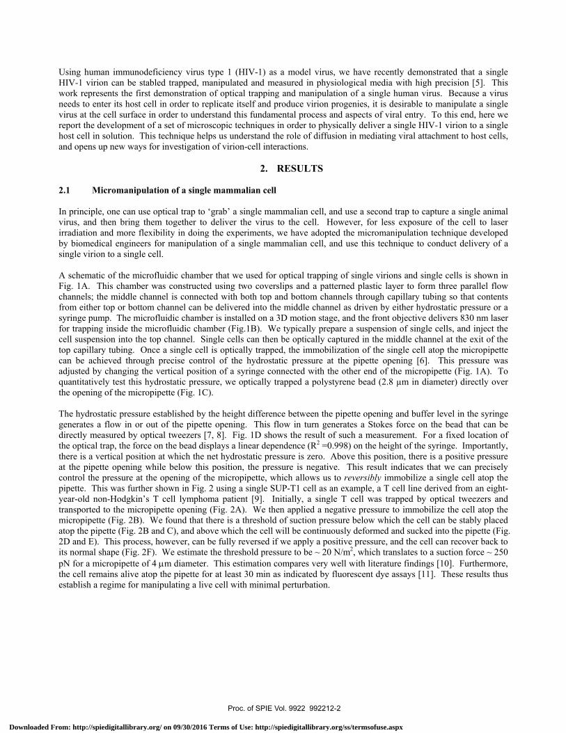
2. RESULTS 2.1 Micromanipulation of a single mammalian cell In principle, one can use optical trap to ‘grab’ a single mammalian cell, and use a second trap to capture a single animal virus, and then bring them together to deliver the virus to the cell. However, for less exposure of the cell to laser irradiation and more flexibility in doing the experiments, we have adopted the micromanipulation technique developed by biomedical engineers for manipulation of a single mammalian cell, and use this technique to conduct delivery of a single virion to a single cell. A schematic of the microfluidic chamber that we used for optical trapping of single virions and single cells is shown in Fig. 1A. This chamber was constructed using two coverslips and a patterned plastic layer to form three parallel flow channels; the middle channel is connected with both top and bottom channels through capillary tubing so that contents from either top or bottom channel can be delivered into the middle channel as driven by either hydrostatic pressure or a syringe pump. The microfluidic chamber is installed on a 3D motion stage, and the front objective delivers 830 nm laser for trapping inside the microfluidic chamber (Fig.1B). We typically prepare a suspension of single cells, and inject the cell suspension into the top channel. Single cells can then be optically captured in the middle channel at the exit of the top capillary tubing. Once a single cell is optically trapped, the immobilization of the single cell atop the micropipette can be achieved through precise control of the hydrostatic pressure at the pipette opening [6]. This pressure was adjusted by changing the vertical position of a syringe connected with the other end of the micropipette (Fig. 1A). To quantitatively test this hydrostatic pressure, we optically trapped a polystyrene bead (2.8 µm in diameter) directly over the opening of the micropipette (Fig. 1C). The hydrostatic pressure established by the height difference between the pipette opening and buffer level in the syringe generates a flow in or out of the pipette opening. This flow in turn generates a Stokes force on the bead that can be directly measured by optical tweezers [7, 8]. Fig. 1D shows the result of such a measurement. For a fixed location of the optical trap, the force on the bead displays a linear dependence (R2 =0.998) on the height of the syringe. Importantly, there is a vertical position at which the net hydrostatic pressure is zero. Above this position, there is a positive pressure at the pipette opening while below this position, the pressure is negative. This result indicates that we can precisely control the pressure at the opening of the micropipette, which allows us to reversibly immobilize a single cell atop the pipette. This was further shown in Fig. 2 using a single SUP-T1 cell as an example, a T cell line derived from an eight-year-old non-Hodgkin’s T cell lymphoma patient [9]. Initially, a single T cell was trapped by optical tweezers and transported to the micropipette opening (Fig. 2A). We then applied a negative pressure to immobilize the cell atop the micropipette (Fig. 2B). We found that there is a threshold of suction pressure below which the cell can be stably placed atop the pipette (Fig. 2B and C), and above which the cell will be continuously deformed and sucked into the pipette (Fig. 2D and E). This process, however, can be fully reversed if we apply a positive pressure, and the cell can recover back to its normal shape (Fig. 2F). We estimate the threshold pressure to be ~ 20 N/m2, which translates to a suction force ~ 250 pN for a micropipette of 4 μm diameter. This estimation compares very well with literature findings [10]. Furthermore, the cell remains alive atop the pipette for at least 30 min as indicated by fluorescent dye assays [11]. These results thus establish a regime for manipulating a live cell with minimal perturbation.
Proc. of SPIE Vol. 9922 992212-2
Downloaded From: http://spiedigitallibrary.org/ on 09/30/2016 Terms of Use: http://spiedigitallibrary.org/ss/termsofuse.aspx
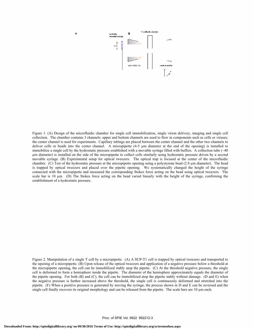
Figure 1. collection.the center deliver ceimmobilizµm diamemovable schamber. is trappedconnected scale bar establishm
Figure 2. Mthe openinthe micropcell is defthe pipettethe negatipipette. (Fsingle cell
(A) Design of th. The chamber cchannel is used
ells or beads intze a single cell byeter) is installed syringe. (B) Exp(C) Test of the
d by optical twed with the micro
is 10 µm. (D)ment of a hydrost
Manipulation ofng of a micropippipette opening, formed to form ae opening. For bve pressure is fF) When a positl finally recovers
he microfluidic contains 3 channfor experiments
to the center chy the hydrostaticon the side of tperimental setuphydrostatic preseezers and plac
opipette and mea The Stokes fortatic pressure.
f a single T cell ette. (B) Upon rthe cell can be
a hemisphere inboth (B) and (Cfurther increasedtive pressure is gs its original mor
chamber for sinnels: upper and bs. Capillary tubihannel. A micrc pressure establthe micropipettep for optical twssure at the micrced over the pipasured the correrce acting on th
by a micropipetelease of the optimmobilized staside the pipette.), the cell can bd above the thregenerated by morphology and can
ngle cell immobbottom channelsings are placed bropipette (4-5 µlished with a mo to collect cells
weezers. The oropipette openingpette opening. esponding Stokehe bead varied
tte. (A) A SUP-tical tweezers anably atop the pip The diameter e immobilized aeshold, the singoving the syringen be released fro
bilization, single s are used to flowbetween the centµm diameter at ovable syringe fi
similarly using ptical trap is fog using a polystyWe systematic
es force acting olinearly with th
-T1 cell is trappend application ofpette. (C) At thof the hemisphe
atop the pipette gle cell is contine, the process shom the pipette. T
virion delivery
w in componentster channel and the end of the
illed with buffershydrostatic pre
ocused at the ceyrene bead (2.8
cally changed thon the bead usinhe height of the
ed by optical twf a negative preshe threshold negere approximatelstably without d
nuously deformehown in D and EThe scale bars ar
, imaging and ss such as cells othe other two chopening) is ins
s. A collection tssure driven by enter of the micµm diameter).
he height of theng optical tweeze syringe, confir
weezers and transssure below a thrgative pressure, tly equals the dia
damage. (D anded and stretchedE can be reversere 10 µm each.
ingle cell or viruses; hannels to stalled to tube (~40 a second
crofluidic The bead e syringe
zers. The rming the
sported to reshold at the single ameter of
d E) when d into the ed and the
Proc. of SPIE Vol. 9922 992212-3
Downloaded From: http://spiedigitallibrary.org/ on 09/30/2016 Terms of Use: http://spiedigitallibrary.org/ss/termsofuse.aspx
2.2 Opt
We have recmanipulate a(Fig. 1A). Tmiddle chann3D motion st
To deliver a a single viriothe virion diavirions weremicropipette distance betwsolid state ladelivered a sleft and thredelivery and remotely throcase, the viriwas shut off all virions dian attachmeDiethylaminoonto host cecollision, repdifference ina single collion virion suinvestigated
Videos 1,2in the prestime. All
tical delivery o
cently developa single cell furTo this end, wenel at the opentage and placed
single virion toon in the middlameter measur
e discarded. Tthrough the m
ween the virionser and the tra
single virion toee deliveries fr
measurementsough a compution was slowlyimmediately, a
issociated froment probabilityoethyl-dextran
ell surface nonpresented by Vn the viral attacision is a ratherurface, or low in the future.
2,3 and 4. Delivsence of 10 µg/mdeliveries were
of a single viru
ped the capabilrther allows us
e injected suspening of the top d the cell on to
o the live T cele channel at thred using the bThe single vir
motorized chamn and the cell pping laser def
o different sporom the top ofs. This deliveter (Fig. 1B). Ty brought into and the virion
m the cell surfay of 0%. Inn (DEAE-dextrnspecifically [1Video 3 and 4, wchment efficienr inefficient prand perhaps
very of a single Hml DEAE-dextradone at 20°C in PBS.
us to a single c
lity to opticalls to deliver a siension of SUPcapillary tubin
op of the microp
ll, we injected he opening of back-focal-planrion was then
mber stage, andwas judged byflection signal ts on the cell sf the cell. Thery strategy waTwo types of dcontact with twas imaged v
ace upon their n the second
ran), which is a12]. Out of a which resultedncy indicates throcess. This loheterogeneous
HIV-1 virion to an (Video 3 and 4
cell
ly trap a singlingle virus to a-T1 cells into ng using opticapipette in the m
diluted HIV-1the bottom cap
ne interferometransferred to
d slowly brougy both the fluoat the objectiv
surface: three he single cell was made possibdelivery experimthe cell surfaceia 488 nm epi initial collision
d scenario, tha polycation ktotal of 77 tri
d in an attachmhat to form a sow efficiency cs receptor dist
a single SUP-T14). Frame rate w
le HIV-1 virioa single cell usthe top channeal tweezers. Wmiddle channel
1 virions into thpillary tubing. try as we desco the vicinity ght into contactorescence imagve back focal pdeliveries fromwas then replable by the 3-axments were cone. Upon virionfluorescence en, represented
he virions weknown to enhanials, 59 virion
ment probabilitypecific contact
can be caused btributions on t
1 cell in the absewas 9.1 Hz for a
on in suspensiosing the microfel, and captureWe then transfel as we illustrat
he bottom chan The single vir
cribed previousof the single
t with the cell ge of the virionplane. Typicalm the right, thraced with a frexis motorized snducted both an and cell encoexcitation. Ouby Video 1 an
ere first incubnce the attachm
ns attached to y of greater that between the vby either low ethe host cell s
ence of DEAE-dall movies and m
on [5]. The afluidic chambed a single T ceerred the cell uted in Fig. 2.
nnel, and opticrion was confisly [5]. Aggre
T cell on topsurface. The
n excited by a lly for a single ree deliveries fesh new cell fstage that is coat 20°C in PBSounter, the opt
ut of a total of 7nd 2, which resbated with 10ment of HIV-1cell surface up
an 77%. This dvirus and the cenvelope glycosurface, which
extran (Video 1 movies were play
ability to er design ell in the using the
cally trap irmed by egates of p of the physical 488-nmcell, we
from the for more ontrolled S. In onetical trap 73 trials, sulted in 0 µg/ml 1 virions pon first dramatic cell upon oproteins h can be
and 2) or yed in real
http://dx.doi.org/10.1117/12.2239051
Proc. of SPIE Vol. 9922 992212-4
Downloaded From: http://spiedigitallibrary.org/ on 09/30/2016 Terms of Use: http://spiedigitallibrary.org/ss/termsofuse.aspx

For those vircell surface tthe fact that that one can pagents, the sivirion internattachment.
2.3 Col
Figure 3. Aand the cea coverslip
The unique athe cell atop we installed after immobioptical tweezpressure throunder a regucollection teadditional cu
Video 5. Araised the positive prgenerated opening oftube at this
rions that were throughout ourat 20°C, neitheprecisely delivingle virion ca
nalization may
llection and re
A single cell wall went into the cp under microsco
advantage of mthe pipette wha collection tu
ilization. We zers, and transough the tube tular microscopechnique is poulturing of the c
A single SUP-T1syringe to generressure and capa torque on the f the collection ts moment, and u
attached onto r experimental er direct fusion
ver a single HIVan be attached
allow observ
ecovery of a si
as trapped by opcollection tube dope (D). The sc
manipulation ushile a positive pube on the sidefirst applied a
sported the celto collect the cpe as shown inotentially usefucell is desired.
1 cell is immobilrate a positive hytured by opticaltrapped cell so
tube using opticupon release of th
the cell surfactime frame. Nn nor endocytoV-1 virion to ato the cell sur
vation of those
ngle cell after
ptical tweezers, adue to the applieale bars are 10 µ
sing micropipepressure can bee of the micro
a positive pressl to the openin
cell (Fig. 3B ann Fig. 3D. A ul for further
lized stably atopydrostatic pressul tweezers indicthat the cell undal tweezers. A nhe optical tweez
e in the presenNo internalizatiosis will occur a single host cerface. Future ee events and m
r an experimen
and delivered toed negative pressµm each.
ette is its reverse used to dislodopipette (Fig. 1sure inside theng of the collend C). As expsimilar procesassay of the
p a micropipette ure inside the miated by the red derwent several negative hydrostzers, the single c
nce of DEAE-don of virions wunder these coll at a desired l
experiments unmeasurement o
nt
o a collection tubsure (B-C). Fina
sibility: negativdge the cell. T1A) and testede micropipette ection tube (Fipected, the singss was shown single cell af
by negative hydicropipette. Thecross. Simultarounds of rotatitatic pressure waell was sucked in
dextran. They rwas observed. onditions. Thelocation. In thnder conditionsof their efficie
be (A). We switally, the cell was
ve pressure allo take advanta
d our ability toto release the g. 3A). We thgle cell was rein Video 5 be
fter an experim
drostatic pressuree cell was immedaneously, the floions. We then tas already genernto the collectio
remained statioThis is consist
ese results demhe presence of as that are condency relative t
tched off the opts recovered and p
ows immobilizage of this reve collect the sincell, captured
hen applied a covered and exelow. This sinment, especial
e. To release thediately released
ow from the mictransported the crated inside the con tube instantan
onary on tent with
monstrate auxiliary ducive to to virion
tical trap, placed on
zation of ersibility, ngle cell it using negative xamined ngle-cell ly when
e cell, we upon this
cropipette cell to the collection
neously. http://dx.doi.org/10.1117/12.2239051
Proc. of SPIE Vol. 9922 992212-5
Downloaded From: http://spiedigitallibrary.org/ on 09/30/2016 Terms of Use: http://spiedigitallibrary.org/ss/termsofuse.aspx
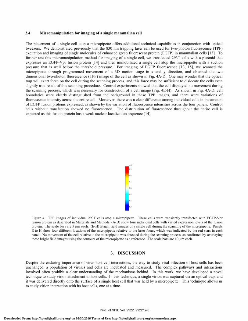
The placemetweezers. Wexcitation anfurther test thexpresses anpressure thatmicropipette dimensional trap will exeslightly as a the scanningboundaries wfluorescence of EGFP fusicells withouexpected as t
Figure 4. fusion proprotein. TE to H shopanel. Nothese brigh
Despite the eunchanged: ainvolved oftetechnique to it was deliveto study virio
ent of a singleWe demonstratend imaging of shis micromani
n EGFP-Vpr fut is well belo
through progtwo-photon flurt force on theresult of this s process, whicwere clearly dintensity acros
ion proteins exut transfection this fusion prot
TPF images ofotein as describedThe scale bars arow four differen
o movement of thht field images u
enduring impoa population oen prohibit a study virion at
ered directly onon interaction w
e cell atop a med previously tsingle moleculipulation methusion protein [w the thresho
grammed movuorescence (TPe cell during thscanning procech was necessadistinguished ss the entire cexpressed, as sh
showed no fltein has a weak
f individual 293d in Materials anre 5 µm each. (Ent locations of thhe cell relative tousing the contour
ortance of viruof viruses andclear understattachment to honto the surface with its host ce
micropipette ofthat the 830 nmles of enhancedod for imaging[14] and then
old pressure. vement of a 3PF) image of thhe scanning produre. Control
ary for construfrom the bac
ell. Moreover, hown by the vafluorescence. k nuclear locali
3T cells atop a mnd Methods. (A-E-H) Bright fieldhe micropipette o the micropipetrs of the micropi
3.
s-host cell inted cells are incuanding of the most cells. In thof a single ho
ells, one at a tim
ffers additionalm trapping lasd green fluoresg of a single cimmobilized For imaging
3D motion stahe cell as showocess, and thisexperiments s
uction of a cellckground in th
there was a cleariation of fluor
The distributiization sequenc
micropipette. T-D) show four ind images of a sinrelative to the l
tte was detected ipette as a refere
DISCUSSIO
eractions, the wubated and memechanisms bhis technique, aost cell that wame.
l technical capser can be usedscent protein (Ecell, we transfea single cell aof EGFP fluo
age in x and wn in Fig. 4A-s force may be showed that thel image (Fig. 4hese TPF imaear difference rescence intension of fluoresce [14].
These cells werendividual cells wngle cell during aser focus, whicduring the scann
ence. The scale
ON
way to study veasured. The ehind. In thisa single virion s held by a mi
pabilities in cod for two-photEGFP) in mamected 293T celatop the microorescence [13,
y direction, aD. One may wsufficient to d
e cell displayed4E-H). As shoages, and theramong individsities across thscence through
e transiently tranwith varied expre
the scanning of ch was indicatedning process, as bars are 10 µm e
viral infection complex path
s work, we hawas captured vcropipette. Th
onjunction withton fluorescencmmalian cells [lls with a plas
opipette with a 15], we scanand obtained wonder that thedislocate the ced no movemenown in Fig. 4Are were varia
dual cells in thehe four panels. hout the entire
nsfected with Eession levels of tf the micropipetted by the red starconfirmed by o
each.
of host cells hhways and inteave developed via an optical this technique a
h optical ce (TPF) [13]. To mid that
a suction nned the the two e optical ells even nt during A-D, cellations of e amount Control e cell is
EGFP-Vpr the fusion e. Panelsrs in each verlaying
has been eractions
a novel trap, and
allows us
2.4 Miccromanipulation for imaging of a single mammalian cell
Proc. of SPIE Vol. 9922 992212-6
Downloaded From: http://spiedigitallibrary.org/ on 09/30/2016 Terms of Use: http://spiedigitallibrary.org/ss/termsofuse.aspx

Brownian diffusion of a virion has been proposed to be one bottleneck in infection of host cells [12, 16-18]. Direct delivery of a single virion to host cell surface bypasses diffusion. However, even when diffusion is bypassed, the efficiency to form specific virion-cell contact is low, suggesting that other barriers instead of diffusion limit virion infectivity. These potential barriers include the envelope glycoprotein density on individual virions [19], and also the density and distribution of receptors on individual host cell surface, the impact of which on viral attachment can be investigated systematically using current experimental design.
The integration of optical tweezers [20, 21] with TPF imaging in a single instrument makes the system versatile. On the one hand, we can use optical tweezers for manipulation of single molecules [22], single viruses and single cells; on the other hand, we can use continuous-wave laser-excited TPF to image live cells without the need of additional laser source. To take advantage of both modalities, we developed a method for imaging of single live cells that grow in suspension (Fig. 4). Conventional fluorescence imaging methods developed for cultured cells are largely limited to cells that can grow on a coverslip surface. There are few reports on imaging of live cells in suspension due to the requirement for noninvasive immobilization. We use optical tweezers to capture a single cell from culture suspension, and developed a micropipette system controlled by hydrostatic pressure to immobilize a single cell atop the micropipette for TPF imaging. The unique advantage of using micropipette for manipulation is that the entire procedure is reversible. On the one hand, a single cell can be immobilized by a negative pressure; on the other hand, the cell can be easily released from the pipette by applying a positive pressure, and transported by optical tweezers for collection and recovery. No damage was incurred to the cell throughout the process. This eliminates the requirement to grow or fix cells on coverslip surface, and is well suited for imaging live cells that are cultured in suspension, such as T cells and B cells. Direct imaging of these cells close to physiological conditions is important. A specific example is provided by HIV-1. Current live cell imaging methods almost exclusively use a HeLa cell derivative that can grow on coverslips for imaging of virion-cell interactions [23], yet the major physiological targets of HIV are T cells. Because different cell types may directly influence virion-cell interactions [24], it is necessary to develop tools for direct imaging of these T cells that are cultured in suspension. As we have shown above, we can work with CD4+ T cells directly instead of engineered HeLa cells.
Lastly, our developments as reported here open up a range of possibilities for single cell-based experiments [25, 26]. For example, we can deliver a virion or a nanoparticle to a single cell using optical tweezers, and use TPF or epifluorescence imaging to follow the recognition or uptake of the particle by the single cell. Subsequent single cell collection and recovery allows further analysis using other techniques to diagnose the consequence of virion internalization. Similarly, we can use optical tweezers to manipulate a single receptor on the cell surface, use TPF imaging to measure reporter gene expression, and further collect cells and use PCR for gene expression analysis to probe the mechanisms of mechanotransduction. The unique feature of this experiment may allow dissection of fundamental events without complication of multiple virions or multiple cells. Literally, this integrated system allows one to use single cells as a test tube, and optical tweezers as a ‘pipetman’, leading to a new approach for the study of biology at single-particle and single cell level.
4. MATERIALS AND METHODS
4.1 Cell culture and production of HIV-1 virions
SUP-T1 cells were cultured at 37°C with 5% CO2 in RPMI supplemented with 10% FBS. HEK 293T/17 cells (ATCC, Manassas, VA) were cultured at 37°C with 5% CO2 in DMEM supplemented with 10% FBS. The procedures to produce HIV-1 virions have been described in detail elsewhere [27]. Briefly, the EGFP-labeled HIV-1 virions were produced by transient transfection of 293T cells with two plasmids using Mirus TransIT LT-1 transfection regent. One of the plasmids encodes HIV-1 proviral DNA pNL4-3 that contains an EGFP flanked in between the matrix and capsid domains of the Gag protein (iGFP) [28] and carries a premature stop codon in gp120/gp41 coding sequence. The other plasmid encodes a fully functional NL4-3 envelope glycoprotein gp120/gp41. At 24 hours post transfection, the supernatant from the cell culture was harvested. Viral particles were filtered through 0.45 µm low-protein binding filter, aliquoted, flash frozen in liquid nitrogen and stored in -80°C freezer.
Proc. of SPIE Vol. 9922 992212-7
Downloaded From: http://spiedigitallibrary.org/ on 09/30/2016 Terms of Use: http://spiedigitallibrary.org/ss/termsofuse.aspx

The trapping of individual virions in solution was done as described previously [5] with a few modifications. Throughout, a home-made OTs instrument using a tapered amplifier diode laser at 830 nm (SYS-420-830-1000, Sacher LaserTechnik LLC, Germany) was used for optical trapping of individual HIV-1 virions [11]. Briefly, a laser power of 130.8 mW at the focus was used throughout for optical trapping and simultaneous TPF excitation. The live virus stock thawed from -80°C was diluted in PBS to a concentration of 0.6 – 1.6 × 10⁸ virions/ml and injected into a microfluidic chamber for optical trapping. The diameter of individual virions was measured as described previously [5]. A Coherent OBIS solid state laser at 488 nm was used for epifluorescence imaging of EGFP-labeled virions. An electron-multiplying charge-coupled device (EMCCD) camera (Evolve, Photometrics) was used for all fluorescence detection including epifluorescence imaging. Individual HIV-1 virions were identified based on an EGFP positive signal and a measured diameter that lies between 96 and 216 nm as described previously [5]. All the trapping and imaging experiments were conducted at a constant temperature of 20.0 ± 0.2 °C.
4.3 Calibration of the micropipette system controlled by hydrostatic pressure
The height difference between the micropipette opening and buffer level in the movable syringe created a hydrostatic pressure at the pipette opening. We placed the optical trap at a distance away from the pipette opening. We tested the hydrostatic pressure quantitatively using an optically-trapped polystyrene bead (2.8 µm diameter). Due to the presence of the hydrostatic pressure, the trapped bead was displaced from its equilibrium position and we measured the force on the bead directly using optical tweezers [11]. For a fixed location of the optical trap, we changed the hydrostatic pressure gradually by adjusting the height of the movable syringe. As shown in Fig. 1D, the measured Stokes force varied linearly with the syringe height, suggesting that the pressure generated at the pipette opening was proportional to height difference between syringe and the pipette opening. The threshold pressure for suction of a single T cell was estimated based on the hydrostatic pressure p=ρg∆h that we established in the system, where ρ is the density of the buffer, g is the acceleration of gravity, and ∆h is the relative height of the syringe away from the balance point at which p=0.
4.4 Single cell collection and recovery
To collect the single cell atop the micropipette, we installed a collection tube (~40 µm inner diameter) on the side of the micropipette (Fig. 1A). After immobilization of a single cell atop the micropipette, we apply a positive pressure to ‘push’ the cell back to the media, capture it using optical tweezers, and transport it to the opening of the collection tube (Fig. 3A). This collection tube was also connected with a movable syringe so that we can apply a negative pressure to collect the cell into the tube (Fig. 3B and C). To recover the cell for further analysis, we found it necessary to approximately estimate the location of the cell inside the collection tube to facilitate its recovery. We estimated that the cell flew into the collection tube at a rate around 100 µm/s based on video microscopy. At this flow rate, it will take 300 – 400 s for the cell to travel through the collection tubing (3-4 cm long), and ~ 83 min to travel through 1 cm of the polyethylene tubing (0.28mm diameter) immediately at the end of the collection tube. Based on these estimations, we waited for ~10 min after cell suction, and directly recovered the cell from the portion of polyethylene tubing at the end of the collection tube. The recovered single cell was then deposited onto a coverslip for examination under a microscope (Fig. 4D). Control experiments show that the cell we collected was indeed the one we immobilized atop the pipette, as repeated mock collection without any cells recovered nothing under the microscope.
4.5 Production of fluorescent cells and TPF imaging
To produce fluorescent cells for TPF imaging using optical tweezers, 293T cells were transiently transfected with a plasmid encoding EGFP-Vpr fusion protein using calcium phosphate method. At 40 hours post transfection, medium was removed. Cells were washed gently with Dulbecco's Phosphate-Buffered Saline (DPBS), and detached from surface using Trypsin-EDTA. At 2 min after trypsin digestion, complete medium (90% DMEM + 10% FBS) was added to stop the reaction and the cells were resuspended in DPBS and ready for experiments. For optical trapping and fluorescence imaging, cells were exchanged into phosphate buffer (100 mM, pH 7.2) by centrifuge, and flown into the upper channel of the microfluidic chamber. We used optical tweezers to capture individual cells near the opening of the upper capillary tubing (Fig. 1A), and placed the cell atop a micropipette (opening around 4-5 µm) controlled by hydrostatic pressure. The hydrostatic pressure was generated by connecting the end of the micropipette with a syringe that can be adjusted vertically (Fig. 1A). This syringe and micropipette system was filled with buffer and we adjusted the pressure at the
4.2 Optical tweezers and epifluorescence imaging
Proc. of SPIE Vol. 9922 992212-8
Downloaded From: http://spiedigitallibrary.org/ on 09/30/2016 Terms of Use: http://spiedigitallibrary.org/ss/termsofuse.aspx
pipette opening through adjustment of the syringe height. When negative pressure was generated, a single cell could be stably immobilized atop the micropipette for fluorescence imaging. When positive pressure was generated, the single cell could be released from the pipette for capture by optical tweezers. To image the single cell atop the micropipette, we placed the laser focus close to the cell, and used a custom-written LabView program to conduct scanning of the micropipette in two dimensions. TPF intensity emitted from the laser focus was recorded by the EMCCD with an exposure time of 1 s and cell images were constructed using Matlab image processing toolbox.
5. ACKNOWLEDGMENTS
This work was supported by NIH Director’s New Innovator Award 1DP2OD008693-01 to WC, NSF CAREER Award CHE1149670 to WC and also in part by Research Grant No. 5-FY10-490 to WC from the March of Dimes Foundation. MCD was supported by a NIH postdoctoral fellowship awarded under F32-GM109771. We thank Professor Benjamin Chen for kindly providing the provirus plasmids encoding iGFP virions. The following reagents were obtained through the AIDS Research and Reference Reagent Program, Division of AIDS, National Institute of Allergy and Infectious Diseases (NIAID), National Institutes of Health (NIH): pEGFP-Vpr from Warner C. Greene; SUP-T1 cells from Dr. James Hoxie.
6. AUTHOR CONTRIBUTIONS
W. C. directed the project; X. H. and M. C. D. conducted manipulation experiments; C. T. prepared and assayed HIV-1virions; W.C. and X. H. wrote the paper.
REFERENCES
[1] A. Ashkin, J. Dziedzic, J. Bjorkholm et al., “First demonstration of stable optical trapping of micron-sizeddielectric objects in three dimensions using a single-beam gradient optical trap.,” Opt. Lett., 11, 288-90 (1986).
[2] X. Hou, and W. Cheng, “Optical Tweezers,” Encyclopedia of Biophysics: SpringerReference, 1800-1807(2013).
[3] M. P. MacDonald, G. C. Spalding, and K. Dholakia, “Microfluidic sorting in an optical lattice,” Nature,426(6965), 421-424 (2003).
[4] A. Ashkin, and J. M. Dziedzic, “Optical trapping and manipulation of viruses and bacteria,” Science, 235(4795),1517-20 (1987).
[5] Y. Pang, H. Song, J. H. Kim et al., “Optical trapping of individual human immunodeficiency viruses in culturefluid reveals heterogeneity with single-molecule resolution,” Nat Nanotechnol, 9(8), 624-630 (2014).
[6] R. M. Hochmuth, “Micropipette aspiration of living cells,” J Biomech, 33(1), 15-22 (2000).[7] R. M. Simmons, J. T. Finer, S. Chu et al., “Quantitative measurements of force and displacement using an
optical trap,” Biophys J, 70(4), 1813-22 (1996).[8] A. van der Horst, and N. R. Forde, “Power spectral analysis for optical trap stiffness calibration from high-
speed camera position detection with limited bandwidth,” Opt Express, 18(8), 7670-7 (2010).[9] S. D. Smith, M. Shatsky, P. S. Cohen et al., “Monoclonal antibody and enzymatic profiles of human malignant
T-lymphoid cells and derived cell lines,” Cancer Res, 44(12 Pt 1), 5657-60 (1984).[10] E. Evans, and A. Yeung, “Apparent viscosity and cortical tension of blood granulocytes determined by
micropipet aspiration,” Biophys J, 56(1), 151-60 (1989).[11] W. Cheng, X. Hou, and F. Ye, “Use of tapered amplifier diode laser for biological-friendly high-resolution
optical trapping,” Opt Lett, 35(17), 2988-90 (2010).[12] E. J. Platt, S. L. Kozak, J. P. Durnin et al., “Rapid dissociation of HIV-1 from cultured cells severely limits
infectivity assays, causes the inactivation ascribed to entry inhibitors, and masks the inherently high level ofinfectivity of virions,” J Virol, 84(6), 3106-10 (2010).
[13] X. Hou, and W. Cheng, “Detection of single fluorescent proteins inside eukaryotic cells using two-photonfluorescence,” Biomed Opt Express, 3(2), 340-53 (2012).
[14] E. Schaeffer, R. Geleziunas, and W. C. Greene, “Human immunodeficiency virus type 1 Nef functions at thelevel of virus entry by enhancing cytoplasmic delivery of virions,” J Virol, 75(6), 2993-3000 (2001).
[15] X. Hou, and W. Cheng, “Single-molecule detection using continuous wave excitation of two-photonfluorescence,” Opt Lett, 36(16), 3185-7 (2011).
Proc. of SPIE Vol. 9922 992212-9
Downloaded From: http://spiedigitallibrary.org/ on 09/30/2016 Terms of Use: http://spiedigitallibrary.org/ss/termsofuse.aspx
[16] Y. J. Kwon, G. Hung, W. F. Anderson et al., “Determination of infectious retrovirus concentration fromcolony-forming assay with quantitative analysis,” J Virol, 77(10), 5712-20 (2003).
[17] U. O'Doherty, W. J. Swiggard, and M. H. Malim, “Human immunodeficiency virus type 1 spinoculationenhances infection through virus binding,” Journal of Virology, 74(21), 10074-10080 (2000).
[18] H. Haim, I. Steiner, and A. Panet, “Synchronized infection of cell cultures by magnetically controlled virus,” JVirol, 79(1), 622-5 (2005).
[19] M. C. DeSantis, J. H. Kim, H. Song et al., “Quantitative Correlation between Infectivity and Gp120 Density onHIV-1 Virions Revealed by Optical Trapping Virometry,” J Biol Chem, 291(25), 13088-13097 (2016).
[20] J. R. Moffitt, Y. R. Chemla, S. B. Smith et al., “Recent advances in optical tweezers,” Annu Rev Biochem, 77,205-28 (2008).
[21] K. C. Neuman, and S. M. Block, “Optical trapping,” Review of Scientific Instruments, 75(9), 2787-2809 (2004).[22] C. Bustamante, W. Cheng, and Y. X. Mejia, “Revisiting the central dogma one molecule at a time,” Cell,
144(4), 480-97 (2011).[23] K. Miyauchi, Y. Kim, O. Latinovic et al., “HIV enters cells via endocytosis and dynamin-dependent fusion with
endosomes,” Cell, 137(3), 433-44 (2009).[24] F. Hladik, P. Sakchalathorn, L. Ballweber et al., “Initial events in establishing vaginal entry and infection by
human immunodeficiency virus type-1,” Immunity, 26(2), 257-70 (2007).[25] X. S. Xie, J. Yu, and W. Y. Yang, “Living cells as test tubes,” Science, 312(5771), 228-30 (2006).[26] T. Kalisky, and S. R. Quake, “Single-cell genomics,” Nat Methods, 8(4), 311-4 (2011).[27] J. H. Kim, H. Song, J. L. Austin et al., “Optimized Infectivity of the Cell-Free Single-Cycle Human
Immunodeficiency Viruses Type 1 (HIV-1) and its Restriction by Host Cells,” PLOS One, 8(6), e67170 (2013).[28] W. Hubner, P. Chen, A. Del Portillo et al., “Sequence of human immunodeficiency virus type 1 (HIV-1) Gag
localization and oligomerization monitored with live confocal imaging of a replication-competent, fluorescentlytagged HIV-1,” J Virol, 81(22), 12596-607 (2007).
Proc. of SPIE Vol. 9922 992212-10
Downloaded From: http://spiedigitallibrary.org/ on 09/30/2016 Terms of Use: http://spiedigitallibrary.org/ss/termsofuse.aspx