optical coherence tomography used for monitoring of pdt treatment
TRANSCRIPT
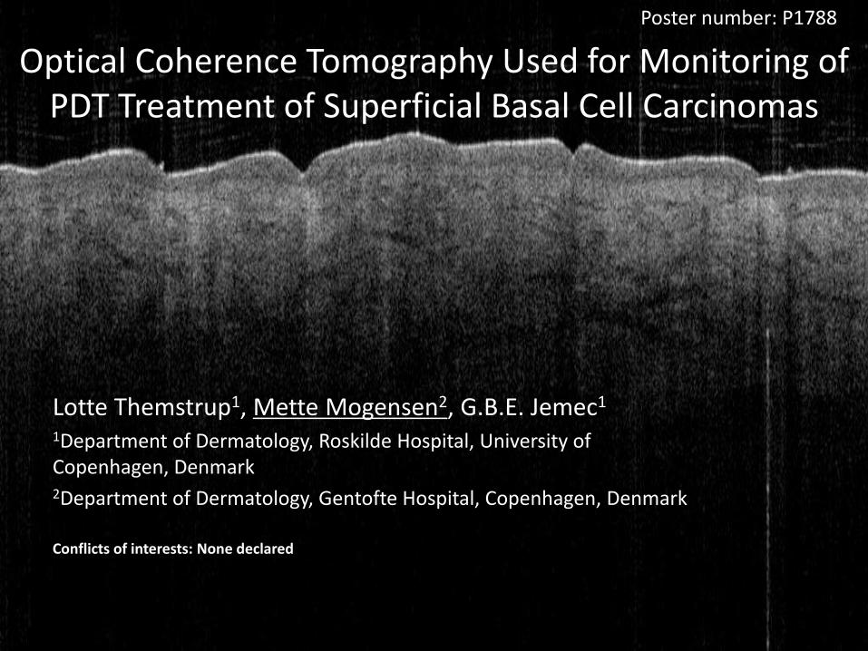
Optical Coherence Tomography Used for Monitoring of PDT Treatment of Superficial Basal Cell Carcinomas
Lotte Themstrup1, Mette Mogensen2, G.B.E. Jemec1
1Department of Dermatology, Roskilde Hospital, University of Copenhagen, Denmark2Department of Dermatology, Gentofte Hospital, Copenhagen, Denmark
Conflicts of interests: None declared
Poster number: P1788

Introduction
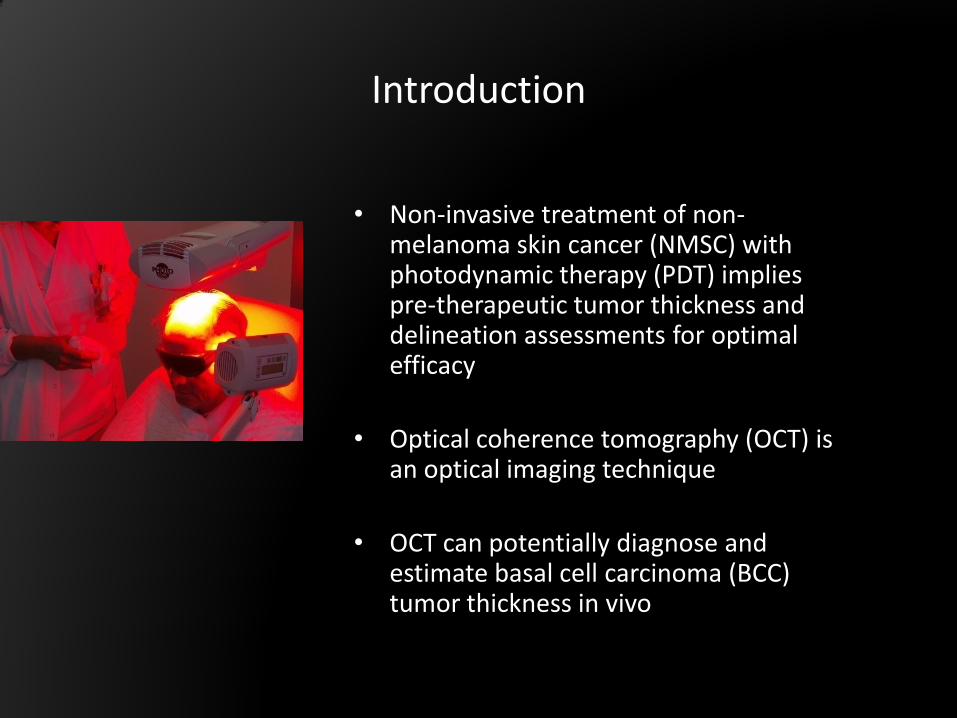
• Non-invasive treatment of non-melanoma skin cancer (NMSC) with photodynamic therapy (PDT) implies pre-therapeutic tumor thickness and delineation assessments for optimal efficacy
• Optical coherence tomography (OCT) is an optical imaging technique
• OCT can potentially diagnose and estimate basal cell carcinoma (BCC) tumor thickness in vivo
Introduction
• Non-invasive treatment of non-melanoma skin cancer (NMSC) with photodynamic therapy (PDT) implies pre-therapeutic tumor thickness and delineation assessments for optimal efficacy
• Optical coherence tomography (OCT) is an optical imaging technique
• OCT can potentially diagnose and estimate basal cell carcinoma (BCC) tumor thickness in vivo

• Sensitivity in clinical diagnosis of non-melanoma skin cancer ranges from 60%-91% and specificity 71-90%Mogensen M, Jemec GB. Diagnosis of nonmelanoma skin cancer/keratinocyte carcinoma: a review of diagnostic accuracy of nonmelanoma skin cancer diagnostic tests and technologies. Dermatol Surg 2007; 33: 1158-74.
• Non-invasive diagnostic methods are warranted as non-invasive treatments are increasingly used for BCC
BCC
PDT
Why study BCC with OCT??

Aim of study
• To describe the OCT morphology in NMSC lesions during PDT treatment
• To assess how OCT morphology before, during and after treatment reflects the treatment out-come at 3 months follow-up

Methods
Patients:
• A total of 20 patients diagnosed with NMSC (BCC and AK) are being monitored by OCT during PDT treatment
• Images will be acquired at 4 time points during PDT treatment and once at 3-months follow-up
• BCC diagnosis is based on histopathology
OCT system:
• Low intensity, 1310nm laser light
• Non-contact, non-invasive
• Real-time imaging
• 2D and 3D visualization

• OCT provides cross-sectional, tomographic, real time imaging on a micrometer scale by infrared scanning of the skin
• OCT resolution 10 µm. Penetration depth 2000 µm
epidermisdermis
OCT image normal skin
Optical Coherence Tomography (OCT)
Courtesy to Peter E. Andersen,
Risø, Denmark
OCT penetration and resolution

• OCT is analogue to ultrasound using infrared light instead of sound
• Variation of reflected infrared light is mapped as a function of depth
dermis
epidermis
Optical Coherence Tomography (OCT)

• Speed of light is too rapid for direct
measurement of pulse transit time
• Low coherence interferomtric
tecnique: only reflected light that
interfere within the coherence length
of light is detected
Outline of OCT system
A BCC lesion BCC
Normal skin

Results
• Normal skin
OCT image of normal skin on the anterior lower arm. Epidermis indicated by ★ ; dermo-epidermal junction (DEJ) indicated by blue arrows. Vessels indicated by fat arrows.
★★
★★

• Non melanoma skin cancer
Characteristic OCT images of:a) Actinic keratosis (AK) b) BCC
a) AK lesion on scalp. Shows disruption of normal skin layering. Thickening of epidermis indicated by white bar. Crusting and ulceration are indicated by ★
a)
b)
a)
b)
b) BCC lesion on scalp. Black necrotic centre indicated by o. Stroma indicated by thick white arrow. BCC islands indicated by triangles. Hairs casting shadows indicated by thin white arrow
o

a) BCC lesion located on back: Central ulceration indicated by Thickening of epidermis marked by white bar , BCC islands marked by thin arrows.
b) Image taken shortly after curettage and PDT treatment: Signal is enhanced, image becomes more homogeneous. Necrosis marked by *.
c) 3 months follow-up: Normal layering is restored. BCC lesion gone. DEJ marked by thin arrows. Vessel marked by blue arrow
a) BCC before PDT
b) BCC after PDT
c) BCC at 3 months follow-up
*
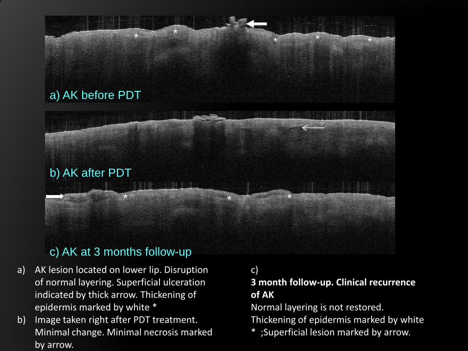
a) AK lesion located on lower lip. Disruption of normal layering. Superficial ulceration indicated by thick arrow. Thickening of epidermis marked by white *
b) Image taken right after PDT treatment. Minimal change. Minimal necrosis marked by arrow.
a) AK before PDT
b) AK after PDT
c) AK at 3 months follow-up
c) 3 month follow-up. Clinical recurrence of AKNormal layering is not restored. Thickening of epidermis marked by white * ;Superficial lesion marked by arrow.
*** **
** *

Conclusion
Lotte Themstrup1, Mette Mogensen2, G.B.E. Jemec1
1Department of Dermatology, Roskilde Hospital, University of Copenhagen, Denmark2Department of Dermatology, Gentofte Hospital, Copenhagen, Denmark
Poster number: P1788
• OCT can visualize skin structures in BCC
lesions in vivo to a depth of 2 mm
• OCT imaging of non-melanoma skin cancer
may provide diagnostic and prognostic
information
• Further evaluation of the potential of OCT as
a monitoring device during NMSC treatment,
as PDT , is warranted
References:Hamdoon Z et al. Photodiagnosis Photodyn
Ther. 2011 Mar;8(1):49-52. 2010 Sep 9.
Mogensen M et al. Br J Dermatol 2009;
160: 1026-33.
Mogensen M, Jemec GB. Dermatol Surg
2007; 33: 1158-74.
Gambichler T, et al. J.Dermatol Sci. 2007;
45: 167-73.
Korde VR et al. Lasers Surg Med. 2007; 39:
687-95.
Olmedo JM et al. J.Am.Acad.Dermatol.
2006; 55: 408-12.
Welzel J et al J.Am.Acad.Dermatol. 1997;
37: 958-63.