optic neuropathies: those that swell may turn pale no ... · ÷ optic nerve edema ÷ ... cerebri...
TRANSCRIPT
4/21/19
1
J O S H U A P A S O L M D N E U R O - O P H T H A L M O L O G I S T
B A S C O M P A L M E R E Y E I N S T I T U T E U N I V E R S I T Y O F M I A M I
M I L L E R S C H O O L O F M E D I C I N E
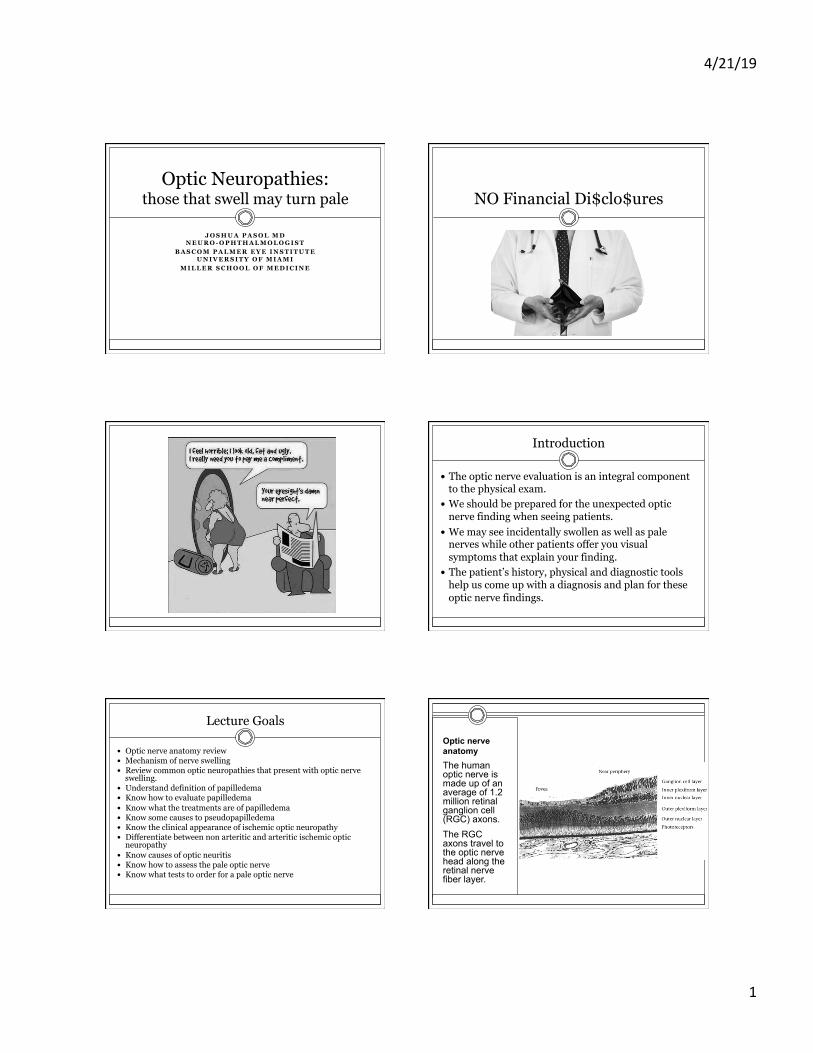
Optic Neuropathies: those that swell may turn pale NO Financial Di$clo$ures
Introduction
� The optic nerve evaluation is an integral component to the physical exam.
� We should be prepared for the unexpected optic nerve finding when seeing patients.
� We may see incidentally swollen as well as pale nerves while other patients offer you visual symptoms that explain your finding.
� The patient’s history, physical and diagnostic tools help us come up with a diagnosis and plan for these optic nerve findings.
Lecture Goals
� Optic nerve anatomy review � Mechanism of nerve swelling � Review common optic neuropathies that present with optic nerve
swelling. � Understand definition of papilledema � Know how to evaluate papilledema � Know what the treatments are of papilledema � Know some causes to pseudopapilledema � Know the clinical appearance of ischemic optic neuropathy � Differentiate between non arteritic and arteritic ischemic optic
neuropathy � Know causes of optic neuritis � Know how to assess the pale optic nerve � Know what tests to order for a pale optic nerve
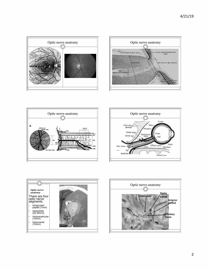
Optic nerve anatomy The human optic nerve is made up of an average of 1.2 million retinal ganglion cell (RGC) axons. The RGC axons travel to the optic nerve head along the retinal nerve fiber layer.
4/21/19
2
Optic nerve anatomy Optic nerve anatomy
Optic nerve anatomy Optic nerve anatomy
Optic nerve anatomy
There are four optic nerve segments: • Intraocular/
papilla (1mm) • Intraorbital
(25-30mm) • Intracanalicular
(10mm) • Intracranial
(10mm)
Optic nerve anatomy
Anterior clinoid
Optic canal Tuberculum
Pituitary fossa

4/21/19
3
Optic nerve anatomy MRI Optic nerve anatomy CT
Mechanism of nerve swelling
� Papilledema due to high cranial pressure ¡ Axoplasmic stasis
÷ Optic nerve edema ÷ Hemorrhages ÷ Retinal edema/exudates ÷ Rare vascular occlusions
� Ischemia ¡ Lack of blood flow leads to:
÷ Optic nerve edema ÷ Hemorrhages ÷ Retinal exudates and retinal
edema
Mechanism of nerve swelling
� Optic nerve swelling due to inflammation or infection: ¡ Inflammatory cells
(lymphocytes, macrophages, plasma cells) penetrate optic nerve tissue via blood vessels leading to nerve swelling, some hemorrhages and possible retinal exudates
¡ Triggered by inflammatory or infectious disease
Optic nerve diseases that swell
Papilledema
� Papilledema is defined as optic nerve swelling due to high cranial pressure.
� High intracranial pressure is due to: ¢ Idiopathic Intracranial Hypertension (IIH)/pseudotumor
cerebri (PTC) ¢ Intracranial mass ¢ Venous sinus thrombosis ¢ Meningitis

4/21/19
4
Papilledema
� Presenting symptoms of papilledema: ¡ None, you just found it looking in the eye ¡ Transient blurred vision with changes in head position
÷ Transient visual obscurations (TVO’s) ¢ Typically last few seconds in one or both eyes (Sadun AA, Currie
JN, Lessell S. Transient visual obscurations with elevated optic discs. Ann Neurol. 1984).
¡ Loss of vision/visual field ¡ Double vision ¡ Headaches ¡ Whooshing sounds in head ¡ Neck or back pain
Case presentation
� 27 year old, 450 pound woman presented with headaches, transient visual obscurations and double vision.
� Exam: ¡ 20/20 OU ¡ Color vision intact ¡ She could not count fingers
inferiorly both eyes and nasally OS
¡ She had a left 6th NP ¡ She had grade 5 papilledema
both eyes
OS OD
OS OD
Evaluation of papilledema
� What are you going to do when you see this on exam
� First thing à do not freak out or scare the patient
� Stay calm � Do as you would with any patient
¡ Take medical history ÷ Anemia, recent weight gain
¡ Ask about medications (new hormones, minocycline, tetracycline, Vitamin A supplements)
¡ Social history such weight gain ¡ Perform your ophthalmic exam ¡ Check blood pressure if available
Evaluation of papilledema
� Visual acuity: ¡ Vision can range from 20/20 to NLP though typically central vision is good
� Visual Fields: ¡ Most important along with visual acuity ¡ Enlargement of the blind spot àinferior arcuate à superior and inferior arcuate
à severe constriction � RNFL OCT:
¡ Complements the eye exam but should not be used alone as if nerves turn pale, thinning of the RNFL isn’t always a good sign.
� B-scan: ¡ Look for buried optic nerve drusen ¡ Needed in nerves with less severe grades of edema where clinical exam can’t
differentiate true vs psedopapilledema � Fluorescein angiogram:
¡ Looks for optic nerve head leakage (one study showed 100% sensitivity in disc edema in children (Chang et al. JAAPOS 2017).
How swollen is the nerve? Frisen grading scale
Grade 1 Grade 2 Grade 3
Grade 4 Grade 5
What to do next?
� There are many factors that determine how to triage a patient with papilledema. ¡ Most important is likely how is the vision?
÷ If vision and visual fields doing well, then not emergent but urgent evaluation needed
¡ Onset of symptoms ÷ Long standing vs acute, as acute more urgent
¡ Social situation ¡ Access to subspecialist

4/21/19
5
What MRI do I need?
� Since papilledema needs imaging to rule out mass and venous sinus thrombosis, we need two types of imaging: ¡ #1 MRI Brain with contrast if possible (current pregnancy,
prior contrast reaction would not likely use contrast) ÷ This rules out a mass
¡ #2 MRV (venogram, no contrast needed for this study though can be done with contrast) ÷ Rules out a venous sinus occlusion
� If for some reason one cannot get an MRI, then CT scan with contrast and CT venogram will suffice.
Frontal lobe mass Venous sinus thrombosis
Abnormal MRI
Scan is normal, now what?
� If the MRI brain and MRV are normal, then you have ruled out a brain mass and venous sinus thrombosis.
� Now what? � Well, the patient then likely has IIH. � What about the LP?
¡ LP can be useful in subtle cases of IIH ¡ LP is needed if there is a suspicion of meningitis
÷ Stiff neck, fever ¡ LP is needed in atypical cases of IIH
÷ Thin ¡ I do not force LP onto people
� LP Opening pressure normal range: ¡ Adults <25cm H2O (clinically correlate) ¡ Children below age 12 <28cm H2O (clinically correlate)
IIH
� IIH a disease of high intracranial pressure typically leading to papilledema (In a cohort of 353 patients with IIH, papilledema was found in 94.3% (Digre et al. Headache 2009).
� Seen in women more often than men � Associated with weight gain/obesity � MRI/MRV negative � LP OP elevated and clear CSF
IIH IIH signs
� Papilledema � Cranial nerve palsy, 6th
nerve palsy most common but other nerve palsies including internuclear ophthalmoplegia (INO) have been reported.

4/21/19
6
IIH treatment weight loss
� Weight loss is the most important treatment in IIH as >90% of IIH patients are obese or overweight (Subramaniam S, Fletcher WA Obesity and Weight Loss in Idiopathic Intracranial Hypertension: A Narrative Review. J Neuroophthalmol. 2017).
� IIH risk increases as a function of body mass index (BMI) and weight gain over the prior year.
� Weight loss typically in the 6%-10% range is recommended.
IIH treatment Diuretics
� Acetazolamide (Diamox) is the main diuretic used in the management of IIH when visual function is non surgical.
� Acetazolamide is a carbonic anhydrase inhibitor that results in reduced CSF production.
� A study of mild papilledema in IIH showed modest improvement in visual function (Effect of Acetazolamide on Visual Function in Patients With Idiopathic Intracranial Hypertension and Mild Visual Loss. The Idiopathic Intracranial Hypertension Treatment Trial. The NORDIC Idiopathic Intracranial Hypertension Study Group Writing Committee. JAMA. 2014) ¡ Improved grade papilledema ¡ Visual field mean deviation ¡ Vision quality of life scale ¡ Greater weight reduction
� Other diuretics are used if intolerant to acetazolamide ¡ Furosemide (Lasix): loop diuretic ¡ Methazolemide (Neptazane): CAI ¡ Topiramate (Topamax): CAI
IIH treatment Surgical
� Surgical treatment is needed when there is significant visual field loss ¡ MD worse than -7
� Typically loss of central vision in IIH is poor sign as central vision is typically spared in IIH.
� Optic nerve sheath fenestration is used with moderate loss of visual fields.
� VP shunt is needed for those with moderate to severe loss of vision.
� Bariatic surgery has been shown to resolve papilledema in 100% of patients who under went surgery (Manfield et al Bariatric Surgery or Non-surgical Weight Loss for Idiopathic Intracranial Hypertension? A Systematic Review and Comparison of Meta-analyses. Obes Surg. 2016)
Clinical monitoring
� Frequency of eye exams based on visual function, severity of edema and visual symptoms.
� Most important objective measurements are visual acuity and visual fields (and of course optic nerve exam).
� Using OCT without visual fields is not advised as some nerves will turn pale in IIH (thinner OCT).
Visual fields and IIH OCT in IIH
� The IIHTT revealed that OCT showed a strong correlation to papilledema Frisen grade (OCT Sub-Study Committee for the NORDIC Idiopathic Intracranial Hypertension Study Group. Baseline OCT Measurements in the Idiopathic Intracranial Hypertension Treatment Trial, Part II: Correlations and Relationship to Clinical Features. Investigative Ophthalmology & Visual Science. 2014;55(12):8173-8179).

4/21/19
7
GCL OCT in IIH
IIH normal GCL IIH thinned GCL
Optic nerve pallor and IIH
Optic nerve diseases that swell
Pseudopapilledema
� Optic nerve head drusen ¡ Seen on optic nerve head ¡ Buried
� Tilted discs ¡ Typically in a myope
� Hyperopic nerves � Congenital anomalous
optic nerves ¡ Small, cupless nerves
� Seen incidentally on exam
� Central vision preserved � May have visual field
defects � Rarely present with acute
loss of vision ¡ Vascular event ¡ TVO’s
� Look for SVP’s
Case presentation
� 12 year old girl in for routine eye exam. � She notes headaches in class and occasional
migraines like her dad. � She refracts to 20/20 OU with slight myopic
correction (-1.00 OU) � You note optic nerves that have blurred edges
OU but note excellent SVP’s. � You suspect pseudopapilledema � VF are normal � What can you do next?
¡ Fundus autofluoresence à look for AF ¡ B scan à calcification
� B scan documented ONHD
Optic nerve drusen
� Optic disc drusen are acellular deposits located in the optic nerve head.
� Likely mechanism is accumulation of axoplasmic transport products in patients with crowded optic nerves
� Chronic accumulation results in worsening of axon function leading to rupture and release of calcifying mitochondria (Rotruck. International Ophthalmology Clinics 2018).
� Can be seen in 1-2% of people � Drusen can be easily visible or buried � In children, drusen maybe non calcified thus more
difficult to see on ultrasound

4/21/19
8
ONHD and VF ONHD and testing
B-Scan CT Fundus
autofluoresence
OCT and ONHD ONHD complications
� ONHD can be a risk for ¡ AION ¡ BRAO and CRAO ¡ Venous occlusions ¡ CNVM
ONHD management
� Consider annual eye exam with VF � No known treatment exists � One may consider brimonidine in
OHTN and ONHD � Consider anti-platelet in ischemic
complications � Unknown if antioxidants have any
role
Optic nerve diseases that swell

4/21/19
9
Ischemic Optic Neuropathy
� ION is a sudden hypoperfusion to the optic nerve head � Typically localized hypoperfusion, rather than an embolic
phenomenon � AION much more common than PION � ION is more commonly non-arteritic than arteritic but
arteritic should be evaluated � Patients present with sudden loss of vision or scotoma � It is typically painless though a small percentage have
pain � Age of onset usually after 50 in NAION but can be seen in
youth as well and over 60 in AAION.
NAION vs AAION
Chalky white nerve in AAION (GCA) NAION
AION VF in AION
OCT and AION AION

4/21/19
10
OCT and AION
Contreras et al. Ophthalmology. 2007;114:2338–2344
AION OD Normal OS
OCTA in AION
AION Management
� Firstly, make sure not GCA (check ESR, CRP in appropriate scenario)
� If GCA, main treatment is prednisone � If not GCA then non-arteritic management includes controlling risk
factors: ¡ Look at med list make sure not an Amiodarone (may cause AION-like bilateral
visual loss with disc edema) ¡ Ask to take blood pressure pills somewhat earlier if possible ¡ Assess for sleep apnea ¡ +/- aspirin (no definite evidence) ¡ Control cholesterol/diabetes/hypertension ¡ Consider avoid erectile dysfunction drugs
� Annual exam � Same eye recurrence rare in NAION � Contralateral eye involvement common throughout lifetime (15% by
5 years)
PION
� Posterior ischemic optic neuropathy ¡ Rare ¡ Seen in three scenarios:
÷ Post-operative blood loss ÷ GCA ÷ Hypoperfusion
¡ Diagnosis of exclusion ÷ Need MRI r/o compressive lesion or optic neuritis
¡ Exam shows reduced va, vf defect, +/- color defect, +APD, but normal fundus exam.
¡ Pallor sets in later
Optic nerve diseases that swell
Optic neuritis
� Optic neuritis (ON) is an inflammation of the optic nerve. � Typical presentation is unilateral loss of vision with eye pain that may be
worse on eye movement. � The clinical exam shows:
¡ Reduced vision (20/20àNLP) ¡ Reduced color vision ¡ Visual field defects (central, nasal, altitudinal, constricted) ¡ APD ¡ possibly a swollen optic nerve typically without hemorrhages
� ON is more common in women vs men � Presents age <40 but can be seen in children and later in adulthood � Causes include:
¡ Idiopathic/post viral ¡ MS related ¡ Neuromyelitis optica (NMO)/Devic’s disease ¡ Sarcoidosis ¡ Other autoimmune

4/21/19
11
VF in optic neuritis Optic nerve in optic neuritis
� Most cases have a normal optic nerve appearance (ie retrobulbar).
� When disc edema occurs, it is usually mild, and visual loss is out of proportion to the optic nerve appearance.
Evaluation of optic neuritis
� All cases likely need an MRI of the brain, with contrast if possible ¡ Looks for multiple sclerosis lesions
� The Optic Neuritis Study Group: Multiple Sclerosis Risk After Optic Neuritis, Final Optic Neuritis Treatment Trial Follow-up. Arch Neurol. 2008.
MRI Brain MS lesions
Evaluation of optic neuritis
� If no MRI lesions, then consider idiopathic or other potential inflammatory etiology.
� MRI orbits useful to assess extent of neuritis and location of neuritis.
� Lab work if pertinent: ¡ ACE ¡ NMO IgG ¡ Anti- MOG ¡ RPR ¡ ANA
� CT chest for sarcoidosis � LP in atypical cases
MRI Orbits optic neuritis
4/21/19
12
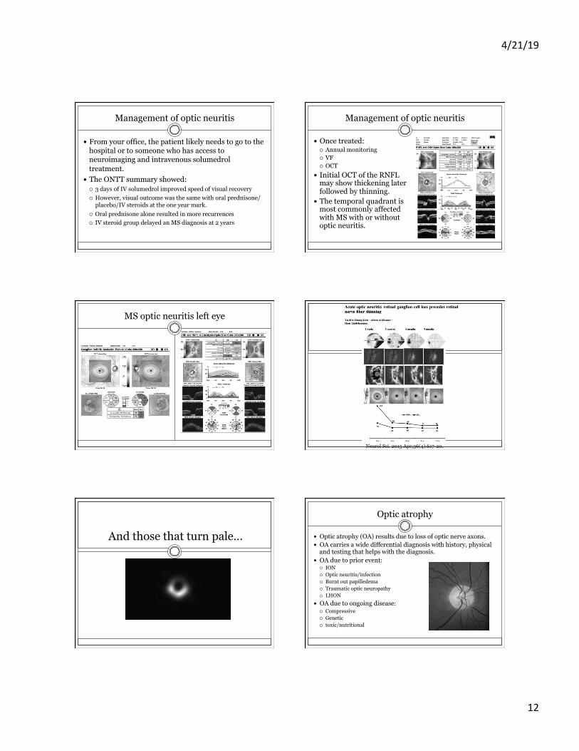
Management of optic neuritis
� From your office, the patient likely needs to go to the hospital or to someone who has access to neuroimaging and intravenous solumedrol treatment.
� The ONTT summary showed: ¡ 3 days of IV solumedrol improved speed of visual recovery ¡ However, visual outcome was the same with oral prednisone/
placebo/IV steroids at the one year mark. ¡ Oral prednisone alone resulted in more recurrences ¡ IV steroid group delayed an MS diagnosis at 2 years
Management of optic neuritis
� Once treated: ¡ Annual monitoring ¡ VF ¡ OCT
� Initial OCT of the RNFL may show thickening later followed by thinning.
� The temporal quadrant is most commonly affected with MS with or without optic neuritis.
MS optic neuritis left eye
Neurol Sci. 2015 Apr;36(4):617-20.
And those that turn pale…
Optic atrophy
� Optic atrophy (OA) results due to loss of optic nerve axons. � OA carries a wide differential diagnosis with history, physical
and testing that helps with the diagnosis. � OA due to prior event:
¡ ION ¡ Optic neuritis/infection ¡ Burnt out papilledema ¡ Traumatic optic neuropathy ¡ LHON
� OA due to ongoing disease: ¡ Compressive ¡ Genetic ¡ toxic/nutritional

4/21/19
13
Case 1
� 42 y/o woman with decreased vision OS and proptosis
� VA ¡ OD 20/30 ¡ OS CF one foot
� RAPD OS
Case 1 CT imaging
Case 2
� 54 y/o with loss of VA OS for 3 months � VA
¡ OD 20/20 ¡ OS 20/30
� RAPD OS
Case 3
� 69 year old AA man decreased vision since age his late 20’s. He has been treated for POAG for many years.
� h/o HTN in anti-hypertensives
� No know prior family history � One of his daughter is a
glaucoma suspect � VA:
¡ 20/100 OU ¡ IOP 13mmHg OU ¡ Color plates 4/10 OU
� Diagnosis: Dominant Optic atrophy
Prior AION OD Evaluation of OA
� Our role should be to look for underlying treatable condition or condition that we can prevent from worsening.
� Onset of visual loss: ÷ Acute ÷ Progressive ÷ Monocular or binocular
¡ Any family history: ÷ Leber’s hereditary
¢ Maternal inherited mitochondrial ÷ Dominant optic atrophy
¢ AD ¡ Medications
÷ Ethambutol ÷ Isoniazid ÷ Linezolid
¡ Gastric bypass ¡ Alcoholism
� Testing: ¡ MRI orbits (with gadolinium
preferred) ÷ Unexplained optic atrophy ÷ Progressive visual loss
¡ Serology (clinically correlate) ÷ CBC ÷ RPR ÷ ANA ÷ ACE ÷ NMO igG ÷ Anti MOG Antibody ÷ DsDNA ÷ ESR ÷ CRP ÷ B12 ÷ Folate ÷ Thiamine ÷ Copper ÷ Heavy metal screens ÷ Paraneoplastic

4/21/19
14
Management of OA
� Mangement is dedicated to the etiology ¡ Compressive:
÷ Remove compression if possible ¡ Genetic:
÷ Await research trials ¡ Prior event without progressive disease (prior AION, TON):
÷ Monitor ¡ Infectious/inflammatory:
÷ Treat with appropriate medication (steroid, antibiotic etc)
� Subsequent follow-up exams is based on disease activity and patient needs
Summary
� Optic neuropathies are common. � Routine eye examinations may reveal cases of optic nerve
swelling or atrophy without patient knowledge. � Prior history can help determine the etiology of optic
neuropathy. � Visual fields help localize etiologies to optic neuropathy. � Many optic neuropathies require neuroimaging to rule
out intracranial processes. � Lab work may be needed in unexplained optic atrophy
though history/exam and visual fields should aid in the diagnosis.