opinion of the scientific committee on food · telex: comeu b 21877. telegraphic address: comeur...
TRANSCRIPT

6&)�&6�&170�0<&����5(9��
1
EUROPEAN COMMISSIONHEALTH & CONSUMER PROTECTION DIRECTORATE-GENERAL
Directorate C - Scientific Opinions&����0DQDJHPHQW�RI�VFLHQWLILF�FRPPLWWHHV��VFLHQWLILF�FR�RSHUDWLRQ�DQG�QHWZRUNV
6&)�&6�&170�0<&����5HY���)LQDO
23,1,21
2)�7+(
6&,(17,),&�&200,77((�21�)22'
21
)86$5,80�72;,16
3$57����7���72;,1�$1'�+7���72;,1
DGRSWHG�RQ����0D\�����
Rue de la Loi 200, B-1049 Bruxelles/Wetstraat 200, B-1049 Brussel - Belgium.Telephone: direct line (+32-2) 296.59.48 / 296.48.70, exchange 299.11.11. Fax: (+32-2) 299.48.91
Telex: COMEU B 21877. Telegraphic address: COMEUR Brussels.

6&)�&6�&170�0<&����5(9��
2
7HUPV�RI�UHIHUHQFH
Although it is acknowledged that there are gaps in the toxicological information available, theScientific Committee on Food is requested
• to assess the health risk associated with exposure to the different Fusarium toxins incereals, taking into the account the current state of knowledge.
• to indicate, on the basis of current knowledge, which of these Fusarium toxins are of mostconcern for public health and for which there is an urgent need for further research and/orneed for measures to reduce the presence of these toxins in cereals.
• to indicate, if possible, the nature of the toxicological studies to recommend in order toelucidate (more) completely the toxicology of these toxins.
In considering these issues the Committee is asked to take note, LQWHU� DOLD, of thecomprehensive report ”Fusarium toxins in cereals – a risk assessment” which has beenprepared for the Nordic Council of Ministers.
%DFNJURXQG
A variety of Fusarium fungi, which are common soil fungi, produce a number of differentmycotoxins of the class of trichothecenes (T-2 toxin, HT-2 toxin, deoxynivalenol (DON) andnivalenol) and some other toxins (zearalenone and fumonisins). The Fusarium fungi areprobably the most prevalent toxin-producing fungi of the northern temperate regions and arecommonly found on cereals grown in the temperate regions of America, Europe and Asia.
Fusarium toxins have been shown to cause a variety of toxic effects in both experimentalanimals and livestock. On some occasions toxins produced by Fusarium species have alsobeen suspected to cause toxicity in humans.
,QWURGXFWLRQ
In the evaluation of Fusarium toxins the criteria for toxin selection have been:• the toxins most commonly found in analytical surveys of cereals• the toxins for which there is a minimum of toxicological data.The first group of toxins to be evaluated is deoxynivalenol (SCF 1999), zearalenone (SCF2000a), fumonisin B1 (SCF 2000b), nivalenol (SCF 2000c), and T-2 toxin and HT-2 toxin.
The present evaluation deals only with T-2 toxin and HT-2 toxin; it is primarily based on thereport prepared for the Nordic Council of Ministers ”Fusarium toxins in cereals – a riskassessment” (Eriksen and Alexander, 1998). It does not address all the terms of referenceoutlined above for fusarium toxins in general. The Committee will address the general aspectswhen all the individual toxins have been considered.

6&)�&6�&170�0<&����5(9��
3
7���DQG�+7���WR[LQ
'HVFULSWLRQT-2 toxin and HT-2 toxin are mycotoxins of the group trichothecenes type A produced byfungi of the )XVDULXP genus,� L�H�� )XVDULXP� DFXPLQDWXP�� )XVDULXP� SRDH� and� )XVDULXPVSRURWULFKLRLGHV� which are commonly found in various cereal crops (wheat, maize, barley,oats, and rye) and processed grains (malt, beer and bread). T-2-and HT-2 toxin often occurtogether in infected cereals. The fungi producing trichothecenes are soil fungi and areimportant plant pathogens which grow on the crop in the field (cited in Eriksen andAlexander, 1998).
&KHPLVWU\T-2 toxin (fusariotoxin T2, insariotoxin, NSC 138780, T-2 mycotoxin, toxin T-2, T2-toxin, T-2 trichothecene): 12,13-Epoxytrichothec-9-ene-3,4,8,15-tetraol 4,15-diacetate 8-(3-methylbutanoate, C24H34O9, MW: 466.58, CAS no.:21259-20-1.
HT-2 toxin (Toxin HT 2): 12,13-epoxytrichothec-9-α,4-β,8-α,15-tetraol 15 acetate 8-isovalerate, C22H32O8, MW 424.54, CAS no.: 26934-87-2
T-2 toxin is rapidly metabolised to HT-2 toxin, which is a major metabolite LQ� YLYR� andtherefore, a common risk assessment for T-2 toxin and HT-2 toxin appears reasonable(Eriksen and Alexander, 1998).
The trichothecenes are in general very stable compounds, both during storage/milling and theprocessing/cooking of food, and they do not degrade at high temperatures (Eriksen andAlexander, 1998).
%LRFKHPLFDO�DVSHFWV�±�FHOOXODU�FKDQJHV�DQG�SRWHQWLDO�PRGHV�RI�DFWLRQ
(IIHFWV�RQ�'1$�DQG�51$�V\QWKHVLVT-2 toxin could inhibit synthesis of DNA and RNA both LQ� YLYR (0.75 mg/kg bw single ormultiple doses) and LQ� YLWUR (> 0.1-1 ng/ml) (Rosenstein and Lafarge-Frayssinet, 1983;reviewed in WHO, 1990).
(IIHFWV�RQ�SURWHLQ�V\QWKHVLVT-2 toxin had a strong affinity for the 60 S ribosomal subunit, and inhibited the activity ofpeptidyl transferase, and consequently also protein synthesis in the initiation phase (Beasley,1989) (see also section on Apoptosis). T-2 toxin inhibited protein synthesis both LQ�YLWUR (0.01ng/ml in suspensions of rat hepatocytes gave 75 % inhibition) and LQ�YLYR (reviewed in WHO,1990)�� ,Q� YLYR inhibition of synthesis of proteins has been demonstrated in cells from bonemarrow, spleen and thymus (0.75 mg/kg bw single dose L�S. in mice) (Rosenstein and
6&)�&6�&170�0<&����5(9��
4
Lafarge-Frayssinet, 1983; Feinberg and McLaughlin, 1989; Thompson and Wannemachter,1990; WHO 1990).
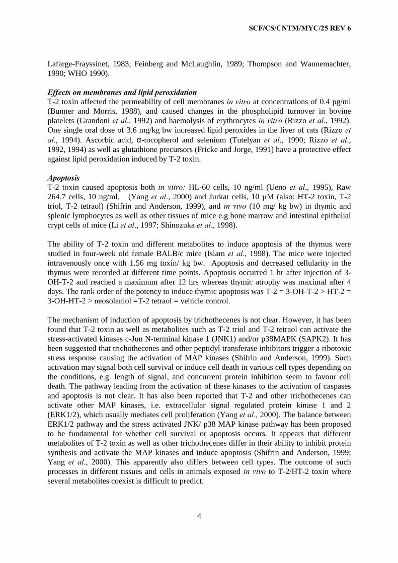
(IIHFWV�RQ�PHPEUDQHV�DQG�OLSLG�SHUR[LGDWLRQT-2 toxin affected the permeability of cell membranes LQ�YLWUR at concentrations of 0.4 pg/ml(Bunner and Morris, 1988), and caused changes in the phospholipid turnover in bovineplatelets (Grandoni HW�DO., 1992) and haemolysis of erythrocytes LQ�YLWUR (Rizzo HW�DO., 1992).One single oral dose of 3.6 mg/kg bw increased lipid peroxides in the liver of rats (Rizzo HWDO., 1994). Ascorbic acid, α-tocopherol and selenium (Tutelyan HW� DO., 1990; Rizzo HW� DO.,1992, 1994) as well as glutathione precursors (Fricke and Jorge, 1991) have a protective effectagainst lipid peroxidation induced by T-2 toxin.
$SRSWRVLVT-2 toxin caused apoptosis both LQ� YLWUR� HL-60 cells, 10 ng/ml (Ueno HW� DO., 1995), Raw264.7 cells, 10 ng/ml, (Yang HW�DO., 2000) and Jurkat cells, 10 µM (also: HT-2 toxin, T-2triol, T-2 tetraol) (Shifrin and Anderson, 1999), and LQ� YLYR (10 mg/ kg bw) in thymic andsplenic lymphocytes as well as other tissues of mice e.g bone marrow and intestinal epithelialcrypt cells of mice (Li HW�DO., 1997; Shinozuka HW�DO., 1998).
The ability of T-2 toxin and different metabolites to induce apoptosis of the thymus werestudied in four-week old female BALB/c mice (Islam HW�DO., 1998). The mice were injectedintravenously once with 1.56 mg toxin/ kg bw. Apoptosis and decreased cellularity in thethymus were recorded at different time points. Apoptosis occurred 1 hr after injection of 3-OH-T-2 and reached a maximum after 12 hrs whereas thymic atrophy was maximal after 4days. The rank order of the potency to induce thymic apoptosis was T-2 = 3-OH-T-2 > HT-2 =3-OH-HT-2 > neosolaniol =T-2 tetraol = vehicle control.
The mechanism of induction of apoptosis by trichothecenes is not clear. However, it has beenfound that T-2 toxin as well as metabolites such as T-2 triol and T-2 tetraol can activate thestress-activated kinases c-Jun N-terminal kinase 1 (JNK1) and/or p38MAPK (SAPK2). It hasbeen suggested that trichothecenes and other peptidyl transferase inhibitors trigger a ribotoxicstress response causing the activation of MAP kinases (Shifrin and Anderson, 1999). Suchactivation may signal both cell survival or induce cell death in various cell types depending onthe conditions, e.g. length of signal, and concurrent protein inhibition seem to favour celldeath. The pathway leading from the activation of these kinases to the activation of caspasesand apoptosis is not clear. It has also been reported that T-2 and other trichothecenes canactivate other MAP kinases, i.e. extracellular signal regulated protein kinase 1 and 2(ERK1/2), which usually mediates cell proliferation (Yang HW�DO., 2000). The balance betweenERK1/2 pathway and the stress activated JNK/ p38 MAP kinase pathway has been proposedto be fundamental for whether cell survival or apoptosis occurs. It appears that differentmetabolites of T-2 toxin as well as other trichothecenes differ in their ability to inhibit proteinsynthesis and activate the MAP kinases and induce apoptosis (Shifrin and Anderson, 1999;Yang HW� DO., 2000). This apparently also differs between cell types. The outcome of suchprocesses in different tissues and cells in animals exposed LQ�YLYR to T-2/HT-2 toxin whereseveral metabolites coexist is difficult to predict.

6&)�&6�&170�0<&����5(9��
5
2WKHU�HIIHFWVT-2 toxin also inhibited the mitochondrial electron transport system in yeast by inhibitingsuccinic dehydrogenase (Khachatourians, 1990). T-2 toxin inhibited gap-junctionalintercellular communication in Chinese Hamster V79 cells (IARC, 1993).
Haematopoietic progenitor cells appears to be a sensitive target for T-2/HT-2 toxin toxicityboth LQ�YLYR�and LQ�YLWUR�(Parent-Massin and Parchment, 1998). However, no mechanisms ofaction have been established except that it is likely that those described above may operate.
7R[LFRNLQHWLFV
No human data on the kinetics of T-2 or HT-2 toxins are available.
T-2 toxin is rapidly absorbed after ingestion in most animal species and it is distributed in theorganism with little or no accumulation in any specific organs. Maximum plasmaconcentrations occurred after about 30 minutes in rodents (reviewed in WHO 1990; IARC,1993). Four hours after intravenous administration of radioactive labelled T-2 toxin to pigs,15-24 % of the radioactivity given was found in the gastrointestinal tract and 4.7-5.2 % in theremaining tissues, mainly muscle and liver (Corley HW�DO., 1986).
The plasma half-life for T-2 toxin is less than 20 minutes. T-2 toxin is rapidly metabolised bydeacetylation, hydroxylation, glucuronide conjugation and de-epoxidation. The mainbiotransformation pathway is deacetylation of the C-4 acetyl group of T-2 toxin, which leadsto HT-2 toxin (the sole metabolite of T-2 toxin with isolated microsomes from liver, kidneyand spleen of various animals). This reaction is catalysed by a non-specific carboxyesterase inseveral tissues, mainly in the liver, but also in blood plasma (Johnsen HW�DO., 1988), intestines(25% of T-2 is converted to HT-2 in the isolated intestinal loop assay) (Conrady-Lorck HW�DO.,1989). HT-2 toxin may be further deacetylated, hydroxylated and conjugated by variousmetabolic pathways to 3’hydroxy HT-2, T-2 triol, 3’hydroxy T-2 triol, 4-deacetylneosolaniolwhich is converted to T-2 tetraol, and glucuronide conjugates of these. T-2 toxin can alsoundergo direct hydroxylation to 3’hydroxy T-2. These metabolites have been observed inseveral rodent species and pigs. Also cells LQ� YLWUR such as lymphoid cells metabolise T-2toxin (WHO, 1990; IARC, 1993).
In cynomolgus monkeys exposed intravenously to one dose of radiolabelled T-2 toxin morethan 22% were found as metabolites in plasma after 5 minutes. After 2 hours 25 % of theamount in plasma was found as T-2, and only 8 % after 24 hours. Less than 2 % of the totalplasma amount was found as HT-2 toxin in plasma at all time points, presumably due to rapidturnover to other metabolites. Major metabolites in plasma were T-2 tetraol and 3’hydroxy T-2. Major metabolites in urine was T-2 tetraol, 3’hydroxy HT-2 and 3’hydroxy-T-2.Metabolites could be observed in urine up to five days after exposure (Naseem HW�DO., 1995).The amount of conjugates, e.g. glucuronide derivatives, was not determined.
In pigs, 63% of the total T-2-metabolites in urine and 77% in bile were glucuronic acidconjugates (Corley HW�DO., 1985). In rodents, liver is the major site of biotransformation of T-2toxin with biliary excretion as the major route (reviewed in IARC, 1993; Matsumoto HW�DO.,1978). T-2 and 3-hydroxylated metabolites in the range from 10-160 µg/kg have been detected

6&)�&6�&170�0<&����5(9��
6
in milk from cows intubated daily with 180 mg T-2 (Yoshizawa, HW�DO., 1981). T-2 toxin israpidly metabolised and secreted and no significant accumulation of T-2 is observed in thespecies tested (e.g. pig, cattle, dog, guinea pig).
HT-2 toxin, 0.06 mg/kg bw, was given in a single oral dose by gavage to mini-piglets andserum concentrations of HT-2 toxin were determined after 0.5, 1, 2 and 4 hours. HT-2 toxinappeared rapidly in serum and reached maximum concentration (1.3 µg/l) after 1 hour. At fourhours the concentration was 0.8 µg/l (Bernhoft HW�DO., 2000).
Since HT-2 toxin is a major metabolite of T-2 toxin (as was shown in LQ�YLWUR studies withliver S-9 fraction from pigs, in which more than 80 % of T-2 toxin was converted to HT-2toxin and metabolites thereof (Kabilka, 1985, see also above)) the same authors (Bernhoft HWDO., 2000) determined HT-2 toxin in serum of mini-piglets following a single oral dose of 0.06µg T-2 toxin /kg bw HT-2 toxin appeared in serum at time points 0.5, 1, 2 and 4 hours. Theserum T-2 toxin- and HT-2 toxin concentrations at 1 hour were approximately 1.3 and 0.6µg/l, respectively. It is therefore reasonable to assume that HT-2 will follow the samemetabolic pathway as T2 toxin (Eriksen and Alexander, 1998).
7R[LFLW\
$FXWH�DQG�VXEDFXWH�WR[LFLW\Acute effects of T-2 toxin occur after oral exposure to 0.06 - 10 mg/kg bw in various species.The effects observed include non-specific symptoms like weight loss, feed refusal, dermatitis,vomiting (cats, dogs, pigs and ducklings), diarrhoea, haemorrhages and necrosis of theepithelium of stomach and intestine, bone marrow, spleen, testis and ovary. Cats appear to beparticularly susceptible to T-2 toxin (see also section on Haematotoxicity) (WHO, 1990;IARC, 1993; Rafai HW�DO., 1995a; Eriksen and Alexander, 1997). A primary target of toxicityfollowing one or more doses in studies on acute toxicity is haematopoetic tissue i.a. in thebone marrow (for further description see section on Haematotoxicity). Toxicity of thegastrointestinal epithelium following acute exposure is also a systemic effect, as it is not onlyobserved following oral - but also parenteral exposure (DeNicola HW� DO., 1978; Pang HW� DO.,1987)
Acute disturbances in the circulatory system (hypotension and arrhythmia), which may resultfrom the general pathophysiological responses to T-2 toxin including a central effect on bloodpressure and catecholamine elevation, have been reported in pigs and rats (reviewed in BubienHW�DO., 1989 and WHO 1990). General arteriosclerosis and hypertension as delayed sequela ofrepeated exposure to T-2 toxin have been reported (Wilson HW�DO., 1982). In a series of studieson rats exposed to a limited course of T-2 toxin doses caused thickening of coronary arteriesincluding myocardial changes. Four L�S� injections of 3 mg T-2 toxin/ kg bw on alternate daysgave vacuolisation and swelling of endothelial cells, basement membrane changes andenlarged medial smooth muscle cells. Upon topical application of T-2 toxin (1-20 µg) to ratskin dilated microvessels packed with erythrocytes and an increased number of degranulatedmast cells were observed (Yarom HW�DO., 1986, 1987a,b).

6&)�&6�&170�0<&����5(9��
7
T-2 toxin has high acute toxicity, with acute oral LD50 values in rodents in the range of 5-10mg/kg bw. Newborn animals seem to be more sensitive than older animals. T-2 toxin andHT-2 toxin have L�S. LD50 in the same dose range in mice (5-10 mg/kg bw). No oral LD50 hasbeen established for HT-2 toxin.
6XEFKURQLF�WR[LFLW\Young pigs were fed 1 to 16 mg purified T-2 toxin/kg feed (equivalent to 0.04 to 0.64 mg/kgbw, source of toxin not given) for 8 weeks (Weaver et. al., 1978). No haematological effectswere seen, while reduced body weight gain was observed at 1 mg/kg feed. The pigs refused adiet containing 16 mg T-2/kg, but not a diet containing 10-12 mg T-2/kg.
Twelve-week-old pigs fed 0.4 to 3.3 mg purified T-2/kg feed (equivalent to 0.016 to 0.132mg/kg bw/day) for 5 weeks had no effects on haematocrit, red blood cell count, cell volume,or haemoglobin concentration. There was a non-significant tendency towards a lowered feedconsumption in the exposed groups (Friend HW�DO., 1992).
Mini-piglets were dosed with 0.012 or 0.06 mg T-2 or HT-2 toxin/ kg bw by gavage oncedaily from age 1 week to 8 weeks (Bernhoft HW�DO., 2000). The piglets were vaccinated withtetanus toxoid, influenza-, herpes- and reo virus at ages 3 and 6 weeks. No effects caused bythe treatment on clinical health, weight gain, haematology and antibody responses tovaccination were observed. Neither were any treatment-related changes found bymacroscopical and histopathological examination at the end of the study.
In a study with seven weeks old pigs given feed containing 0.5, 1.0, 2.0, 3.0, 4.0, 10.0 or 15.0mg pure T-2 toxin/kg feed for 3 weeks, lowered levels of glucose, inorganic phosphorus andmagnesium were observed at doses from 1.0 mg/ kg feed (equivalent to 0.06 mg/kg bw).Except for a 10 % reduction in feed intake no effects on weight gain or other parameters werefound among pigs fed 0.5 mg/kg feed (equivalent to 0.03 mg/kg bw) (Rafai HW�DO., 1995a).
In a another study that was reported by Rafai and co-workers (Rafai HW�DO., 1995b), on seven-week-old pigs exposed to T-2 toxin in the feed for three weeks immunotoxicity was observedat doses of 0.03 and 0.06 mg/ kg bw (see section on Immunotoxicity for further description).
Male rhesus monkeys (7 animals) were given 0. 1 mg T-2 toxin/kg bw by gavage daily for 4-5weeks. There were no controls. Blood was drawn before the start of the experiment. At anearly stage of treatment 3 animals developed complication notably vomiting, haemorrhage,respiratory infection, and died. In the remaining animals effects observed were a 40%reduction in leukocyte counts, reduction in phagocytosis of (��FROL and a reduction in B-cellsand T-cell lymphocytes (Jagadeesan HW�DO., 1982).
&KURQLF�WR[LFLW\�DQG�FDUFLQRJHQLFLW\In mice fed 10 and 15 mg T-2/kg feed (equivalent to 1.5 and 2.25 mg/ kg bw) for 12 months,lesions (hypoplasia, hyperkeratosis and acanthosis) in the oesophageal region were observed.Such changes in the squamous epithelium were�observed after 13 weeks of feeding and duringthe rest of the experimental period.�The lesions were reversible and most had subsided 3months after the exposure was terminated (Ohtsubo and Saito,�1977). In Wistar rats given 5 to

6&)�&6�&170�0<&����5(9��
8
15 mg T-2/kg feed for 4 weeks, lesions similar to those observed in mice in the epithelium ofthe oesophageal region of the stomach were observed in rats receiving both 10 and 15 mg T-2toxin/kg feed. These lesions were negligible in rats fed 5 mg/kg feed (equivalent toapproximately 0.5 mg/ kg bw) (Ohtsubo and Saito, 1977). No adverse effect was observed inHoltzman albino rats fed 10 mg T-2/kg feed for 8 months (Marasas HW�DO., 1969).
Mice (50 males and 50 females per group) were given feed containing 0, 1.5 or 3,0 mgpurified T-2/kg feed (approx. 0.23 or 0,45 mg/kg bw/day) for 16 months. In males theincidence of pulmonary adenomas was 10% (4 animals) in controls, 15% (7 animals) for thelow dose and 23.3 % (11 animals) for the high dose. Two controls and three high-dose maleshad pulmonary adenocarcinomas. In addition, the incidence of hepatocellular adenomas inhigh-dose male mice was increased compared to controls (control 3 animals (7%), low dose 3(6%) and high dose 10 (21 %)). Significant differences in incidences of pulmonary andhepatocellular adenomas were found between males in the high dose and control groups (p <0.05). A dose-related increase was observed in epithelial hyperplasia in the forestomach witha statistically significant difference between all treatment levels in both male and female mice.A dose related increase in heart weight was found in�males, statistically significant for thehighest exposed group, but not female CD-1 mice. No effects were found in other organs,haematological parameters or in T-dependent humoral immunity tests (response towardsSRBC) (Schiefer HW�DO., 1987).
The Committee was aware of a study in mice (Yang and Xia, 1988 cit. IARC, 1993) and twostudies in rats (Li HW� DO., 1992 (in Chinese) cit. IARC, 1993; Schoental HW� DO., 1979) whereforestomach papillomas (and forestomach and stomach epithelial hyperplasia orhyperkeratosis in the latter rat study) were observed, but could not use the informationbecause of lack of information in the protocols.
T-2 toxin was negative in both initiator and promoter tests of skin tumour induction (reviewedin WHO, 1990; IARC 1993).
The Committee noted that IARC (1993) concluded that no data were available on thecarcinogenicity to humans of toxins derived from )XVDULXP� VSRURWULFKLRLGHV� and� there islimited evidence in experimental animals for the carcinogenicity of T-2 toxin. The overallconclusion by IARC was that toxins derived from )XVDULXP� VSRURWULFKLRLGHV are notclassifiable as to their carcinogenicity to humans (Group 3).
*HQRWR[LFLW\
T-2 toxin was assayed in several LQ�YLWUR and LQ�YLYR tests (see Haschek, 1989 and IARC, 1993for reviews). T-2 toxin did not induce DNA damage in bacteria, gene mutations in 6DOPRQHOODW\SKLPXULXP or mitotic crossing-over or mitochondrial petite mutations in 6DFFKDURP\FHVFHUHYLVLDH with and without exogenous metabolic system. In cultured Chinese hamster V79cells induction of gene mutations (0.1 µg T-2 toxin/ml) (activation by rat hepatocytes), andsister chromatid exchanges (activation by rat oesophageal epithelium), chromosomalaberrations and micronuclei (with and without exogenous metabolic system) were observed atvery low concentrations. T-2 toxin slightly increased sex chromosome loss in 'URVRSKLODPHODQRJDVWHU, but the results for sex-linked recessive lethal mutations were inconclusive. T-2

6&)�&6�&170�0<&����5(9��
9
induced chromosomal aberrations in cultured human lymphocytes in the absence of anexogenous metabolic system, but did not induce sister chromatid exchanges.
T-2 did not induce UDS in primary cultures of human gastric epithelial cells (Ji HW�DO., 1994).In human fibroblasts, no UDS was observed after exposure to HT-2 toxin without metabolicactivation. With liver S-9 activation the highest dose of HT-2 toxin caused UDS (seeHaschek, 1989 and IARC, 1993 for review). T-2 toxin (0.005-0.5 µg/ml) and T-2 tetraol (0.1-10 µg/ ml), induced unscheduled DNA synthesis (UDS) in cultured human fibroblasts(Oldham HW�DO., 1980). The UDS induction did not show any dose dependence, possibly due toconcurrent cytotoxicity at all dose levels; moreover, at the highest doses protein and DNAsynthesis are strongly inhibited.
Chinese hamsters treated in vivo with T-2 toxin had a slight increase in the frequency ofchromosomal aberrations (1.70 mg/kg bw L�S.), but no increase of micronuclei (3.00 mg/kgbw L�S.). In mice 3 mg/kg bw L�S. (50% of LD50) T-2 toxin caused a slight increase in single-strand breaks in spleen and thymus, but not in the liver (IARC, 1993). Bilgrami and co-workers (1995) studied the effect vitamin C (10 mg/ kg bw) on chromosomal changes in bonemarrow cells of mice fed 0.10 ± 0.02 mg T-2 toxin /kg feed in the diet (equivalent to 0.015mg/kg bw). T-2 toxin given for 12 weeks in the feed induced chromosomal abnormalities(mostly fragmentation, pulverisation and polyploidy, and no increase in chromosomalstructural abnormalities was reported) and vitamin C reduced these effects. No dose responseof T-2 toxin was investigated.
,Q�YLWUR studies showing clastogenic effects have been carried out at concentrations between0.0001 and 2.3 µg T-2 toxin/ml (IARC, 1993). Inhibition of protein synthesis has been foundin rat hepatocytes at 0.00001 µg T-2 toxin/ml (see section on Effects on protein synthesis).DNA damage or clastogenicity were seen LQ�YLYR at doses of T-2 toxin also inducing generaltoxic effects.
,PPXQRWR[LFLW\
Various studies with T-2 toxin demonstrate effects on the production of antibodies, both inassays measuring T-dependent responses (e.g. response to sheep red blood cells, SRBC) andT-independent response (e.g. dinitrophenyl-bovine serum albumin or lipopolysaccharides,LPS) having effects on both humoral and cellular immune responses (Rafai and Tuboly, 1982;Taylor HW�DO., 1989; WHO, 1990). Trichothecenes, as demonstrated for T-2 toxin and DON,can cause both suppression and stimulation of immunoglobulin production, depending on thedoses and timing of exposure (WHO 1990, Pestka and Bondy 1994).
Rafai and co-workers (1995b) fed four groups of seven-week-old pigs weighing about 9 kg forthree weeks with T-2 toxin. The concentrations were 0.5, 1.0, 2.0 or 3.0 mg/kg feed(prestarter), corresponding to average daily intakes of 0.38, 0.81, 1.24 or 1.43 mg,respectively. (Based on a corresponding experiment by the same authors (Rafai HW�DO., 1995a)the mean weight during the experiment was calculated and it was found that the meanexposures would be equivalent to 0.03, 0.06, 0.1 and 0.13 mg T–2 toxin/kg bw, respectively.)At the first and fourth days of treatment the pigs were immunised with horse globulin. Blood

6&)�&6�&170�0<&����5(9��
10
samples were drawn at days 7, 14 and 21. The two highest doses caused a significantreduction in the number of red blood cells at day 21. Leukocyte counts, particularly T-lymphocytes, were reduced at all doses. A reduced synthesis of antibodies towards horseglobulin was seen at all doses and at all time points. Upon histopathological investigation oflymphoid organs at the end of the study the authors report dose dependent depletion oflymphoid elements in the thymus and spleen, but in the lymph nodes this was difficult toquantify. Even at the lowest dose (0.03 mg toxin/kg bw) the reduction in leukocyte count was20 %, the response to horse globulin was reduced by 29 % and the blastogenic response oflymphocytes to PHA was reduced by 25 %. It should be noted that the feed intake wasreduced by 10 % without any change in weight gain in this group (Rafai HW�DO., 1995a). Thiscould be due to potential confounding effects, as pair-fed controls were not used.
In another study with mini-piglets 0.012 and 0.06 mg T-2 – or HT-2 toxin / kg bw had noconsistent imunotoxic effects (Bernhoft HW� DO., 2000, see section on subchronic toxicity). Inother studies in pigs no haematological changes were found with doses equivalent to 0.1 and0.5 mg/kg bw (Weaver HW�DO., 1978, Friend HW�DO., 1992).
Thymus atrophy has been found in mice and rats after oral and intraperitoneal exposure. Thelowest oral dose shown to induce thymus atrophy, particularly some subpopulations ofthymocytes, in mice is 0.75 mg/kg bw (LOAEL = 0.75 mg/kg bw) (Smith HW�DO., 1994).
Increased resistance towards bacteria in mice was observed after 0.5-3 mg T-2 toxin/kg bw(Corrier and Ziprin, 1986a, b, Corrier HW�DO., 1987, Cooray and Jonsson, 1990). No effect onresistance towards bacteria were found after oral exposure to T-2 toxin to 0.75 mg/kg bw dailyfor a week prior to inoculation with bacteria, but an enhanced macrophage activity wasobserved (Atroshi HW�DO., 1994, Cooray and Jonsson, 1990).
(IIHFWV�RI�7���WR[LQ�RQ�WKH�KXPDQ�LPPXQH�V\VWHPT-2 toxin reduced responses to mitogens in human lymphocytes LQ�YLWUR (IC50: 0.9-1.3 nM).Combined with nivalenol an additive effect was seen whereas with DON a less than additiveeffect was seen (Thuvander HW�DO., 1999). No LQ�YLYR data from humans with known exposuresare available, but effects on lymphocytes were recorded in Russia in persons affected by thealimentary toxic aleukia, an epidemic where T-2 toxin is suspected to be the causative agent(see section on Human studies) (Taylor HW�DO., 1989, WHO 1990).

6&)�&6�&170�0<&����5(9��
11
+DHPDWRWR[LFLW\
The effects of T-2- and HT-2 toxin on red cell-, leukocyte- and platelet progenitor cells frommice, rats and humans LQ�YLWUR have been investigated with respect to effects on proliferation,differentiation and cytotoxicity. Such cells seem to be sensitive to toxic effects of both T-2and HT-2 toxins as inhibitory effects on both parameters appear to occur in concentrationsbetween 10-7 and 10-10M (0.05-50 ng/ml) (Dugyala HW�DO., 1994; Lautraite HW�DO., 1995,1996;Rio HW�DO., 1997). In foetal liver haematopoietic precursor cells from mice T-2 toxin (10-7 M,50 ng/ml) resulted in highly selective and nearly complete toxicity to a subpopulation ofCD45R+ B-lymphocytic cells (Holladay HW�DO., 1995).
Human circulating blood cells LQ� YLWUR are less sensitive to T-2- and HT-2 toxin than otherprogenitor blood cells (see section on effects on membranes and lipid peroxidation).
Haematopoietic tissue LQ�YLYR is a target of toxicity in several animal species such as mice,rats, cats, rabbits and guinea-pigs following acute exposure to one ore more doses of T-2 – orHT-2 toxin (WHO, 1990).
A single intramuscular injection of 0.65 mg T-2/kg bw (LD20) in cynomolgus monkeys causedtransient leukocytosis, prolongation of prothrombin time and a decrease in coagulation factors(Cosgriff HW�DO., 1986).
Adult rhesus monkeys weighing 2-3 kg were given T-2 toxin semi-purified by extractionsfrom )XVDULXP LQFDUQDWXP grown on rice (Rukmini HW�DO., 1980). The final extract containedonly T-2 toxin and no other trichothecenes. In the control group there was three females andthree males and the treated group consisted of three males and two females. The treated groupwas given 1 mg T-2 toxin/ kg bw/day for four days in milk by stomach tube. The males startedto vomit on the second day and became weak and the daily dose was reduced to 0.5 mg /kgbw from day 5-15. The male monkeys had skin haemorrhages and died from respiratoryfailure and lung congestion. Severe leukopenia as well as a decrease in haemoglobinconcentration and platelet count were observed in all treated animals by the end of week oneand in the surviving females at day 15. After 30 days of recovery the two females and twoadditional male monkeys were given 0.1mg/kg bw/day for 15 days. Leukopenia developedslowly in this group and was marked at day 15; also the haemoglobin concentration wasreduced. Autopsies and histopathology were not reported for the low dose experiment.
Four groups of cats with 4-6 animals in each group were exposed to T-2 toxin from )�VSRURWULFKLRLGHV�No. 921 a strain associated with a fatal outbreak of alimentary toxic aleukia(ATA) (Lutsky HW�DO., 1978). A crude extract of T-2 toxin, a crude extract free of T-2 toxin andpurified T-2 toxin were prepared. The dissolved toxin was administered in gelatine capsuleson alternate days. Groups of six and four cats received 0.08 or 0.1 mg purified T-2 toxin/kgbw, respectively. And groups of four and six cats received 0.06 or 0.076 mg T-2 toxin incrude extract/ kg bw, respectively. Crude extract from which T-2 toxin had been removed wasgiven to two cats and diethylene glycol-ethanol-water solution was given to two cats, the lattertwo groups serving as controls. Following 16 doses the controls groups were healthy. Exceptfor one cat receiving 0.076 mg T-2 toxin from crude extract all the other T-2 toxin exposedcats died between day 6 and 40. The mean survival time was dose dependent and varied from

6&)�&6�&170�0<&����5(9��
12
13.5 to 34.5 days in the highest and lowest dose. The signs included emesis, anorexia, bloodydiarrhoea, ataxia and weight loss. The lesions included multiple haemorrhages in the skin,intestinal tract, lymph nodes and heart, necrosis of gastrointestinal epithelium and decreasedcellularity of bone marrow, lymph nodes and spleen. A slight leukocytosis occurred earlyfollowed by pancytopenia with a marked leukopenia after two weeks.
Cats appear to be extremely susceptible to T-2 toxin induced haematotoxicity and generaltoxicity with high lethality in comparison with other experimental species. The reason for thiscould be that cats in contrast to several other species, e.g. rodents, monkeys, pigs and humans,lack the ability to form glucuronide conjugates particularly of planar phenolic compounds(Timbrell, 1991; Court and Greenblatt, 1997 a,b). UDP-glucuronosyltransferase (UGT) 1A6 isa pseudogene in the cat and there is a reduced diversity of expressed hepatic UGT1A isoforms(Court and Greenblatt, 2000). Glucuronidation of various hydroxylated T-2 and HT-2metabolites followed by urinary and biliary excretion are important detoxificationmechanisms of these toxins in several other species, e.g. rats, mice, pigs. Thebiotransformation of T-2 toxin in cats has, however, not been examined.
In guinea-pigs 0.9 mg T-2 toxin/ kg bw and day for 27 days caused erythropenia, leukopenia,lymphopenia and reduced lymphocyte content of the bone marrow. More moderate changeswere seen in animal given 0.5 mg T-2 toxin/ kg bw and day for 21 days followed by 0.75 mgT-2 toxin/ kg bw/day for 21 days (DeNicola HW�DO., 1978).
Mice fed T-2 toxin in the diet, 20 mg/ dry diet (equivalent to about 3 mg/ kg bw) for 21 and41 days showed hypoplastic lymphoid tissues, bone marrow and splenic red pulp resulting inanaemia (WHO, 1990). 59Fe incorporation of circulating erythrocytes was strongly inhibitedand granulocyte macrophage colony-forming cells in the bone marrow were transientlydepleted by T-2 toxin in a single subcutaneous dose of 0.3 mg/kg (Faifer HW�DO., 1992).
In mice treated with a single subcutaneous dose of 2 mg T-2 toxin/kg bw it was foundfollowing a potent initial inhibition, that spleen activity rapidly recovered while bone marrowremained significantly depressed for a much longer time (Velazco HW�DO., 1996).
Rafai and co-workers (1995b) fed four groups of seven-week-old pigs weighing about 9 kg forthree weeks with T-2 toxin. The pigs showed signs of haematotoxicity, see description insection on Immunotoxicity.
Pregnant mice were exposed to 1.2 or 1.5 mg T-2 toxin /kg bw by oral gavage on gestationaldays 14-17 and haematopoietic cells in foetal livers were investigated on gestational day 18.Subpopulations of cells with surface markers CD441o, CD451o, CD45R+ were decreased. Inadult mice exposed to 1.75 mg T-2 toxin /kg bw for four consecutive days bone marrowcellularity was reduced including cells with surface marker CD45R+ but not cells with markerCD44+ (Holladay HW�DO., 1995).
6&)�&6�&170�0<&����5(9��
13
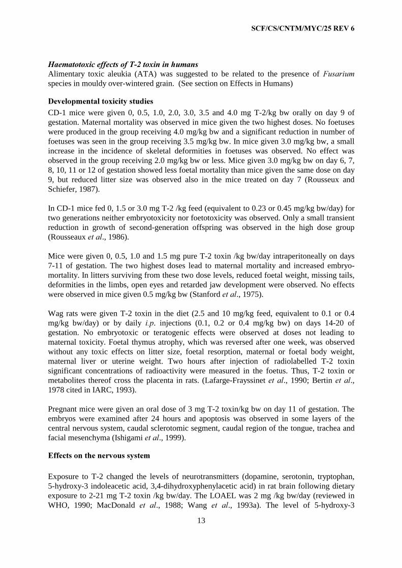
+DHPDWRWR[LF�HIIHFWV�RI�7���WR[LQ�LQ�KXPDQVAlimentary toxic aleukia (ATA) was suggested to be related to the presence of )XVDULXPspecies in mouldy over-wintered grain. (See section on Effects in Humans)
'HYHORSPHQWDO�WR[LFLW\�VWXGLHVCD-1 mice were given 0, 0.5, 1.0, 2.0, 3.0, 3.5 and 4.0 mg T-2/kg bw orally on day 9 ofgestation. Maternal mortality was observed in mice given the two highest doses. No foetuseswere produced in the group receiving 4.0 mg/kg bw and a significant reduction in number offoetuses was seen in the group receiving 3.5 mg/kg bw. In mice given 3.0 mg/kg bw, a smallincrease in the incidence of skeletal deformities in foetuses was observed. No effect wasobserved in the group receiving 2.0 mg/kg bw or less. Mice given 3.0 mg/kg bw on day 6, 7,8, 10, 11 or 12 of gestation showed less foetal mortality than mice given the same dose on day9, but reduced litter size was observed also in the mice treated on day 7 (Rousseux andSchiefer, 1987).
In CD-1 mice fed 0, 1.5 or 3.0 mg T-2 /kg feed (equivalent to 0.23 or 0.45 mg/kg bw/day) fortwo generations neither embryotoxicity nor foetotoxicity was observed. Only a small transientreduction in growth of second-generation offspring was observed in the high dose group(Rousseaux HW�DO., 1986).
Mice were given 0, 0.5, 1.0 and 1.5 mg pure T-2 toxin /kg bw/day intraperitoneally on days7-11 of gestation. The two highest doses lead to maternal mortality and increased embryo-mortality. In litters surviving from these two dose levels, reduced foetal weight, missing tails,deformities in the limbs, open eyes and retarded jaw development were observed. No effectswere observed in mice given 0.5 mg/kg bw (Stanford HW�DO., 1975).
Wag rats were given T-2 toxin in the diet (2.5 and 10 mg/kg feed, equivalent to 0.1 or 0.4mg/kg bw/day) or by daily L�S. injections (0.1, 0.2 or 0.4 mg/kg bw) on days 14-20 ofgestation. No embryotoxic or teratogenic effects were observed at doses not leading tomaternal toxicity. Foetal thymus atrophy, which was reversed after one week, was observedwithout any toxic effects on litter size, foetal resorption, maternal or foetal body weight,maternal liver or uterine weight. Two hours after injection of radiolabelled T-2 toxinsignificant concentrations of radioactivity were measured in the foetus. Thus, T-2 toxin ormetabolites thereof cross the placenta in rats. (Lafarge-Frayssinet HW�DO., 1990; Bertin HW�DO.,1978 cited in IARC, 1993).
Pregnant mice were given an oral dose of 3 mg T-2 toxin/kg bw on day 11 of gestation. Theembryos were examined after 24 hours and apoptosis was observed in some layers of thecentral nervous system, caudal sclerotomic segment, caudal region of the tongue, trachea andfacial mesenchyma (Ishigami HW�DO., 1999).
(IIHFWV�RQ�WKH�QHUYRXV�V\VWHP
Exposure to T-2 changed the levels of neurotransmitters (dopamine, serotonin, tryptophan,5-hydroxy-3 indoleacetic acid, 3,4-dihydroxyphenylacetic acid) in rat brain following dietaryexposure to 2-21 mg T-2 toxin /kg bw/day. The LOAEL was 2 mg /kg bw/day (reviewed inWHO, 1990; MacDonald HW� DO., 1988; Wang HW� DO., 1993a). The level of 5-hydroxy-3

6&)�&6�&170�0<&����5(9��
14
indoleacetic acid was increased in rats given 2.5 and 10 ppm T-2 in the feed (equivalent to 0.1and 0.4 mg/kg bw/day) (Wang HW�DO., 1993a).
In various behavioural tests rats given a single oral dose of 2.0 mg/kg bw showed reducedmotor activity and performance in the passive avoidance test. No effect (NOAEL) wasobserved in rats receiving 0.4 mg/kg bw (Sirkka HW�DO., 1992).
'HUPDO�HIIHFWV
T-2 toxin produces oedema, intradermal haemorrhage and necrosis of the skin. Guinea pig isthe most sensitive species. The effect on skin has been used as a biological assay for detectionof trichothecenes. T-2 can be detected at 0.2 µg with a skin necrosis assay. The minimumeffective amount needed to elicit irritation is much less. The mechanism for skin toxicity hasnot been established (reviewed in WHO, 1990).
(IIHFWV�LQ�KXPDQV
Alimentary toxic aleukia (ATA) that occurred in the USSR in the period 1941-47 wassuggested to be related to the presence of )XVDULXP species in mouldy over-wintered grain.An association to )��SRDH and )�� VSRURWULFKLRLGHV, which in later fungal cultures have beenfound to produce several trichothecenes including T-2 – and HT-2 toxin, was established.Extractions of the suspected wheat were also tested on the skin of animals, and showed toxicdermal effects. The most severe outbreak of the disease was in 1944, but outbreaks have alsobeen reported in 1952, 1953, and 1955 particularly in people consuming over-wintered wheat.Clinical symptoms include vomiting, abdominal pain and diarrhoea followed by leukopenia,bleeding from the nose and throat, depletion of the bone marrow and fever. Depending onseverity necrotic lesions in the oral cavity, oesophagus and stomach may occur and thelethality may be high. ATA in the second stage is characterised by leukopenia, haemorrhagicdiathesis, granulopenia, bone marrow aplasia and sepsis. Although the symptomscharacterising ATA resemble symptoms seen in cats and rodents exposed to crude extracts ofinfected wheat or T-2 toxin, no epidemiological studies have been reported to linktrichothecenes to ATA (reviewed in Beardall and Miller, 1994; Joffe, 1974; WHO, 1990;IARC, 1993).
In an outbreak of toxicosis in a Chinese county 165 subjects consumed rice infected with)XVDULXP�KHWHURVSRUXP�and ). *UDPLQHDUXP��About 50 % of the persons consuming the ricefell ill with symptoms consisting of nausea, dizziness, vomiting, abdominal distension andpain, chills and diarrhoea. Samples of the suspected rice were analysed for T-2 toxin using anELISA assay and a level of 180 - 420 µg/kg was found. Analysis for other toxins was notreported (Wang HW�DO., 1993b). It has to be noted that these Fusarium species are not known toproduce T-2 toxin, but are known to produce several other trichothecenes such as DON,nivalenol and acetyl-DON (Eriksen and Alexander, 1998).

6&)�&6�&170�0<&����5(9��
15
Other outbreaks of trichothecene-related disease have also been reported, one in China1984/85 and one in India in 1987. Each involved several hundred cases. Typical symptomswere throat irritation, nausea, vomiting, abdominal pain diarrhoea and some with blood instools. In the first case DON and zearalenon were detected, but not T-2 toxin. In the outbreakin India DON, acetyl-DON, nivalenol and T-2 toxin were detected. No information on bloodcells were reported and no fatalities occurred (WHO, 1990).
(YDOXDWLRQ�DQG�&RQFOXVLRQ
The general toxicity, haematotoxicity and immunotoxicity of T-2 toxin are considered to bethe critical effects.
T-2 toxin potently inhibits synthesis of proteins and, at higher doses, DNA and RNA, both LQYLWUR and LQ�YLYR�
There is limited evidence for tumourigenicity of T-2 toxin in experimental animals. It inducedhepatocellular- and pulmonary adenomas in male mice. Several studies in mice and ratsindicate that T-2 toxin cause toxicity and proliferative changes in the oesophagus- andforestomach epithelium.
Several conventional tests for genotoxicity LQ� YLWUR and in rodents LQ� YLYR� in particular forclastogenic effects, were positive for T-2- and HT-2 toxin� These effects were observedprimarily at concentrations also known to inhibit protein- and DNA-synthesis and producecytotoxicity. Therefore, the Committee considered that the effects observed LQ� YLWUR and LQYLYR were most likely secondary.
Only those studies considered to be of good quality and addressing relevant endpoints aresummarised in Table I.
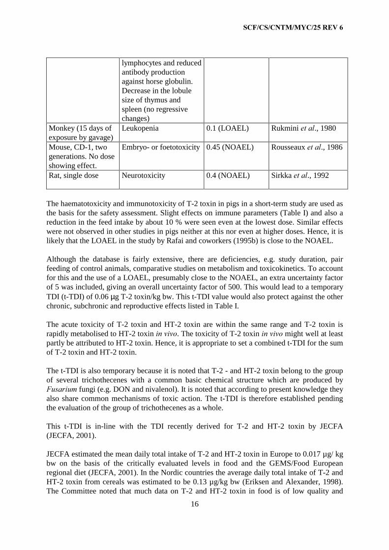
7DEOH�,��/2$(/V�DQG�12$(/V�RI�7���WR[LQ
6WXG\ &ULWLFDO�(IIHFW /2$(/�12$(/�PJ�NJ�EZ�GD\�
5HIHUHQFH
Mouse, 16 months Pulmonary adenomas
Hepatocellular adenomas
Forestomach epithelialhyperplasia
0.23 (NOAEL)
0.23 (NOAEL
0.23 (LOAEL)
Schiefer HW�DO., 1987
Rat, 4 weeks Forestomach epithelialhyperplasia
0.5 (NOAEL) Ohtsubo and Saito,1977
Mouse, 5 days Thymus atrophy,decreased number of T-and B- cells
0.75 (LOAEL) Smith HW�DO., 1994
Pig, 3 weeks(subacute)
Reduced number ofleukocytes and
0.03 (LOAEL) Rafai HW�DO., 1995b
6&)�&6�&170�0<&����5(9��
16
lymphocytes and reducedantibody productionagainst horse globulin.Decrease in the lobulesize of thymus andspleen (no regressivechanges)
Monkey (15 days ofexposure by gavage)
Leukopenia 0.1 (LOAEL) Rukmini HW�DO., 1980
Mouse, CD-1, twogenerations. No doseshowing effect.
Embryo- or foetotoxicity 0.45 (NOAEL) Rousseaux HW�DO., 1986
Rat, single dose Neurotoxicity 0.4 (NOAEL) Sirkka HW�DO., 1992
The haematotoxicity and immunotoxicity of T-2 toxin in pigs in a short-term study are used asthe basis for the safety assessment. Slight effects on immune parameters (Table I) and also areduction in the feed intake by about 10 % were seen even at the lowest dose. Similar effectswere not observed in other studies in pigs neither at this nor even at higher doses. Hence, it islikely that the LOAEL in the study by Rafai and coworkers (1995b) is close to the NOAEL.
Although the database is fairly extensive, there are deficiencies, e.g. study duration, pairfeeding of control animals, comparative studies on metabolism and toxicokinetics. To accountfor this and the use of a LOAEL, presumably close to the NOAEL, an extra uncertainty factorof 5 was included, giving an overall uncertainty factor of 500. This would lead to a temporaryTDI (t-TDI) of 0.06 µg T-2 toxin/kg bw. This t-TDI value would also protect against the otherchronic, subchronic and reproductive effects listed in Table I.
The acute toxicity of T-2 toxin and HT-2 toxin are within the same range and T-2 toxin israpidly metabolised to HT-2 toxin LQ�YLYR. The toxicity of T-2 toxin LQ�YLYR might well at leastpartly be attributed to HT-2 toxin. Hence, it is appropriate to set a combined t-TDI for the sumof T-2 toxin and HT-2 toxin.
The t-TDI is also temporary because it is noted that T-2 - and HT-2 toxin belong to the groupof several trichothecenes with a common basic chemical structure which are produced by)XVDULXP fungi (e.g. DON and nivalenol). It is noted that according to present knowledge theyalso share common mechanisms of toxic action. The t-TDI is therefore established pendingthe evaluation of the group of trichothecenes as a whole.
This t-TDI is in-line with the TDI recently derived for T-2 and HT-2 toxin by JECFA(JECFA, 2001).
JECFA estimated the mean daily total intake of T-2 and HT-2 toxin in Europe to 0.017 µg/ kgbw on the basis of the critically evaluated levels in food and the GEMS/Food Europeanregional diet (JECFA, 2001). In the Nordic countries the average daily total intake of T-2 andHT-2 toxin from cereals was estimated to be 0.13 µg/kg bw (Eriksen and Alexander, 1998).The Committee noted that much data on T-2 and HT-2 toxin in food is of low quality and

6&)�&6�&170�0<&����5(9��
17
should not be used for intake estimations. Furthermore, the sensitivity of the analyticalmethods used was low in many cases, which can lead to an overestimation of intakedepending on the method of for calculating intakes. Hence, comparison of intake estimates isvery uncertain.

6&)�&6�&170�0<&����5(9��
18
1HHGV�IRU�IXWXUH�VWXGLHV
The haematotoxicity and immunotoxicity induced by T-2/HT-2 toxin should be investigatedby the use of sensitive endpoints with respect to low dose-effects and time course in longer-term exposure studies where feed intake is controlled (e.g. 60 days in pigs).
Comparative studies on metabolism and toxicokinetics in cats, rodents and pigs might allowinsight into species differences in toxicity. Studies of metabolism of T-2 toxin in humansusing LQ�YLWUR models would also be helpful.
There is a need for more adequate information on the exposure to T-2 toxin and HT-2 toxin(and other trichothecenes), i.e. occurrence in food commodities and intakes.
5HIHUHQFHV
Atroshi, F., Rizzo, A.F., Veijalainen, P., Lindberg, L.A., Honkanen-Buzalski, T., Andersson.K., Hirvi, T. and Saloniemi, H. (1994) The effect of dietary exposure to DON and T-2 toxinan host resistance and serum immunoglobulins of normal and mastitic mice. J Anim. Physiol.Anim. Nutrition 71, 223-233
Beardall, J.M. and Miller, J.D. (1994) Diseases in humans with mycotoxins as possiblecauses. In: 0\FRWR[LQV�LQ�*UDLQ��&RPSRXQGV�RWKHU�WKDQ�DIODWR[LQ. pp 487-539. Miller J.D andTrenholm, H.L (eds) Eagan press, St. Paul, Minnesota, USA
Beasley, V.R. (ed.) (1989) 7ULFKRWKHFHQH� P\FRWR[LFRVLV�� 3DWKRSK\VLRORJLF� HIIHFWV. CRCPress, Boca Raton, Florida, USA
Bernhoft, A., Modestas, K., Langseth, W., Åkesson, C.P., Oswald, I.P. and Larsen, H.J.S.(2000) A study on immunotoxicity of HT-2 and T-2 toxins in minipigs. Abstract (Poster)presented at: X International IUPAC Symposium on Mycotoxins and Phycotoxins, Brazil,May, 2000
Bilgrami, K.S., Masood, A. And Rahman, M.F. (1995) Cumulative effect of T-2 toxin andvitamin C on chromosomal abnormalities in the bone marrow cells of mice (0XV�PXVFXOXV).Cytobios., 81, 171-174.
Bubien, J.K., Lundeem, G., Templeton, C. and Woods, W.T. (1989) Effects on the circulatorysystem. In: 7ULFKRWHFHQH� 0\FRWR[LFRVLV�� 3DWKRSK\VLRORJLFDO� HIIHFWV, Vol I, pp. 3 )2-33,Beasley., VR,(ed.), CRC Press, Boca Raton, Florida, USA.
Bunner, D.L. and Morris, E.R. (1988). Alteration of multiple cell membrane functions in L-6myoblasts by T-2 toxin: an important mechanism of action. Toxicol. Appl. Microbiol. 92,113-121.
Conrady-Lorck, S., Strugala, G.J., Feng, X.C. and Fichtl, B. (1989) Intestinal metabolism ofT-2 toxin, a major trichothecene mycotoxin. Prog. Pharmacol. Clin. Pharmacol. 7, 319-326
6&)�&6�&170�0<&����5(9��
19
Cooray, R. and Jonsson, P. (1990) Modulation of resistance to mastitis pathogens by treatmentof mice with T-2 toxin. Food Chem. Toxicol. 28, 687-692
Corley, R.A., Swanson, S.P. and Buck, W.B. (1985) Glucoronide conjugates of T-2 toxin andmetabolites in swine bile and urine. J. Agric. Food Chem. 33, 1085-1089
Corley, R.A., Swanson, S.P., Gullo, G.J., Johnson, L., Beasley, V.R., Buck, W.B. (1986)Disposition of T-2 toxin, a trichothecene mycotoxin in intramuscularly dosed swine. J. Agric.Food Chem. 34, 868-875
Corrier, D.E and Ziprin, R.L., (1 986a) Immunotoxic effects of T-2 toxin on cell-mediatedimmunity to listeriosis in mice: comparison with cyclophosphamide. Am J. Yet. Res. 47,1956-1960
Corrier, D.E. and Ziprin, R.L. (1986b) Enhanced resistance to listeriosis induced in mice bypreinoculation treatment with T-2 mycotoxin. Am. J Vet. Res��47, 856-858
Corrier, D.E. and Ziprin, R.L. and Mollenhauer, H.H. (1987) Modulation of cell-mediatedresistance to listeriosis in mice given T-2 toxin. Toxicol. Appl. Pharmacol., 89, 323-331
Cosgriff, T.M., Bunner, D.R, Wannemachter, R.W., Hodgson, L.A. and Dinterman, R.E.(1986) The hemostatic dearangement produced by T-2 toxin in Cynomolgus monkeys.Toxicol. Appl. Pharmacol., 82, 532-539
Court M. H., Greenblatt D.J. (1997a) Molecular basis for deficient acetaminophenglucuronidation in cats. An interspecies comparison of enzyme kinetics in liver microsomes.Biochem Pharmacol., 53,1041-7
Court M.H., Greenblatt DJ. (1997b) Biochemical basis for deficient paracetamolglucuronidation in cats: an interspecies comparison of enzyme constraint in liver microsomes.J Pharm Pharmacol., 49,446-9
Court M. H., Greenblatt D.J. (2000) Molecular genetic basis for deficient acetaminophenglucuronidation by cats: UGT1A6 is a pseudogene, and evidence for reduced diversity ofexpressed hepatic UGT1A isoforms. Pharmacogenetics 10,355-69
DeNicola, D.B., Rebar, A.H. and Carlton, W.W. (1978) T-2 toxin mycotoxicosis in theguinea-pig. Fd. Cosmet. Toxicol., 16, 601-609.
Dugyala, R.P., Kim, Y.P. and Sharpa, R.P. (1994) Effects of aflatoxin B1 and T-2 toxin on thegranulocyte-macrophage progenitor cells in mouse bone marrow cultures. Immunopharmacol.,27, 57-65.
Eriksen GS, Alexander J (eds.), 1998. Fusarium toxins in cereals – a risk assessment. NordicCouncil of Ministers; Tema Nord 1998, 502, pp. 7-44; Copenhagen.
Faifer, G.C., Zabal, O. and Godoy, H.M. (1992) Further studies on the hematopoietic damageproduced by a single dose of T-2 toxin in mice. Toxicology., 75, 169-174.

6&)�&6�&170�0<&����5(9��
20
Feinberg, B. and McLaughlin, C.S.(1989) Biochemical mechanism of action of trichothecenemycotoxin. In: 7ULFKRWHFHQH� 0\FRWR[LFRVLV�� 3DWKRSK\VLRORJLFDO� HIIHFWV, Vol 1, pp. 27-35,Beasley., V.R.,(ed.), CRC Press, Boca Raton, Florida, USA
Fricke, R.F and Jorge, J. (1991) Methylthiazolidine-4-carboxylate for treatment of acute T-2toxin exposure. J. Appl. Toxicol., 11, 135-140
Friend, D.W., Thompson, B.K., Trenholm, H.L., Boermans, H.J., Hartin, K.E. and Panich, P1(1992) Toxicity of T-2 toxin and its interaction with deoxynivalenol when fed to young pigs.Can J. Anim. Sci., 72, 703-711
Grandoni, K.M., Gentry, P.A., Holub, B.J. and Yagen, B. (1992) Trichothecene mycotoxinsinhibit phosphoinositide hydrolysis in bovine platelets stimulated with platelet activatingfactor. Toxicol.,72, 5160
Haschek, W.M. (1989) Mutagenicity and carcinogenicity of T-2 toxin. In�� 7ULFKRWHFHQH0\FRWR[LFRVLV��3DWKRSK\VLRORJLFDO�HIIHFWV, Vol I, pp. 32-33, Beasley., V.R.,(ed.), CRC Press,Boca Raton, Florida, USA.
Holladay, S.D., Blaylock, B.L., Comment, C.E., Heindel, JJ. and Luster, M.I. (1993) Fetalthymic atrophy after exposure to T-2 toxin: Selectivity for lymphoid progenitor cells. Tox.Appl. Pharmacol., 121, 8-14.
IARC, 1993. Monographs on the evaluation of carcinogenic risks to humans; Vol. 56: Somenaturally occurring substances, food items and constituents, heterocyclic aromatic amines andmycotoxins. International Agency for Research on Cancer, World Health Organization, pp397-333; Lyon.
Ishigami, N., Shinozuka, J., Katayama, K., Uetsuka, K., Nakayama, H., Doi, K., (1999)Apoptosis in the developing mouse embryos from T-2 toxin-inoculated dams�� Histol.Histopathol., 14.729-33
Islam, Z., Nagase, M., Ota, A.,Ueda, S., Yoshizawa, T., Sakato, N. (1998) Structure-functionrelationship of T-2 toxin and its metabolites in inducing thymic apoptosis in vivo in mice.Biosci. Biotechnol. Biochem., 62, 1492-1497.
Jagadeesan, V., Rukmini, C., Vijayaraghovan, M. and Tulpule, P.G. (1982) Immune studieswith T-2 toxin: effect of feeding and withdrawel in monkeys. Food chem. Toxicol., 20, 83-87
JECFA (2001) Summary and Conclusions of the Fifty-sixth meeting Geneva, 6- 15 February2001. Mycotoxins. http://www.fao.org/waicent/faoinfo/economic/esn/jecfa/jecfa56.pdf
Ji, X., Ning, K. Y., Liang, Y., Wang, D. and Shi, G. (1994) Effects of sterigmatocystin andT-2 toxin on the induction of unscheduled DNA synthesis in primary cultures of humangastric epithelial cells. Nat. Toxins, 2, 115-119

6&)�&6�&170�0<&����5(9��
21
Joffe, A.Z. (1974) Toxicity of )XVDULXP� SRDH� and F. VSRURWULFKLRLGHV� and its relation toalimentary toxic aleukia. In: Purchase, I.F.H., ed., Mycotoxins, Amsterdam, Elsevier, pp. 229-262
Johnsen, H., Odden, E., Johnsen, B.A. and Fonnum, F. (1988) Metabolism of T-2 toxin byblood cell carboxylesterases. Biochem. Pharmacol., 37,3193-3197
Kabilka, G.(1985) Zum nachweis von T-2 toxin in blutserum, ham und kot. PhD-thesis,Munich, Germany
Kanai, K. and Kondo, E. (1984) Decreased resistance to mycobacterial infection in mice fed atrichothecene compound (T-2 toxin). Japan. J. Med. Sci. Biol., 37, 97-104
Khachatourians G. G. (1990) Metabolic effects of trichothecene T-2 toxin. Can. J. Physiol.Pharmacol., 68, 1004-1008
Lafarge-Frayssinet, C., Decloitre, F., Mousset, S., Martin, M., Frayssinet, C. (1981) Inductionof DNA single-strand breaks by T2 toxin, a trichothecene metabolite of fusarium: effect onlymphoid organs and liver. Mutat. Res.,88, 115-23
Lafarge-Frayssinet, C., Chakor, K., Lafont, P. and Frayssinet, C. (1990) Transplacentaltransfer of T2toxin: pathological effect. J. environ. Pathol. Oncol., 10, 64-68
Lautraite, S., Parent-Massin, D., Rio, B. and Hoellinger H. (1995) Comparison of toxicityinduced by T-2 toxin on human and rat granulo-monocytic progenitors with an in vitro model.Hum. Exp. Toxicol., 14, 672-678.
Lautraite, S., Parent-Massin, D., Rio, B. and Hoellinger H. (1996) Comparison of toxicityinduced by HT-2 toxin on human and rat granulo-monocytic progenitors with an in vitromodel. Hum. Exp. Toxicol., 15, 208-213.
Lutsky, I., Mor, N., Yagen, B. and Joffre, A.Z. (1978) The role of T-2 toxin in experimentalalimentary toxic aleukia: a toxicity study in cats. Toxicol. Appl. Pharmacol., 43, 111-124.
Li, G. Shinozuka, J., Uetsuka, K., Nakayama, H. And Doi, K. (1997) T-2 toxin-inducedapoptosis in intestinal crypt epithelial cells of mice. Exp. Toxicol. Pathol., 49, 447-450
MacDonald, E.J., Covan, K.R. and Smith, T.K. (1988) Effect of acute oral doses of T-2 toxinon tissue concentrations of biogenic amines in the rat. J. Anim. Sci., 66, 434-441
Marasas, W.F.O., Barnburg,, JR, Smalley, E.B., Strong, F.M., Ragland, W.L. and Degurse,P.E., (1969) Toxic effects on trout, rats and mice of T-2 toxin produced by the fungusFusarium tricincturn. Toxicol. Appl. pharmacol., 15,471-482
Matsumoto, H., Ito, T. and Ueno, Y. (1978) Toxicological approaches to to the metabolites offasaria. XII. Fate and distribution of T-2 in mice. Jpn. J. Exp. Med., 48, 393-399
6&)�&6�&170�0<&����5(9��
22
Naseem, S.M., Pace, J.G., Wannemacher, R.W.Jr. (1995) A high-performance liquidchromatographic method for determining [3H]T-2 and its metabolites in biological fluids ofthe cynomolgous monkey. J. Anal. Toxicol., 19, 151-156
Ohtsubo, K and Saito, M. (1977) Chronic effects of trichothecene toxins. In Rodricks, J.V.,Hesseltine C.W. and Mehlman, M.A. eds. 0\FRWR[LQV� LQ�+XPDQ� DQG� $QLPDO�+HDOWK. ParkForest South, Illinois, Pathotox Publishers, pp. 255-262.
Oldham, J.W., Allred, L.E., Milo, G.E., Kindig, O. And Capen, C.C. (1980) The toxicologicalevaluation of the mycotoxins T-2 and T-2 tetraol using human fibroblasts in vitro. Toxicol.Appl. Pharmacol., 52, 159-168.
Parent-Massin, D. and Parchment, R.E. (1998) Haematotoxicity of mycotoxins. SynthèseScientifique. Rev. Méd. Vét., 149, 591-598.
Pestka, J.J. and Bondy, G.S. (1994) Immunotoxic effects of mycotoxins. In:�0\FRWR[LQV� LQJUDLQ��&RPSRXQGV�RWKHU� WKDQ�DIODWR[LQ. pp. 339-358, Miller, J.D. and Trenholm, H.L (eds)Eagan press, St. Paul, Minnesota, USA
Rafai, P. and Tuboly, S. (1982) Effect of T-2 toxin on adrenocortical function and immuneresponse in growing pigs. Zbl. Vet. Med., B29, 558-565
Rafai, P., Bata, A., Vanyi, A., Papp, Z., Brydl, E., Jakab, L., Tuboly, S. and Tury, E. (1995a)Effect of various levels of T-2 toxin on the clinical status, performance and metabolism ofgrowing pigs. Vet. Rec., 136, 485-489
Rafai, P., Tuboly, S., Bata, A., Tilly, P., Vanyi, A., Papp, Z., Jakab, L. and Tury, E. (1995b)Effect of various levels of T-2 toxin in the immune system of growing pigs. Vet. Rec., 136.511-514
Rio, B., Lautraite, S. and Parent-Massin, D. (1997) In vitro toxicity of trichothecenes onhuman erythroblastic progenitors. Hum. Exp. Toxicol., 16, 673-679.
Rizzo, A., Atroshi, R., Hirvi, T., Saloniemi, H. (1992) The hemolytic activity ofdeoxynivalenol and T-2 toxin. Nat. Toxins, 1, 106-110
Rizzo, A., Atroshi, F., Ahotupa, M., Sankari, S. and Elovaara, E. (1994) Protective effect ofantioxidants against free radical-mediated lipid peroxidation induced by DON or T-2 toxin. J.Vet. Med. A 41, 81-90
Rukmini, C., Prasad, J.S. and Rao, K. (1980) Effects of feeding T-2 toxin to rats andmonkeys. Food Cosmet. Toxicol., 18, 267-269
Roderiks, J.V., Hasseltine, C.W. and Melham, M.A. (1977) 0\FRWR[LQV�LQ�+XPDQ�DQG�$QLPDO+HDOWK. Pathotox Publishers Inc., Park Forest South, IL.
Rosenstein, Y. and Lafarge-Frayssinet, C., (1983) Inhibitory effect of Fusarium T-2 toxin on
6&)�&6�&170�0<&����5(9��
23
lymphoid DNA and protein synthesis. Toxicol. Appl. Pharmacol., 70,283-288
Rousseux, C.G. and Schiefer, H.B. (1987) Maternal toxicity, embryolethality and abnormalfetal development in CD-1 mice following one oral dose of T-2 toxin. J. Appl. Toxicol., 7,281-288
Rousseux, C.G., Schiefer, H.B.and Hancock, D.S. (1986) Reproductive and teratologicaleffects of continous low-level dietary T-2 toxin in female CD-1 mice for two generations. J.Appl. Toxicol., 6, 179184
Schiefer B.B., Roussaux, C.G., Handcock, D.S. and Blakely, B.R. (1987) Effects of low-levellongterm oral exposure to T-2 toxin in CD-1 mice. Food Chem. Toxicol., 25, 593-601
Schoental, R, Joffe, A2., and Yagen, B. (1979) Cardiovascular lesions and various tumorsfound in rats given T-2 toxin, a trichothecene metabolite of Fusarium. Cancer Res., 39,2179-2189
SCF (1999) Opinion on Fusarium toxins – part 1: Deoxinivalenol (DON), expressed on 2 December1999. http://europa.eu.int/comm/food/fs/sc/scf/out44_en.pdf
SCF (2000a) Opinion on Fusarium toxins – part 2: Zearalenon (ZEA), expressed on 22 June 2000.http://europa.eu.int/comm/food/fs/sc/scf/out65_en.pdf
SCF (2000b) Opinion on Fusarium toxins – part 3: Fumonisin B1 (FB1), expressed on 17 October2000. http://europa.eu.int/comm/food/fs/sc/scf/out73_en.pdf
SCF (2000c) Opinion on Fusarium toxins – part 4: Nivalenol, expressed on 19 October 2000.http://europa.eu.int/comm/food/fs/sc/scf/out74_en.pdf
Shifrin, V.L. and Anderson, P. (1999) Trichothecene mycotoxins trigger ribotoxic stressresponse that activates c-Jun N-terminal kinase and p38 mitogen-activated protein kinase andinduces apoptosis. J. Biol. Chem., 274, 13985-13992.
Shinozuka J, Suzuki M, Noguchi N, Sugimoto T, Uetsuka K, Nakayama H, Doi K (1998)T-2 toxin-induced apoptosis in hematopoietic tissues of mice. Toxicol. Pathol.,26, 674-81
Sirkka, U., Nieminen, S.A. and Ylitalo, P. (1992) Acute neurobehavioural toxicity oftrichothecene T-2 toxin in the rat. Pharmacol. Toxicol., 70,111-114
Smith, B.J., Holladay, S.D. and Blaylock, B.L, (1994) Hematopoietic alterations afterexposure to T-2 mycotoxin. Toxicon., 32, 1115-1123.
Stanford, G.K., Hood, R.D., and Hayes, A.M. (1975) Effect of prenatal administration of T-2toxin to mice. Res. Commun. Chem. Pathol. Pharmacol., 10, 743-746
6&)�&6�&170�0<&����5(9��
24
Swanson, S.P. and Corley, R.A. (1989) The distribution, metabolism and excretion oftrichothecene mycotoxins. In: 7ULFKRWHFHQH�0\FRWR[LFRVLV��3DWKRSK\VLRORJLFDO�HIIHFWV, Vol 1,pp. 32-33, Beasley., V.R.,(ed.), CRC Press, Boca Raton, Florida, USA.
Taylor, MJ., Pang, V.F. and Beasley, V.R. (1989) The immunotoxicity of trichothecenemycotoxins. In: Trichotecene Mycotoxicosis: Pathophysiological effects, Vol II, pp. 1-37,Beasley., V.R.,(ed.), CRC Press, Boca Raton, Florida, USA
Thompson, W.L. and Wannemachter, R. W. (1990) In vivo effects of T-2 mycotoxin onsynthesis of proteins and DNA in rat tissues. Toxicol. Appl. Pharmacol., 105, 483-491
Thuvander A, Wikman C, Gadhasson I. (1999) In vitro exposure of human lymphocytes totrichothecenes: Individuql variation in sensitivity and effects of combined exposure onlymphocyte function. Fd. Chem. Toxicol., ,37,639-648
Timbrell, J.A. (1991) 3ULQFLSOHV�RI�%LRFKHPLFDO�7R[LFRORJ\� Taylor & Francis, London, UK
Tutelvan, V.A., Kravchenko, L.V., Kuzmina, E.E, Avrenieva, L.I. and Kumpulainen LT.(1996) Dietary selenium protects against acute toxicity of T-2 toxin in rats. Food Addit. Cont.,7, 821-827
Ueno, Y., Umemori, K., Niiini, E., Tanuma, S., Nagata, S., Sugamata, M., Ihura, T., Sekijima,M., Kawai, K., Ueno, 1. and Tashiro, F. (1995) Induction of apoptosis by T-2 toxin and othernatural toxins in HL-60 human promyelotic leukemia cells. Natural Toxins, 3, 129-137
Velazco, V., Faifer, G.C. and Godoy, H.M. (1996) Differential effects of T-2 toxin on bonemarrow and spleen erythropoiesis in mice. Fd. Chem. Toxicol., 34, 371-375.
Wang, L, Fitzpatrick, D.W. and Wilson, J.R. (1993a) Effect of dietary T-2 toxin on biogenicmonoamines in discrete areas of the rat brain. Food Chem. Toxicol., 31, 191-197
Wang, Z. G., Feng, J. and Tong, Z. (1993b) Human toxicosis caused by moldy ricecontaminated with Fusarium and T-2 toxin. Biomed. Environ. Sci., 6, 65-70
Weaver, G.A., Kurtz, H.J., Baresv, F.Y., Chi, M.S., Mirocha, C. J., Behrens, J.C. andRobison, T. S. (197 8) Acute and chronic toxicity of T-2 mycotoxin in swine. Vet. Rec.,103,531-535
WHO (1990) Selected mycotoxins: Ochratoxins, Trichothecenes, Ergot��(QYLURQPHQW�+HDOWK&ULWHULD 105. Geneva
Wilson, C.A., Everard, D.M. and Schoental, R. (1982) Blood pressure changes andcardiovascular lesions found in rats given T-2 toxin, a trichothecene secondary metabolite ofcertain )XVDULXP microfungi. Toxicol. Lett., 10,339-341.
Yagen, B and Bialer M (1993) Metabolism and pharmacokinetics of T-2 toxin and relatedrichothecenes. Drug Metabol.Rev., 25,281-323.

6&)�&6�&170�0<&����5(9��
25
Yang, G-H., Jarvis, B.B., Chung, Y-J., Pestka, J.J. (2000) Apoptosis induction by satratoxinsand other trichothecene mycotoxins: Relationship to ERK, p38 MAPK, and SAPK/JNKactivation. Toxicol. Appl. Pharmacol., 164, 149-160.
Yang, S. and Xia, O.J. (1988) Papiloma of the forestomach induced by )XVDULXP�T-2 toxin.(Chin.) Chin. J. Oncol., 10, 339-341 (cited in IARC, 1993)
Yoshizawa, T., Mirocha, C.J, Behrens, J.C. and Swanson, S.P. (1981) Metabolic fate of T-2toxin in a lactating cow. Food Cosmet. Toxicol., 19, 31-39
Yarom, R. and Yagen, B. (1986) T-2 toxin effect on the ultrastructure of myocardial micro-vasculature. J. Exp. Path., 67, 55 - 63.
Yarom, R., Bergmann, F. and Yagen B. (1987) Cutaneous injury by topical T-2 toxin:involvement of microvessels and mast cells. Toxicon,, 25, 167-174.
Yarom, R., Sherman, Y., Bergmann,F., Sintov, A. and Berman, L.D. (1987) T-2 toxin effecton rat aorta: Cellular changes in vivo and growth of smooth muscle cells in vitro. Exp. Mol.Pathol., 47, 143–153