operative vaginal delivery: the art of...
TRANSCRIPT

Operative Vaginal Delivery: The Art of Obstetrics
EF “Pat” Magann MD FACOG FRANZCOG MFM Division and Fellowship Director University of Arkansas for the Medical Sciences Little Rock, Arkansas

Objectives
Define the classification of operative vaginal delivery performed. Understand the indications for performing
operative vaginal delivery. Properly apply both forceps
No disclosures

Timing of Delivery
I am a fetus in the womb I fear it may become my tomb If only I could give a shout To make my doctor get me out Unknown medical student, Dublin Ireland BJOG

Overview of Forceps
700+ varieties 3 categories Classical Parallel shanks: Simpson, DeLee, Irving,
Hawks-Dennen Overlapping shanks: Elliott, Tucker-McLane Rotational (Kielland, Leff) Special (Piper)
Types of Forceps
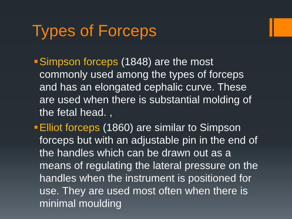
Simpson forceps (1848) are the most commonly used among the types of forceps and has an elongated cephalic curve. These are used when there is substantial molding of the fetal head. , Elliot forceps (1860) are similar to Simpson forceps but with an adjustable pin in the end of the handles which can be drawn out as a means of regulating the lateral pressure on the handles when the instrument is positioned for use. They are used most often when there is minimal moulding

Types of Forceps
Kielland forceps (1915, Norwegian) are distinguished by an extremely small pelvic curve and a sliding lock. The most common forceps used for rotation. The sliding lock is helpful in asynclitic Kielland forceps lack traction because they have almost no pelvic curve Wrigley's forceps are used in low or outlet delivery
and in cesarean section delivery where manual traction is proving difficult. The short length results in a lower chance of uterine rupture. Piper's forceps have a perineal curve to allow
application to the after-coming head in breech delivery.

Anatomy of the Forceps: Elliott and Simpson Forceps

Anatomy of the Forceps

Forceps Locks
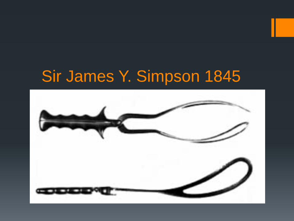
Sir James Y. Simpson 1845

Luikart-Simpson • Luikart R. A modification of the Kielland, Simpson, and Tucker-McLane forceps to simplify their use and improve function and safety. Am J Obstet Gynecol 1937;34:686
• pseudofenestrated blade

Elliot •Overlapping shanks
•Short, rounder cephalic curve
•Set screw between handles (reduce cephalic compression)
•For unmolded heads

Anatomy of the Forceps:Elliot Forceps with Adjustable Pin

Tucker-McLane 1880’s
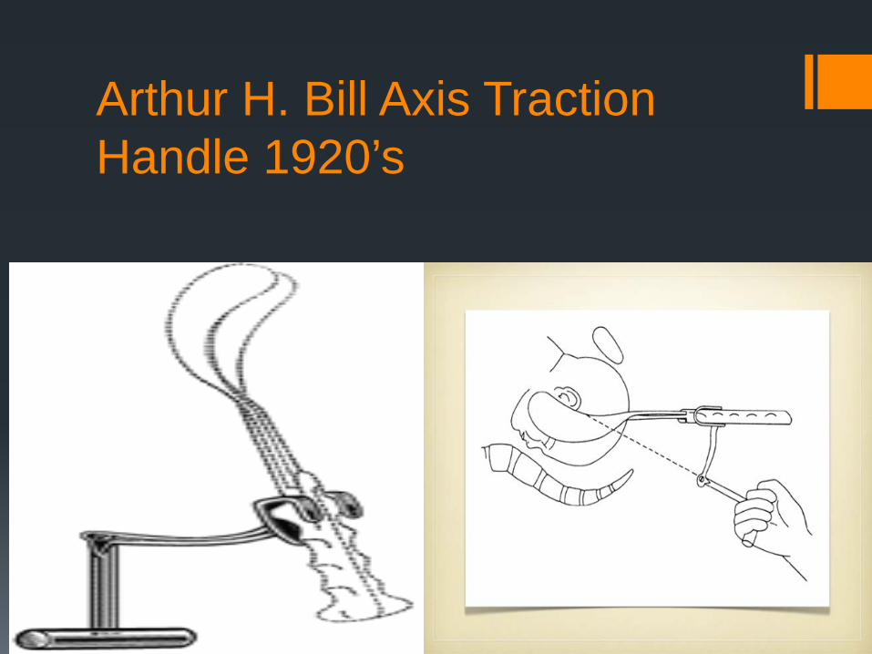
Arthur H. Bill Axis Traction Handle 1920’s

Christian Kielland 1915

Edmund B. Piper 1924
Long shanks of the forceps are curved backwards like a reverse pelvic curve This design drops the handles below the blades The unique construction of the shanks provide more spring to
the blades and results in less head compression

Operative Vaginal Delivery
Applying direct traction to the fetal skull (forceps) or the fetal scalp (vacuum) along with maternal expulsive efforts to effect a vaginal delivery Incidence estimated at 8-15% Fetal head must be engaged Membranes ruptured Cervix completely dilated Bladder empty of urine
Operative Vaginal Delivery
Indications for operative vaginal delivery Prolonged second stage of labor (nulliparous 3
hours with regional anesthesia or 2 hours without) multiparous (2 hours with regional anesthesia and 1 hour without regional anesthesia) Fetal compromise Shorten of the second stage of labor for maternal
indications

Prerequisites for Forceps Delivery The head should be engaged and the station of the
head accurately known The cervix should be completely dilated.
The exact position of the head should be known.
Occasionally ultrasound may help if the degree of molding creates confusion. The type of pelvis should be known. Certain pelvic
types will not allow for rotation. For example, a fetus in a posterior position in an android or anthropoid pelvis is best delivered in the occipitoposterior (OP) position.

Prerequisites for Forceps Delivery The operator should be familiar with the
advantages and disadvantages of the different forceps. There should be adequate anesthesia for the
forceps delivery contemplated. A low or outlet forceps delivery can be performed under pudendal block ; a forceps rotation of greater than 45° or a midforceps procedure requires a good epidural The bladder should be empty.
This is an operative procedure, and it should be
accorded the same respect and care for aseptic technique as any other operative procedure.

Contraindications for Operative Vaginal Delivery
Unengaged fetal head In ability to determine fetal position Malpresentation (face or brow) CPD actual or suspected Prematurity (< 34 weeks vacuum) Repeated scalp pH (vacuum) Inability to apply instrument correctly Incompletely dilated cervix

Position of the fetal head

Position of the Fetal Head

Outlet Forceps
Scalp is visible at the introitus without separating the labia Fetal skull has reached the pelvic floor Sagittal suture in A-P diameter, or Right or Left
anterior or posterior position Fetal head is at or near the perineum Rotation does not exceed 45 degrees ACOG

Outlet Forceps Lift out deliveries and at CS
•The fetal scalp is visible without separating the labia •The fetal skull has reached the pelvic floor •The sagittal suture is in the antero–posterior diameter or right
or left occiput anterior or posterior position (rotation does not exceed 45 degrees) •The fetal head is at, or on, the perineum RCOG
Wrigley's forceps

Low Forceps
Leading point of skull at or > +2 and not on pelvic floor Rotation is 45 degrees or less (R or L ant to OA or
R or L posterior to OP) Rotation is > 45% ACOG

Midforceps Stations is above +2 but the head is engaged High forceps are no longer included in the
classification
ACOG
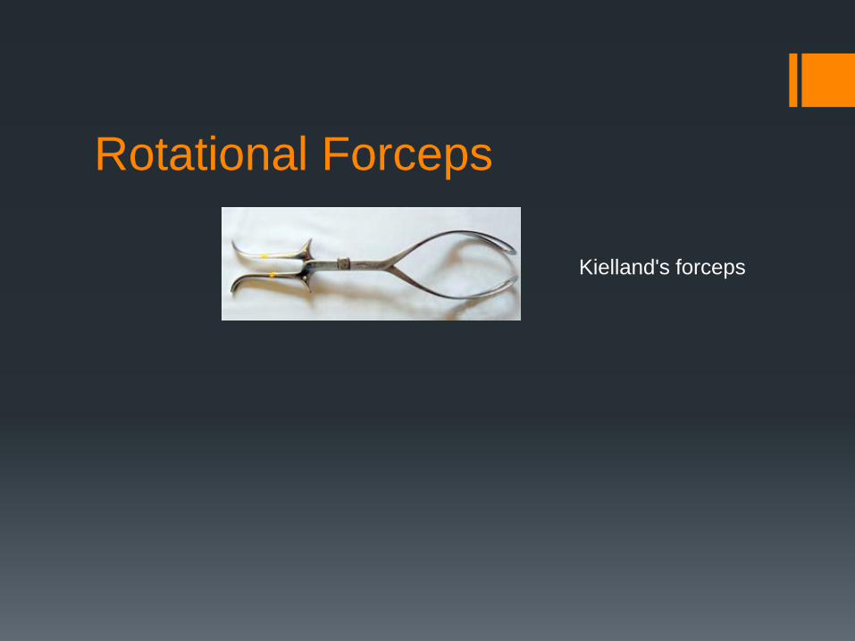
Rotational Forceps
Kielland's forceps

Forceps Delivery OA, LOA, ROA


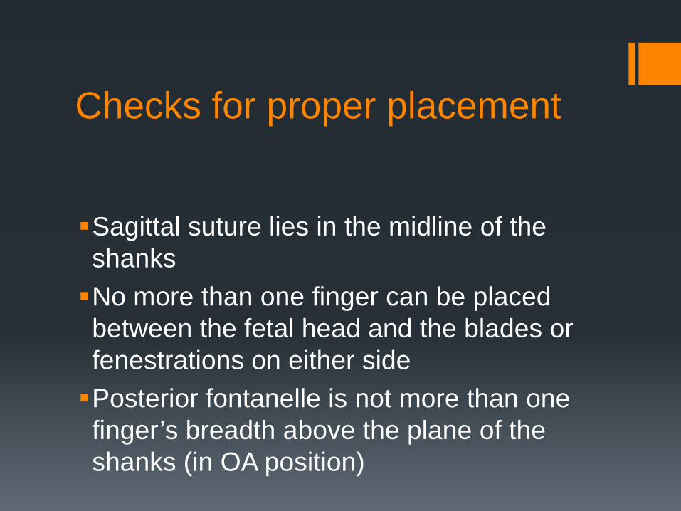
Checks for proper placement
Sagittal suture lies in the midline of the shanks No more than one finger can be placed between the fetal head and the blades or fenestrations on either side Posterior fontanelle is not more than one finger’s breadth above the plane of the shanks (in OA position)

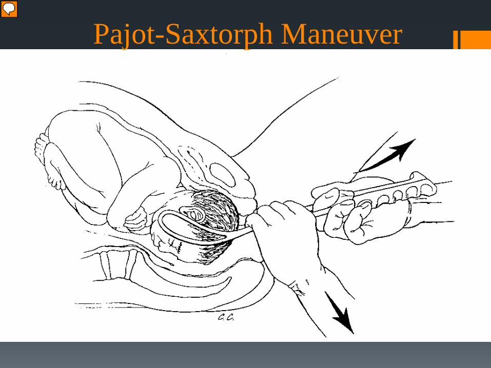
Pajot-Saxtorph Maneuver


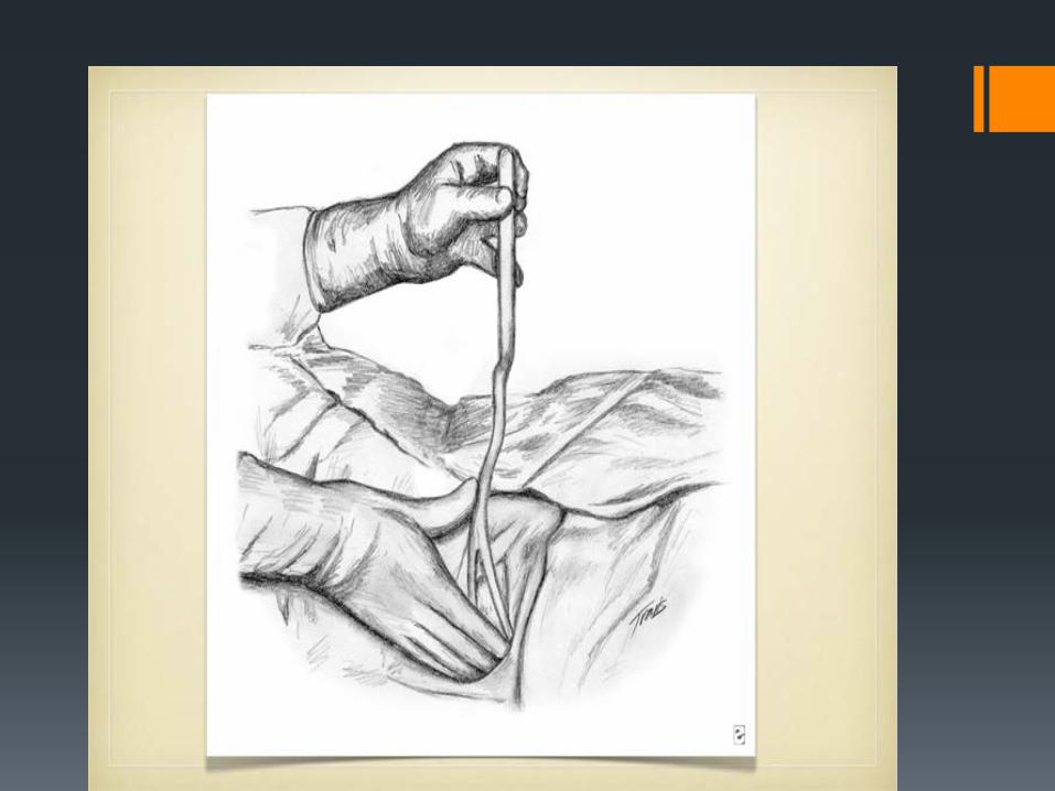
Rotation with Forceps

Recent Publication Obstet Gynecol 2013;121:1032-9 (May issue)


Rotational Forceps
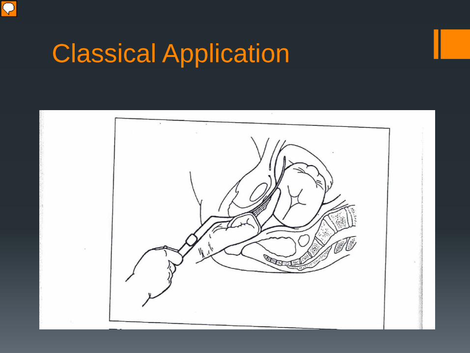
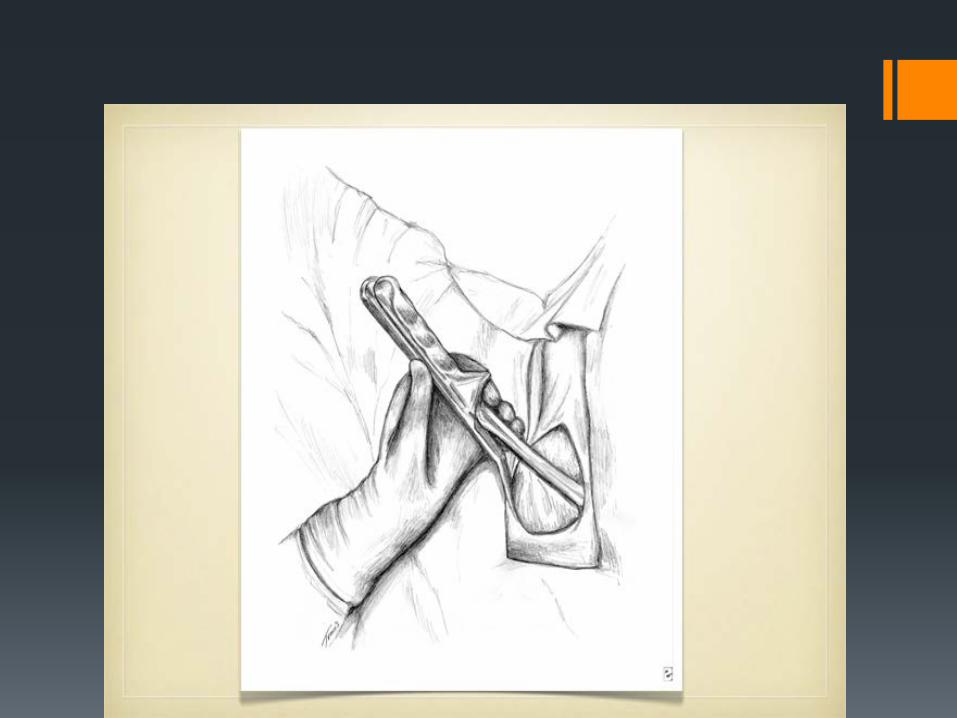
Classical Application

Classical Application

Classical Application

Classical Application
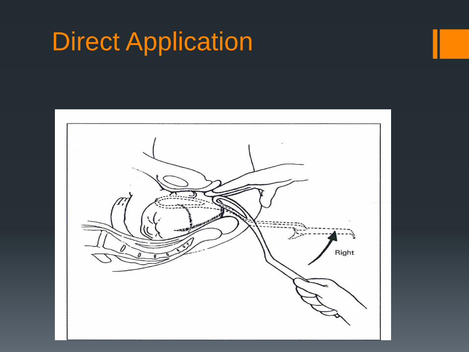
Direct Application
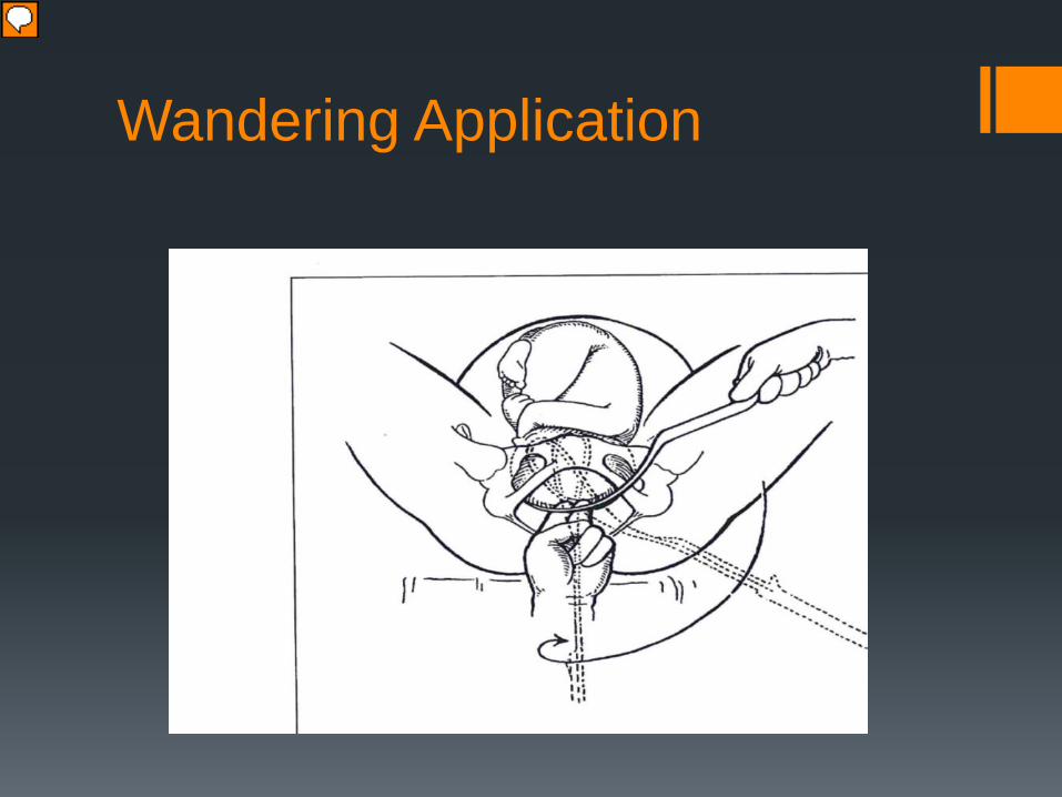
Wandering Application

Piper Forceps
Application to the after-coming head in a breech delivery Overall morbidity decreased by 50% with use of forceps Controls the flexion attitude of the head, avoids
hyperextension, no traction on the cervical spine or trunk Avoid delayed descent and possible hypoxia

Piper Forceps: Technique
Application: shoulder and arms delivered with head in pelvis Infants body is held horizontal by an assistant (towel sling) Operator assumes low sitting or kneeling position

Piper Forceps: Technique
Insertion from below and upward the handle is swept in a
downward arc toward the midline L blade inserted into L side of pelvis over infants R ear (Direct Application) Application of the blade guided by 2 fingers of the R hand

Piper Technique
The fetus is then swung towards the maternal L thigh and the R blade is inserted from below and upward, the R handle being swept in a downward arc toward the midline with the R blade inserted into R side of pelvis over infants L ear (Direct Application) Application of the blade guided by 2 fingers of the R hand If resistance is met, the toe of the blade is introduced more
posteriorly and wandered into place

Piper Forceps: Technique
After the shanks are locked the infant is straddling the forceps The handles rest in the upturned palm with middle finger in
the space between the shanks The neck is splinted by the fingers of the operators L hand

Piper Forceps: Technique
The fetus is then delivered over the perineum by flexion without removal of the forceps ( Note how the application of the forceps keeps the head flexed and prevents deflexion/extension of the fetal head Mauriceau Maneuver: index and middle finger are applied
over the maxiall to flex the fetal head while the body is elevated towards to abdomen

Mauriceau Maneuver
Index and middle finger are applied over the maxilla to flex the fetal head while the body is elevated towards to abdomen
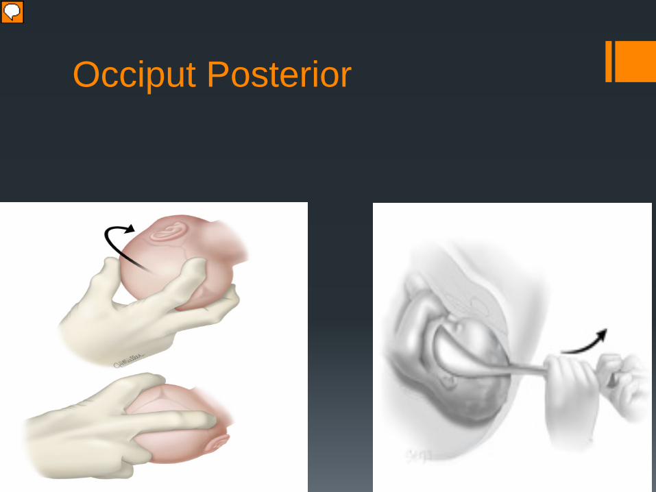
Occiput Posterior