open conformation of human dopa decarboxylase reveals the ... pnas.pdf · open conformation of...
TRANSCRIPT

Open conformation of human DOPA decarboxylasereveals the mechanism of PLP additionto Group II decarboxylasesGiorgio Giardinaa, Riccardo Montiolib, Stefano Giannic, Barbara Cellinib, Alessandro Paiardinia,Carla Borri Voltattornib,1, and Francesca Cutruzzolàa,d,1
aDipartimento di Scienze Biochimiche ‘A. Rossi Fanelli’, Sapienza Università di Roma, Piazzale Aldo Moro 5, 00185 Rome, Italy; bDipartimento di Scienzedella Vita e della Riproduzione, Sezione di Chimica Biologica, Facoltà di Medicina e Chirurgia, Università degli Studi di Verona, Strada Le Grazie, 8,37134 Verona, Italy; cIstituto di Biologia e Patologia Molecolari del Consiglio Nazionale delle Ricerche, Dipartimento di Scienze Biochimiche ‘A. RossiFanelli’, Sapienza Università di Rome, Piazzale Aldo Moro 5, 00185 Rome, Italy; and dIstituto Pasteur Fondazione Cenci Bolognetti, Dipartimento diScienze Biochimiche ‘A. Rossi Fanelli’, Sapienza Università di Roma, Piazzale Aldo Moro 5, 00185 Rome, Italy
Edited by Harry B. Gray, California Institute of Technology, Pasadena, CA, and approved October 3, 2011 (received for review July 14, 2011)
DOPA decarboxylase, the dimeric enzyme responsible for the synth-esis of neurotransmitters dopamine and serotonin, is involved insevere neurological diseases such as Parkinson disease, schizophre-nia, and depression. Binding of the pyridoxal-5′-phosphate (PLP) co-factor to the apoenzyme is thought to represent a centralmechanism for the regulation of its activity. We solved the structureof the human apoenzyme and found it exists in an unexpected openconformation: compared to the pig kidney holoenzyme, the dimersubunits move 20 Å apart and the two active sites become solventexposed. Moreover, by tuning the PLP concentration in the crystals,we obtained two more structures with different conformationsof the active site. Analysis of three-dimensional data coupled to akinetic study allows to identify the structural determinants of theopen/close conformational change occurring upon PLP bindingand thereby propose a model for the preferential degradation ofthe apoenzymes of Group II decarboxylases.
apoprotein ∣ Shiff base ∣ stability ∣ kinetics ∣ FRET
Dopamine, serotonin, GABA, and histamine are synthesizedby three structurally related pyridoxal-5′-phosphate (PLP)
dependent enzymes: 3,4-dihydroxy-L-phenylalanin (L-DOPA)decarboxylase (DDC), the glutamic acid decarboxylase (GAD),and the histidine decarboxylase (HDC). These enzymes belongto the Group II of Fold Type I aspartate aminotransferase family.Surprisingly, despite these three decarboxylases are involved insevere diseases [e.g., Parkinson’s disease, Tourette’s syndrome,schizophrenia, depression and cancer (1–5)], the regulation oftheir activity is still shrouded in mystery.
In the brain, where the three enzymes are highly expressed(6, 7), the equilibrium between apo- and holoenzymes is depen-dent on PLP availability (8–10). Binding of PLP to the apoen-zymes is thought to represent an essential mechanism of theirregulation (11) and altered ratio of holo- vs. apoenzymes, as wellas preferential degradation of the latter, have been implied ina number of diseases and disorders such as DDC deficiency(12–14). PLP crosses the blood brain barrier as its vitamin B6 pre-cursors, Pyridoxal (PL), Pyridoxine (PN), or Pyridoxamine (PM)and then is phosphorylated again by the pyridoxal kinase (PLK)(15, 16). Therefore vitamin B6 deficiency in the central nervoussystem (CNS) or inherited defects in the gene encoding PLK,are also responsible for the alteration in the equilibrium betweenapo- and holo-decarboxylases, and represent the cause of severalneurological pathologies [e.g., epilepsy, EEG abnormalities,psychiatric disorders (13, 17)]. However, there is still little under-standing of the mechanism of PLP binding to the apoenzymes. Ithas been suggested that, upon PLP binding, the three decarbox-ylases undergo a large conformational change, which leads to amore compact and less solvent accessible conformation (18–21).Nevertheless there are still many obscure points: (i) What is the
exact mechanism of PLP binding to Group II apo- decarboxy-lases? (ii) How large is the conformational difference betweenthe holo- and apo-forms? (iii) What is the reason behind the pre-ferential degradation of the latter? (iv) Does the apoenzyme bindto free PLP in vivo or does this happen through a protein-proteininteraction with PLK? To date, no clues to address these puzzlingissues have come from structural biology, because no experimen-tally determined apo-structure of the Group II enzymes had beenobtained so far.
Here we present the crystal structure of an apoenzyme ofGroup II decarboxylases; the human apoDDC. Based on struc-tural and kinetic data, we could identify the structural determi-nants of the conformational change occurring upon PLP bindingand propose a model for the preferential degradation of theapoenzyme. Our results force to reconsider the mechanism ofPLP addition to the apoenzymes of Group II decarboxylases ina unique perspective. Insights coming from this structural workmay thus prove fundamental in understanding the regulation ofthe homeostasis of this key class of enzymes in the CNS and in theperipheral tissues, and will hopefully help to develop new ther-apeutical approaches.
ResultsGroup II decarboxylases are homodimers with each monomercomposed of three domains (22). The first two, a large domain(L-domain) formed by a central seven-stranded β-sheet sur-rounded by α-helices and the C-terminal domain (small domainor C-domain), are typical of the aspartate aminotransferase fa-mily (Fold Type I). The third N-terminal domain (N-domain)is formed by three α-helices that fold upon dimerization and ischaracteristic of Group II decarboxylases (22, 23). In the holostructures the active site is buried in the central part of the dimerand accessible only through a tight gorge (18, 24, 25).
In an attempt to investigate, by X-ray crystallography, thestructural basis of PLP binding to human apoDDC, we setup anumber of crystallization trials using both the apoenzyme andthe holoenzyme, the latter in the presence of primary amines.We obtained crystals by mixing the holoDDC with Jeffamine®
Author Q:1contributions: G.G., C.B.V., and F.C. designed research; G.G., R.M., S.G., B.C., and F.C.performed research; R.M., B.C., and C.B.V. contributed new reagents/analytic tools; G.G.,S.G., A.P., C.B.V., and F.C. analyzed data; and G.G., S.G., A.P., C.B.V., and F.C. wrotethe paper.
The authors declare no conflict of interest.
This article is a PNAS Direct Submission.
Data deposition: The atomic coordinates have been deposited in the Protein Data Bank,www.pdb.org (PDB ID codes 3RBF; 3RCH; and 3RBL).1To whom correspondence may be addressed. E-mail: [email protected] [email protected].
This article contains supporting information online at www.pnas.org/lookup/suppl/doi:10.1073/pnas.1111456108/-/DCSupplemental.
Q:2
www.pnas.org/cgi/doi/10.1073/pnas.1111456108 PNAS Early Edition ∣ 1 of 6
BIOCH
EMISTR
Y

ED-2001 [O,O-Bis (2-aminopropyl) polypropylene glycol]. Thisprecipitant contains primary amino groups attached to the termi-nus of a polyether backbone. The exchange reaction led tothe release of the PLP cofactor bound to the holoprotein yieldingcrystals of the apoenzyme.
Human apoDDC Crystal Structure: an Open Conformation. Compari-son of the human apoDDC (apo-hDDC) with that of the closeorthologue pig kidney holoDDC [holo-pkDDC—89% sequenceidentity (25)] reveals the existence of unexpected conformationbecause the apo-dimer is completely open with the two largedomains moving by up to 20 Å with respect to the holo-enzymeas shown in Fig. 1. The open conformation of the enzyme isachieved through a rigid body quaternary rearrangement of thedimer because the fold of each domain is essentially unchanged inthe apo-hDDC. Accordingly, the comparison of near-UV CDspectra as well as of intrinsic and 1-anilinonaphtalene-sulfonicacid (ANS) fluorescence spectra of holo and apo-hDDC provideevidence for different conformations of the two forms (Fig. S1).Such an open form of the apoenzyme could hardly be predicted,because this dramatic conformational rearrangement results in acomplete exposure of the active site and hydrophobic regionsof the apoDDC. Surprisingly, the dimer interface of the openconformation comprises only the helices of the N-domain (bluein Fig. 1). These helices form an extended helix-bundle with thepartner subunit (Fig. S2, Movie S1), while the central part of theL-domain (yellow in Fig. 1) is completely exposed to the solvent.Overall, we can compare the apo-hDDC structure to an open bi-valve shell, with the interface between the N-domains functioningas the hinge. Given the reduced dimer interface, the open form ofthe enzyme may well be representative of a system of conforma-tions where the two monomers are substantially mobile in vivo.
An Asymmetric Structure: Trapping Different Conformations of theActive Site. The crystal form of the open hDDC obtained in
Jeffamine® proved to be an extremely powerful system to inves-tigate PLP binding, uncoupling local structural changes in the ac-tive site from quaternary conformational changes (i.e., open-closed transition). This uncoupling occurs because, within thecrystal lattice, the dimer is trapped in the open conformation byintermolecular crystal contacts (Fig. S3A). Moreover, the struc-ture of the two subunits is not symmetrical, with chain-A beingmore closed than chain-B (Fig. S3B).
Indeed, in the first structure that we solved (structure 1 inFig. 2), the PLP present in the crystallization solution (equimolarto the protein) is still able to bind to the active site, and electrondensity compatible with PLP was observed in one monomer(chain-A) and was included in the final model with occupancy of0.5 (Fig. 2). Excitingly, the structural asymmetry of the two sub-units is also reflected at the level of the active site, with chain-Aappearing able to better accommodate the cofactor than chain-B.This initial observation was confirmed by varying the PLPconcentration in the crystals of the open DDC, by soaking. Weobtained two more structures of the open enzyme with PLP
Fig. 1. The apo hDDC open conformation. Surface and cartoon representa-tion of the apo-hDDC structure in the open conformation. The domain orga-nization of one monomer is highlighted using different colors (Blue—N-domain; Yellow—Large domain; Red—C-domain) while the other mono-mer is shown in White. The structure of the holo-pkDDC (pdb id: 1js6) inthe closed conformation is shown for comparison in the same orientation;the three domains are colored differently for clarity (Purple—N-domain;Green—Large domain; Orange—C-domain)
Fig. 2. The asymmetric structure of the open hDDC dimer. The upper part ofthe figure schematically illustrates the two asymmetric monomers of theopen structures of hDDC (Red—chain-B, open conformation; Purple—chain-A, intermediate conformation; see also Fig. S3) and the PLP occupancyin the four alternative conformations observed for the active site in the threestructures of hDDC obtained before (structure 1) and after soaking (structure2 and 3). Each conformation is shown with different colors and ordered(from A to E) as illustrated in the scheme, going from the open to the closedmonomer (Blue—holo-pkDDC, pdb id: 1js6) and from 0 to 1 PLP occupancy.The lower part of the figure shows a stick representation of the side chains ofthe active site residues in the five alternative conformations, including theclosed conformation. The colors used in each box from A to E are the sameof the upper scheme. Throughout the boxes a dashed line indicates the posi-tion of the backbone residues of loop1: Red—apo state; Blue—holo state.
2 of 6 ∣ www.pnas.org/cgi/doi/10.1073/pnas.1111456108 Giardina et al.
bound to both monomers and with no PLP bound respectively(structures 2 and 3 in Fig. 2).
As a consequence of the conformational asymmetry, fromthe three structures that we solved we identified four alternativeconformations of the active site, as illustrated in Fig. 2: (Fig. 2A)no PLP (apo); (Fig. 2B) 0.5 occupancy of PLP in the open mono-mer (no internal aldimine formed yet); (Fig. 2C) 0.5 occupancyof PLP in the intermediate monomer (no internal aldimineformed yet) and; (Fig. 2D) PLP bound with 1.0 occupancy (inter-nal aldimine formed) to the intermediate monomer. These fourstructures of the active site together with that of the closed-holo-pkDDC, represent a set of five alternative conformations. Wehave ordered the five structures of the active site according to twoparameters: the increase of PLP occupancy and the openingdegree of the single monomer (going from Fig. 2 A to E). Thesestructural snapshots of the active site within the open dimerallowed us to highlight some important structural determinantsof the conformational rearrangement upon PLP binding (seeMovie S2 online, for a five step morphing from state A to E,and Fig. S4 for a stereo representation of the electron densityof the 4 different conformations).
The starting and the ending point of this set of conformationsare the open-apo state (chain-B—structure 1—Fig. 2A) and theclosed-holo state (Fig. 2E—pkDDC structure) respectively.Chain B of structure 1 shows a clear electron density for all theresidues in the active site. No density is observed for PLP and thusthis subunit may be regarded as a model for the apoprotein openactive site. Comparison with the holoprotein (pkDDC) showsthat all the major differences are related to the alternative con-formation adopted by the last seven residues of loop1 (residues66–84; connecting the N-domain to the L-domain) and especiallyby Tyr-79 and Phe-80 in the apo structure. In the apoprotein, Tyr-79 is making a hydrogen bond with His-302 that, as a result, isfound in a different conformation than in pkDDC. The side chainof the PLP-binding residue Lys-303 is shifted by 6 Å, swappingits position with that of the rotated side chain of Tyr-274. As aconsequence of these changes the favorable aromatic stackinginteraction between Trp-304, Tyr-274 and Phe-80, present inthe holoenzyme, is disrupted.
Between the open-apo and the closed-holo state, we havetrapped three more conformations of the active site (Fig. 2 B–D).In the second conformation (Fig. 2B—chain-B—structure 2) thesteric hindrance of the pyridine ring of PLP, entering in the activesite of the open monomer, forces the side chain of Tyr-274 toswap position with Lys-303, which becomes available for aldimineformation.
Moving to the intermediate monomer (Fig. 2C—chain-A—
structure 1) PLP is bound as in B but, as a consequence of themore closed conformation, the catalytic loop (residues 300 to310) is closer to the PLP molecule. His-302 is back to the positionobserved in the holo state interacting with the phosphate group,and Tyr-79 is released from the hydrogen bonding interaction.Two out of the three residues involved in the aromatic stackinginteraction (Trp-304 and Tyr-274) are positioned as in the holostate of pkDDC, while the third (Phe-80) is in the apo state. Loop1is still in the apo conformation but is now more mobile as reflectedby the increase of the temperature factors and by the reducedoccupancy of both Tyr-79 and Phe-80 side chain atoms.
The same monomer after soaking with PLP (Fig. 2D—chain-A—structure 2) shows a clear electron density for all PLP atomsand for the double bond of the Shiff base. The structure of theactive site is now almost identical to the holo state of pkDDC.Loop1 has completely rearranged to yield the holo state confor-mation and Phe-80 side chain is now forming the aromatic stack-ing interaction with Trp-304 and Tyr-274, like in the closedpkDDC structure. The only difference with the closed-holo stateis the position of the Tyr-79 side chain, which in the open struc-tures is solvent exposed and thus not well defined.
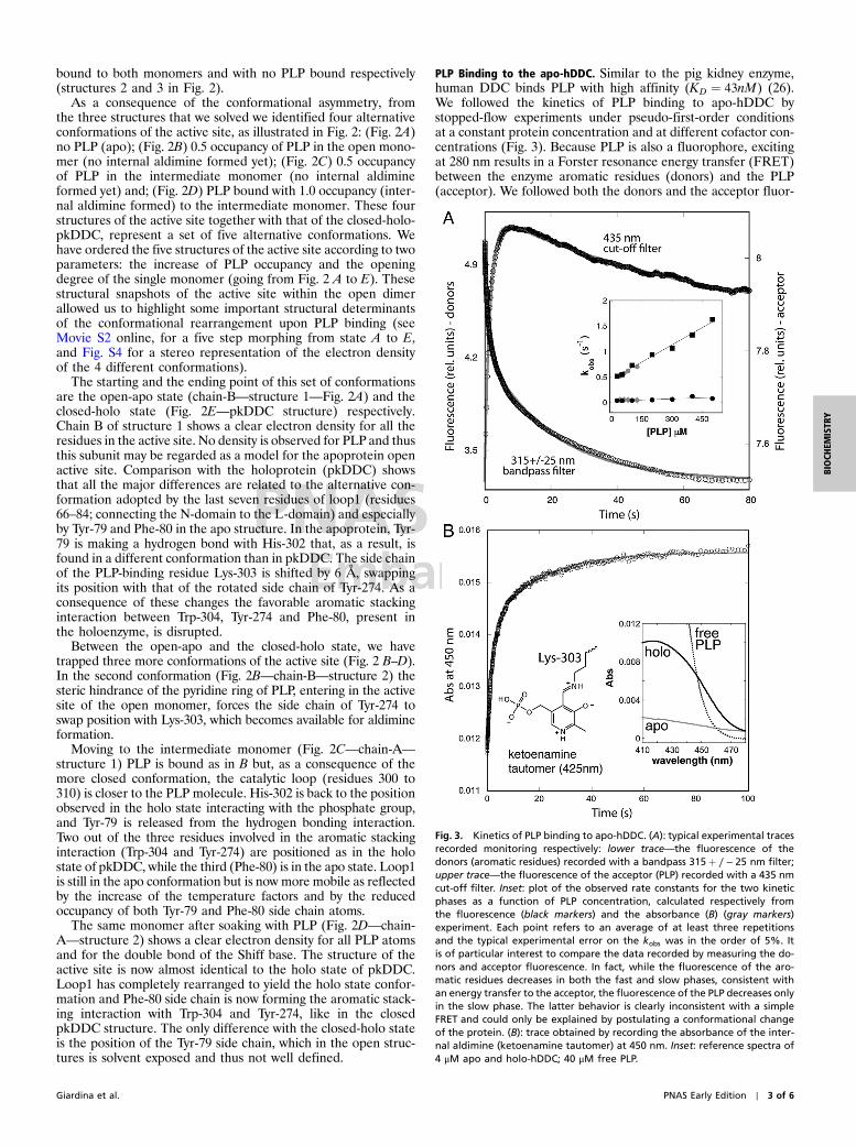
PLP Binding to the apo-hDDC. Similar to the pig kidney enzyme,human DDC binds PLP with high affinity (KD ¼ 43nM) (26).We followed the kinetics of PLP binding to apo-hDDC bystopped-flow experiments under pseudo-first-order conditionsat a constant protein concentration and at different cofactor con-centrations (Fig. 3). Because PLP is also a fluorophore, excitingat 280 nm results in a Forster resonance energy transfer (FRET)between the enzyme aromatic residues (donors) and the PLP(acceptor). We followed both the donors and the acceptor fluor-
Fig. 3. Kinetics of PLP binding to apo-hDDC. (A): typical experimental tracesrecorded monitoring respectively: lower trace—the fluorescence of thedonors (aromatic residues) recorded with a bandpass 315þ ∕ − 25 nm filter;upper trace—the fluorescence of the acceptor (PLP) recorded with a 435 nmcut-off filter. Inset: plot of the observed rate constants for the two kineticphases as a function of PLP concentration, calculated respectively fromthe fluorescence (black markers) and the absorbance (B) (gray markers)experiment. Each point refers to an average of at least three repetitionsand the typical experimental error on the kobs was in the order of 5%. Itis of particular interest to compare the data recorded by measuring the do-nors and acceptor fluorescence. In fact, while the fluorescence of the aro-matic residues decreases in both the fast and slow phases, consistent withan energy transfer to the acceptor, the fluorescence of the PLP decreases onlyin the slow phase. The latter behavior is clearly inconsistent with a simpleFRET and could only be explained by postulating a conformational changeof the protein. (B): trace obtained by recording the absorbance of the inter-nal aldimine (ketoenamine tautomer) at 450 nm. Inset: reference spectra of4 μM apo and holo-hDDC; 40 μM free PLP.
Giardina et al. PNAS Early Edition ∣ 3 of 6
BIOCH
EMISTR
Y

escence emission. In all cases, the observed kinetics displayed abiphasic behavior (Fig. 3A). Overall, whereas the rate constantfor the fast phase appears to increase linearly with increasingligand concentration (Fig. 3A inset, black squares), the rateconstant for the slow phase is essentially independent of PLP con-centration (black circles). This result indicates the presence of amonomolecular reaction following initial ligand recognition, con-sistent with a conformational change occurring after the initialbinding of PLP to the apoenzyme.
We next performed single wavelength stopped-flow experimentsrecording the absorbance of the sample at 450 nm (Fig. 3B). Underthese conditions, we monitored the formation and local environ-ment of the Schiff base between the PLP and the ϵ-nitrogen ofLys-303 of the apoenzyme. The PLP-Lys-303 Shiff base undergoesa keto-enol tautomeric equilibrium with the ketoenamine and theenolimine tautomers peaking at 425 and 335 nm, respectively. Theratio between the two species is affected by the chemical environ-ment and by pH (21, 27). In the absorbance experiment at 450 nm,where we only monitor the formation of the ketoenamine tauto-mer (28, 29), we observed the same two phases as in the FRETexperiments (the observed rate constants as a function of PLPconcentration are plotted in the inset of Fig. 3A—gray markers).This result indicates that the fast and slow kinetic phases cannotbe assigned to (i) a first encounter between PLP and the enzymefollowed by (ii) the formation of the Schiff base.
Overall, our kinetic analysis suggests the existence of a com-plex mechanism whereby cofactor binding and Schiff base forma-tion occur simultaneously in the fast phase, which depends onPLP concentration. These events are followed by a major confor-mational change, whose apparent kinetics does not depend oncofactor concentration, and where the local environment of PLPis altered, as detected by absorbance and emission properties ofthe complex.
DiscussionThe structure of apo-hDDC indicates that this protein, which hasbeen considered to date as a tightly associated dimer, is able toassume an open conformation. This evidence, while explainingdata indicating that a large conformational transition is occurringto Group II decarboxylases upon PLP binding (18–21), forces toreconsider the mechanism of cofactor addition as well as the dataavailable on the preferential degradation of the apoenzymes in anew perspective.
The crystals of the asymmetric dimer of the open DDC allowedus to take structural snapshots of the cofactor-free apoproteinand of three alternative conformation of the active site at differentPLP saturations (Fig. 2). In the fourth conformation (Fig. 2D:chain-A—structure 2) PLP is covalently bound through the Shiffbase and loop1 is in the holo conformation. This evidence indicatesthat complete achievement of the closed conformation of the di-mer is not essential for Shiff base formation and that PLP bindingto the intermediate monomer is able to induce rearrangement ofloop1. On the other hand, it is also evident that covalent binding ofthe cofactor can only be achieved after an initial rearrangementtowards the closed conformation, because, even at saturating PLPconcentration, no Shiff base is observed in the active site of theopen monomer (Fig. 2B: chain-B—structure 2).
Coupling the structural analysis with the kinetic experiments,our data strongly suggest that PLP binding to the open structure,which may exist in solution as a dynamic ensemble of conforma-tions, triggers an initial conformational change at the level of theactive site yielding the rearrangement of loop1 that favors thetransition to the closed conformation. Because loop1 in theclosed conformation is making direct interaction with loop2 ofthe partner subunit (residues 100′ to 110′) which in turn contactsthe flexible loop (also of the other subunit—loop3′), as illustratedin Fig. 4, Movie S3, we think that the conformational change ofloop1 is transmitted to loop2′ and then to loop3′.
Indeed both loop2 and 3 in the open conformation of hDDCare solvent exposed and appear to be mobile and therefore notvisible in the electron density of both subunits, while in the closedform of the enzyme they are buried and well structured (with theexception of 13 residues of the flexible loop). The hypothesis thatloop1 acts as a conformational relay and can be considered as akey structural element is also confirmed by the architecture of thesmall C-domain. The backbone of loop1, in fact, makes a doubleturn due to the presence of Pro-74. This conformation is furtherstabilized by an additional aromatic stacking interactions with re-sidues belonging to the C-domain (Fig. 4). The double turn allowsTrp-71, and other residues of loop1, to interact directly withloop2′ by forming an hydrophobic cluster comprising, amongother residues, Phe-103′ and Tyr-79 as shown in Fig. 4. Noticethat loop1, loop2 (in particular Phe-103), and the flexible loopall contain residues involved in substrate binding (25, 30, 31); in-dicating that the structural rearrangement described here and theenzyme catalytic activity are tightly interconnected. Furthermore,of the 23 point mutations of DDC identified in patients withvarious neurotransmitters disorders 16 are part of the structuraldeterminants involved in the conformational change that wepropose, or make direct interaction with them (Table S1). In
Fig. 4. Proximity relation of loop1 with loop2 and the flexible loop. The topbox is a cartoon representation of the closed-holo-pkDDC dimer (Pdb id: 1js6)illustrating the proximity relation of loop1 (Cyan) with loop2 (Gray) and theflexible loop (Black) of the partner monomer. The three domains are coloredas in Fig. 1. In this structure, loop1 (Cyan) directly interacts with loop2′ (Gray)which in turn contacts the flexible loop3′ (Black). The position of someimportant residues of loop1 in the holo state (i.e. Tyr-79 and Trp-71) andthe position of the two aromatic stacking interactions (Phe-80 with Tyr-274and Trp-304 of the large domain and of Phe-77 with Phe-388 and Phe-448from the C-domain) is also shown. Notice that in the structure of the open-apo-form of hDDC (bottom box), due to the separation of the two subunits,the residues of loop2 and of the flexible loop are not visible, and only thearomatic stacking interaction with the residues of the C-domain is conserved.
4 of 6 ∣ www.pnas.org/cgi/doi/10.1073/pnas.1111456108 Giardina et al.

particular, eight of these mutants belong to or contact loop1.Among them, the Y79C mutant appears particularly significantbecauseTyr-79, which switches position with Phe-80 when loop1changes conformation from the apo to the holoenzyme, is one ofthe key residues in our proposed mechanism.
This study allows us to speculate over a debated aspect ofthe mechanism of PLP addition to the apoenzyme. It has beenproposed that, in the cell, PLP is directly transferred from pyr-idoxal kinase (PLK) to the apoenzyme by a channeling mechan-ism (32) but, although direct interaction between PLK andapoGAD has been demonstrated (33), the only access to the ac-tive site in the closed pkDDC dimer is a small 7 Å wide positivelycharged gorge (Fig. S5). The open conformation described here istherefore the form of the enzyme most likely able to interact withPLK, because the active site in this conformation is solvent ex-posed and easily accessible (Fig. 1). In this sense it is temptingto speculate on the astonishing structural complementarity be-tween the PLK dimer and the huge cleft of the open-apo-hDDCstructure: in a putative complex between the two proteins the twoPLP molecules bound to PLK would face exactly the active sitesof the open DDC (Fig. S6).
Finally, our results shed light on another crucial issue: there isevidence that apoDDC and apoGAD are selectively degraded invivo. ApoDDC is degraded at least 20-fold faster than the holoen-zyme in rat brain cells (11), while it has been demonstrated thatholoGAD has a much higher stability than apoGAD (20). Indeedpreferential degradation by the ubiquitin-proteosome system(UPS) has been suggested for apoDDC and demonstrated forapo tyrosine aminotransferase (13, 32). On the basis of the holostructures available for these enzymes (25, 30) these data couldhardly be interpreted and most of the current hypothesis focuseson the mobility of the flexible loop. On the contrary, the existenceof the open structure of hDDC would allow to present a struc-tural interpretation of the lower stability of the apoenzymes invivo. Indeed it is accepted that ubiquitylation is often driven byconformational and/or by an order-disorder transition. In parti-cular, ubiquitin-ligases recognize unstructured zones of the targetprotein, or newly exposed surfaces. The opening of the apo-hDDC dimer implies that: (i) the solvent accessible surface areaincreases of 14%, (ii) the active site and a large part of the dimerinterface becomes exposed, (iii) three important loops becomemobile or unstructured and, (iv) the global enzyme volume andshape changes. Based on these observations, we propose a simplemechanism where the UPS regulates the levels of the total en-zyme in the CNS, based on PLP availability (Fig. 5). In this hy-pothesis, which will need further experimental evidence, theconcentration of PLP in the brain is directly affecting the ratiobetween holo and apoenzyme which, because of their differentconformation, may have different fates within the cell.
Materials and MethodsDDC Expression and Purification. Human DDC was heterologously expressedin Escherichia coli and purified as described in ref. 34. The apoenzyme wasprepared as described in ref. 34.
Crystallization and Data Collection. Single crystals of hDDC were grown at21 °C by hanging drop vapor diffusion method; 1 μL of protein solution(holo-hDDC 5 mg∕mL in 50 mM Hepes pH 7.4 buffer) was mixed with 1 μLof reservoir (500 μL containing 0.1 M Hepes pH 7.0 and 28–30% JeffamineED 2001 pH 7.0). Crystal grew in 5–8 d. No cryoprotectant was used beforeflash freezing in liquid nitrogen. Datawere collected to 2.9 Å resolution. Crys-tals belong to the tetragonal space group P41212. Data were integrated/scaled usingMOSFLM (35) and SCALA (36). The asymmetric unit (AU) containstwo monomers (one noncrystallographic dimer) with a solvent contentof 54%.
Crystals of the open hDDC with 1.5 equivalents of PLP bound (structure-2,Fig. 2) and no PLP bound (structure-3, Fig. 2) were obtained by soaking theprevious described crystals in a solution containing 0.1 M Hepes pH 7.0; 15%Jeffamine ED 2001 pH 7.0; 5 mM PLP for 72 h and in a 2 μL droplet of reservoirfor 24 h respectively. Diffraction data were collected up to 2.8 Å and 3.2 Å forthe +PLP and −PLP crystals respectively. See Table S2 for complete data col-lection statistics.
Phasing and Refinement. Phases were obtained by Molecular Replacementmethod with MOLREP (37) using one monomer of the holo-pkDDC as searchmodel (PDB id: 1js6).
Building and refinement was iteratively carried out using COOT (38) andREFMAC5 (39). Nonvisible side chain atoms were included in the final modelwith zero occupancy only for residues with visible main chain atoms, other-wise the entire residue was omitted. The geometrical quality of the threemodels was assessed using PROCHECK (40) and MolProbity (41). Final statis-tics are reported in Table S2.
PLP Binding to apo-hDDC. The time evolution of binding to PLP was followedon a stopped-flow apparatus by monitoring either the absorbance at 450 nmor the fluorescence emission (exciting the sample at 280 nm and monitoringemission using either a 315� 25 nm band pass or a 435 nm cut-off filter).Enzyme concentration (monomer) after mixing was 4 μM and 1 μM respec-tively for the single wavelength and the fluorescence experiment. Measure-ments were performed on an SX18-MV stopped-flow instrument (AppliedPhotophysics) using symmetric mixing. The traces were recorded in100 mM Hepes buffer at pH 7.4, at 25 °C.
Kinetic traces were extracted and analyzed using Kaleidagraph softwarepackage.
ACKNOWLEDGMENTS. We thank European Synchrotron Radiation Facility(ESRF) and Berliner Elektronenspeicherring-Gesellschaft für Synchrotron-strahlung (BESSY) staff for beam time allocation and for technical assistance.Maurizio Brunori, Anna Tramontano and Roberto Contestabile (Rome, IT) arealso gratefully acknowledged for critical reading of the manuscript. Fundsfrom the Ministero della Università of Italy [RBRN07BMCT, 20094BJ9R7_001] and from the University of Rome La Sapienza to F.C. are gratefullyacknowledged. This study was also supported by funds from MIUR (Prin2007) to C.B.V.
1. Lanoue AC, Dumitriu A, Myers RH, Soghomonian JJ (2010) Decreased glutamic acid
decarboxylasemRNA expression in prefrontal cortex in Parkinson’s disease. Exp Neurol
226:207–217.
2. Kontos CK, Papadopoulos IN, Fragoulis EG, Scorilas A (2010) Quantitative expression
analysis and prognostic significance of L-DOPA decarboxylase in colorectal adenocar-
cinoma. Br J Cancer 102:1384–1390.
Fig. 5. Proposed regulatory model of DDC levels in the brain: Only Pyridoxal(PL), Pyridoxine (PN), or Pyridoxamine (PM) can cross the blood brain barrier,mostly at the choroid plexi. PLP is first cleaved to pyridoxal by nonspecificmembrane-associated alkaline phosphatases. Once in the brain cell, B6
vitamers are converted again in PLP by PL kinase, and directly or indirectlytransferred to apoenzymes. PLP transfer may be facilitated by the open con-formation of the apoenzyme presented in the present paper. The active holo-form is responsible for the synthesis of biogenic amines, while the apo- openconformation, which exposes a wider protein surface and has more unstruc-tured regions (loop2 and the flexible loop), is recognized by ubiquitin ligasesand preferentially degraded by the ubiquitin proteasome system (UPS).
Q:3
Giardina et al. PNAS Early Edition ∣ 5 of 6
BIOCH
EMISTR
Y

3. Karolewicz B, et al. (2010) Reduced level of glutamic acid decarboxylase-67 kDa in theprefrontal cortex in major depression. Int J Neuropsychoph 13:411–420.
4. Ercan-Sencicek AG, et al. (2010) L-histidine decarboxylase and Tourette’s syndrome.N Engl J Med 362:1901–1908.
5. Abdel-Salam OM (2008) Drugs used to treat Parkinson’s disease, present status andfuture directions. CNS Neurol Disord-Dr 7:321–342.
6. Blechingberg J, Holm IE, Johansen MG, Borglum AD, Nielsen AL (2010) Aromaticl-amino acid decarboxylase expression profiling and isoform detection in the devel-oping porcine brain. Brain Res 1308:1–13.
7. Kitahama K, et al. (2009) Aromatic L-amino acid decarboxylase-immunoreactive struc-tures in human midbrain, pons, and medulla. J Chem Neuroanat 38:130–140.
8. Guilarte TR, Wagner HNJ, Frost JJ (1987) Effects of perinatal vitamin B6 deficiency ondopaminergic neurochemistry. J Neurochem 48:432–439.
9. Siow YL, Dakshinamurti K (1985) Effect of pyridoxine deficiency on aromatic L-aminoacid decarboxylase in adult rat brain. Exp Brain Res 59:575–581.
10. Rahman MK, et al. (1982) Effect of pyridoxal phosphate deficiency on aromatic L-ami-no acid decarboxylase activity with L-DOPA and L-5-hydroxytryptophan as substratesin rats. Jpn J Pharmacol 32:803–811.
11. Matsuda N, Hayashi H, Miyatake S, Kuroiwa T, Kagamiyama H (2004) Instability of theapo form of aromatic L-amino acid decarboxylase in vivo and in vitro: implications forthe involvement of the flexible loop that covers the active site. J Biochem 135:33–42.
12. Allen GF, et al. (2010) Pyridoxal 5′-phosphate deficiency causes a loss of aromaticL-amino acid decarboxylase in patients and human neuroblastoma cells, implicationsfor aromatic L-amino acid decarboxylase and vitamin B(6) deficiency states. J Neuro-chem 114:87–96.
13. Allen GF, Land JM, Heales SJ (2009) A new perspective on the treatment of aromaticL-amino acid decarboxylase deficiency. Mol Genet Metab 97:6–14.
14. MyersMA, et al. (2003) A diabetes-related epitope of GAD65: a major diabetes-relatedconformational epitope on GAD65. Ann N Y Acad Sci 1005:250–252.
15. di SalvoML, Contestabile R, SafoMK (2011) Vitamin B(6) salvage enzymes: Mechanism,structure and regulation. Biochim Biophys Acta 1814:1597–1608.
16. Li MH, et al. (2002) Crystal structure of brain pyridoxal kinase, a novel member of theribokinase superfamily. J Biol Chem 277:46385–46390.
17. Clayton PT (2006) B6-responsive disorders: a model of vitamin dependency. J InheritMetab Dis 29:317–326.
18. Moya-Garcia AA, Medina MA, Sanchez-Jimenez F (2005) Mammalian histidine decar-boxylase: from structure to function. Bioessays 27:57–63.
19. Rodriguez-Caso C, et al. (2003) Local changes in the catalytic site of mammalianhistidine decarboxylase can affect its global conformation and stability. Eur J Biochem270:4376–4387.
20. Chen CH, Wu SJ, Martin DL (1998) Structural characteristics of brain glutamate dec-arboxylase in relation to its interaction and activation. Arch Biochem Biophys349:175–182.
21. Moore PS, Dominici P, Borri Voltattorni C (1996) Cloning and expression of pig kidneydopa decarboxylase: comparison of the naturally occurring and recombinant enzymes.Biochem J 315:249–256.
22. Schneider G, Kack H, Lindqvist Y (2000) The manifold of vitamin B6 dependentenzymes. Structure 8:R1–6.
23. Sandmeier E, Hale TI, Christen P (1994) Multiple evolutionary origin of pyridoxal-5′-phosphate-dependent amino acid decarboxylases. Eur J Biochem 221:997–1002.
24. Fenalti G, et al. (2007) Molecular characterization of a disease associated conforma-tional epitope on GAD65 recognized by a human monoclonal antibody b96.11. MolImmunol 44:1178–1189.
25. Burkhard P, Dominici P, Borri-Voltattorni C, Jansonius JN, Malashkevich VN (2001)Structural insight into Parkinson’s disease treatment from drug-inhibited DOPAdecarboxylase. Nat Struct Biol 8:963–967.
26. Dominici P, Moore PS, Castellani S, Bertoldi M, Voltattorni CB (1997) Mutation ofcysteine 111 in Dopa decarboxylase leads to active site perturbation. Protein Sci6:2007–2015.
27. Zhou X, Toney MD (1999) pH studies on the mechanism of the pyridoxal phosphate-dependent dialkylglycine decarboxylase. Biochemistry 38:311–320.
28. Hill MP, Carroll EC, Toney MD, Larsen DS (2008) Rapid photodynamics of vitamin B6coenzyme pyridoxal 5′-phosphate and its Schiff bases in solution. J Phys Chem B112:5867–5873.
29. Malerba F, Bellelli A, Giorgi A, Bossa F, Contestabile R (2007) The mechanism of addi-tion of pyridoxal 5′-phosphate to Escherichia coli apo-serine hydroxymethyltransfer-ase. Biochem J 404:477–485.
30. Fenalti G, et al. (2007) GABA production by glutamic acid decarboxylase is regulatedby a dynamic catalytic loop. Nat Struct Mol Biol 14:280–286.
31. Bertoldi M, Gonsalvi M, Contestabile R, Voltattorni CB (2002) Mutation of tyrosine332 to phenylalanine converts dopa decarboxylase into a decarboxylation-dependentoxidative deaminase. J Biol Chem 277:36357–36362.
32. Kim YT, Kwok F, Churchich JE (1988) Interactions of pyridoxal kinase and aspartateaminotransferase emission anisotropy and compartmentation studies. J Biol Chem263:13712–13717.
33. Cheung PY, et al. (2003) Interaction between pyridoxal kinase and pyridoxal-5-phos-phate-dependent enzymes. J Biochem 134:731–738.
34. Montioli R, Cellini B, Borri Voltattorni C (2011) Molecular insights into the pathogeni-city of variants associated with the aromatic amino acid decarboxylase deficiency.J Inherit Metab Dis, Epub ahead of print. (doi: 10.1007/s10545-011-9340-6).
35. Leslie AGW (1992) Mosflm. Joint CCP4 and ESF-EACBM newsletter on protein Crystal-lography 26:27–33.
36. Evans PR (1997) Scala. Joint CCP4 and ESF-EACBM newsletter on protein Crystallogra-phy 33:22–24.
37. Vagin A, Teplyakov A (2010) Molecular replacement with MOLREP. Acta Crystallogr D66:22–25.
38. Emsley P, Cowtan K (2004) COOT: model-building tools for molecular graphics. ActaCrystallogr D 60:2126–2132.
39. Murshudov GN, Vagin AA, Dodson EJ (1997) Refinement of macromolecular structuresby the maximum-likelihood method. Acta Crystallogr D 53:240–255.
40. Laskowski RA, MacArthurMW,Moss DS, Thornton JM (1993) PROCHECK: a program tocheck the stereochemical quality of protein structures. J Appl Crystallogr 26:283–291.
41. Davis IW, et al. (2007) MolProbity: all-atom contacts and structure validation for pro-teins and nucleic acids. Nucleic Acids Res 35:W375–83.
6 of 6 ∣ www.pnas.org/cgi/doi/10.1073/pnas.1111456108 Giardina et al.

Supporting InformationGiardina et al. 10.1073/pnas.1111456108
Fig. S1. Near-UV CD spectra, intrinsic and ANS fluorescence spectra of holo and apo-hDDC. (A). Near-UV and Vis CD spectra of 5 μM holo (Black) and apoen-zyme (Green). (B). Fluorescence emission spectra of 1 μM holo (Green) and apoenzyme (Blue). Excitation at 280 nm (C). Fluorescence emission spectra of 1 μMholo (−) and apoenzyme (- - -) incubated in the presence of 15 μM 8-Anilino-1-naphthalenesulfonate (ANS) at 25 °C for 1 h, registered upon excitation at365 nm. All measurement were made in 0.1M Potassium Phosphate buffer pH ¼ 7.4.
Giardina et al. www.pnas.org/cgi/doi/10.1073/pnas.1111456108 1 of 7

Fig. S2. Structural organization of the N-terminal hinge (the dimer interface). Organization of the 12 helices cluster that arises from the domain swappingof the N-terminal domain which is peculiar of the Group II decarboxylases. These helices are organized as an extended four helix bundle network and buildthe dimer interface; i.e., the hinge of the bivalve shell (see Fig. 1). Color code: Blue—N-terminal domain; Yellow—Large domain; Red—Small domain; residuesthat are not part of the interface were removed for clarity. The capital letters used after the helix number denote the domain to which each helix belongs(i.e., α-1N—N-terminal, α-1L—Large, α-1C—C-terminal). The lower part of the figure shows a helical-wheel diagram and a cartoon representation of the centralfour helix bundle (α-1N − α-1L + α-1N’ − α-1L’) illustrating the strong hydrophobic interaction of the core and the network of hydrogen bonds of the surfaceresidues.
Giardina et al. www.pnas.org/cgi/doi/10.1073/pnas.1111456108 2 of 7

Fig. S3. Intermolecular crystal contacts and asymmetry of the two dimer subunits. (A). Tetrameric assembly of hDDC in the crystal lattice. Each dimer in theopen conformation bites a symmetry related mate, corresponding to the equivalent position y, x, -z of an adjacent cell. The space group is P41212. (B). Top—Superposition of the two monomers of hDDC on the N-domain helices (Red: chain-B; Purple: chain-A) reveals that the structure of the two subunits is notsymmetrical, with chain-A being more closed than chain-B. The monomers are shown in the same orientation as in (A). Bottom—superposition, on the N-domain helices, of the open monomer (chain-B) of hDDC (Red) with the pkDDC (pdb id: 1JS6) closed monomer (Blue). Open, Intermediate and Closed refer tothe different conformations of the monomer as illustrated in Fig. 2.
Giardina et al. www.pnas.org/cgi/doi/10.1073/pnas.1111456108 3 of 7

Fig. S4. Electron density of the alternative conformations of the active site. On the left: Fo-Fc omit map calculated for PLP and contoured at 2.0σ. On the right:Stereo representation of the 2Fo-Fc map contoured at 1.3σ.
Giardina et al. www.pnas.org/cgi/doi/10.1073/pnas.1111456108 4 of 7

Fig. S5. Access to the active site in the closed pkDDC dimer. Surface representation colored by electrostatic potential of the active site access in the closedconformation (pkDDC—pdb id: 1js6). Only the phosphate group of the cofactor is visible inside the 7 Å wide gorge.
Fig. S6. Structural complementarity between the PL Kinase (PLK) dimer and the open apo-hDDC. On the left, a top view of the open apo-hDDC dimer showingthe position of the exposed active sites (magenta); on the right, top and bottom views of the human PLK dimer (pdb id: 3keu). The transparency in the top viewillustrates the position of the PLP and the direction of the access channels to the substrate binding site, which are visible in the bottom view. In a possiblecomplex between the two proteins the two PLP molecules bound to PLK would face exactly the active sites of DDC.
Movie S1. Domain organization of the apo hDDC dimer. The first part of the animation shows the Tertiary and Quaternary structure of the apo-hDDC dimer inthe open conformation. The second part illustrates the organization of the dimer’s hinge; the domain swapping of the N-domains.
Movie S1 (MOV)
Giardina et al. www.pnas.org/cgi/doi/10.1073/pnas.1111456108 5 of 7

Movie S2. Five step morphing through the alternative conformations of the active site of DDC. The animations is a five step morphing through the differentconformations of the active site of DDC as ordered in Fig. 2. Letters corresponding to the five different states appear (top left corner) each time the morphingcorresponds to a crystal structure. Notice that morphing is a simple animation from the starting coordinates to the ending coordinates. No kinetic, thermo-dynamic or molecular dynamic calculation is employed.
Movie S2 (MOV)
Movie S3. Morphing through the different monomer conformations of DDC. The animation shows the morphing from an hypothetical symmetric dimer withboth monomers in the open conformation (Red) to the closed conformation (Blue—pkDDC structure), passing through the intermediate conformation (Pur-ple). The second part of the movie focuses on the conformational transition of loop1, after PLP binding, followed by folding of the mobile loop2 and loop3residues, which are structured only in the closed conformation. Notice that during the animation, for technical reasons, PLP is not morphing together with theprotein and its position, in the starting and ending points, is shown only for clarity. Notice that morphing is a simple animation from the starting coordinates tothe ending coordinates. No kinetic, thermodynamic or molecular dynamic calculation is employed.
Movie S3 (MOV)
Giardina et al. www.pnas.org/cgi/doi/10.1073/pnas.1111456108 6 of 7

Table S1. List of 23 point mutation of DDC identified in patients with various neurotransmitters disorders. The mutationsare taken from BIOMDB database (http://www.biopku.org/BioPKU_databasesBIOMDB.asp). Mutants which are part ormake direct interaction with the structural determinants involved in the conformational change discussed in the paperare highlighted. A brief description of their role or nature of the interaction is also provided
Mutation Position Interaction Type of interaction (conformation)
Y79C Loop1 swapping position with F80 going from the apo to the holo stateH72Y Loop1 adjacent to W71 (see Fig. 4)T69M Loop1 same as aboveH70T Loop1 same as aboveR462P Loop1 H-bond with Y75 (open)R447H Loop1 H-bond with the C ¼ O of Y79 (closed)E25K Loop1 interacting with Y75 (closed)L408I Loop1 interacting with F80 (closed)G102S Loop2 involved in substrate bindingA110Q Loop2F309L Loop2 Loop3 involved in substrate binding; contacts both Loop2 and
Loop3 residues in the closed structure; it is mobile in the open structureR347Q Loop3 involved in substrate bindingR358H Loop3 H-bond with D310 of the catalytic Loop (open)R7X N-ter. Hinge (α-1N) H-bond with N19 and E22 (see Fig. 2)A91V N-ter. Hinge (α-1L) hydrophobic core of the central 4-helix bundleL38P N-ter. Hinge (α-2N) disruption of α-2NP47HS147RS250FA275TR285WV460GR412W
Table S2. Data collection and refinement statistics for structure 1–3 (see Fig. 2)
Structure 1 Structure 2 Structure 3
Coordinates 3RBF 3RCH 3RBLData collection *Beamline BESSY (BL-1) ESRF (ID14-1) ESRF (ID14-1)Space group P41212 P41212 P41212Cell dimensionsa ¼ b;c (Å) 175.84, 74.96 177.00, 74.83 179.64, 74.95Resolution (Å) 2.90 (3.06–2.90) 2.80 (2.95–2.80) 3.25 (3.40–3.25)Rsym or Rmerge 11.2 (47.9) 13.3 (61.2) 13.1 (40.1)I∕σI 15.7 (3.6) 13.6 (3.5) 13.8 (3.8)Completeness (%) 99.3 (99.3) 100 (100) 99.8 (100)Redundancy 8.8 (6.0) 7.1 (7.2) 6.4 (6.2)RefinementResolution (Å) 45.0–2.9 30.0–2.8 30.0–3.25No. reflections 26,369 28,290 18,961Rwork∕Rfree 21.2∕26.6 20.1∕25.9 22.1∕28.3No. atomsProtein (chain A/B) 3;474∕3;480 3;504∕3;461 3;469∕3;480PLP (chain A/B) 16∕− 15∕16Ion 2 1Water 28Mean B-factor (Å2)Protein (chain A/B) 18.1∕18.2 25.9∕26.2 48.0∕45.3PLP (chain A/B) 35.2∕− 34.8∕34.9Ion 40.1 56.4Water 16.8rmsdBond lengths (Å) 0.011 0.011 0.011Bond angles (°) 1.263 1.318 1.301Ramachandran: n. residues (%)favored 710 (90.7) 699 (89.7) 721 (92.2)allowed 71 (9.1) 78 (10.0) 60 (7.7)disallowed 2 (0.3) 2 (0.3) 1 (0.1)Molprobity scores (percentile) 2.62 (89th) 2.94 (69th) 2.83 (91st)
*Values in parentheses are for highest-resolution shell.
Giardina et al. www.pnas.org/cgi/doi/10.1073/pnas.1111456108 7 of 7

AUTHOR QUERIES
AUTHOR PLEASE ANSWER ALL QUERIES 1
Q: 1_Please review the information in the author contribution footnote carefully. Please makesure that the information is correct and that the correct author initials are listed. Note thatthe order of author initials matches the order of the author line per journal style. You mayadd contributions to the list in the footnote; however, funding should not be an author'sonly contribution to the work.
Q: 2_PNAS does not allow statements of novelty or priority. Please approve the edit from “new”to “unique.”
Q: 3_The prefix “non” is only hyphenated when before a proper noun, an animal, or a protein orantibody and when part of a multiple-word term.