one-dimensional assembly and selective orientation of lander molecules on an o–cu template
TRANSCRIPT

AngewandteChemie
Communications
The growth of individual molecular building blocks can be carefullycontrolled by small modifications to a template surface. In theCommunication by F. Besenbacher and co-workers on the followingpages, the width of the lines of a Cu–Cu/O supergrating are shown todetermine the orientation in which molecules adsorb to the surface,and thus steer their alignment along the template.
2091Angew. Chem. Int. Ed. 2004, 43, 2091 DOI: 10.1002/anie.200353586 � 2004 Wiley-VCH Verlag GmbH & Co. KGaA, Weinheim

Nanotechnology
One-Dimensional Assembly and SelectiveOrientation of Lander Molecules on an O–CuTemplate**
Roberto Otero, Yoshitaka Naitoh, Federico Rosei,Ping Jiang, Peter Thostrup, Andr� Gourdon,Erik Lægsgaard, Ivan Stensgaard, Christian Joachim,and Flemming Besenbacher*
Properly functionalized organic molecules are promisingbuilding blocks for nanoscale electronic circuits.[1] A majorchallenge is to develop a novel technology to assemble suchmolecular elements in a planar conformation into a prede-termined architecture with atomic-scale precision.[2] Thisrequires the ability to create an ordered pattern for themolecular adsorption sites and to steer the adsorptionorientation of the molecules into a geometry that willenable the interconnection of the molecules in a circuit.Here we demonstrate that the striped periodic supergratingcreated by a controlled oxidation of a Cu(110) surface[3] is asuitable nanoscale template for the assembly of individualmolecular building blocks, such as the so-called “Landermolecules”, into well-ordered arrays of long molecular chains.Furthermore, we show that by controlling the width of thenanotemplate we can select the adsorption orientation of themolecules, and thereby steer their alignment along thespecific direction of the template.
The supergrating is created by exposing a clean Cu(110)surface to 4–6 Langmuir (1 L= 10�6 Torr s) of oxygen at625 K. This procedure leads to a self-organized reconstructionwith alternating one-dimensional (1D) bare Cu stripes and
(2 5 1)-O reconstructed regions, consisting of Cu–O addedrows, both aligned along the [001] surface direction (Fig-ure 1a).[3] By controlling the oxidation process, it is possible toadjust the dimensions of the supergrating. For instance,
increasing the temperature while keeping the exposureconstant leads to an increase in the distance between thestripes, which also become broader, whereas increasing theexposure at the same temperature leads to narrower stripes.The finest nanopattern consists of bare Cu troughs 2.0�0.3 nm wide, separated by Cu–O regions 5� 2 nm wide (seeFigure 1b). The Cu–O template exhibits long-range order onthe length scale of several hundred nanometers.
Onto this alternating Cu–O/Cu template, we have depos-ited “Single Lander”[4] molecules (SL, C90H98). The SLs arecomposed of a polyaromatic hydrocarbon central board withfour lateral 3,5-di-tert-butylphenyl substituents acting as
Figure 1. a) 70 � 70 nm2 STM image of the Cu–O/Cu periodic nanopat-tern resulting from O2 exposure. A certain fraction of the derivedimage has been added to enhance the resolution; b) 14 � 14 nm2 STMimage of a partially oxidized Cu(110) surface recorded at room temper-ature. 2 � 1 reconstructed oxidized areas can clearly be distinguished,separated by bare Cu stripes running along the [001] direction. Theimage is rotated so that the [001] direction is vertical. The originalscan direction can be easily recognized as the direction of the spikenoise originated from the fast diffusion of kink atoms in the Cu–Oedges at room temperature.
[*] Dr. R. Otero, Dr. Y. Naitoh, Prof. Dr. F. Rosei,+ P. Thostrup,Dr. E. Lægsgaard, Prof. Dr. I. Stensgaard, Prof. Dr. F. BesenbacherInterdisciplinary Nanoscience Center (iNANO)Center for Atomic-scale Materials Physics (CAMP) andDepartment of Physics and AstronomyUniversity of AarhusNy Munkegade, 8000 Aarhus C (Denmark)Fax: (+45)8612–0740E-mail: [email protected]
Dr. P. Jiang, Dr. A. Gourdon, Dr. C. JoachimCEMES–CNRS29 rue J. Marvig, P.O. Box 434731055 Toulouse Cedex (France)
[+] Present address: INRS-EMTUniversity of Quebec, 1650 Boulevard Lionel BouletJ3X 1S2 Varennes, QC (Canada)
[**] We acknowledge financial support from the Danish NationalResearch Foundation through the Center for Atomic-scale MaterialsPhysics (CAMP), from the VELUX foundation, from the EU network“Manipulation of individual atoms and molecules” and the IST-FETproject “Bottom Up Nanomachines”, and from the Danish Ministryfor Science, Technology and Innovation through the iNANO Center.R.O. acknowledges financial support from the EU through a MarieCurie Individual Fellowship. F.R. is grateful to the FQRNT (Provinceof Quebec) and to the Canada Research Chairs Program for salarysupport.
Communications
2092 � 2004 Wiley-VCH Verlag GmbH & Co. KGaA, Weinheim DOI: 10.1002/anie.200353586 Angew. Chem. Int. Ed. 2004, 43, 2092 –2095
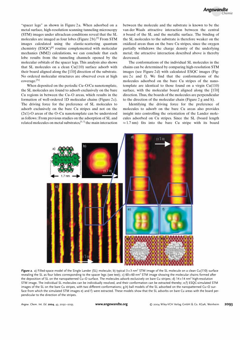
“spacer legs” as shown in Figure 2a. When adsorbed on ametal surface, high-resolution scanning tunneling microscopy(STM) images under ultraclean conditions reveal that the SLmolecules are imaged as four lobes (Figure 2b).[5] From STMimages calculated using the elastic-scattering quantumchemistry (ESQC)[6] routine complemented with molecularmechanics (MM2) calculations, we can conclude that eachlobe results from the tunneling channels opened by themolecular orbitals of the spacer legs. This analysis also showsthat SL molecules on a clean Cu(110) surface adsorb withtheir board aligned along the [11̄0] direction of the substrate.No ordered molecular structures are observed even at highcoverage.[5c]
When deposited on the periodic Cu–O/Cu nanotemplate,the SL molecules are found to adsorb exclusively on the bareCu regions in between the Cu–O areas, which results in theformation of well-ordered 1D molecular chains (Figure 2c).The driving force for the preference of SL molecules toadsorb exclusively on the bare Cu stripes and not on the(2x1)-O areas of the O–Cu nanotemplate can be understoodas follows: From previous studies on the adsorption of SL andrelated molecules on metal substrates,[5, 7] the main interaction
between the molecule and the substrate is known to be thevan der Waals attractive interaction between the centralp board of the SL and the metallic surface. The binding ofthe SL molecules to the substrate is therefore weaker on theoxidized areas than on the bare Cu stripes, since the oxygenpartially withdraws the charge density of the underlyingmetal; the attractive interaction described above is therebydecreased.
The conformations of the individual SL molecules in thechains can be determined by comparing high-resolution STMimages (see Figure 2d) with calculated ESQC images (Fig-ure 2e and f). We find that the conformations of themolecules adsorbed on the bare Cu stripes of the nano-template are identical to those found on a virgin Cu(110)surface, with the molecular board aligned along the [11̄0]direction. Thus, the boards of the molecules are perpendicularto the direction of the molecular chain (Figure 2 g and h).
Identifying the driving force for the preference ofmolecules to adsorb on the bare Cu areas also providesinsight into controlling the orientation of the Lander mole-cules adsorbed on Cu stripes. Since the SL (board length� 1.7 nm) fits into the bare Cu stripe with its board
Figure 2. a) Filled-space model of the Single Lander (SL) molecule; b) typical 3 � 3 nm2 STM image of the SL molecule on a clean Cu(110) surfacerevealing the SL as four lobes corresponding to the spacer legs (see text); c) 60 � 60 nm2 STM image showing the molecular chains formed afterthe deposition of SL on the nanopatterned Cu–O surface. The molecules adsorb exclusively on bare Cu stripes; d) 14 � 14 nm2 high-resolutionSTM image. The individual SL molecules can be individually resolved, and their conformation can be extracted thereby; e,f) ESQC-simulated STMimages of the SL on the bare Cu stripes, with two different conformations; g,h) ball models of the SL adsorbed on the nanopatterned Cu–O sur-face from which the simulated STM images e) and f) were extracted. These models show that the SL adsorbs on bare Cu areas with the board per-pendicular to the direction of the stripes.
AngewandteChemie
2093Angew. Chem. Int. Ed. 2004, 43, 2092 –2095 www.angewandte.org � 2004 Wiley-VCH Verlag GmbH & Co. KGaA, Weinheim
perpendicular to the direction of the oxygen-induced nano-template, no energetic reasons exist for the SL to change itsadsorption geometry on the nanotemplate with respect to theone adopted on a virgin Cu(110) surface, that is, aligned alongthe [11̄0] direction of the substrate. If, on the other hand, newLander molecules were synthesized with a board longer thanthe width of the stripe, and the molecules were still orientedperpendicular to the template, part of the board would bepositioned on top of the oxidized area. In this case one wouldanticipate the Lander molecules to adopt a more energeticallyfavorable orientation in which the board would fit completelywithin the bare Cu area of the nanotemplate.
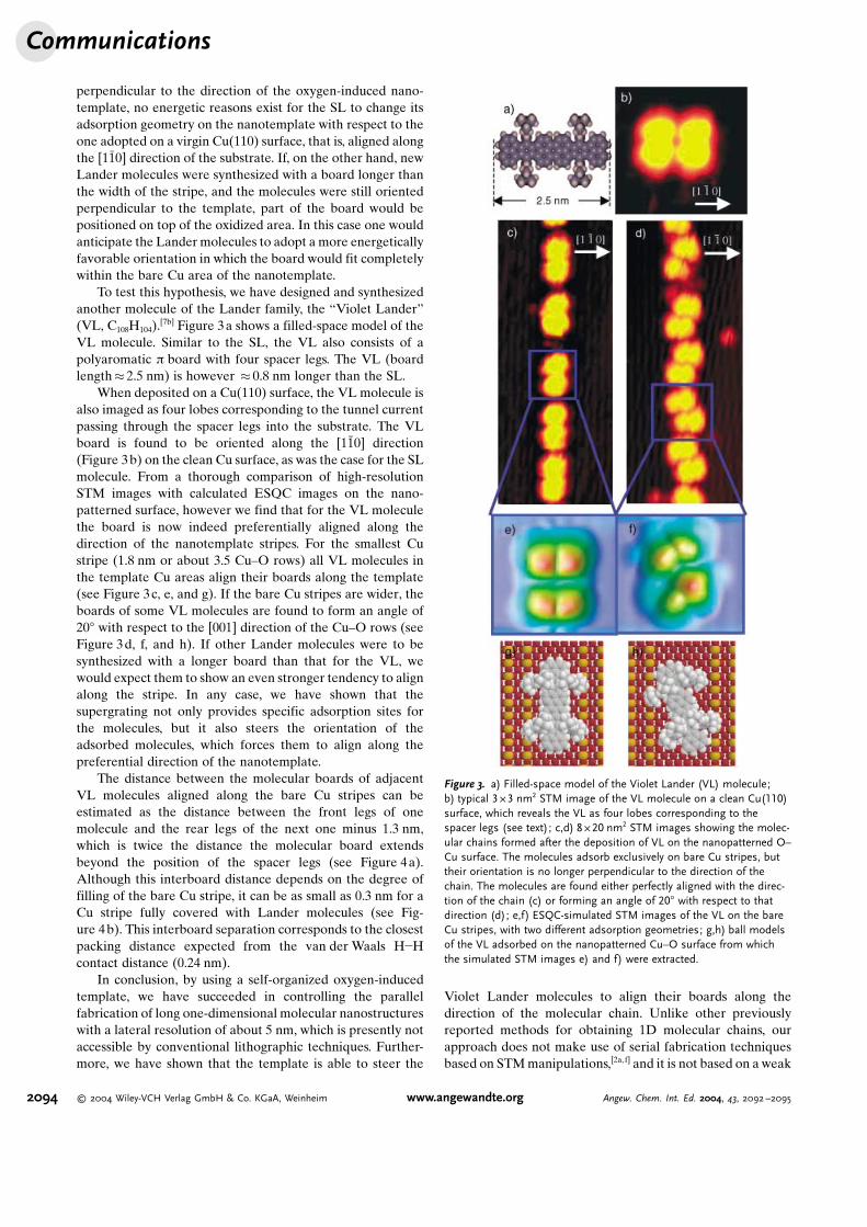
To test this hypothesis, we have designed and synthesizedanother molecule of the Lander family, the “Violet Lander”(VL, C108H104).
[7b] Figure 3a shows a filled-space model of theVL molecule. Similar to the SL, the VL also consists of apolyaromatic p board with four spacer legs. The VL (boardlength� 2.5 nm) is however � 0.8 nm longer than the SL.
When deposited on a Cu(110) surface, the VL molecule isalso imaged as four lobes corresponding to the tunnel currentpassing through the spacer legs into the substrate. The VLboard is found to be oriented along the [11̄0] direction(Figure 3b) on the clean Cu surface, as was the case for the SLmolecule. From a thorough comparison of high-resolutionSTM images with calculated ESQC images on the nano-patterned surface, however we find that for the VL moleculethe board is now indeed preferentially aligned along thedirection of the nanotemplate stripes. For the smallest Custripe (1.8 nm or about 3.5 Cu–O rows) all VL molecules inthe template Cu areas align their boards along the template(see Figure 3c, e, and g). If the bare Cu stripes are wider, theboards of some VL molecules are found to form an angle of208 with respect to the [001] direction of the Cu–O rows (seeFigure 3d, f, and h). If other Lander molecules were to besynthesized with a longer board than that for the VL, wewould expect them to show an even stronger tendency to alignalong the stripe. In any case, we have shown that thesupergrating not only provides specific adsorption sites forthe molecules, but it also steers the orientation of theadsorbed molecules, which forces them to align along thepreferential direction of the nanotemplate.
The distance between the molecular boards of adjacentVL molecules aligned along the bare Cu stripes can beestimated as the distance between the front legs of onemolecule and the rear legs of the next one minus 1.3 nm,which is twice the distance the molecular board extendsbeyond the position of the spacer legs (see Figure 4a).Although this interboard distance depends on the degree offilling of the bare Cu stripe, it can be as small as 0.3 nm for aCu stripe fully covered with Lander molecules (see Fig-ure 4b). This interboard separation corresponds to the closestpacking distance expected from the van der Waals H�Hcontact distance (0.24 nm).
In conclusion, by using a self-organized oxygen-inducedtemplate, we have succeeded in controlling the parallelfabrication of long one-dimensional molecular nanostructureswith a lateral resolution of about 5 nm, which is presently notaccessible by conventional lithographic techniques. Further-more, we have shown that the template is able to steer the
Violet Lander molecules to align their boards along thedirection of the molecular chain. Unlike other previouslyreported methods for obtaining 1D molecular chains, ourapproach does not make use of serial fabrication techniquesbased on STMmanipulations,[2a, f] and it is not based on a weak
Figure 3. a) Filled-space model of the Violet Lander (VL) molecule;b) typical 3 � 3 nm2 STM image of the VL molecule on a clean Cu(110)surface, which reveals the VL as four lobes corresponding to thespacer legs (see text); c,d) 8 � 20 nm2 STM images showing the molec-ular chains formed after the deposition of VL on the nanopatterned O–Cu surface. The molecules adsorb exclusively on bare Cu stripes, buttheir orientation is no longer perpendicular to the direction of thechain. The molecules are found either perfectly aligned with the direc-tion of the chain (c) or forming an angle of 208 with respect to thatdirection (d); e,f) ESQC-simulated STM images of the VL on the bareCu stripes, with two different adsorption geometries; g,h) ball modelsof the VL adsorbed on the nanopatterned Cu–O surface from whichthe simulated STM images e) and f) were extracted.
Communications
2094 � 2004 Wiley-VCH Verlag GmbH & Co. KGaA, Weinheim www.angewandte.org Angew. Chem. Int. Ed. 2004, 43, 2092 –2095

hydrogen-bond-directed assembly.[2b–d] This greatly enhancesthe thermal stability of the molecular structures up totemperatures of 350 K. In general, the interplay betweenself-organized chemical patterning of the substrate and arational design of molecular structure can be exploited to self-fabricate integrated nanoelectronic devices.
Received: December 19, 2003 [Z53586]
.Keywords: molecular electronics · nanostructures ·scanning probe microscopy · self-assembly · surface analysis
[1] a) C. Joachim, J. K. Gimzewski, A. Aviram, Nature 2000, 408,541 – 548; b) F. Rosei, M. Schunack, Y. Naitoh, P. Jiang, A.Gourdon, E. Lægsgaard, I. Stensgaard, C. Joachim, F. Besen-bacher, Prog. Surf. Sci. 2003, 71, 95 – 146; c) R. L. Carroll, C. B.Gorman,Angew. Chem. 2002, 114, 4556 – 4579;Angew. Chem. Int.Ed. 2002, 41, 4378 – 4400.
[2] a) Y. Okawa, M. Aono, Nature 2001, 409, 683 – 684; b) T.Yokoyama, S. Yokoyama, T. Kamikado, Y. Okuno, S. Mashiko,Nature 2001, 413, 619 – 621; c) M. BNhringer, K. Morgenstern,Phys. Rev. Lett. 1999, 83, 324 – 327; d) J. V. Barth, J. Weckesser, N.Lin, A. Dmitriev, K. Kern,Appl. Phys. A. 2003, 76, 645 – 652; e) S.Lukas, G. Witte, C. WNll, Phys. Rev. Lett. 2002, 88, 28301; f) G. P.Lopinski, D. D. M. Wayner, R. A. Wolkow, Nature 2000, 406, 48 –51; g) P. W. Murray, I. M. Brookes, S. A. Haycock, G. Thornton,Phys. Rev. Lett. 1998, 80, 988 – 990; h) J. K. Gimzewski, C.Joachim, Science 1999, 283, 1683 – 1688.
[3] a) K. Kern, H. Niehus, A. Schatz, P. Zeppenfeld, J. Goerge, G.Comsa, Phys. Rev. Lett. 1991, 67, 855 – 858; b) F. Besenbacher, F.Jensen, E. Lægsgaard, K. Mortensen, I. Stensgaard, J. Vac. Sci.Technol. B 1991, 9, 874 – 878.
[4] A. Gourdon, Eur. J. Org. Chem. 1998, 12, 2797 – 2801.[5] a) V. L. Langlais, R. R. Schittler, H. Tang, A. Gourdon, C.
Joachim, J. K. Gimzewski, Phys. Rev. Lett. 1999, 83, 2809 – 2812;b) F. Rosei, M. Schunack, P. Jiang, A. Gourdon, E. Lægsgaard, I.Stensgaard, C. Joachim, F. Besenbacher, Science 2002, 296, 328 –331; c) M. Schunack, F. Rosei, Y. Naitoh, P. Jiang, A. Gourdon, E.Lægsgaard, I. Stensgaard, C. Joachim, F. Besenbacher, J. Chem.Phys. 2002, 117, 6259 – 6265; d) J. Kuntze, R. Berndt, P. Jiang, H.Tang, A. Gourdon, C. Joachim, Phys. Rev. B 2002, 65, 233405;e) L. Gross, F. Moresco, M. Alemani, H. Tang, A. Gourdon, C.Joachim, K.-H. Rieder, Chem. Phys. Lett. 2003, 371, 750 – 756.
[6] P. Sautet C. Joachim, Chem. Phys. Lett. 1991, 185, 23 – 30.[7] a) T. Zambelli, P. Jiang, J. Lagoute, S. E. Grillo, S. Gauthier, A.
Gourdon, C. Joachim, Phys. Rev. B 2002, 66, 75410; b) T.Zambelli, H. Tang, J. Lagoute, S. Gauthier, A. Gourdon, C.Joachim, Chem. Phys. Lett. 2001, 348, 1 – 6.
[8] E. Lægsgaard, L. Osterlund, P. Thostrup, P. B. Rasmussen, I.Stensgaard, F. Besenbacher, Rev. Sci. Instrum. 2001, 72, 3537 –3542.
Figure 4. a) Cartoon illustrating how to estimate the interboard distan-ces from the distances between the legs of consecutive VL molecules;b) 3 � 6 nm2 STM image showing two VL molecules aligned along thebare Cu stripe in Van der Waals contact, as determined from the inter-board distances estimated from the scan line in c); c) scan along themolecular boards of the molecules shown in b).
AngewandteChemie
2095Angew. Chem. Int. Ed. 2004, 43, 2092 –2095 www.angewandte.org � 2004 Wiley-VCH Verlag GmbH & Co. KGaA, Weinheim