on the road to therapeutic cloning
TRANSCRIPT
N E W S A N D V I E W S
in the same range as those obtained with thewell-established NPTII gene and kanamycinselection.
But the trump card of DAAO could proveto be its combination with site-specific excision systems: after positive selection oftransgenics followed by initiation of recombi-nation, a switch to negative selection couldverify the removal of the now redundantmarker (Fig. 1b). This scheme has yet to bedemonstrated, however, and claims of pana-ceas should be postponed until the function-ality of the selection principle in other plantspecies has been proven.
Although A. thaliana appears to lackDAAO, we know little about endogenousDAAO enzyme activity in other plants, orabout the distribution of its substrates amongplants (and plant eaters). The enzyme shows ahigh specificity for the D enantiomer, but itssubstrate specificity is less strict and sideeffects of overexpression will have to be care-fully excluded from recipient plants.
Nevertheless, the marker gene is of eukary-otic origin and encodes one of the best-known enzymes. The activity of DAAO wasdiscovered more than 60 years ago, and it is amodel flavoprotein for which the catalytic
mechanisms and a three-dimensional struc-ture have been described in detail3,4. D-aminoacids, although not incorporated into pro-teins and peptides of eukaryotes, are presentin natural and processed food5 and are proba-bly easy to handle safely under laboratoryconditions.
The potential of the DAO1 marker will trig-ger rapid, broad trials in various transforma-tion systems in combination with differentregulatory elements, provided that the use ofDAO1 is not inhibited by lengthy regulatorynegotiations. The results of such experimentswill also allow thorough examination of bio-safety aspects. If the selection principle can betransferred to crop plants and subsequentintegration and excision prove feasible, wewill have gained an elegant and importantnew tool for the plant biotechnology work-shop.
1. Erikson, O., Hertzberg, M. & Näsholm, T. Nat.Biotechnol. 22, 455–458 (2004).
2. Miki, B. & McHugh, S. J. Biotechnol. 107, 193–232(2004).
3. Mattevi, A., Vanoni, M.A. & Curti, B. Curr. Opin.Struct. Biol. 7, 804–810 (1997).
4. Pilone, M.S. Cell. Mol. Life Sci. 57, 1732–1747(2000).
5. Friedman, M. J. Agric. Food Chem. 47, 3457–3479(1999).
cloned embryos showed abnormalities andnone was viable past a very early developmen-tal stage2. At the same time, it should be rec-ognized that another key to the achievementof Hwang et al. may lie in the enthusiasm withwhich research on regenerative medicine isgreeted in South Korea, which gave theresearchers access to the 242 human oocytesused in the study and allowed them to con-duct their work in a relatively permissive regulatory environment—a much morefavorable climate than in many countries inEurope and North America.
As has been the case with some previousreports of experimental cloning, commenta-tors may challenge Hwang et al.’s ES cells asthe products of experimental error, suggest-ing that they may be parthenogenetic in ori-gin rather than derived from a bona fidecloned embryo. Those with experience incloning know, however, that the chance ofsuch an error occurring is vanishingly small.Nuclei are always positioned under the polarbody, and it is no more likely for a scientist tomisidentify a failed enucleation than for aprofessional chef to miss the fact that a yolkand egg white have failed to separate. Indeed,Hwang et al. confirmed enucleation byHoechst staining and UV irradiation, whichin the end may have actually damaged thecloned embryos and worked to lower theirsuccess rate. They report that only one line ofES cells was established from 30 original blas-tocysts, giving a success rate of 3.3%.Moreover, the donor somatic cell and oocytewere taken from the same individual, a req-uirement that, if not overcome through tech-nical advances, would mean that only patientspossessing a supply of healthy oocytes couldbenefit from therapeutic cloning.
Although the technique of generatingsomatic cell–derived ES cell lines by nucleartransfer (which we refer to as ntES cells) is stillrelatively new, studies in mice have shownthat ntES cells can be established using nucleifrom fibroblasts—a plentiful and nearly ubiq-uitous source—and, furthermore, that suchntES cells can be derived at efficiencies of10–20% using cells from donors of eithergender and from lines that have never beensuccessfully used to produce a live-borncloned individual3 (Fig. 1). If the same provestrue for humans, improvements in therapeu-tic cloning techniques may enable any patientto act as his or her own ntES cell donor. Inmice, success rates for deriving ntES cell linescan be as much as an order of magnitudehigher than those for reproductive cloning,suggesting that the scientific barriers to thera-peutic cloning may be lower than the barriersto creating a new individual by cloning.
NATURE BIOTECHNOLOGY VOLUME 22 NUMBER 4 APRIL 2004 399
The political tussles surrounding human EScells have overshadowed discussion of the sci-entific challenges that lie ahead if the promiseof ES cells for regenerative medicine is to berealized. A recent report by Hwang et al.1 inScience of ES-cell derivation from a clonedhuman embryo invites reflection on theobstacles that remain in the development ofthis technology.
Human ES cells are envisioned as anunlimited source of cells for cell replacementtherapies. However, as with any allogeneicmaterial, ES cells derived from fertilized blas-tocysts, and the progeny of such cells, riskimmune rejection after transplantation. It has
been proposed that ES cells derived fromembryos cloned from a patient’s own cellsrepresent a solution to the problem of rejec-tion, as any replacement cells would be genet-ically identical to the patient. The work ofHwang et al. provides an important proof ofprinciple of the feasibility of creating humanES cells using a patients’ own cells as a source.But many challenges remain, including repro-gramming without induction of abnormali-ties, induction and targeting of differentiation,the control of stem cell proliferation and thedilemma of persistent re-rejection in cases ofautoimmune disease.
The success of Hwang et al. did not rest ona major technical breakthrough; rather, itresulted from a persistent and painstakingoptimization of the many seemingly minor,but actually critically important, experimen-tal factors involved in mammalian cloning. Asimilar experiment was attempted in theUnited States in 2002, but in that study the
On the road to therapeutic cloningTeruhiko Wakayama
Human embryonic stem (ES) cells have been derived from cloned embryos,an important step in the development of therapeutic cloning.
Teruhiko Wakayama is at the Center forDevelopmental Biology, RIKEN, 2-2-3Minatojima Minamimachi Chuo ku, Kobe,Hyogo 650 0047, Japan.e-mail: [email protected]
©20
04 N
atur
e P
ublis
hing
Gro
up
http
://w
ww
.nat
ure.
com
/nat
ureb
iote
chno
logy
N E W S A N D V I E W S
That said, several important issues remainto be addressed. Animals produced by cloningconsistently show a variety of developmentalabnormalities, ranging from placental anom-alies to obesity to premature mortality ofunknown cause. It has been suggested thateven apparently ‘normal’ cloned animalsexhibit epigenetic defects4,5. Similar deficien-cies—such as abnormal gene expression patterns6 and the retention of somaticcell–like features7—have also been reportedin cloned animal blastocysts. These abnor-malities have been attributed to insufficientreprogramming of donor nuclei, perhaps dueto flaws in nuclear transplantation technol-ogy. Whatever the case, little is known aboutreprogramming even in normal or in vitrofertilization, despite the central biologicalimportance of this process. At this point, it isunclear whether cloned blastocysts are inher-ently different from their ‘natural’ counter-parts or whether such differences areartifactual and therefore might be resolved byimprovements in technique.
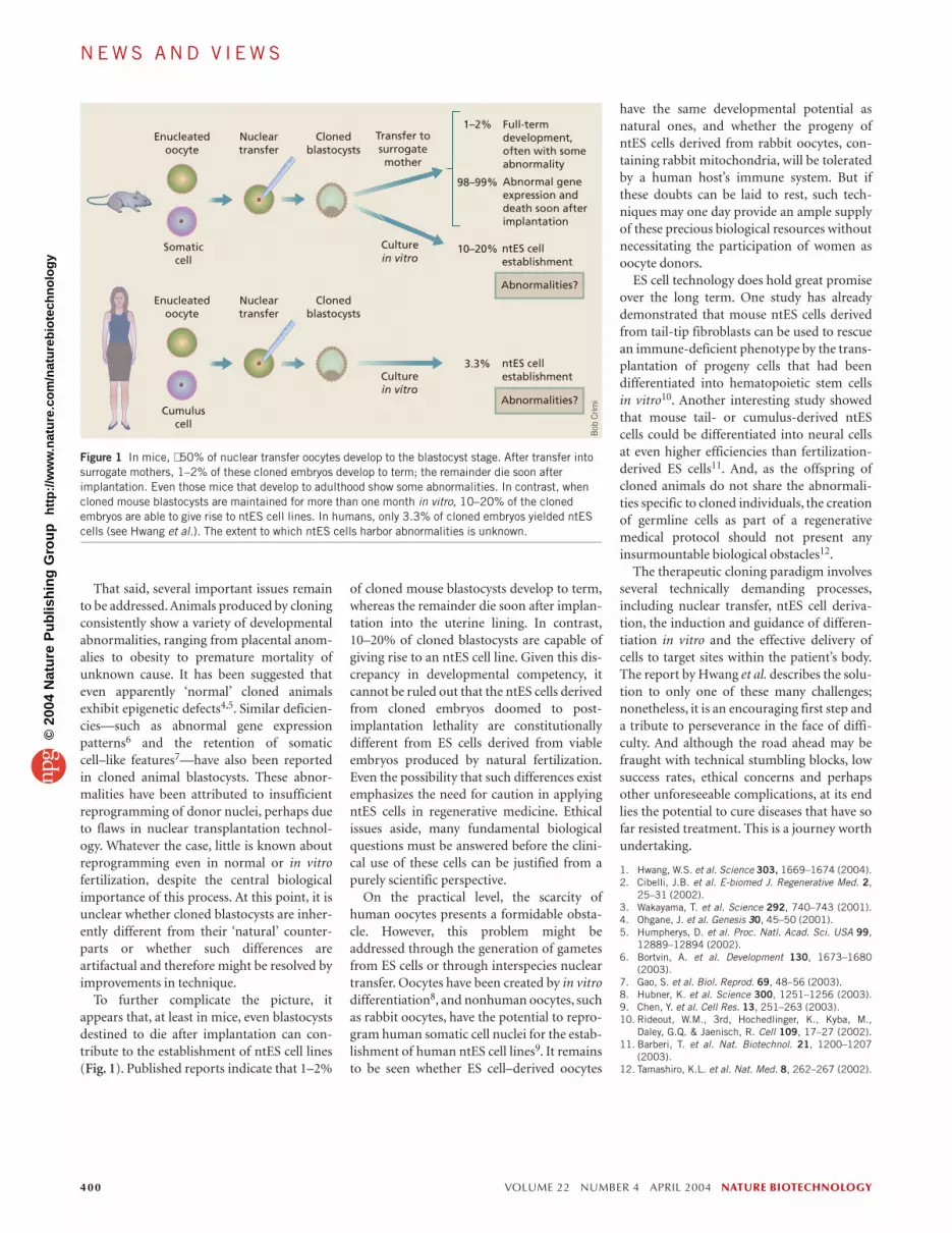
To further complicate the picture, itappears that, at least in mice, even blastocystsdestined to die after implantation can con-tribute to the establishment of ntES cell lines(Fig. 1). Published reports indicate that 1–2%
of cloned mouse blastocysts develop to term,whereas the remainder die soon after implan-tation into the uterine lining. In contrast,10–20% of cloned blastocysts are capable ofgiving rise to an ntES cell line. Given this dis-crepancy in developmental competency, itcannot be ruled out that the ntES cells derivedfrom cloned embryos doomed to post-implantation lethality are constitutionallydifferent from ES cells derived from viableembryos produced by natural fertilization.Even the possibility that such differences existemphasizes the need for caution in applyingntES cells in regenerative medicine. Ethicalissues aside, many fundamental biologicalquestions must be answered before the clini-cal use of these cells can be justified from apurely scientific perspective.
On the practical level, the scarcity ofhuman oocytes presents a formidable obsta-cle. However, this problem might beaddressed through the generation of gametesfrom ES cells or through interspecies nucleartransfer. Oocytes have been created by in vitrodifferentiation8, and nonhuman oocytes, suchas rabbit oocytes, have the potential to repro-gram human somatic cell nuclei for the estab-lishment of human ntES cell lines9. It remainsto be seen whether ES cell–derived oocytes
have the same developmental potential asnatural ones, and whether the progeny ofntES cells derived from rabbit oocytes, con-taining rabbit mitochondria, will be toleratedby a human host’s immune system. But ifthese doubts can be laid to rest, such tech-niques may one day provide an ample supplyof these precious biological resources withoutnecessitating the participation of women asoocyte donors.
ES cell technology does hold great promiseover the long term. One study has alreadydemonstrated that mouse ntES cells derivedfrom tail-tip fibroblasts can be used to rescuean immune-deficient phenotype by the trans-plantation of progeny cells that had been differentiated into hematopoietic stem cellsin vitro10. Another interesting study showedthat mouse tail- or cumulus-derived ntEScells could be differentiated into neural cellsat even higher efficiencies than fertilization-derived ES cells11. And, as the offspring ofcloned animals do not share the abnormali-ties specific to cloned individuals, the creationof germline cells as part of a regenerativemedical protocol should not present anyinsurmountable biological obstacles12.
The therapeutic cloning paradigm involvesseveral technically demanding processes,including nuclear transfer, ntES cell deriva-tion, the induction and guidance of differen-tiation in vitro and the effective delivery ofcells to target sites within the patient’s body.The report by Hwang et al. describes the solu-tion to only one of these many challenges;nonetheless, it is an encouraging first step anda tribute to perseverance in the face of diffi-culty. And although the road ahead may befraught with technical stumbling blocks, lowsuccess rates, ethical concerns and perhapsother unforeseeable complications, at its endlies the potential to cure diseases that have sofar resisted treatment. This is a journey worthundertaking.
1. Hwang, W.S. et al. Science 303, 1669–1674 (2004).2. Cibelli, J.B. et al. E-biomed J. Regenerative Med. 2,
25–31 (2002).3. Wakayama, T. et al. Science 292, 740–743 (2001).4. Ohgane, J. et al. Genesis 30, 45–50 (2001).5. Humpherys, D. et al. Proc. Natl. Acad. Sci. USA 99,
12889–12894 (2002).6. Bortvin, A. et al. Development 130, 1673–1680
(2003).7. Gao, S. et al. Biol. Reprod. 69, 48–56 (2003).8. Hubner, K. et al. Science 300, 1251–1256 (2003).9. Chen, Y. et al. Cell Res. 13, 251–263 (2003).10. Rideout, W.M., 3rd, Hochedlinger, K., Kyba, M.,
Daley, G.Q. & Jaenisch, R. Cell 109, 17–27 (2002).11. Barberi, T. et al. Nat. Biotechnol. 21, 1200–1207
(2003).12. Tamashiro, K.L. et al. Nat. Med. 8, 262–267 (2002).
400 VOLUME 22 NUMBER 4 APRIL 2004 NATURE BIOTECHNOLOGY
Figure 1 In mice, ∼ 50% of nuclear transfer oocytes develop to the blastocyst stage. After transfer intosurrogate mothers, 1–2% of these cloned embryos develop to term; the remainder die soon afterimplantation. Even those mice that develop to adulthood show some abnormalities. In contrast, whencloned mouse blastocysts are maintained for more than one month in vitro, 10–20% of the clonedembryos are able to give rise to ntES cell lines. In humans, only 3.3% of cloned embryos yielded ntEScells (see Hwang et al.). The extent to which ntES cells harbor abnormalities is unknown.
Enucleatedoocyte
Somaticcell
Nucleartransfer
Clonedblastocysts
Transfer tosurrogatemother
Full-termdevelopment,often with someabnormality
Abnormal geneexpression anddeath soon afterimplantation
ntES cellestablishment
Abnormalities?
Abnormalities?
Culturein vitro
1–2%
98–99%
10–20%
ntES cellestablishment
3.3%Culturein vitro
Enucleatedoocyte
Cumuluscell
Nucleartransfer
Clonedblastocysts
Bob
Crim
i
©20
04 N
atur
e P
ublis
hing
Gro
up
http
://w
ww
.nat
ure.
com
/nat
ureb
iote
chno
logy