on a new canker-disease of prunus yedoensis, p. mume …
TRANSCRIPT

Instructions for use
Title ON A NEW CANKER-DISEASE OF PRUNUS YEDOENSIS, P. MUME AND OTHER SPECIES CAUSED BYVALSA JAPONICA MIYABE et HEMMI sp. n
Author(s) HEMMI, Takewo
Citation The journal of the College of Agriculture, Tohoku Imperial University, Sapporo, Japan, 7(4), 257-319
Issue Date 1916-08-25
Doc URL http://hdl.handle.net/2115/12540
Type bulletin (article)
File Information 7(4)_p257-319.pdf
Hokkaido University Collection of Scholarly and Academic Papers : HUSCAP

ON A NEW CANKER-DISEASE OF
PRUNUS YEDOENSIS, P. MUME AND
OTHER SPECIES CAUSED BY
VALSA JAPONICA MIYABE et HEMMI SPa n. BY
Takewo Hemmi, Nogakltslli
(With Plates VII-X)
1. Introduction.
The canker-disease of PnllZltS yedoensis MATSUM. (Somei-yosltillo) was
first noticed in 19' 3. by Prof. K. M IYABE in the Botanic Garden of our Univer-
sity, and was proved by him to be due to a species of Valsa. It was in the
early spring- of 19 [4, that my attention was drawn to the seriousness and
widespread occurrence of the disease in the vicinity of Sapporo, when, at the
suggestion of Prof. K. MIYABE I resolved to undertake the investigation of the
disease. Since that time, many samples of the diseased branches and stems
have been collected on different species and varieties of Prunus. Among
those affected by the disease, PrulZltS Mlt11te S. et Z. seems to be very suscep
tible to the disease, and its causal fungus was proved to be the same as that
of Pntlllls yedoensis by my cultural and inoculation experiments. The peach
tree (PntllllS Per sica S. et Z.) was also proved to be sometimes attacked by
the same fungus, but the extent of its damage was not investigated in my
. study. According to my observations, Pnllllls saclta!illellsis KOIDZ. ((Jyal1la
:;akllra or Ye'[;o-7lO-~yallla.~a!:"lra) seems to have a strong resistance against the
disease, though not immune.' As to the other host plants my observations
and studies are not yet complete, and I wish to defer their report to a future
occasion. The fact that the canker disease has not yet been found on the
European Cherry tree (PntllllS Ceraslts L.) in the vicinity of Sapporo, notwith-
[Jour. of the College of Agr., Tohoku Imp. Univ .• Sapporo, Vol. VII, Pt. 4. August, 1916]

25 8 ON A NEW CANKEl~-DISEASE OF PRUNUS YEDOENSIS, etc_
standing its wide cultivation, shows the plant is immune to the disease. As
the result of my studies this destructive canker disease was proved to be
caused by the attack of a new species of Valsa acting as a wound parasite.
I wish to express here my heartiest thanks to Prof. K. MIYABE for his
constant and kind direction; and to Assistant Prof. S. ITo to whom I am also
indebted for his many valuable suggestions. I wish also to express my
sincere thanks to Prof. H. KORIBA, Messrs. Y. KUDO, S. NISHIDA and oth:2r
gentlemen, who have kindly helped me in many ways.
2. Historical Review of Valsa-Diseases.
Valsa is a large Pyrenomycetous genus, having more than 4C>0 species,
most of which are generally described as saprophytes in mycological litera
ture. Up to the present time only five species have been recognized as the
cause of plant diseases. They are, namely, -- Va/sa /euo-ostoma (PEf{S_) FR.,
Va/sa oxysto11la REHM, Va/sa ambiens FR., Valw (Eutypdla) Pnmastri (PERS.)
FR. and Valsa }lilali MIYABE et YAMADA.
Among them, Valsa leucostoma (PElZS.) FR. is most widely knowl1 as the
cause of a destructive disease of the drupaceous trees throughout Europe,
Australia and N. America. This fungus attacks the cherry, peach and other
drupaceous fruit-trees, causing the disease known as the "Die-back" of the
twigs, and also the cankers upon the trunks and limbs. In the Rhine-district,
from about the year 1899, the cherry trees have been so severely affected by
the fungus as to arouse the attention of many phytopathologists. FRA~K
(1899) V) studied the disease and ascribed it to Cytospora ntbcscens FR. On
the other hand, GOETHE (189;» 11), ZAPFE (1899) 35), SO!{AUER (1900) 2;) RASCHE~
(1900)21) and LABo:-n-k (I90:)f5) considered the disease to be due to some un
favorable climatic conditions or to other physiological derangements. It was
by ADEf{HOLD ([9:)3) 1) that this disease was most thoroughly investigated; and
he proved it to be caused by Va/sa /cucostoma, the gcnerally ascrived climatic
causes to be merely its promoting agencies, and C:J'tospora rubcscclls, which
was observed by Fl{A~K (1899) 9>, to be the pycnosporous stage of this fungus.

TAKEWO HEMMI 259
In Australia, McALPINE ([902) li) described the same pycnosporous stage, by
the name of Cytospora leucostoma, on almond, peach, plum and cherry twigs.
In America it was first described by ROLFS (1907) 23), who found it upon peach
and Japanese plum.
Valsa oxystoma REHM is known as the cause of a disease of the alder-tree
in Europe. It was first described by REHM (r882)n), \vho thought it to be a
saprophyte. But in 1893, TUBEUF 30) ascribed the death of the twig of Aillus
viridis to the attack of this fungus in Germany, and in 1899 the same fungus
was observed by P. NYPELS'19) in Belgium to cause the disease of Alnus glltti-
llosa.
l/'alsa amoiells FR. was shown by M. C. COOKE 7) to seriously affect the
living bark of apple-trees in Europe, although the fungus is usually considered
as a pure saprophyte. Valsa Pnmastri (PERS.) FR. is known to be the cause
of a serious disease of plum, apricot, peach and apple trees in Europe.
In our country there is a species of Valsa causing the destructive canker
disease on apple trees. The disease was first discovered in the Aomori Pre
fecture, but it is now widely spread in Hokkaido. Prof. K. MIYAllE and Prof.
G. YAMADA studied the fungus and named it Valsa Mlli MIY. et YAM. It is
an endemic species. Its description appeared for the first time in I90;! in IDE
TA'S Hand book of the Plant-Diseases in Japan 12).
There are, besides, two more very destructive Valsa diseases in our
country, which have not yet been reported. One is on Paulownia * and the
other is on the Japanese flowering cherry-trees, Jap:mese apricot and peach
trees. The latter was taken up as the subject of my present investigations,
as it is the more important from the economic standpoint.
3, Symptoms of the Disease.
a. General Appearance of the Diseased Trees.
It is easy enough to distinguish this disease on the bark of the branches
* From our studies, this fungus abo seem3 to be new to science. vVe named it, therefore, Va/sa Pau/owniae MIYABE et HE:\IMI. Its diagnosis willl>e given in another paper.

260 ON A NEW CANKER-DISEASE OF PRlJNUS YEDOENSIS, etc.
or trunks throughout the year, if careful search is made_ Although the effect
of the disease on the general appearance of the tree is noticeable at any time
during the growing season, it is most conspicuous during the spring months,
at least in the vicinity of Sapporo, when the causal fungus is under most
favorable conditions for its growth.
In the vicinity of Sapporo, where the disease is common, it is not rare to
find the entire tree or some of its branches which are in blossom or in leaf
suddenly wither and then dry up at the beginning of May. The brown,
shri veled flowers and leaves are readily seen even at a distance, while the
healthy part of the tree is still in blossom or in foliage. If one examines the
base of such a withered branch, a comparatively large canker or a part entire
ly girdled by the disease will be found. Such symptoms are also noticed in
summer, although not numerous. If the point of infection is near the base
of the trunk, as is most freq ucntiy the case, one side of the tree will wilt first,
then a month or so later the rest of the tree. From the point of entrance, the
fungus hyphae pass rather more slowly toward the center of the tree than
they do up and down through the water ducts, causing the formation of a
gummy substance, which plugs up the lumina of the ducts. and cuts o~f the
water supply for the transpiration organ of the tree.
The wilted dead leaves or flowers remain clinging to the branches for a
long time. If the plugging of the ducts by the gum or the girdling of a limb
by the fungus takes place during the winter, the plant will appear in the
spril~g as if it had been killed by a very low temperature.
b. Appearance of the Diseased Portions of the Bark.
On the smooth bark, by the death of the tissues of the cambium and
inner bark, a canker formation takes place, accompanied generally by the
exudation of gum around the affected portion. These cankered portions are
slightly sunken, and distinguished from the healthy bad,;: by a dark-brown or
blackish-brown color, whereas the normal bark is more of a chestnut-brown or
light-brown color. Often the bark on these cankered spots is more or. less

TAKEWO HEMMI 261
cracked at the marginal portion. These cankers occur on branches of all
sizes,-rarely, however, on young branches. The usual shape of the canker
is elliptical, being longer in the direction of the long axis of the branch.
The margin of the canker is usually fairly regular, but it may be irregular.
According to my observations, the cankers formed on the large branch be
come deeper year by year as healthy wood is formed about them, thus causing
a condition similar to the apple-tree canker or the American chestnut-tree
canker. When the fungus affects the rough bark of the trunks, the cankers
do not show very distinctly, since the change in color of the bar~ is not
evident and the shape of the canker is mostly irregular. It is frequently
distinguished from the healthy parts only by the lenticel-like openings of'the
stromata. But the cankers on the rapidly growing branches are usually out
lined by a distinct ridge of slightly hypertrophied tissue. It seems to me
that there are two different types of the diseased branches. The one is the
cankered type (PI. VII, Fig. 2-4) as already described, and the other is the
girdled type (PI. VIII, Fig. I) which does not produce the canker. The latter
takes place mostly in the case of the weakened trees or when the rathcr fine
branches or twigs are infected in spring. The girdling is caused by the rapid
growth of the mycelium without inducing the formation of a definite canker;
and the fruiting pustules break out rather scatteringly all over their surface.
Such a diseased portion extends sometimes t{) the greater part of the branch,
which at once withers and dies. Although this girdling is seen more or less
on all host plants, it is most cOlllmon on Primus sacltalinellsis KOIDZ. (PI.
VIII, Fig. I), which is rarely attacked by this disease and on which I have
not yet found the cankered type.
The fruiting pustules are at n.rst covered by the periderm, which becomes
lifted up and finally ruptured, exposing the brown or blackish brown stromata
of the fungus. These slits appear at first glance like the lenticels, for they
are usually elongated horizontally. During the growing season, when the
weather is damp, from these pustules are pushed out very fine reddish threads
or spore-horns which are composed of inllumerable Pycllospores. In the

262 ON A NEW CANKER-DISEASE OF PRUNUS YEDOENSIS, etc.
early spring the red crusts composed of masses of ascospores are someti ;11I::S
seen on the stromata.
c. Development of the Canker.
The canker appears first as a more or less discolored area about the point
of infection, which takes place most frequently through cracks and wounds
on the bark, or where a branch or twig has been pruned, or killed frOIll some
cause, as winter injury. It is also not seldom that the fungus gets a start
from the wound which is given by an insect such as Sesia hector BUTL. or a
crack in the crotch of the branches. Judging from my observations, the
disease may also arise through the injured buds. The affected area of the
branch soon becomes sunken, making the boundary between the dead and
living tissues very marked. Gum pockets are often formed at this point.
The gummy exudation pushing through the tissue of the bark appears on its
surface as a copious gum How. The bark of such a diseased portion gradual
ly dries up and at last produces numerous pustules which are the beginning
of the stromata. Callus soon pushes out from the edge of the canker, but it
is very soon attacked by the fungus and produces a large new canker sur
rounding the previous one, requiring for its formation just one year. One
can see, therefore, at times on a large tree cankers as large as five or six feet
long, having three or four concentric ridges. Such injuries are gradually ex
tended, often girdling the branches and even the trunks of the tree. The
gummosis is also constantly associated \vith these cankers, being especially
conspicuous on PrltlZllS 111u1JZe.
d. Age and Parts of the Hosts affected.
According to my observations, the disease attacks the large trees more
frequently than the smaller ones. This is evidently caused by the facts that
the larger trees have a much greater chance of getting wounded by insects
and other causes than the young trees, that the healing power of the former
is not so strong as in the latter, and that moreover the younger it is, the

TAKEWO HEM:\II
greater its resistibility to this disease.
Even on a single tree the disease is most frequently found on the thick
branches or trunks, and the cases on the twiglets are very rare, if ever attacked.
When the disease affects the young twig, the pustules of the stromata are very
small, covered by the periderm for a long time without being ruptured; and
the spores are in such a case very rarely produced, for the twigs die and dry
up very rapidly. In the case of PntllllS Mlt11le, the canker is generally pro
duced on comparatively small branches, having the diameter of one and a half
centimeters or more.
4. Host Plants.
The disease in question was first found on przmus ye£ioeJlsis, which is most
severely attacked. By extending our observations to other species of Prunus
we found a similar disease on most of them. The trees affected by the disease
are PntllltS sacltali71eJlsis KOIDZ. (/f ;t, -\? ~ -'+}"~ :7), P. Koidzu11lii lVIATSUM. (A
V-\? -;T' 57 'j), P. scrrltlata LnmL. ( -\? '? -'+}"'p :7), P. sermlata LI~DL. var. !lobilis
(KOIDZ.) f. Yakihi (A 111 f~ t~ ill: -[{e.), P. kltrilellsis MIYABE (7- v'? -tf.p :7),
P. MUllle S. et Z. ('7 ~ ), and P. Persica S. et Z. (=£: ... ).
The identity of the fungus ~vhich attacks P. sachalimlZsis, P. j/1lt1lZe, and
P. Pasica causing tile canker-disease was proved by cultural and inoculation
experiments.
On PtltllltS serrltlata, the pycnidial stage only of the fungus has been ob
served. On Pntlllts serrltlata LINDL. var. !lob/lis, the canker is most beauti
fully formed, showing apparently the weak power of its resistance to the
disease. The specimen on PntllltS kltrile;zsis having both ascosporous and
pycnidial stages was collected by Prof. S. ho in May of 1915 on the dead
twig. The specimen on PntlZlts Koidzll71zii was collected by myself on the
dead twig, but the fungus is lacking in the ascosporous stage, and its parasi
tism is not clear. In regard to the causal fungus of the disease on these host
plants, I ha1:e not been able to undertake the cultural and inoculation experi-
ments,

264 ON A NEW CANKER-DISEASE OF PRUNUS YEDOENSIS. ctc.
In July of 1914, I collected a species of Cytospora on the dead twiglets
of the European cherry, Primus Cerasus L. The morphological charac-
ters of its spores are very much like those of the pycnospores of our fungus;
but its cultural characters seem to be more or less different, and the inocula
tion experiments have also shown negative results.
5. Distribution of the Disease and Extent of the Damage.
This disease is common not only in the vicinity of Sapporo, but seems also
to be widely distributed throughout Hokkaido.
In September of 1914. I found diseased branches on PmlZltS J'edoe7lsis at
Asahigawa, and an examination showed that it also was due to the same
fungus. In February of 1915, Mr. S. NISHIDA collectcd the s'ame fungus, also
on Pnmus yedoe7lsis, at the Hakodate Park, and in May of the same year he
again visited the same place for me and counted about thirty trees attacked
by this disease.
From other parts of Japan we have not yet been informed of the outbreak
of this disease. I received negative answers to all my inquiries to various
places in Honshu. In spite of careful search for the disease made by my
friends Mr. M. YATAGAI and Mr. Y. TAKENOUCHI and me during our journey
through Honshu, we did not find it. Among the specimens collected by Mr.
M. YATAGAI in Tokyo, there is one on a cherry tree which resembles our
disease in its external appearance, but whose microscopical characters are
very different. Through the kindness of Prof. K. Mn'ABE, I was able to
examine the diseased brancl~es of the peach which wcre sent to him from
Gifu. It is due to the attack of a certain species of Cytospora, the morpho
logical characters of which are quite different from those of our fungus.
Judging from these facts, it is safe to concl ude for the present that the disease
0:1 the species of Prunus is limited to Hokkaido.
In the vicinity of Sapporo the damage caused by this disease is by no
means small. Almost all aged trees of Pnmns J1cdoellsis have their branches
more or less attacked by this fungus. There are, indead, numerous branches

TAKEWO HEMMI 265
and even whole trees, that were cut off in consequence of the disease. On
Pnf.llllS sacllalinellsis, however, we have very few cases of this malady. On
PntlZlts Mume the disease seems to be not uncommon, but its damage is not
so great as in the case of Pntlllts yedoCilsis. Judging from the extent of the
damage, we may safely infer that the disease had existed in Hokkaido for
many years without drawing the attention of pathologists. I may further
more infer that the causal fungus is indigenous to Hokkaido growing on the
wild Pntlllts sadtalillCilsis or other species of Primus, having a comparatively
strong resistance, and that it has recently found more congenial hosts in the
cultivated cherry and apricot trees introduced from other parts of Japan.
6. Morphology of the Causal Fungus.
a. Stromata.
The infected tissues do not show at first any external signs of the fungus
itself; but in time, on the smooth bark, numerous fruiting pustules gradually
protrude through the horizontal rent of the periderm, having generally a small
lenticel-like appearance. In the young stage such pustules are covered by
the cork layers of the bark and on young twigs, especially on PntlZlts 1I1ltlllC,
they remain covered for a long- time. On rough bad{s or in special cases, as
in Pmlllts lWzt1lze, they break out from irregularly ruptured periderm.
In the young stage, a section through the pustule shows the compactly
united mass of the hyphae under the periderm (PI. IX, Fig. 8.). It is at first
quite small and more or less flattened, but in time it becomes subs[.lherical,
ellipsoidal or irregularly oblong in shape. These young stromata increase
their size little by little until they reacll the matured conical or wart-like
shape. The stromata are composed mostly of the fungus hyphae with some
host tissues intermixed. From about the time when the stroma is exposed by
the rupture of the periderm, a pycnidial cavity beg-ins to be produced. It is
at first composed of a' s:11all simple chambel", but gradually it increases its
size, irregularly ramifying and filling up the stroma, having a single coml11on
exit. The exit or opening of the pycnidiul11 appears to the naked eye as a

266 ON A NEW CANKER-DISEASE OF PRUNUS YEDOENSIS, etc.
black point at the center of the surface of the stroma.
Such a stroma is said to be "ectostroma" by .RUHLAxD (19:::>:::» 21), separating
it from "entostroma", in which perithecia are produced. The color varies
with age, being white or light greenish black at first, later becoming grayish
black or light blackish brown, finally greenish brown or black and sometimo:s
lighter-colored near the center. The size of sllch "ectostroma" varies also
with external conditions, and generally in moist situations they seem to
develop much larger than in drier places. The stromata formed on the large
branches are also much larger than those on the twigs. The matured ecto-
stromata are usually elongated horizontally and their average size is about
2.2 mm. in breadth, 1.5 mm. in height and 1.0 mm. in depth.
In autumn the entostroma is produced uncleI' the ectostroma on the old
canker, increases its size dispfacing the ectostroma, and at last takes posses
sion of its site completely. Of course it is not infrequently happens that the
. entostroma is independently produced without any connection with the ecto
stroma.
In the formation of the entostroma, the initial stage of its development
IS exactly the same as in the case of the ectostroma. It is also elongated
horizontally and has a conical or wart-like shape, having a round or elliptical
base. Its average size is about 3-5 mm. in breadth, 2-4 mm. in height and
1-1.5 mm. in depth. The entostromata also vary widely in size with the
environment and season; they become much larger in moist situations than
in dry surroundings. The color also varies with age, being brownish gray
at first, later becoming gray or light black on the surface, but always gray or
yellowish gray near the center. The stromata afe subcoriaceous and easily
torn apart.
The matured entostroma has on the exposed surface numerous minute
black papi.1lae which project scarcely above its surface. On a single stroma
from only a few to thirty or more of these papillae may be found. Each
papilla is the opening of a long neck that forms a canal from the perithecium
buried in th:,: stroma.

TAKEWO HEMMI
In the systematic studies on the genus Valsa, a great importance is
attached to the structure of the stroma especially at its basal portion. In the
case of the present fungus there is no black boundary line, so called "Con
ceptaculum", between the stroma and the host-tissue. The presence of such
a boundary line is the most essential character of the Subgenus LeucostollZa,
which was founded by NITSCHKE (1867) 1,') and accepted by many subsequent
authors. But in the case of our fungus, the stroma is composed mostly of
the mycelial tissue in which disintegrated cells of the host are more or less
intermixed; while stromata of many other species belonging to the Subgenus
Elt7Jalsa consist mostly of the host tissues, especially at their basal portion.
Some sections of the stromata in our fungus have an appearance as if the
whole body of the stroma is composed entirely of mycelial tissue, and that
there is a distinct boundary between the hyphal portion and host tissue. A
careful observation, however, showed at once that the above mentioned
appearance is caused by the compact arrangement of the numerous perithecia
in a single stroma. vVe do not find here a black compact stratum or bounda
ry line, and moreover, the closer observations revealed the presence of the
host tissue scattered deeply in the stroma, even in the portion above the bodies
of the perithecia (PI. IX, Fig. 3, 5.). We can more easily see the relation of
such structures of the stroma by means of microtome sections of the material
imbedded in celloidin.
b. Pycnidia and Pycnospores.
On the smooth bark of the diseased twigs or the young cankers, especial
ly in summer, the cork layer is l'aised in numerous little pustules, from the
apices of which slender, reddish, waxy curling threads may be frequently
seen in moist weather. As already stated, such pustules are the ectostromata
in which the pycnidia are produced. The pycnidium appears at first as only
a small cavity in the young stroma, If a cross-section is made through the
young stroma, a lighter colored portion, which is composed of a loose gt'owth
of white or slightly yellowish mycelium, is seen at first, which gradually

268 ON A NEW CANKER-DISEASE OF PRUNGS YEDOENSIS, ctc.
increases in size and at last produces a pycnidium. The pycnidia, even 11l
the matured stage, are only cavities in the stromata, and their wall is not
clearly distinguished from the surrounding tissue of the stroma. Their size
and shape are also various and irregular. The pycnidial cavity is almost
single in a cross-section of a stroma at first. In a single cross-section of a
matured stroma, a few or several disconnected irregular shaped cavities are
usually to be seen (PI. IX, Fig. [.). But if the entire stroma is cut into serial
sections, it shows clearly that there is but a single pycnidium with a number
of communicating chambers, having a single exit.
The conidiophores form a. dense hymenium on the inner wall of th:::
pycnidium, extending directly out into the cavity from every point of the
wall. They are of uneven lengths, the majority being [4-28 plong, and are
about 1.75-2.1/1. in diameter. They are much longer in the pycnidial cavity
produced on pure cultures, n::aching about 40-5:) f1 in lcngth. Thcy may be
simple or branched (PI. IX, Fig. 9.). Spores are cut off successi vely from the
conidiophores and soon fill the cavity, but since the production of the spores
does not cease when the cavity is filled, they are forced out through an ostiole
at the top in a reddish spore horn. The spore horn is usually spirally twisted
into coils o~ other various shapes. In cultures, the spores are exuded in drops
instead of horns because of the too great moisture present.
The reddish tendrils are composed entirely of the small hyaline pycno
spores held together by a binding substance. When placed in water, the
tendril first swells and turns white, then the binding substance dissolves and
the spores float away free from each othec They commonly measure 5.25-
10.50 x 1.4-2. I f1 in size. But the sizes of the pycnospores are very variable in
accordance with the grade of their maturation; and the difference between the
maximum and minimum sizes is remarkably wide, c.g., 3.5-15.75 x 1.0-2.63/1..
Such vacillation of the spore size is most commonly seen on pure cuI tures.
They are cylindrical, allantoid with rounded ends, usually curved but
sometimes straight. The spore-wall is smooth and colorless (.PI. IX. Fig. 2.).
From the characters of the pycnidia and the Pycllospores as described

T AKEWO HEMMI
above, we easily recognize our fungus to be a species belonging to the genus
Cptospora of Sphaerioldaceae.
c. Perithecia.
The matured stromata on the old~r cankers have numerous projecting
papillae on the external surface, especially near the margin. A cross-section
of a stroma shows the subglobose sac-like perithecia each with a long black
neck (PI. IX. Fig. 3, 5.). Generally, there are fifteen to forty perithecia in a
stroma, but the number varies greatly. The bodies of the perithecia arrange
themselves compactly forming the base of a stroma.
The perithecial wall is gray or black and sometimes light brownish black
in color under a microscope. But when seen under a hand-lens or with the
naked eye, the perithecial wall ,appears jet black. The mature perithecia
measure about 3sc-S88 p. in diameter and are mostly spherical, but the shape
is often modified by the pressure of the adjoining perithecia.
Since the perithecia are always in the bottom of the stroma, or even
partly in the host tissue, the length of the neck varies with the luxuriance of
the stroma; but, in general, it is 1.5-3 times the diameter of the body. In a
microtome section the wall of the perithecia on artificial cultures is seen to
be composed of eight or ten layers of thick-walled cells. Inside these, there
are a few layers of thin-walled cells, from the inner surface of the basal part
of which the asci grow out into the cavity. But the wall of the neck is
composed of densely interwoven, septate, thick-walled hyphae running longi
tudinally, parallel with the long axis of the neck.
The hyphal branches, or periphyses, project out free into the c·anal and
they are especially prominent at the swallen tip of the neck where the canal
is also enlarged (PI. X, Fig. 9.).
d. Asci and Ascospores.
vVben mature the cavity of the peri theci um is filled with asci, each con
taining eight allantoid spores. The asci are formed from the base of the

270 ON A :t:\EW CANKER-DISEASE OF PRUNUS YEDOEi'\SIS, etc.
perithecial cavity. The cavity is finally f.lled with detached asci. The asci are
cylindrical or rarely clavate and subsessile, measuring 6::>.0-96.0 x 8.8-16.0/1 ..
The wall of the ascus is hyaline and more or less thickened at the tip (PI. IX,
Fig. 7.). When moul1ted in water or potash, they swell irregularly at first,
then dissolve gradually and for this reason it is hard to make out their
structure, unless they are mounted in acetic acid or stained. In nature, the
asci usually dissolve themselves while they are in the perithecia. And it
often happens that the whole perithecial cavity is filled with the spores presen
ting an appearance of a pycnidium. Indeed, it is only when the perithecium is
young or new that one sees perfect asci in it; and in the old perithecia one
occationally finds even the germinating ascospores. (PI. IX, Fig. 4.).
The ascospores are arranged in an ascus mostly biseriately, sometimes
irregularly or rarely uniseriately. They are allantoid, with rounded ends and
one-celled (PI. IX, Fig. 6.). They measure about 10.0-28.0 x 3.2-7.2 fl (most
commonly 18.0-22.0 x 4.0-4.g fl); and their contents are d:::nse and homogene
ous, and occasionally guttat.e. These ascospores are hyaline when young,
but in the most matured stage some of them become slightly darker.
e. Mycelium.
The individual hyphae are septate and branched, the branching being
always monopodia!. The hyphae are not uniform in diameter, but vary
from I to 8 fl. They are ramifying in the tissue of the bark, destroying the
parenchyma cells, and they also spread deeply into the woody tissue. The
color of the myceli urn is almost hyaline in the tissue of the host; but in most
cultures the mycelium becomes yellow or brown after a few weaks, dJle to
the production of pigments.
The relation between the myceli um and host cells I shall consider in
another chapter.

TAKEWO HEMMI 2JI
7. Cultural Studies of the Causal Fungus.
I have grown the causal fungus on various cultural media under observa
tion for a year, and for the sake of comparison I have now and then had
cultures of Valsa Mali on the apple tree and Valsa PauloZCJlliae, which had
been isolated from Paulowllia tomelltosa. I have grown many hundreds of
these cultures on ~ variety of media in test tubes, Petri dishes and Erlen
meyer's flasks, though for most purposes, tubes of apricot-juice or host-bark
decoction agar have proved the most satisfactory.
a. Isolation.
The fungus is most readily isolated by removing the pycnospores from
the host plants to agar media, when the spore-formation is most vigorous.
If a piece of such a diseased branch is kept in a moist condition, a red spore
horn oozes out from each stroma. If the fungus is in the ascosporous stage,
the spores may be permitted to fall on sterilized plates or on the stromata
themselves to form reddish crust~ after natural ejection. It can be done only
in the case of fresh materials. Therefore either kind of the spores may be
sOlVn on the agar, or streaks may be made on agar slants with the spore
horns. If the material is not fresh or the causal fungus is immature, the
fungus is isolated by removing, after sterilization of the exposed surface, a
small piece of the diseased tissue of the inner bark, especially in the youngest
part of the canker, and transferring it to aga!' tubes.
b. General Cultural Characters.
In the saprophytic condition the fungus in question seems to be almost
omnivorous. Rut it likes on the whole such a comparatively high acidic
medium containig sugar as the juice of fruits j and on such media the fruiting
pustules are not generally produced in spite of the vigorous growth of the
mycelium. In the cases of fungi which form the stromata, it is reported by
many authors that they have not succeeded in prod\lcing the ascospore stage
in culture, even on sterilized twigs, but I have succeeded in producing such

27 2 ON A NEW CANKER·DISEASE OF PRUNUS YEDOENSIS, etc.
as cos pores of this causal fungus with cultures of the sterilize'Ll twigs. Si)cak
ing generally, the present fungus has the tendency to form the crecping
mycelium rather than the aerial onc. I can advocate, therefore, the cultural
characters, such as the color of the mycelium and of the spore-masses, or
the characteristic hyphal growth, as a means of distinguishing this species
from the other very closely related species of Valsa. The chief cultural
di fferences of our three species of Valsa arc as follows:
( I ) In general, the mycelial growth of Valsa Mali IS comparati vel y
poor in artificial cultures, while the growth of Valsa Paulown/ae is most
vigorous, and our present fungus shows an intermediate growth between the
above two species.
(2) For most cases the growth of the aerial mycelium is most vigorous
in cultures of Valsa Paltlowniac, compared with cultures of the other two.
(3) The mycelium of the present fungus on cultures turns yellow, green
ish yellow or light brown in color with age, especially on fruits-juice-agar
or fruits-juice-gelatine. But the white color of mycelium is retained for a
long time in Valsa lI;[aZi. In Valsa Paulowlliae the mycelium shows, after a
while, light flesh or light pink color on the same medium.
(4) The color of the mllsses of pycnospores is a most conspicuous
characteristic to distinguish our species from the others. The color of the
spore masses in the three fungi is as follows :--.
Valsa Mali Mn:ABE et yAMADA ........................ Yellow.
Valsa PallloU'lliae M IYABE ct HElVlMI ............... Greenish black.
Valsa sp. on Prullus .................................... Red or flesh color.
c. Cultural Characters on Different Media.
([) Sterilized Twigs.
On all sterilized twigs of PntllilS yeciol?llsis, Pntllus Mume, Pntll1lS Persica
and Pinls MalliS, the causal fungus seems to grow. On the twigs of Pints
Malus, however, the growth of the hyphae was rather poor, and only a few
stromata and spores had developed in most cases. On all of them, the fungus

TAKEWO HEMMI 273
produces at first a white, web-like growth over the surface of the twig as
well as in the bark. In about two weeks after inoculation, thick globose
masses of mycelium, where the pycnidia or the perithecia are to develop, are
produced in large numbers all over the surface of the twigs. Here ane! there
the creeping mycelium anel the globose masses turn brownish-yellow, especial
lyon the cut surface. Only under moist condition in the dishes do the
pycnidia thus formed push out very long red threads, composed of innumer
able pycnospores. If one observes closely the development of such globose
masses of mycelium, it is seen that the stromata of the fungus are first pro
duced under the periderm and then the excessive growth of the mycelium
takes place, rupturing the periderm. The pycnidiul11 is formed either in the
globose mass of mycelium or in the stroma under the periderm; while only
under the periderm of the sterilized twig can the perithecia develop, having
long necks which form the canals through such a mycelial mass. The globose
masses are composed of the entangled mycelium and are not so compact as
the stromata in nature. Unless CJ.reful observations be made, the develop
ment of the perithecia may be overlooked, for they are produced in the bark.
The sterilized twig was the only medium on which I have succe~ded in
producing ascospores in the artificial cultures.
(2) Bark-Decoction Agar.
When streak cultures are made on bark decoction agar slants with the
spores from a spore-horn, the myceli um begins to spread along the streak
as a white or light brown weft and spreads rapidly toward the edge. The
mycelium is apt to creep on the surface, and the aerial mycelium is scant. But
in a week, at ordinary room temperature, the brown color begins to appear
along the streak, and it broadens until the whole surface of the slant is cover
ed with such brown mycelium. After a while, the small bunches of the
myceli um appear over the surface of the medi um, and soon some of them turn
into the compact stromata which produce numerous pycno:"pores, forming
beautiful reddish masses. But I have not yet been able to get ascospores on

274 ON A NEW CANKER-DISEASE OF PRUNUS YEDOENSIS. etc.
this medium.
When the spores are inoculated on the central portion of the same medium
in Petri-dishes or Erlenmeyer's flasks, the creeping mycelium spreads radially
toward the marginal portion of the medium, and the color of the mycelium is
also brown. This character is one of the marks which can be used in dis-
tinguishing the present fungus from other species of Valsa. The fungus,
isolated from Primus ycdoeJZsis, grows on both bark-decoction agar of Primus
yedoC1Zsis and Pnmus Mume with the undistinguishable characters; and the
fungus, isolated from PnUlus Mumc, indicates the same characters. This fact
may prove that those two fungi belong to one species. But I had no oppor
tunity to make a culture of the fungi isolated from other host plants on the
same medium.
(3) Apple-Fruit Slice.
On this medium, the white mycelial growth of the causal fungus is at
first very active, but soon it turns grayish yellow or yellow. On the same
medium Valsa Mali also grows actively, but the mycelium is white or gray
in color and the substratum gradually turns black ;- while in the case of our
fungus the color of the substratum remains unchanged.
Up to the present time, neither kind of spores and stromata of the fungus
IS produced on this mediUIll, while Valsa lI;[tlli produced them on some of
the same cultures.
(4) Fruit-Juice Agar.
I used apple, apricot or pear to make this culture; and the cultural
characters are, on the whole, similar in all cases. The mycelial growth was
most vigorous and rapid. The color of the mycelium showed also a comr;aon
characteristic on these media. Cultures containing agar showed at first a
>",hite cottony mycelial growth, but it gradually turns yellow or greenish
yellow, while in Valsa Mali the mycelium retains for a long time its white
color, and in Valsa Paulowiliac it gradually turns from snow white to light

T AKEWO HEMMI 275
flesh color. The mycelial growth is apt at first to be closely limited to a
small area, whence it spreads toward the margin of the medium. Although
such cultural media containing agar are prone to become darkened by the
fungus growth, beginning from the marg-in and spreading toward the center,
it is not so conspicuous as with Valsa Mali on the same medium. But Valsa
Paulowlliae never causes darkening of the same media and the mycelium is
apt to extend loosely over its whole surface, without making any cottony
growth on a small area. N one of these three fungi seems able to dissolve
the agar.
The present fungus and Valsa Mali do not produce any spores on those
cultures, though only on pear juice agar and gelatine were produced the
mycelial bunches which have somewhat the appearance of the stromata. But
Valsa PaulowlZiae produced numerous stromata and greenish black masses of
pycnospores throughout the surface of apricot-juice-agar cultures.
(5) Fruit-Juice Gelatine.
Cultures containing gelatine showed at first the same color and mode ·~f
the growth of the mycelium, but at last the color turns a deeper yellow than
in the case of the agar. The gelatine is more or less dissol ved and becomes
darkened by the growth of any of these three fungi.
(6) Corn-Meal Agar.
This medium was used by the author as the standal-d medium of the
tannic acid cultures, which will be described in the following chapter. Cultures
containing no tannic acid showed a vigorous growth of the mycelium, which
was, however, very loosely entangled, continuing as the aerial mycelium. and
the growth of the fungus caused no dat-kening of the medium. Although the
color of the mycelium is white for a long time on the plane cultures in the \
Erlenmeyer's flasks, the slant cultures of the same medium show a very light
yellow color after three months and in a few tubes appear the round bunches
of myceli urn, turning into the com9act stromata. But even after a period of

276 ON A NEW CANKER-DISEASE 0:1" PRUNUS YEDOENSIS, etc.
four months, the spores ,have not been observed in those stromata.
(7) Bean Agar Slant.
On this medium the growth of the mycelium is very slow, and causes no
darkening of the medium. But the mycelial growth is more or less thick,
and after six months some of the tubes produced the stromata and pycno
spores. The color of the mycelium is at first snow-white, but gradually
turns to a very light yellow color.
(8) Soy Agar after Miyoshi.
The use of Japanese soy as a cultural medium of the fungi was at first
proposed by Dr. M. lVIIYOSHI. The formula, which I used in this study, is
as follows :-
Soy ..................... 20 c. c. Conc. boiled onion juice ......... 25 c. c.
Cane sugar............ 5 gr. Dist. \Vater ........................ 50 c. c.
Agar .................. 1.5 %
On this medium the growth of both aerial and creeping mycelium is very
vigorous, covering the whole surface of the medium with a very thick hypha 1
layer, and its entanglement is more or less close. But both stromata and
spores are not produced, and the color of the mycelium is at first white and
then gradually turns into very light yellow or gray.
(9) Potato Agar.
This medium was also used as the standard medium of the tannic acid
cultures. Cultures containing no tannic acid or citric acid showed a very
poor growth of the fungus, lacking the aerial mycelium, and producing no
stromata and spores. The growth of the fungus caused no darkening of the
. medium. Cultures containing low percentages of such acids showed more
or less good results as the cultural media and about them I shall explain in
detail in the following chapter. Although the color of the mycelium was
white on slants in test tubes, containing no such acids, the plain cultures of

TAKEWO HEMMI 277
the same medium in Petri-dishes showed a special characteristic, namely,
that the creeping mycelium spreads toward the margin from the inoculated
point, forming concentric rings of a dirty or purplish brown color.
(10) Oat Agar Slant.
On this medium the causal fungus grew very vigorously and also pro
duced numerous pycnospores. At first the white aerial mycelium grew active
ly and after a while its color gradually turned more or less yellow. Then the
fung'us produced a cotton-like growth over the whole surface of the medium.
From the first, the mycelial growth was very thick and in about eighty days
the comparatively large stromata appeared and numerous pycnospores were
produced from them as reddish masses.
On the same medium, Va/sa 111ali and Valsa PaltloZIJlliae also grew active
ly and produced pycnospores.
(I I) Synthetic Solution.
The synthetic solution, which I used as the cultural medium, has the
following formula:-
KHzP04 ........................ 0.50 MgS04 ························O.25
N03 NH4 ........................ 1.')0 FeS04 ........................ trace
Cane sugar ..................... 5.00 'NateI' ........................ 100.00
The pycnospores, when sown in this cultural medium, gel'minated readily,
and after a while, a poor thin growth of the mycelium was formed at the
bottom of the flask. But the growth was very poor and it soon died without
growing up to the surface of the liquid.
8. Effects of Tannic Acid on the Causal Fungus.
a. Tannin and its Relationship to the Fungi.
In nature, the causal fungus grows in the bark which contains tannin and
in artificial cultures its spore production was most rapid and vigorous on
twigs and bark decoction agar. From my histological studies of the diseased

278 ON A NEW CANKER-DISEASE OF PRUNUS Y,EDOENSIS, ctc.
branch, it was found that the amount of tannin is more or less increased in
the diseased bark. It was thought desirable therefore to study the causal
fungus in artificial cultures containing different percentages of tannin, to
determine how it affected their vigor, growth and spore production. Accord
ing to the previous researches, tannin is said to occur in practically all parts
of the plant. but reaches its maximum in the bark of trees. It varies in
amount in different parts of the plant, and at different seasons of the year.
It also differs in different plants, but all tannins, from whatever source, have
many properties in common. Tannin has generally been supposed to be
largely a waste product, which serves more or less as a protective ag'ent against
animal and fungus attack. A few writers have raised the question whether
or not it might serve some use in the physiological activities of the plant,
possibly in the way of food. For instance, PFEFFER (1897) 20) and a few
other authors are inclined to consider the tannin as something more than a
by-product.
PFEFFER (1897) 20) says that fungi can assimilate many aromatic bodies
such as tannin, resorcin, hydroquinone, phloroglucin, etc., but, except in the
·case of quinic acid, most of these afford very poor food materials; and again,
he adds, tannins and glucosides are undoubtedly produced for definite pur
poses, and are not mere by-products produced under all circumstances.
COOK (1911) G), who studied the effects of tannic acid on different species
of fungi in artificial cultures, says that the results of his experiments in some
respects confirm the investigations of MASSEE, WARD and others concerning
the positive and negative chemotactic action on the germ tubes of the fungi.
By his report, it appears that tannin is an important factor and that its im
pOl·tance varies according to the other substances with which it is associated
in the cells of the host plant; and while tannin no doubt serves as a protective
agent, its efficiency in this direction will vary somewhat with the character
of the other substances within the cell. He thinks this may account for the
variation in power of resistance between species, varieties, and individual
plants. He further says: "The fact that plants which produce large quantities

TAKEWO HEMMI 279
of tannin are subject to disease is no argument against the preceding. The
organism may live in tissues which bear little or no tannin, or which contain
other substances that in a measure counteract the influence of the tannin.
Furthermore, some species of fungi are much more resistant to tannin than
others, and the species which attack these high tannin-bearing plants no doubt
possess this quality."
CLINTON (1913)5>, who studied the effect of tannic acid on the chestnut
blight fungus, Endotltia parasitica, and saprophytic Endotltia gyrosa in arti
ficial cultures, asserts in his summary that both fungi can use tannic acid, at
least in small amounts, as food. He also says: "To the writer it has occurred
that possibly tannin may serve as an unusual source of food for certain trees
rich in this product under unfavorable conditions for active formation of their
normal food supply, such as drought years, and that such a use would lessen
the supply of tannin laid down in the annual growth of wood formed in these
years. Or possibly if not used for food, thf'se unusual conditions do 1I0t
favor its normal production. In any case, if tannin content bears a relation
to the blight disease, it is not the tannin of the whole tree that counts so much
as the tannin of the bark and wood of that year's growth. If it bears any
relation to the chemical activity of the tree, we can readily see that it could
easily vary from year to year according to external conditions more or less
favorable for its production."
b. Tannic Acid Cultures.
To prove the above statements by COOK and CLINTON, I made three series
of cultures; one was prepared after Clinton's method which was used in
his study of the chestnut blight fungus, and in the two others I used the
corn-meal-agar after COOl('S formula. I have always used for these cultural
experiments the pycnospores produced on the fresh cultures of the causal
fungus isolated from PruJlus J1edoellsis.
. EXPERIlIIEN'l' 1.
In this case, I used potato-juice-agar as the standard medium. At first

280 ON A NEW CANKER-DISEASE OF PRUNUS VEDOENSIS, etc.
500 grams of peeled potatoes were sliced as thin as possible and cooked in
500 c. c. of water for one hour, then strained through cloth. J 5 grams of
agar were melted separately in 500 c. c. of water. The two were mixed and
enough water was added to make a total of 1000 c. c. It was boiled suf
ficiently in Koch's steam sterilizer and then filtered through cotton. Even
if prepared by the same method, when the standard media had been made
in many different flasks, they were thoroughly mixed just befNe they were
divided into small flasks, in order to add the tannic acid.
I grew the causal fungus and the other two fungi mostly on slant test
tubes and partly on the plain standard medium in Petri-dishes or Erlenmeyer's
flasks, as checks for comparison, and also on the same medium to which hac!
been added the following percentages of tannic acid: 0.1, 0.2, 0-4, 0.8, [.2,
2.0,5.0,8.0, I 1.0, I4.0. These cultures vvere made in November, 19[4 'lne!
the observations were continued to April, I9[5. From the results of these
investigations I obtained the following informations:
([) The growth of any of these fungi causes no darkening of the
standard medium, when tannic acid is not added; while if it is added, even
as little as O.I %, a darkening of the medium takes place. Such a fact was
observed by CLI?iTON 5" in the case of cultures of the chestnut-blight-fungus,
ane! he proposed that this darkening indicates an oxidation of the tannic acid
by the fungus, since those tubes without the introduction of the fungus remain
un darkened except with the higher percentages, when they color as soon as
made, upon cooling.
(2) The medium in the tannic acid tubes remains liquefied when 0.8%
or more of tannic acid is added.
(3) Cultures of this causal fungus in media containing O.I, 0.2, 0.4 % of tannic acid show a more vigorous growth than the check cultures of potato
juice-agar without tannic acid. In tubes, which contain 0.8 % of tannic acid,
the fungus also grows vigorously, and tends to form a more or less firm
coating over the surface, after the manner of growth on the solid medium;
but it is only after a long time that it begins to show the ordinary mode of

TAKEWO HEMMI 28r
growth on the liquefied medium.
(4) On the medium containing 0.2 % of tannic acid, the pycnospores are
produced at about the ninetieth day after inoculation, and the spore develop
ment is not vigorous.
(5) In the liquefied tubes, from 0.8 0J0 to 2 % tannic acid, the growth
of the causal fungus becomes less evident, generally appearing in floating
patches, embedded masses, or lateral growth along the side of the tube.
But it never makes a continuous coating over the surface of the medium. It
generally fails entirely to make any growth at above 5 %, and only in a few
tubes of 5 % did a very poor growth take place. Finally, at the highest
percentages, 8 to 14, growth or even germination entirely ceases.
(6) Valsa Mali and Valsa Pmtlowniae generally show a poor growth on
these cultures, and spores have not been produced. In the high percent
ages of tannic acid, Valsa Mali shows an enfeebled growth sooner than does
the causal fungus of this disease, since at 0.8 % it makes comparatively little
growth, which corresponds to the condition shown by the causal fungus at
about I.2 to 2 %. Valsa Mali generally fails to make any growth at above
1.2 %, or only a very poor growth is made in a few tubes up to 2 %. At
above 5 % the growth or even the germination of Valsa ]vIali entirely ceases.
Generally, the toxicity of tannic acid against Valsa Paulowlliae seems to
be more conspicuous than against Valsa Mali. The culture solution con
taining tannic acid in which the Paulownia fungus distinctly shows its
growth are only up to 0.2 %, although up to 1.2 % in some tubes a very
poor growth could be noticed. In the Petri-dish or Erlenmeyer's flask cul
tures with the same media, I obtained almost the same results. The results
of this experiment are given in Table 1.
Table 1.
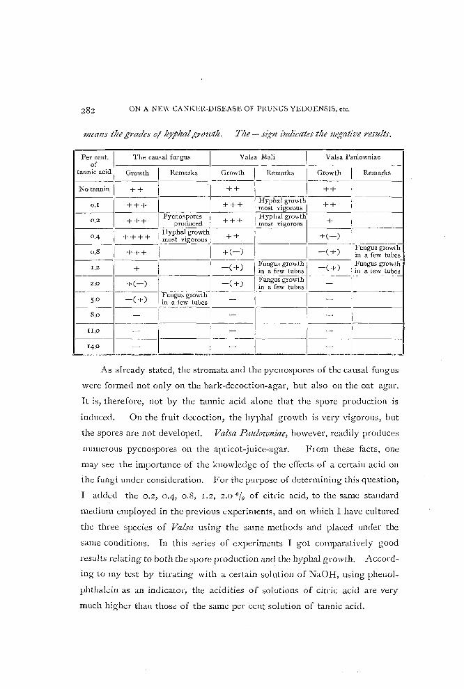
Table sltowillg- the different debtrJ'ees of gr:J'ivtlt of tlte causal fungus and two
other species of Valsa on tlte stalldard medium containing different percentages
of tlte tallnic acid. Tlte + sig-Il indicates tlte growth of the fungus and its 1lItmber
282 ON A NEW CANKER-DISEASE OF l'RU~US YEDOENSIS, etc.
mealls the grades oj hJphal/:,o'loth. Tlte - Sl~rryl indicates the negative results.
The causal fungus Val;a Mali Valsa Paulowuiae Per cent. of I-----c-------I------- ---- --- - - -
tannic acid Growth Remarks Growth \ Remarks Growth I Remarks ------1----- -------
No tannin ++ ++ 1 I ++ , 0.1 --+-+-+--1------1----+-+--+--1 Hyphal growth --+-+-- --- ----I
most vigorouo 1 ___________ 1
1
Pycnospores + I Hyphal growth + I 0.2 + + + produced + + mo"t vigorous I 0,4 + + + + I Hyphal growth --;=;:-,---------+-(---)-1------
1
__ _ __ I _____ -m-o--st-vlgorOUS _____ ,~ __ -" +(_) I -(+) - Fungus growth
0.0 + + + 1 in a few tuhes I---+--+------I--_-C-+-)-I ~'ungus growth l-=C-) -( !ungus growth-
1.2 _____ III a few tuhes + III a few tubes
2.0 + C -) [:-----, -( +) 1 t~u:g~:;r~~~~ 1-~ -- - - - --1-----1------ Fu~gusgrO\vth------ 1 I
5.0 -( + ) in a few tuhes - -
-8.0-1----1 I - - 1---1
-----
1_II.O_I ___ [_1 ___ -__ 1 __ 11
_ ---- 1 __
14.0 - -______ .---' __ . _...c.. ____ ..J.-_____ '--~ ___ ~__'__. ____ ._~
As already stated, the stromata and the pycnospores of the causal fungus
were formed not only on the bark-decoction-agar, but also on the oat agar.
It is, therefore, not by the tannic acid alone that the spore production is
induced. On the fruit decoction, the hyphal growth is very vigorous, but
the spores are not developed. Valsa Paulozvlliae, however, readily produces
numerous pycnospores on the apricot-juice-agar. From these facts, olle
may see the importance of the knowledge of the effects of a certain acid on
the fungi under consideration. For the purpose of determining this question,
I added the 0.2, 0.4, 0.8, 1.2, 2.0 % of citric acid, to the same standard
medium employed in the previous experiments, and on which I have cultured
the three species of Valsa using the same methods and placed under the
same conditions. In this series of expel-iments I got comparatively good
results relating to both the spore production and the hyphal growth. Accord
ing to my test by titrating with a certain solution of NaOH, using phenol
phthalein as an indicator, the acidities of solutions of citric acid are very
much higher than those of the same per cent solution of tannic acid.

TAKEWO HEMMI
From these experiments I obtained the following results:
(I) All the media remain liquefied when citric acid is added, but some
of them were at last solidified in consequence of the vigorous growth of the
hyphae.
(2) The growth of any of these fungi causes for the most cases no
darkening of the media even when citric acid is added.
(3) The causal fungus grows in all the cultures containing citric acid
except 2 %, showing a more vigorous growth than in the check cultures. The
,cultures containing 2.0 % of citt'ic acid show a comparatively poor growth
of the causal fungus and a darkening of the medium takes place in some
of them,
(4) Some of the cultures of this fungus containing 0.4 % of citric acid
produce the pycnO'ipores at about the 120th day after inoculation, and the
spore development is conspicllollsly vigorous all over the surface of the
medium.
(5) The color of the mycelium of this fungus gradually turns from white
to light yellow on the cultures containing citric acid, while on the cultures
containing tannic acid it turns to dirty brown.
(6) Although Valsa Mali grows in all percentages, its growth is general
ly poorer than in the case of the causal fungus; and the spores are not pro
duced.
Although Valsa Paulozvniae grows vigorously in all percentages, yet in
the tubes containing 0.2 and 0.4 % of citric acid its growth is most vigorous.
The cultures of the fungus containing 0.8, 1.2 and 2.0 % of citric acid
produce numerous pycnospores at about the I 20th day after inoculation, and
spore development is also conspicuously vigorous, with the exception of 2 % culture, in which the development is rather poor.
The results of this series of experiments are given in Table II, with the
same signs as in the preceding Table.

284 ON A NEW CANKER-DISEASE OF PRUNUS YEDOENSIS, etc.
Table II.
Table siwzvil1g tlte effect 0/ tlte differeNt percelltages of citric acid ill the
standafd medium Oil tile dez,eloj11lent 0/ tile causal/ungus and two otller species
0/ Valsa.
Per cent. I The causal fungus I VaLa Mali I Valsa Paulowniae of ,
citric acid I Growth I Remarks Growth Reluarks Growth I Remarks ------- -~--
I I No acid + I + + I ---- ------
0.2 ++ 1- + ++++ I Hypha~ growth most vlgorous
0.4 ++ I Pycnos pores + ++++ I Hyphal. growth produced lno;.;t vIgorous
0.8 ++++ I.HYPha~growthl +++ Hyphalgrowth +++ I
Pycno.;pore.; most vigorous _ mo~~igo~us ------
produced
1.2 ++ I ++ .++ I Pycnospores produced
Some orculture:; --
PycnOS1)Ore.:; 2.0
, +(-) I turn to black +(-) + I produced
EXPERIMENT II.
The next experiment was made with only the cultures of the causal
fungus, using the standard medium of corn-meal-agar to which had been
added the following percentages of tannic acid: 0.1, 0.4, 0.7, 1.0. To make
the standard medium I used the following method :-
15 grams agar melted in 500 c. c. water.
15 grams corn meal cooked ill 500 C. c. water for one hour.
Strain through cloth.
Mix the two and filter through cotton.
In this experiment, I used Erlenmeyer's flasks containing 25 c. c. of the
media in each. The cultures were started on the 2yd of December, I914.
The observations were continued to April, 1915, and the following results
were obtained.
(1) The growth of the fungus causes no darkening of the check-medium
containing no tannic acid; but when tannic acid is added, even as loW' as 0.1 %, the growth of the fungus causes a darkening of the medium, as already stated
in the case of the first experiment.

TAKEWO HE~1MI
(2) The medium containing tannic acid remains liquefied when 0.7 or
r .0 % is added.
(3) On all cultures containing tannic acid, the mycelium turns to a dirty
brown color, while on checks it has remained colodess.
(4) Check cultures containing no tannic acid show a vigorous growth
of mycelium, which is, hmyever, very loose, sending up the aerial myceliu~.
Spores have not been produced in these check cultures.
(5) Cultures containing o. I % of tannic acid also show a VlgOroUS
growth of mycelium, but its entanglement is a little closer, and some of them
produced numerous red masses of pycnospores at about the 50th day after
inoculation. In this case, the spore production is more vigorous than in the
cultures containing 0-4 % of tannic acid.
(6) Cultures containing 0.4 % of tannic acid show the most vigorous
growth of mycelium in this series of experiments, forming a close coating
over the surface of the media. Some of them produced also nurnerous red
masses of pycnospores at about the 50th day after inoculation.
(7) The growth of hyphae on the cultures containing tannic acid seems
to be slow at first, but later becomes more luxuriant, compared with the
cultures without it.
(8) Some of the cultures containing 0.7 % of tannic acid also show a
vigorous growth of mycelium, but it takes a long time for the mycelium to
appear and grow on the surface, due to the liquid of the medium; and at last
the growth of the fungus tends to form a more or less close coating over the
surface. But in some of these cultures the fungus entirely failed to grow. I
(9) All cultures containing 1.0 % of tannic acid fail to allow the growth
of the fungus.
The results of the second series of experiments are given in Table III.
Table III.
SllOwillg tlte effect of tile dijjerellt percClZtages of tannic acid ilt tlte standard
medium Oil tlte growtlt of tlte causal fungus.

286 ON A NEW CANKER-DISEASE OF PRUNUS YEDOENSIS, etc.
Per cent. Growth of Mycelium Production of of
tannic acid Grade Remarks pycnospores ---- -----
No tannin + + loose -
0.1 + + close + +
0.4 + + + closest +
0·7 + or- clo.;e -
1.0 - I -
EXPERum;>;T II L
In this experiment the same media and methods were used as in the case
of the second experiment. It was started at the beginning of May, 1915. In
this case, Valsa Mali and Valsa PaulozVlziae were taken up in addition to the
causal fungus, for the sake of comparison.
following table.
Table IV.
The results are given in the
Showing the effect of tlte different percmtages of tallnic acid ill the standard
medium on the growtlt of tlte causal fungus and two other sPecies of Valsa.
! Per cent. The causal fungus , Vaha Mali Valsa Paulowniae
tannic acid Growt-h--,-Production- Growth Production Growth' Producti~ I of I ____ _ of pycnospores ~ _____ ~fpycno,;p~r-,e,,-s'I _____ ofpycnos;)Qres
No tannin ++ I ++ +++ ~------I------,---------I---
0.1 ++ + ++ + --·---1-----------1-----·------1--- ---I------l
0.4 ++ I -• + - + or - -
I 1.0
- I -
I - I -I
-+ or - - - -
- I - - -
0.7
c. Conclusion.
From the foregoing experiments on the effect of tanni~ acid on the de
velopment of the causal fungus and other species of Valsa, the foilowing con
clusions may be drawn:-

TAKEWO HEMMI
(I) The fungi can use tannic acid, at least in a small amount, as food,
shown by the blackening of the media through oxidation, and by a more
luxuriant growth, with a low percentage of the tannic acid added, than in
the case without it.
(2) Higher percentages of tannic acid (1.2 % and above) are detri
mental to a vigorous growth of the causal fungus, and finally (8 to [4 %)
entirely inhibit its growth.
(3) Different species of the same genus may vary in power of resistance.
The resistant power of the causal fungus against tannic acid is the highest
among our three species of Valsa. I agree, therefore, with COOK'S opinion 6)
that some species of fungi are much more resistant to tannin than others, and
the species which attack the high tannin-bearing plants no doubt possess this
quality.
(4) High percentages of tannin have the tendency to retard or inhibit
the growth of fungi. But the growth of the fungi is frequently increased by
the use of low percentages of tannin, and in this case the hyphal growth is
closer than without it.
(5) In some cases the growth was at first retarded by certain percent
ages of tannin, but later became as good or better than on the medium without
it.
(6) The formation of spores was stimulated, at least in the case of the
causal fungus, by low percentages of tannin.
(7) Citric acid also stimulates the spore formation of this fungus, pro
bdbly rather more than tannic acid. But on the cultures containing tannic
acid, the time required for frUiting was shorter than on the cultUl'es contain
ing citric acid.
(8) The fruiting of this fungus is not determined by the acidity of the
medium alone, but it seems to me to be due to a combination of various
factors that constitute the real cause of the stimulation.

288 ON A NEW CANKER-DISEASE OF PRUNUS YEDOENSIS, etc.
9. Confirmation of the Genetic Relation between Pycno- and Ascosporous Stages.
On the inf!':cted area of the branches and trunks, two spore forms, pycno
and ascospores, usually develop in different stages, as already described. It
has been demonstrated by various authors that some species of Cytospora
belong to Valsa as its pycnidial stages. In my case also, the pycnidial stage
of the causal fungus is a species of C;,tospora.
Inoculations made with pure cultures of our Valsa and Cytospora produced
the same results. The characters of pure cultures of these two types of the
spores are quite identical. As a direct proof, the ascospores of Valsa
were inoculated on the sterilized twigs of PmlZlts yedomsis. As I had
anticipated, a (ytospora was at first produced which is identical in every respect
with one on the natural host, and finally the Valsa stage was reproduced.
Moreover, when the Cytospora spores were inoculated on the sterilized twigs
of the same plant, the same Cytospora was reproduced, and after a long time
a Valsa form was produced which is identical with the causal fungus.
10. Drop-Cultures.
a. Germination of pycnospores.
The pycnospores were not made to germinate well 111 pure water.
Although I sowed the spores in a drop of distilled water over and over again,
I could not succeed in making them germinate well. But once in a while
I noticed that a few spores put out a slender germ-tube at one end, without
conspicuous swelling, but soon died for want of nutrient. I also got the
same negative results with rain-water, tank-water, and various kinds of sugar
in such low percentages as I, 2, 5 or 10 010' I have found the most satis
factory medium for this purpose to be a decoction made by boiling host-'bark
and diluted pear juice.
The time required for germination vanes with the temperature. At
room temperature in summer, which ranges from 20° to 28°C, the germina
tion occurs in thirty to sixty hours. At lower temperatures the process often

TAKEWO HEMMI
requires four or five days. From these facts, we infer that infection by the
pycnospores can occur only in the warm period of summer.
The process of germination begins with an enormous swelling of the
spores, especially in width. The spores measuring 5·25-I2.25 x 1.75-2.63 f1
before germination were found, at the end of twenty-four hours in a bark
decoction, to measure 6.I3-£4.0 x 4.72-7.0 f1; and not infrequently they
reached 15.75 x 7.88 f1 just before germination. In such cases, the spores
took various irregular forms,- ellipsoid, ovoid, obovoid, subglobular, etc. In
, a few cases I noticed that some spores were divided into two cells just before
germination (PI. X, Fig. 4-5.). The spores germinate in thirty to sixty hours,
throwing out one to three germ-tubes. Usually a germ tube grows out from
one end, and this is followed later by a second one from the opposite end (PI.
X, Fig. 5-7.). The germinating hyphae are at first hyaline,' about 3.2-4.0 f1 in
width and occasionally swollen in an irregular shape. The branching and
septation of the germ tube or mycelium then take place; and the old hyphae
often turn gradually to a yellow or light brown color after three or four days.
The germinating spores and hyphae are at first granular in contents and after
a while vacuolization occurs in many cases.
In the experiments described above, I took all the spores from pure
cultures. Such swelling and manner of germination of the pycnospore are
not infrequent in many genera belonging to the Ascomycetes. ADERHOLD
(I90 3) I) also described the same process in the case of Valsa leltcostoma
(PERS.) FR.; but such two-celled spores have not been observed by him.
According to DE BARY (1887) 8\ the cause of such enormous swelling of the
spores before germination is attributed to absorption of water. On the con
trary, ANDERSON and RANKIN (I9I4) 2) speaking about the phenomenon in the
case of Endothia parasitica (MURR.) ANDERS., the chestnut blight fungus,
write:- "The swelling of the spores is due, not merely to a mechanical
imbibition of water, but also to a process of growth. Pycnospores stained
just before the germ tube is started show that the increase in size is accompa
nied by active nuclear division, two to six nuclei then being present. The

290 ON A NEW CANKER-DISEASE OF PRUNUS YEDOENSIS, etc.
nuclei pass out into the germ tubes almost as soon as they start. The waIl,
also, has increased in thickness until it almost equals the diameter of the
resting spore." This is surely a very interesting observation, but I can not
yet assert whether it is correct or not, at least in the case of the present
fungus.
b. Germination of AscosjJores.
Unlike the py<;nospores, the ascospores germinate readily in pure water.
After two or three days, the germinating hyphae grow weaker and weaker, and
at last die in water. The time required for germination is much shorter
than for the pycnospores in a nutritient solution or even in pure water. At
comparatively low temperatures, ranging from 14° to 24°C, it occurs perfectly
within the first twenty hours. I even observed the septation and branching of
the germ tube at the end of twenty-four hours. From such facts, we can
safely infer that if they are placed under more favorable circumstances, germi
nation will take place within six or twelve hours, and that therefore infection
by the ascospore is most considerable and dangerous.
Like the pycnospores, the ascospores swell before germination, but not
to so great an extent as in the case of the former. The ascospores, measur
ing J 6.0-22.0 X 3.2-4.8 fl at first, were found, just before germination in a
bark decoction and pear juice, to measure 20.0-36.0 x 4.0-10.0 fl. But
according to Illy observations, they always swell uniformly, keeping their
original allantoid forIll. This character differs from that of Valsa leucostoma
in which ADERHOLD (1903) 1) observed the spores to swell to eIlipsoidal or
globular shape. The first germ-tube usually appears at one end; the next
one COIlles from the other end or from one side; and these are occasionally
followed by another one or two, making a total of one to four germ tubes.
They are most commonly thrown out from both ends, but rarely from the
convex side of a spore. About 10% to 15 % of the germinating ascospores are
divided into two cells as in the case of the PYCllospores (PI. X, Fig. 1-2.).
In 1891, such two-celled ascospores before germination were already observed

TAKEWO HEMMI
by BREFELD3) in the case of germination of Valsa ceratophora TuL.
The germinating hyphae are hyaline at first, about 3.2-6.0/,. in width,
and often the old parts turn light brown or light yellow. In a drop of bark
decoction, it was occasionally observed that some germinating hyphae stopped
their growth when they reached about 32.0-80.0 p. in length, swelling a little,
and that soon the fine horn-like hyphae developed again from their swelled
ends (PI. X, Fig. 3.).
In the eXDeriments, I obtained pure ascospores from the perithecia on the
natural host by letting them eject the spores on a slide. In the process of
germination of the pycno- and ascospores, the various behavior of the hyphae
and changes produced in their contents at-e very similar. The ascospores of
our present fungus do not require a 'period of rest, but germinate directly
after maturity, if placed unda favorable conditions.
c. Observations on the Hyphae in Drop Cultures.
If the surrounding conditions are favorable, the hyphae started from
the germ-tube proceed with their growth in a radiating manner. But the
rate of growth varies considerably with the temperature and the kinds of
nutritient solutions; and in such an acid solution as fruit juice, the hyphae
gl-0W most actively. In the bark decoction, the growth is not so active, but
the dense and short branching, which is said by many authors to be a young
stage in the development of the pycnidium, is apt to be quickly produced.
The growing hyphae are always hyaline, although their basal parts turn
yellow or light brown; and the hyphae are also not of uniform diameter, but
vary, according to my observation, from 2.0 to 8.0 p..
Under the microscope, we often see the hyphae anastomosing in many
ways; and in consequence, they at last make the hyphal network. After more
than ten days, it is not an infrequent sight to see them entangling in a mass.
Occasionally I saw a substance being secreted on the tip of the hyphal branch,
which is at first hyaline and at last turns grayish yellow or dirty yellow. But
the nature of this secreted substance is not yet known to me.

292 ON A NEW CANKER-DISEASE OF PRUNUS YEDOENSIS .. ctc.
11. Resistant Power of the Hyphae against Solutions of Corrosive Sublimate, Copper Sulphate, and other Chemicals.
Solutions of corrosive sublimate and copper sulphate are generally used
for fungicides, and occasionally soda-solutions are used for the same purpose
in our country. vVhen surgical operations of various canker-diseases are
performed, they are also used to wash the exposed surface of the wound. In
order to determine first their killing power against the hyphae of the present
fungus, I poured each of these poisonous solutions into a test-tube, in which
the hyphae had been growing up actively. After certain intervals, as indi
cated in the following tables, I took out the pieces of hyphae with a platinum
needle, and washed them in distilled water which had been previously steri
lized, and again inoculated on a new cultural medium.
Of course such experiments are not precise, because the chemicals can
not sometimes touch the hyphae, owing to the air layer which is surrounding
them. It was most conspicuous in the case of copper sulphate. Even if the
results of my experiments were not conclusive, for the one purpose of deter
mining th~ir value as fungicides, we can safely summarize as follows :--
I. Corrosive sublimate has a strong sterilizing power for the hyphae of the
present fungus, being effective even in such a dilute solution as 0.0! %.
But as a fungicide, it is safer to use a 0.05 % or o.! % solution.
2. Copper sulphate and carbonate of soda did not give good results for the
same purpose, probably owing to the disturbance of the air layer.
3. Caustic soda has sterilizing power for the hyphae of the present fungus,
when such a concentrated solution as 10 0/ 0 or 20 0/ 0 is used. Even with
the 5.0 % solution, we can probably obtain good results for the same pur
pose.
The results of these experiments are given in the following tables. In
these tabl~s the plus sign means the presence of the living hyphae and the
minus sign means their death.

TAKEWO HEMMI
Table V. Results of experiment with corrosive sublimate.
Time of soakage 0.01 % 0.025,;6 0.05% 0.1% (in minutes)
0 + + + +
I - - - -
5 - - - -
10 - - - -
30 - - - -
Table VI. Results of experiment with copper sulphate.
Time of soakage 0.5%
I 1.0% 2.0~~ 3.0 % 4.0 % (in minutes)
0 + + + + +
1 + + + - + - + -
5 + + + - + - + -10 + - + - + - - -
30 + - + - + - - I -
60 + - + - - - I -
Table VII. Results of experimmt with caustic soda.
Time of soakage
I 1.0%
I 2.0% 5.0% 10.0% 20.0% (in minutes)
0 + I + + + + I 1 + +
I
+ - - -
5 + - + - - -
10 + - + - I - - -
3 0 - - I - - -i I
Table VIII. Results of experiment with "carbonate of soda".
I Time of soakage 5.0 % 10.0% 20.0% 30 .0 ~G 40 .0 % (in minutes)
0 + + + + +
1 + + + + + -- --
5 + + - + + + -10 + + - + + - -30 + - + - + - + - -

294 ON A NEW CANKER·DlSEASE OF PRUNUS YEDOENSIS, etc.
12. Systematic Position and Nomenclature of the Causal Fungus.
From the morphological characters, we may easily recognize our fungus
to be a species of Valsa. It has pycnidia of the Cytospora type.
The genus Valsa was first described by FIUES( I 849) :JG); and then NITSCHKE
(1867) IR), who made a new family Valsaceae out of it, studied the genus ex;
haustively in his work "Pyrenomycetes Germanici". In ENGLER and PRANTL'S
"Die N atiirlichen Pflanzenfamilien", L I~mAU (1897) 16) di vided the genus into
ten subgei1era. Most of these subgenera were founded by NITSCHKE and
endorsed by WINTER (I887)34) and SCHKOTER ([9:)8)26). They are distin
guished from one another chiefly by the construction of the stromata. In
SACCAI{DO'S "Sylloge Fungorum", however, only two subgenera, Ellvalsa and
Leucostoma, are treated as belonging to this genus and all other subgenera
were raised to independent genera. On the ground of the morphological
characters of the stromata, I have come to the conclusion that the present
fungus is a species of Eltvalsa. NITSCHKE divided this subgenus further into
two groups, .!VIollostieltae and Circinatae, and our fungus belongs to the latter,
which corresponds to Macrosporae in SACCAIWO'S system.
Up to the present time, more than fifteen Valsa and still more Cytospora
were found both saprophytically and parasitically on the twigs and branches
of the various species of Primus in the world; but most of them differ in their
systematic position and also in other essential characteristics from our fungus.
With some of them, owing to the brevity of their descriptions, I could
scarcely make any comparison with our causal fungus.
Valsa leucostoma (PERS.) Flc which causes the disease of many drupace
ous trees, known as "die back", throughout Europe, Australia and America,
not only has the "conceptaculum" between the stroma and the host tissue,
but differs also from our present fungus in many other essential points, as
shown in the following table :-

TAKE\,\'O HEMMI 295
Table IX.
Valsa leucos toma The Causal Fungus
Numher of perithecia 3-10 (to 20 after some anthors) Commonly 15-40 in a stroma
-- ---- --48-56 X6-81'-
6v-96 X8.8-161'-Asci (35 -45 X 7 -81'- after STEVENS, ) 40-62X5-SI'- after ADERHOLD.
------- ---1O-18X2.5-3.51'- IO-28x3.2-7.21'-
Ascospore, (9.3-14X2.3-2.61'- (mostly 1O-12X2.5) (COmmOnly 18-22X4.0-' 1'-) after ADERHOLD, 4.8 1'- ) 9-12 X2-2.5 po after STEVENS.
4-5X1\1- 3.5- 15.75 X 1.0-2.63 \1 Pycnospores (J.5-9.3XO.87-2.3\1(COmmOnIY ) (CommonlY 7.0-8.75 X) 4.5-6x 1-1.5 \1) after ADERHOLD,
5.5-7.5 X 1.0- 1.5 \1 after McALPINE. 1.4- 1.751'-
Valsa Prztllastri (PEKS.) FR., reported as the cause of a serious disease of
apple, plum and other drupaceous trees in England, belongs to the subgenus
l:"lttypella, which was raised by SACCAIZDO to an independent genus, and is too
distinct from our fungus for the purpose of comparison.
Valsa ambiens (PE[{S.) FR., which is described as widely distributed on
various kinds of trees and which belongs systematically to the same subgenus,
seems to be nearly allied to our causal fungus. But one can still find various
differences, as seen in the following table, especially in the measurement of
the pycnospores and the structure of the stromata.
Table X.
I Valga ambiens The Causal Fnngus
I 3- 5 mm. in breadth, Size of stromata
I 1.5 - 3.0 mm. in diameter at base. 2-4 mm. in height,
and 1-1.5 mm. III depth.
:K umber of perithecia I 4-20 Commonly 15-40 in a stroma
---- --"-- -
Construction of I Lower half of the stroma consists mostly The greater part of the stroma consists of the mycelial tissue,
stromata II of the hOot-tissue. and the host-cells are scattered in it.
---~ ----
I
oblong or clavate, Cylindrical or rarely clavate, Asci
40 - 88 XS-16I'-,4-8 spored. 60-96 X8.8-16\1, always 8 spored .
-- --- . -~-
(16-24X3-6 \1 in 8 spored asci,) 24-36x5-8~. in 4 spored asci
JO-28X3.2-7.2 1'-after \VINTER and SCHROETER. Asco.;pores
(16-18X3-4\1 in8 spored aSCi') 20-20XS-61'- in 4 spored asci (Commonly 18-22X4.0-4.8\1-)
after SACCARDO.
I Pycnospores I 3.5- 15.75 X 1.0-2.631'-
5 -7 X 1\1- (Most commonly 7.0-8.75X I 1.4- 1.751'-

296 ON A NEW CANKER-DISEASE OF PRUNUS YEDOENSIS, etc.
Further we were fully convinced that these two fungi are quite different,
when we examined and compared our fungus with the European specimen of
Valsa ambiClls preserved in the Herbarium of our University.
As we have not been able to find any species of Valsa or Cytospora which
agrees exactly with the present fungus, either in mycological or phytopa
thological literature, we consider the fungus in question to be quite new to
science. The species may be characterized as follows :-
Valsafapollica MIYABE et HEMMI sp. n.
Stromata scattered, produced at first under periderm, then erumpent,
forming lenticel-like openings, which are slightly elongated horizontally,
conical or wart-like with round or elliptical bases, gray or brownish-gray
with many black ostioles of perithecia on the surface, gray or yellowish gray,
subcoriaceous in the inside, consisting for the most part of the mycelial tissue,
with a few host cells scattered deeply in it, about 3-S mm. in average breadth,
2-4 mm. in average height and I-I.S mill. in depth; perithecia immersed,
subglobose with long neck, compactly arranged in concentric circles, 15-40
in a stroma, 350-S8o,u in diameter; asci cylindrical or rarely clavate, subsessile
or short stalked, 60-96 x 8.8-16.0,u, hyaline, eight-spored, evanescent in old
perithecia; spores mostly biseriate, sometimes irregularly arranged, allantoid
with rounded ends, almost hyaline, slightly curved, 10.0-28.0 x 3.2-7.2,u (com
monly 18.0-22.0 x 4.0-4·8 ,u).
Pycnidia: stromata scattered, produced at first under periderm as sub
spherical or ellipsoidal cushions, then erumpent, turning to conical or wart
like shape, average about 2.2 mm. in breadth, 1.S mm. in height and 1.0 mm.
in depth; internally divided into cavities with a single common exit; pycno
spores hyaline, cylindrical, rounded at both ends, generally curved but occa
sionally straight, 3.50-15.75 x 1.00-2,,63,u (mo9t commonly 7.00-8.75 x 10⁴-
I.75,u), oozing out in reddish curls when moist; basidia hyaline, simple or
branched, variable in length but measuring 14-28 x 1.75-2. 1/1..
Hab. On the bark of Pnmlts yedoCllsis. MATSUM.

TAKEWO HEMMI 297
Provo Ishikari; Sapporo and its vicinity (I & II. * from 19[3 to [915.
K. MIYABE, S. ITo, T. HE:\1MI), Asahikawa (I. Sept. 26, [914. T.
HEMMI).
Provo Oshima; Hakodatc (II. Feb. 14, [915. S. NISHIDA).
On the bark of Pntlllts sachalillelZsis KOIDZ.
Provo Ishikari; Sapporo and its vicinity (I & II. Apr.-Nov. 1914.
T. HE~Ii\lI).
On the twigs of PyltllltS Koidzumii MAKINO.
Provo Ishikari; Sapporo (1. May [8, 19 [5. T. HnDlI).
On the bark of Pntlllts serrulata LINDLEY.
Provo Ishikari; Sapporo (I. August 15, 1914. T. HE~IMI).
On the bark of Pmlllts sen-ulata LINDL. var. lzobilis (KOIDZ.) f.
Yokilzi.
Provo Ishikari; Sapporo (II. May, 1915.1. NAMIKAWA).
On the t\vigs of PritIlUS kltrilensis MIYABE.
Provo Ishikari; Sapporo (I & II. May 12,1915. S. ITo).
On the bark of Pmlllts Mume S. et Z.
Provo Ishikari; Sapporo (I & II. 19[4-1915. K. MIYABE, S. ITo, T.
l-rEM~lI ). . On the bark of PntllliS Persica. S. et Z.
Provo Ishikari; Sapporo (I & II. Feb. 8, 1915; I & II. Apr. 8,1914.
T. HEMMI).
13. Inoculation Experiments.
These experiments were undertaken over and over again 111 order to
determine the parasitic habit of this fungus. Although I had failed in many
cases with these inoculations, I succeeded at last uncler a special condition to
produce the disease artificially. The most important reason for such failures
seems to be the powerful resisting power of the seedlings, which were used in
these experiments for convenience' sake.
* 1= Pycnidial stltge. II = Ascosporous stage.

298 ON A NEW CANKER DISEASE OF PRUNUS YEDOENSIS, etc.
EXPERIlIIENTS I.
It is rather difficult in making a series of inoculations in the field to
provide conditions which correspond to those of natural infection, and also
by such experiments to draw the conclusion that the disease is caused by the
fungus used.
From April to June, 1914, I inoculated the fungus on PrUl1lts yedoeJZsis,
using various methods in the field. But all these inoculations resulted in
failure with one exception, that is, when I inoculated with the germinating
ascospores in the apple juice into a wound and covered it up with moist cotton
and paraffine paper for a few days. After two months I found the character
istic appearance of the disease around the inoculated point. The failures of
other inoculations were due chiefly to the want of moisture or to the washing
away of the spores by a heavy rain.
EXPERIMENTa II.
In this case, the seedling trees of Prltll1ts yedoensis, a little more than two
years old, grown in pots, were used. In July, 1914, the inoculations were
made partly with pycnospores from artificial cultures, and partly with asco
spores from a natural host. Ordinarily, a small slit in the bark was made with
a sharp sterilized scalpel, and the spores were introduced with a sterilized
needle. The wound was covered with moist absorbent cotton, and then all
was bound up with paraffine paper. The seedlings, inoculated by the same
method, were partly placed in a moist condition under a bell-glass for a few
days. As checks, uninjured sound bark and lenticels were also inoculated.
Although in all these experiments the moisture was supplied ill sufficient
quantity, in no case was the infection accomplished.
EXPFRHfENTS III.
Again in September (28-30), 1914, the seedlings of Pmlllts yedoeJZsis,
a little more than two years old, grown in puts, were used for inoculation.
In these experiments, for the place of inoculation, a small portion of the bark

TAKEWO HEMMI 299
was burned in addition to the same methods used in Exs>eriment II. The
experiment was based upon the idea that the fungus may first require the
dead cells for the penetration of its germ-tubes. Upon such a hypothesis,
infection experiments were performed by ADERHOLD J) with great success in
the case of Valsa leucostoma. To keep it moist, I placed the pot under a
bell-glass for one week. The results are given in the following table.
Table XI.
Inoculated bark I Inoculum I Results
Smooth bark ! Pycnospores from culture ---- --------
T.enticel I Pycnospores and mycelium from culture -
Cut and burnt bark I Pycnospores from culture 1--+ -
I Cut and burnt bark
I Mycelium from culture +
New cut wound I )iseased bark I -New cut wound I Ascos pores from natural host -
--"- - - --- - ----- --New cut wound I Pycnospores from culture --- -
New cut wound ! Mycelium from culture ----~-~
New cut wound I Pycnospores and mycelium from culture I -
The seedling which had been inoculated with the mycelium to its burnt
portions suddenly wilted in about two weeks after inoculation, and the seed
ling which was inoculated with the pyCllospores to a similar wound also
suddenly wilted in about three weeks after inoculation. But even on the same
seedling" the twigs above a burnt wo~nd which was treated in the same way,
with the exception that no spores \~cre introduced into the wound, did not
show any change for a long time. In the former case, the cork layer of the
infected portion was raised in numerous little blisters. Sections. through
such a dead portion showed the tissues of the bark as well as the water ducts
of the woody portion to be traversed by the mycelium. Under each blister
was a compaCt mass of the mycelium, which is no doubt a young stage in
the development of the stro;na. But in no blisters were pycnidia devc:loped,
whose non-formatio~ is chieAy due to the resistibility of the young host.

(I)
(2)
(3)
ON A NEW CANKER-DISEASE OF PRUNUS YEDOENSIS, etc.
EXPERBlENTS IV.
As a result of the above experiments, it was necessary to try inoculations
on aHarge branch. On January 19th, 19 [5, a large branch of Primus yecloensis,
about four centimeters in diameter, was cut and put in water in the labora
tory; and the water was renewed every day in order to prevent the multiplica
tion of micro-organisms in it. On about the 20th of February the branch was
in full blossom. On the day when the branch was cut, it was inoculated with
the fungus in various ways, and on the 13th of February the results were as
follows :-
Table XII.
Inoculated portion I
Inoculum I Results - - --
New cut wound with scalpel Ascospores from nalural host i Bark slightly sunken and gUln , Howed out _._---
" Check I No change
New cu~ wound with saw I l'ycnospores and mycelIUm from I No cha!lge culture
" Check I No change
Hurnt wound Ascospores from natural host I Bark sunken, guin-flowed out and stromata developed
Pycnospores and mycelium from I Barl< sunken, gum tlowed out and " culture stromata devel:,ped
" I Check I No change
In these experiments, the wounds were kept moist by covering them with
moist cotton, and then binding them up with paraffin paper for a week.
In the first case, the portion around the inoculated point sto?ped its
growth, forming an elliptical sunken sp6t; and the gum was exuded from the
inoculated wound. But in such a sunken area, the progress of the disease
soon ceas.ed, and no stroma of the fungus developed in it. But on the COI1-
trary, the sunken area of the thit'd case gradually increased its extent and the
stromata of the fungus were developed sparingly upon it. When the branch
was in full-bloom, thirty-live days after the inoculations, it began to wilt,
showing a diseased appearance. In April, numerous small pustules of the
fungus stromata appeared, scattered throughout the surface of such a dead
I
I

T A KEWO HEMMI 30r
branch, showing the symptom of a girdled branch. Similar experiments
were also undertaken with smaller branches, and the same results were
obtained.
Besides these experiments, I also inoculated the same fungus on the
branch of Przmus Cerasus, using the same method, but the results were
negative.
EXPERIMEN'l'S V.
In this case, I used two comparatively aged potted trees of Pnmus Mume
which were at that time in full-bloom in a green house, and on each tree
small burnt areas and slit wounds by a scalpel were made just before inocula
tion. In the injured portions of the one I inoculated the mycelium from a
culture which was isolated from P. Mtmze, on February 12,1915. On another
one I inoculated the mycelium from a culture which was isolated from Pmlllts
1'edoellsis on the same day. To keep it moist, I placed the pot under a bell
glass for one week, and then it was placed for a long time in a warm labora
tory.
On the first tree, the bark of an inoculated branch changed its color
around the burnt portion, then gum flowed out, and at last many small pus
tules of the fungus stromata app~ared rather scatteringly, while the checks
indicated no change. The discolored portion of the bark gradually extended
its area, which was sunken slightly, but up to the end of June the branch
was still in foliage, except a small twig which was branched near the infected
portion. From July to September, the upper jJortion of the branch from the
infected point died gradually, and the fungus stromata also produced scatter
ingly all over its surface. On the other branch which was inoculated in the
slit wounds, the results were negative.
On the second tree, the bark in the vicinity of both burned and injured
portions on which inoculations had been made, first changed color, then gum
flowed out, and at last the upper two twigs died, when there appeared also
small pustules of the fungus stromata. The wilted flowers remained clinging

302 ON A NEW CANKER-DISEASE OF PRUNUS YEDOEKSIS, ctc.
on the dead twig for a long time (PI. VIII. Fig. 3.). But the lowet- branches
were still in foliage up to the end of September, although the diseased area
had slowly been extending.
EXPERIMENTS VI.
From January to April, 1915, I several times tried various kinds of
inoculation on the three-year seedlings of P. yedoeJZsis and also the com
paratively old potted trees of a kind of P. sermlata. And the conclusion
was reached· that this fungus most easily infects the hosts at the old wounds
which have a layer of dead cells on the exposed surface. In these exoeri
ments the pots were placed under bell-glass in order to keep them moist fOl- a
week. The details of these experiments are shown in the following table:-
Table XIII.
Inoculated portion Time of
Inoculum Results inoculation
I. Sound Luds of seedling Jan. 26 Pycnospores from culture - Lut infected Luds wilted
---- ---~- ----- -2. New slit wounds on seedling Jan. 26 Ascospores from natural host -
3· Older wounds on seedling March 13 Mycelium from culture + --
N<,;w slit wounds 4. potted tree
on aged Fehr. 12 Mycelium from culture --- -------.---~--
5· Burnt portion on aged potted Febr. 12 Pycnospores from culture 1-tree
- ---~---- -~
6. Burnt portion on aged potted April 16 Pycnospores from culture I + tree
In the first case, it seemed to me that the germ-tubes of the inoculated
spores had the power to kill the buds. But on the sound bark, the infection
never took place. In the third case, four slit wounds were given with a steri
lized scalpel on the bark of the three-yeat--old seedlings, about two months
before inoculation. On March 13th, 1915, mycelium with a little piece of the
cultural m~dium was inoculated in the npper three wounds. and the lowest
wound was treated as a check. About the 9th of April, the upper-most
shoot wilted, and at the end of the same month the second shoot wilted,
developing at the same time many small pustules of the young stage of the

TAKEWO HEMMI
fungus stromata on the bark. Its appearance on the 1st of May is shown in
the photograph of PI. VIII, Fig. 2. In the sixth case, the upper twigs wilted
about the middle of May, and the pustules of the stromata began to be pro
duced at the end of the same month.
EXPERIMENTS VII.
In April, 1915, several inoculation experiments were made with myce
lium from cultures, isolated from various host plants, on the burned bark
of the seedlings of P. yedomsls. The results of these experiments are as
follows ;-
Table XIV.
Host of inoculated fungus Time of Results I inoculation
Prunus Persica S. et Z. I
April 9 On May 10 the uppermost shoot wilted and
on ahout June 4 the stromata developed.
Prunus sachalinensis KOlI>z. April 9 On May 3 the uppermost shoot wilted and
at the end of May stromata developed.
Prunus Mume S. et Z. April 30 Negative
Prunns Cerasus L. April 16 Negative
l'rUl1Us yedoensis MATSUM. April 9 On May 1 the uppermost two shoots wilted
and at the end of May stromata developed.
14. Entrances and Promoting Agencies of the Disease.
Judging from the results of the inoculation experiments, we may safely
infer that Valsa iaponica is the direct cause of this canker-disease, and that
the fungus is a wound parasite requiring for its infection various kinds of
wounds. When the spores germinate in a wound, the mycelium derived
from the germ tubes thrives on the injured and dead cells until it has produced
a mass of mycelium. Then, gradually accumulating strength as it increases,
the mycelium en masse pushes out into the living tissue of the bark. Single
hyphae do not seem to possess the power of penetrating into the living cells,
but the in vasion is accomplished by the force of mass action. Starting from

304 ON A NEW CANKER-DISEASE OF PRUNUS YEDOENSIS, etc.
an infected point, the hyphae gTOW and spread in all directions, completely
destroying parenchyma, collenchyma and cambium cells as they go. These
facts are not open to question, judging from the results of the inoculation and
anatomical studies. I shall now give a brief explanation, based upon my
own observations, of the mode of the entrance of the hyphae, and its pro
moting agencies.
a. Crotch of branches.
The crotches of branches have many chances to get cracked by wind,
snow and various other causes. There are commonly many rumples which
are composed of dead tissues, especially in the Case of old branches. Such
places are also under such a condition as to remain moist for a comparati vely
long time. It is therefore probable that such crotches serve as places of
entrance of the parasite. Indeed, I have observed many cases in which the
disease spreads into both branches, starting from a crotch, as shown in the
photograph in PI. VII, Fig. I.
In the case of Valsa Mali, it was also suggested by Prof. K. MIYABE and
other observers that the crotch infection of the fungus takes place in the
apple tree.
b. Dead twigs.
I do not hesitate to suggest a dead twig as a channel of the entrance of
the disease, since I often find it at the center of cankers, especially of young
cankers, as shown in the photograph in PI. VII, Fig. 2 and Fig. 6.
Although the cause of the death of the twigs was not investigated by
me, winter injury may in most cases be the cause of its death in a cold, snowy
country such as Hokkaid6. Indeed in spring, one can find numerous dead
twigs on the tree, especially on Pnmus Mume. But unskilful pruning and
injuries caused by men, and by birds and other animals may occasionally be
promoting agencies of such death.

TAKEWO HEMMI
c. Buds.
As already shown by the inoculation experiment, the germ-tubes and
hyphae of this causal fungus have the power to kill the buds of the host plant,
although I failed to secure infection of the bark of the twig.
In the case of Valsa leucostoma PERS., the causal fungus of the "die
back" of the peach tree, ROLFS (I9'J7) 23) also suggested it to be a channel of
entrance of the disease.
d. Insects.
Insects are found in great numbers both in and on the bark of the host
plants in the vicinity of Sappo1'O, and it is reasonable to believe that they
may crawl over sticky spore horns and carry the spores away to deposit them
on wounds, and thus start new cankers. Indirectly, insects may be connected
with the spread of the disease by making wounds in the bark, where the
spores may gain entrance after having been carried by some other agents.
I am now of the opin ion that this is the way in which insects are most closely
reldted to the dissemination of the disease. In the fall of 1914, I found many
larvae of Sesia Ileelol' BUTL., commonly known by the name of" Kosltl.:aslliba"
in Japan, feeding 'on the diseased portions in almost all infected parts of the
smooth bark 011 trunks. According to Profs. MATSU~1URA and NIIJIMA,
Sesia Ilector BUTL. is the most common noxious insect, whose larvae inhabit
the bark and wood of the trees, which belong to the genus PrltIZUS, in Japan.
It is also written that this insect is found mostly on the trunks of aged trees
and causes the gummosis of the host plant. The fact that the present disease
is very common on the trunks of old trees shows beyond all doubt that infec
tion on such a smooth bal'k originated for the most part in the exit holes of
this insect.
There are, besides, two or three kinds of bark borers in the vicinity of
Sapporo, found on the living and dead trees of the hosts.

306 ON A NEW CANKER·DISEASE OF PRUNUS YEDOP;;-;SIS, etc.
e. Pruning.
It is a very rare practice in Japan to prune Primus yedoensis and other
Flowering Cherry trees, while it is quite common to prune Primus Mumc and
Primus Persica. The cut surface of the branches is most likely to become
a place of entrance for the disease. An instance of such infection is shown
in the photograph in PI. VII, Fig. 3.
f. Rain and Wind.
As the pycnospores appear to be sticky, there is no evidence that they
are transmitted by wind except where they are washed down into the dust
and blown about with it. But rain dissolves the mucilaginous matrix of the
spore horns, and the pycnospores may be splashed to other trees that are in
close proximity to diseased ones; particularly they are washed down fro;11
twig infections to the lower parts of the tree where they lodge in wounds
and produce cankers. But the ascospores can be caught up by the wind and
carried for considerable distances and may well be responsible for a large part
of the infection, for they are forcibly ejected into the air. 'Also the asco
spores, which sometimes ooze out instead of being forcibly ejected, may be
carried dowll in the same manner as the pycnospores.
15. Relation between the Present Disease and Gummosis.
Gummosis is a disease common to a number of trees, atid is specially
prevalent in the genus PrltlZus. The name gummosis is applied to a condition
in which an exudation of the gummy substance takes place through the bark.
The gummosis of Prullus has been the subject of study by many investigators,
but it was most thoroughly studied by BUTLER (19 1 I) 4). Dr. S. KUSANO
(1911) J.;) also studied the gummosis at the basal portion of the witches' broom
in PruJllts st'rruiata.
It is generally said that such plants arc susceptible to gummosis whenever
conditions are favourable for the active growth of the cambium. It is also said
that the disease may be produced either autogenously or by external agents.

TAKEWO HEMMI 30 7
Although the external agents causing gummosis must be many, it has been
proved by various authors that this phenomenon is produced by a variety of
parasitic fungi. In the case of our disease, it is al ways acco:upanied by the
gum flows as already stated; and the fact that in the inoculation eX.Jeriments
the gum flowed out most vigorously only from the injured bark ill which the
infection occurred, shows the gum Aow of our disease to be surely produced,
or at least its quantity to be increased, by the fungus attack.
The cross-sections of the diseased branch al ways show many gum pockets,
arranged in one or several rows, in the spring wood of the anIlual rings which
were formed after the fungus attack, es~ecially of the callus wood. Such gum
pockets are al ways produced in the embryon ic woody tissue developed special
ly from the cambium layer. As has already been shown by BUTLER (191 I) j)
and other authors, the cambium lays down centripetally cells rich in granular
protoplasm, and the tissue thus formed constitutes the embryonic woody tissue.
But such embryonic woody tissue is later buried deeply in wood, in con
sequence of the formation of normal woody tissue. By absorption of water,
the membranes of the embryonic wood cells increase in bulk and turn into a
semi-Auid gummy substance. The gum thus formed is accumulated in small
cavities or pockets. The gurnmosis now spreads more and more deeply into
the circumambient tissues. The cells bordel-ing the pocket are sloughed off
from the subjacent cells, which become convex on their free ends and finally
loosened and freed by a process exactly similar to that which brought about
the f.rst formation of the gum cavity and which may continue until all the
tissue capable of gummy degeneration has been destroyed. In young gum
pockets, cells will sometimes be observed floating in the gu:n. But these
cells immediately vanish upon the addition of water. In general, the medul
lary ray cells seem to remain longest unchanged into the gum. The above
observations agree entirely with those of BUTLER.4) I have occasionally ob
served that the cambium itself is destroyed and changed into the gum, and
that the gum pockets are thereby produced also in the inner baric
The existence of the gum in the normal tissue, especially in vessels, of

308 ON A NEW CANKER-DISEASE OF PRUNUS YEDOENSIS, etc.
the diseased branch requires special attention, as it has an important bearing
upon the disease. In general, the gum appears in the wood vessels of the
diseased branch in extraordinary amounts, as shown in PI. X, Fig. 11-12. The
gum also gradually replaces the starch and other contents in the medullary
ray cells, wood parenchyma and other tissues; and the wood vessels are slowly
plugged up by the deposits of the gum. In consequence, the water supply
is cut off from the distal portion of the tree, causing the death of a large
branch or the whole tree when it is affected by this disease. The fact that a
qranch of the tree, having a small canker of this disease, wilts in a compara
tively early stage, may also be caused by the gum-formation.
i6. Anatomical and Histological Studies of the
Diseased Branch.
The observations and investigations recorded below were undertaken
with a view to determine the nature of the changes produced in the bark and
sap-wood of PrulluS yedoCllsis, when it is invaded by this fungus. FOI' this
purpose a comparative study of normal and diseased tissues was made. Special
attention was also paid to determine the distribution of the mycelium in the
host tissues. In the microscopical studies free hand sections of fresh material
were used when possible; but sections requiring more uniformity vvere made
by means of a Thoma-Jung sliding microtome, the material having been
previously imbedded in celloidin. In making studies of the fungus hyphae
in the host tissues, the use of the stain known as "PIANEZE III b" 32) has been
found n~ore or less satisfactory in differentiating the fungus from the plant
substratum.
Starting from a point of infection, the hyphae grow out, destroying
parenchyma, collenchyma and at last cambium cells as they go. The diseased
branch ceases, therefore" its growth on the attacked side, and the callus
formation soon begins to appear from the edge of the canker. The width of
the annual ring becomes thereby irregular, and in consequencc, the cross
sections show various shapes.

T AKEWO HEMMI
In 1914, KEEFER 1:1) reported his microchemical and histological studies
of the effect of the fungus (Elldotitia parasitica) on the bark and the wood of
the chestnut canker. I tried the same studies on our cherry canker, but the
results were not so conspicuous as in the case of the chestnut tree, chiefly
owing to the disturbance caused by the destruction of the cell-walls by the
fungus and the formation of the gum. But Igot, on the whole, the following
results :-
Tlte primary cortex. The cork cells are practically unaffected by the
fungus, with the exception of the rupture which is apparently produced by
mechanical pressure from below. No hyphae were seen penetrating the cell
walls or passing between them. Perhaps the only change to be observed was
with respect to the arrangement of the tissue. At points where the stromata
are forming beneath the periderm, there is a bulging of the layer, followed
by a complete rupture during the maturation of the perithecia.
The sclerenchyma, which have highly lignified walls enclosing a very
narrow empty cell lumen, are also not affected by the fungus,-the structure,
size, arrangement, and chemical nature being unchanged.
The collenchyma and the thin \,\called parenchyma composed of cells
having more or less pure cellulose membrane, are destroyed by the fungus,
and the mycelium is penetrating and ramifying throughout those tissues.
As KEEFER has shown, I have also found that the cellulose cell walls become
partially lignified. The degree of lignification, however, is very poor in my
case. Such lignified ctlls are only sparingly found by a microchemical test.
As to the cbange in the cell contents, I could not get clear results, but it is
apparent, according to the test, that the tannin is increased more or less in
those cells.
Tlte bast zone. This is chiefly composed of sclerenchyma in the form
of bast fibers and sieve tubes, as \vell as of phloem parenchyma and medullary
rays. The sieve tubes and the parenchyma, which have more or less pure
cell ulose cell-walls, are ec1sily destroyed by the fungus. Those cells are so
strongly dissolved that their original shapes are lost, and the mycelium is

3IO ON A NEW CANKER-DISEASE OF PRUNUS YEDOENSIS, etc.
found penetrating and ramifying among those tissues. The lignification of
the cell-walls occurs also more or less, and is rather rare as in the case of the
primary cortex.
No change of importance were noticed in the sclerenchyma. A partial
process of lignification takes place also in the medullary ray cells when they
are approached by the mycelium. In the case of the chestnut canker, KEEFE({!~)
reported that the cells of the medullary rays are not individually affected 01-
broken, but there is an increase in the number of medullary ray cells, due
probably to a stimulating action by the fungus. In our case also, the medul
lary rays seem to remain for a long time without being injured by the fungus,
but at last they also are destroyed. I did not, however, find an increase in
the number of the medullary ray cells, as KEEFER 13) observed.
The cambIUm. The cambium is easily killed by the fungus attack, and
a partial lignification takes place in the wall of the cambium upon the invasion
of the fungus. No hyphae were seen penetrating the cell-walls or going
between them as reported by KEEFER IH).
Tlte woody portion. As ·already stated, 111 the woody portion, the em
bryonic tissue and the gum pockets are formed as the result of the disease.
Although the mycelium is seen even in the deeper portion, the walls of the
other woody elements apparently show no change. According to my studies,
the mycelium of this fungus is commonly seen in the wood vessels, medullary
ray cells, and occasionally in the wood parenchyma, but it is not so numerous
as to plug up those cells (PI. X. Fig. ro-12.). An experiment was made to
determine to what depth the fungus really does penetrate into the wood.
New disease~ logs were selected and cut into moderate sizes. The exposed
surface was passed through the flame of an alcohol lamp to guard against
contamination. They were at once placed in moist sterilized Petri-dishes.
After one or two days, the white mycelium grew out even from a deep
portion of the wood, and occasionally from the central portion of the branch
of about five centimeters in diameter. The mycelium gradually turned to the
proper yellowish-brown color, and in the course of a few weeks even stromata

TAKEWO HEMMI 3 11
were produced 011 the cut surface. The presence of the mycelium in the
deep wood is also easily proved under a microscope in the stained sections.
17. Prevention and Cure.
(a) Cut off all dead or diseased branches, and burn them up as soon as
possible. vVe can not cure them by a surgical operation as in the case of (b),
for the mycelium of the causal fungus is living deeply in the woody portion.
If the cut surfaces are large, paint them at once.
(b) If the diseased portion is on the large limb or the trunk, we can
cure them only in the case of the young stage of the canker. Carve off the
affected bark and even the woody tissue of the diseased portion with a sharp
knife or with other cutting instruments as thoroughly as possible. vVash the
cut surface at once with a powerful fungicide such as 0.05 or 0.1 % corrosive
sublimate, and, after a few days, paint the surface with a thick paint. In this
operation, the cut bark and woody tissue must be burnt up at once, without
leaving them in the garden or the orchard.
(c) Take all possible care to prevent external injuries to the healthy
trees.
(d) Take care to prevent injuries to the bark by insects, such as Sesia
!tector.
(e) Cut off all dead twigs and other dead 'portions, and keep the trees
clean.
(f) To prevent the infection of the disease, spray the trees with the
Bordeaux Mixture or some other fungicide before the leaves or the flowers
come out in the spring. But in the case of ornamental trees, a dirty fungi
cide such as the Bordeaux Mixture should be avoided.

3I2 ON A NEW CANKER-DISEASE OF PRUNUS YEDOENSIS, ete.
18. General Summary.
1. The present canker disease was first noticed in Sapporo in 19 [3 and its
distribution is at present limited to Hokkaid6.
2. The canker disease is not only common on Pmnus yedofmis, P. sadtali
lle7Zsis and P. MU11lf, but it is also found on a number of other species
of Pnmus. The identity of the fungi which were found parasitic on
P. yedoe7Zsis, P. sachalil1e7Zsis, P. Jl,lume and P. Persica was proved by
cultural and inoculation experiments.
3. There are two different types of symptoms in the diseased branch. The
one is the cankered type and the other is the girdled aile, which never
forms a canker.
4. The causal fungus belongs to the subgenus Elt1Jalsa and is new to science.
It is described under the name of Valsa japonica MIYABE et HE~fMI.
5. In the saprophytic condition, Valsa japolZica seems to be almost omnivor
ous. On a comparatively high acid medium such as fruit-juice, the
fruiting pustules are not generally produced, in spite of the vigorous
growth of its mycelium. The fruiting pustules are, however, most
readily produced on the host-bark-decoction-agar, oat-juice-agar, and
also on the sterilized twigs.
6. The mycelium of the present fungus on artificial cultures turns yellow,
greenish-yellow or light brown color with age.
7. The fungus produces a blackening of the media and grows more luxuri
antly on cultures containing a low percentage of tannic acid than without
it. But the high percentages of tannic acid inhibit its growth entirely.
Tl1e fruiting of the fungus is also stimulated by low percentages of
tannin.
S. Corrosive sublimate has the strongest sterilizing power for the hyphae
of the fungus, and a 0.05 % or O. I % solution is the most effective. But
copper sulphate and carbonate of 'soda are not effective for the same
purpose.

TAKEWO HEMMI
9. Judging from the resuLts of the inoculation experiments, we can infer
that Vaisa japollica is the direct {;_ause of this disease, ~ut the fungus
requires a wound to secure infection most easily, and for that purpose,
the wOllnd must not be new. It requires a layer of dead cells on the
exposed surface, on which a mass of mycelium is first formed, and by
accumulated strength it penetrates the living tissues below.
10. This disease is always accompanied with gummosis. As to the process
of the gum formation, I entirely agree with BUTLER and other investi
gators.
I I. The causal fungus grows in the bark, destroying the cellulose cell-walls
of collenchyma, parenchyma, sieve tubes and at last the medullary ray
cells. But it produces no change on the lignified membrane of scleren
chyma. In the woody portion, the hyphae penetrate into the wood
vessels, medullary ray-cells and wood part:nchyma.
October 3, I9 I 5·
The Botanical institute,
College of Agriculture,
Tohoku imperial University,
Sapporo, .Japan.

3[4 ON A ':-;EW CANKER-DISEASE OF PRUNUS YEDOENSIS, etc.
19. Literature Cited.
1. ADERHOLD, R.: Ueber das Kirschbaumsterben am Rhein, seine Ursachen und seine Behandlung.
(Arb. a. d. BioI. Allt. f. Land- und Forstw. a. Kais. Gesundheitsamt. Bd. Ill. S. 309. 1903).
2. ANDERSON, P. J. and RANKIN, W. H.: Endothia Cauker of Chestnut. (Cornell Univ. Agr. Exp.
Sta. Bull. 347. p. 531. (914).
3. BREFELD, 0.: Untersuchungen a. d. Gesammtgebiete der Mykologie. X. Ascomyceten II. S. 241,
Taf. 8. Fig. 173. 1891.
4. BUTLER, O. : A Study on Gummosis of Prunus and Citrus. (Ann. of Bot. Vol. XXV, p. 107. 19II).
5. CLINTON, G. P.: Chestnut Bark Disease. (Connecticut Agr. Exp. Sta. Rept. 36 (for 19 (2): P.359.
1913).
6. COOK, M. T.: The Relation of Parasitic Fungi to the Contents of the Cells of the Host Plants. 1.
The Toxicity of Tannin. (Delaware Agr. Exp. Sta. Bull. 91: 21. 19I1).
7. COOKE, M. C.: Fungoid Pests of Cultivated Plants. p. 120. Tal>. X. Fig. 7.
8. DE BARY, A.: Comparative Morphology and Biology of the Fungi, Mycetozoa and Bacteria. p. 109-
1I8. 1887.
9. FRANK, A. B.: Das Kirschbaumsterben am Rhein. (Deutsche Landw. Presse. S. 249. 1899).
10. FRANK, A. B.: t;elungene Infektionsversuche mit dem Pilze des rheillischen Kirschbaumsterbem;.
(Deutsche Landw. Presse. S. 1024. lYOO).
II. GOETHE, R.: Das Ahsterhen der Kirschenbaume in den Kreisen St. Goar, St. Goarschauseu und
Unterlahn. (Deutsche Landw. Presse. S. II II. (899).
12. IDETA, A. : Hand·Book of the Plant-Diseases of Japan. p. 295. 1909. (In Japanese).
13. KEEFER, W. E.: Pathological Histology of the Endothia Canker of Che.;tnut. (Phyto!,athology
Vol. IV. p. 191. (914).
14. KUSANO, S. : On the Abnormal Thickening and Gummosis of the Basal Portion of the vVitches'
Broom of Cherry Tree. (Miyabe-Festschrift. p. II7. 19I1. In Japanese).
IS. LABONTE: Die Krankheit der Kirschl>aume am Rhein und ihre verschiedenen Ursachen. (Mittheil
ullgen libel' 05t- und Gartenbau. S. 102. lyOO).
16. LINDAU. G. in ENGLER und PRANTL: Die Natiirlichen Pflanzenfamilien. Teil I. Abt. I. S. 456. 1897.
17. McALPINE, D.: Fungus Diseases of Stone-Fruit 'free in Australia and their Treatment. p. 67, lI5.
1902 •
18. NITSCHKE, TH.: Pyrenomycetes Germanici. 1867.
19. NYPELS. P.: Une Maladie epidemique de I'Aune commun, Alnus glu!inosa. (Bull. d. Soc. Beige de
Microscopie. XXV, p. 95, (899).
20. PFEFFER, W. : Pflanzenphysiologie. Bd. I. S. 4Yl. 1897.
21. RASCHEN: Kirschl>aumsterben und Kalkdlingung. (Deutsche Landw. Presse. S. 7. (900).
22. REHM, H. : Ascomyceten. n. 270. Diagno.,i5. p. 70, 1882.
23. RoLl'S F. M.: Die·Rack of Peach Trees. (Science. Vol. XXVI. p. 87. 1907).
24. RUHLAND, W.: Untersuchungen zu einer Morphologie der Stromabildenden Sphaeriales auf
entwicklungsgeschichtlicher Grundlage. (Hedwigia . .!:ld. 39. S. I. 1900).

T AKEWO HEMMI
25. SACCARDO, P.: Sylloge Fungorum omnium hucusque cognitorum. Vol. 1. p. 108. 1882.
26. SCHROETER, P.: Die Pilze Schlesiens. II. Ascomyceten. S. 398. 1908.
27. SORAUER, P.: Das Kirschbaumsterben am Rhein. (Deutsche Landw. Presse. S. 201. 1900).
28. SORAUER, P.: HandLuch der Pflanzenkrankheiten. Auf. III. Bd. I. S. 693, Bd. II. S. 26]-:265. (1908-
1909).
29. STEVENS, F. L.: The Fungi which cause Plant Disease. p. 278. 1913.
30. TUBEuF, K.: Mitteilungen tiLer einige Prlanzenkrankheiten. (Zeitschr. f. Pflanzenkr. III. S. 141.
1893).
3 1. TUBlmF, K.: Pflanzenkrankheiten durch Kryptogame Parasiten verursacht. S. 239. 1895.
32. VANGHAU, R. E. : A method for the differential Staining of fungus and hoot cells. (Ann. of Mis
souri Bot. Gard. Vol. r. p. 241. 1914).
33. WEHMER, C.: Zum KlrschIJaumsterLen am Rhein. (Deutsche Landw. Presse. S. 1080. 1899).
34. WINTER, G.: Die Pilze Deutschlands, Oesterreichs und der Schweiz. II. Aht. Ascomyceten. S. 67.
1887.
35. ZAPFF.: Kirschenzucht und Kirschhaumkrankheit in Camp. a. Rh. 14 Jahrg. S. 473. 1899.
36. FRIES, E. M. : Summa VegetaIJilium Scandinaviae. p. 410. 1849.

3 I 6 ON A NEW CANKER-DISEASE OF l'RUNUS YEDOENSIS, etc,
Fig.
Fig.
Fig.
Fig.
Fig.
Fig.
20. Explanation of Plates.
The microscopical drawings were done with the aid of a camera lucida.
I.
2.
3·
4·
5· 6.
Plate VII.
A young stage of the canker at the crotch of Prunus yedoensis.
Cankered branch of Prultus yedoensis caused by Valsa jap01zica.
Diseased branch of Pnmus Mume caused by Valsa faponica.
Cankered branch of Pnmus yedoensis caused by Valsa faponica.
Diseased branch of Pnmus yedoensis caused by Valsa fapollica.
Diseased branch of Pnmus Mone caused by Valsa japonica.
Plate VIII.
Fig. I. Diseased branch of Pnmus sachalillensis caused by Valsa faponica.
Fig. 2. Result of inoculation experiment on t4e seedling of Primus
yedoensis with the causal fungus isolated from the same host, (Ino
culated in the old slit wounds.)
P ., , ....... '., ................... ,Inoculated point.
C ............................. ... Check wound.
Fig. 3. Result of inoculation experiment on Pnmus Mume with the causal
fungus isolated from Prunus yedoensis.
p ................................ .Inoculated point.
C .... . '" ............... , ....... ' . ,Check wound.
Plate IX.
Fig. I. Section of matured ectostroma and pycnidi urn. (Pnmlts JJedoensis).
Zeiss A x 2.
Fig. 2. Pycnospores. (Primus yedoensis). Zeiss F x 4.
Fig. 3. Section of stroma'showing perithecia and a pycnidium. (Pnmus
Mume). Leitz I x 3.
Fig. 4. Ascospores and germinating ascospores in the cavity of perithecia.
(Pnmlts Mime). Zeiss F x 2.

TAKEWO HEMMI
Fig. 5. Section of matured entostroma and perithecia. (Pnt1Zus J'edoensis).
Leitz 1 x 3.
Fig. 6. Ascospores. (Prunus yedoensis). Zeiss F x 2.
Fig. 7. Asci and ascospores. (PnilZus J1edoensis). Zeiss DD x 4.
Fig. 8, Section of stroma in young stage. (Prunus yedoellsfs). Leitz 1 x 3.
Fig. 9. Conidiophores and pycnospores. (Pnf1lU8 yedoensis). Zeiss F x 4.
Plate X.
Fig. 1-3. Germinating ascospores. (Pnmus yedoensis). Zeiss DD x 4.
Fig. 4-7. Stages in the germination of pycnospores. (Prltnlts yedoensis).
Zeiss DD x 5.
Fig. 8. Hyphal branches in drop culture. Zeiss DD x 4.
Fig. 9 .. Longitudinal section of the neck of a perithecia. (PnJ1llts yedoensis).
Zeiss A x 4.
Fig. 10. Cross sections of the wood vessels showing the hyphae in their
cavities. (Pmrztts yedoCllsis). Zeiss DD x 4.
Fig. 11. Longitudinal section of a wood vessel showing the hyphae and
gummy substance in its cavity. (Pntltus yedoensis). g. Gummy
substance. Zeiss DD x 4.
Fig. 12. Showing the hyphae in the medullary ray cells and gummy sub
stance in the wood vessel. (Prunus yedoensis). g. Gummy sub
stance. Zeiss DD x 4.

318 ON A NEW CANKER-DISEASE OF PRUNUS YEDOENSIS, etc.
CONTENTS PAGE.
r. Introduction ...................................................................... 257
2. Historical Review of Val sa-Diseases ...................................... 258
3. Symptoms of the Disease ............... , ...................................... 259
a. General Appearance of the Diseased Trees ...... ' ............. 259
b. Appearance of the Diseased Portions of the Bark ............. 260
c. Development of the Canker. ....................................... 262
d. Age and Parts of the Hosts affected ............................ 262
4. Host-Plants ....................................................................... 20 3
5. Distribution of the Disease and Extent of the Damage ................ 264
6. Morphology of the Causal Fungus .......................................... 265
a. Stromata ... , ......................................... , .. ' ........ , .. , " 265
b. Pycnidia and Pycnospores ......................................... 267
c. Perithecia .............................................................. 269
d. Asci and Ascospores ........................ , ....................... 269
e. Mycelium .............................................................. 270
7. Cultural Studies of the Causal Fungus .................................... 27 I
a. Isolation ...................... , .. ' '" .. , ..... , .... " .. , .. , .. , .. ' ..... , " 27 r
b. General Cultural Characters ...................................... 271
c. Cultural Characters on Different Media ........................ 272
8. Effects of Tannic Acid on the Causal Fungus ........................... 277
a. Tannin and its Relationship to Fungi ........................... 277
b. Tannic Acid Cultures ........ , '.' ................................... 279
Experim<:nts. I-III ............................................. 279
c. Cone! usion .. , ........ , .. , ................... , .. , '.' .. , .. " ." ........ " 285
9· Confirmation of the Genetic Relation between Pycno- and Asco-
sporous Stages ... " . " ...... , ........... '" . '" ...................... , '" . .. .. 288
10. Drop-Cultures .................................................................. 288
a. Germination of Pycnospores ........................................ 288
b. Germination of Ascospores......................................... 290

TAKEWO HEMMI
c. Observations on the Hyphae in Drop Cultures............... 29 [
I r. Resistant Power of the Hyphae against Solutions of Corrosive
Sublimate, Copper Sulphate, and other Chemicals ................... 292
12. Systematic Position and Nomenclature of the Causal Fungus ........ 294
13. Inoculation Experiments....................................................... 297
Experiments. I-VII ...... , ........ , ........... ' .. , .. , ..... , ... '. 298
14. Entrances and Promoting Agencies of the Disease ..................... 303
a. Crotch of Branches ... " ...... , .. ' .. , ............ , ., .. ' .. ' .. ' . .. . .... 304
b. Dead Twigs ........................................................... 304
c. Buds .......................................................... '.' ........ 30$
d. Insects .................................................................. 305
e. Pruning ................................................................. 306
f. Rain and \Vind ....................................................... 306
15. Relation between the Present Disease and Gummosis .................. 306
16. Anatomical and Histological Studies of the Diseased Branch ....... 308
17. Prevention and Cure ............................................................ 311
J 8. General Sun1mary ................................. ,............................ 312
19· Literature Cited ..... , ......... , . " .................. '., ..... , . .. . .. . .. . .. . .. ..... 3 I 4
20. Explanation of Plates .......................................................... 316

Jour. ColI. Agric. T. 1. U. Sapporo. Vol. VII. PI. VII.
I, a 3,
4, 5, 6,
S. Nislzida photo

Jour. ColI. AgTic. T. I. U. Sapporo. Vol. VII. PI. VIII.
S. Nishida photo

Jour. ColI. T. 1. U. SaPJ>oro. Vol. VII. PI. IX.
n I'
T. Hl2.m1';ri M.

__
l~~~VO~I'V;II~. ____ -=~ __ . 'I. U. Sapporo. C 11 A0nc. 1. Jour. o. I:> Pl. X.
3.
---- -'-<d / ~b~WI ,::""
;/
T.H~ cU2.