oligandrin. a proteinaceous molecule produced by the
TRANSCRIPT

Oligandrin. A Proteinaceous Molecule Produced by theMycoparasite Pythium oligandrum Induces Resistance toPhytophthora parasitica Infection in Tomato Plants1
Karine Picard, Michel Ponchet, Jean-Pierre Blein, Patrice Rey, Yves Tirilly, and Nicole Benhamou*
Laboratoire de Microbiologie et Securite Alimentaire, Universite de Brest, Technopole Brest-Iroise, 29200Plouzane, France (K.P., P.R., Y.T.); Unite de Recherche Santé Végétale et Environnement, Phytopathologie,Institut National de la Recherche Agronomique, BP 2078, 06606 Antibes, France (M.P.); Institut National dela Recherche Agronomique, Unite Mixte de Recherche 692, Laboratoire de Phytopharmacie et Biochimie desInteractions Cellulaires, BV 1540, 21034 Dijon cedex, France (J.-P.B.); and Recherche en Sciences de la Vie et dela Sante, Pavillon Charles-Eugene Marchand, Laval University, Sainte-Foy, Quebec, Canada G1K 7P4 (N.B.)
A low-molecular weight protein, termed oligandrin, was purified to homogeneity from the culture filtrate of the myco-parasitic fungus Pythium oligandrum. When applied to decapitated tomato (Lycopersicon esculentum Mill. var. Prisca) plants,this protein displayed the ability to induce plant defense reactions that contributed to restrict stem cell invasion by thepathogenic fungus Phytophthora parasitica. According to its N-terminal sequence, low-molecular weight, acidic isoelectricpoint, ultraviolet spectrum, and migration profile, the P. oligandrum-produced oligandrin was found to share somesimilarities with several elicitins from other Phytophthora spp. and Pythium spp. However, oligandrin did not inducehypersensitive reactions. A significant decrease in disease incidence was monitored in oligandrin-treated plants as comparedwith water-treated plants. Ultrastructural investigations of the infected tomato stem tissues from non-treated plants showeda rapid colonization of all tissues associated with a marked host cell disorganization. In stems from oligandrin-treated plants,restriction of fungal growth to the outermost tissues and decrease in pathogen viability were the main features of thehost-pathogen interaction. Invading fungal cells were markedly damaged at a time when the cellulose component of theircell walls was quite well preserved. Host reactions included the plugging of intercellular spaces as well as the occasionalformation of wall appositions at sites of potential pathogen entry. In addition, pathogen ingress in the epidermis wasassociated with the deposition of an electron-opaque material in most invaded intercellular spaces. This material, lining theprimary walls, usually extended toward the inside to form deposits that frequently interacted with the wall of invadinghyphae. In the absence of fungal challenge, host reactions were not detected.
In the past two decades, various strategies havebeen considered by plant pathologists toward en-hancing resistance of plants to disease. With the de-velopment of more and more pesticide-resistantstrains, the replacement of chemicals by the con-trolled use of alternative agents and/or products hasbecome the focus of considerable interest in the con-text of a sustainable, economically profitable agricul-ture. As a consequence, a number of biological ap-proaches have been proposed and much attentionhas been focused recently on the introduction of al-ternatives that could be efficient, reliable, and safe forthe environment (Chet, 1993; Lyon and Newton,1997). Among the microbial agents that have shownsatisfactory degrees of control against root rot patho-gens, Trichoderma spp. (Chet, 1993) and fluorescentpseudomonads (Kloepper, 1993) have been reportedto reduce disease incidence by inhibiting pathogen
growth and development in the rhizosphere and byinducing plant defense reactions (Tuzun and Kloep-per, 1995; Benhamou et al., 1996; Yedidia et al., 1999).Another mycoparasite that is receiving increasingattention as a promising biocontrol agent of a num-ber of soilborne plant pathogens is Pythium oligan-drum Dreschsler (Martin and Loper, 1998). Recentinvestigations have provided the first conclusivedemonstration that, in addition to exerting a strongantagonistic activity against a wide range of fungalpathogens (Benhamou et al., 1999), P. oligandrum dis-played the ability to penetrate the tomato (Lycopersi-con esculentum Mill. var. Prisca) root system withoutinducing extensive cell damage (Rey et al., 1998) andto trigger an array of structural defense-related reac-tions upon challenge with Fusarium oxysporum f.sp.radicis-lycopersici (Benhamou et al., 1997). Beside themycoparasitic activity exerted in the rhizosphere andin planta, the formation of structural and biochemicalbarriers, which adversely affected pathogen growthand development in the host plant, was found to be amajor component of the observed induced resistance.
In spite of extensive research on P. oligandrum-mediated induced resistance in tomato plants, the
1 This work was supported by grants from the Fonds Quebecoispour la Formation de Chercheurs et l’Aide a la Recherche and fromthe Natural Sciences and Engineering Council of Canada and bythe Brittany Regional Council (France).
* Corresponding author; e-mail [email protected]; fax 418 –656 –7176.
Plant Physiology, September 2000, Vol. 124, pp. 379–395, www.plantphysiol.org © 2000 American Society of Plant Physiologists 379 www.plantphysiol.orgon February 20, 2018 - Published by Downloaded from Copyright © 2000 American Society of Plant Biologists. All rights reserved.
exact mechanisms underlying the process of elicita-tion are not yet understood, although it appears re-alistic to believe that perception of pathogen signalsby the host plant might account for the activation ofdefense responses. Among the fungal signals thathave long been reported to elicit plant defense reac-tions, oligosaccharides including (1-3,1-6)-b-glucans,chitin, and chitosan oligomers (Cote and Hahn, 1994;Benhamou, 1996), Man-rich glycopeptides (DeWitand Spikman, 1982), phospholipids (Creamer andBostock, 1986), and/or fatty acids such as the arachi-donic acid (Ebel and Scheel, 1992) have all beenidentified as potential elicitors capable of initiatingthe cascade of events leading to the activation ofplant defense genes (Lyon et al., 1995). In recentyears, another class of fungal proteinaceous mole-cules with signaling properties, the so-called elicitins(Ricci et al., 1989), has attracted much attention be-cause of its ability to induce hypersensitive reactions(HR) as well as systemic acquired resistance againstfungal and bacterial pathogens in some plant speciesincluding tobacco and radish (Kamoun et al., 1993;Bonnet et al., 1996). Unlike other resistance elicitors,elicitins have been reported to induce systemic resis-tance via their ability to be readily translocatedthrough the vascular system (Devergne et al., 1992;Zanetti et al., 1992).
The identification and characterization of severalelicitin-like proteins in some Pythium spp. (Huet etal., 1995; Panabieres et al., 1997) recently have led tothe concept that production of such molecules was acommon feature shared by the fungal genera Pythiumand Phytophthora in the Pythiaceae family. Althoughthe structure of these Pythium spp.-produced metab-olites has been deeply studied (Panabieres et al.,1997), their biological functions in terms of potentialinduction of plant disease resistance have not beenwell defined. In an attempt to bring new insights intothe mechanisms underlying P. oligandrum-mediatedinduced resistance in tomato (Benhamou et al., 1997),we became interested in finding out whether P. oli-gandrum produced a metabolite similar to thoseidentified in other Pythium spp. and whether thismetabolite could trigger a resistance response. Anexperimental model, consisting of decapitated plantsinfected by Phytophthora parasitica (Ricci et al., 1989),was chosen to investigate the potential effect of the P.oligandrum metabolite on the rate and extent of to-mato plant colonization.
Our results provide the first conclusive evidencethat P. oligandrum secretes a low-molecular mass pro-tein, termed oligandrin, which induces resistanceagainst P. parasitica in tomato. Data are presenteddemonstrating that treatment with the oligandrintriggers ultrastructural and biochemical modifica-tions in tomato stem cells, and that such changescorrelate with marked alterations of the invading fun-gal cells. To our knowledge, this is the first detailed
report on the cytologically visible consequences in-duced by a protein of fungal origin in tomato.
RESULTS
Oligandrin Purification
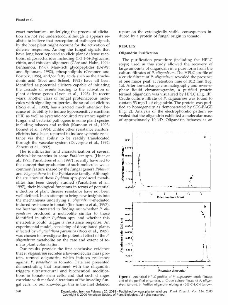
The purification procedure (including the HPLCsteps) used in this study allowed the recovery oflarge amounts of oligandrin in a pure form from theculture filtrates of P. oligandrum. The HPLC profile ofa crude filtrate of P. oligandrum revealed the presenceof one major peak at retention time of 10.2 min (Fig.1a). After ion-exchange chromatography and reverse-phase liquid chromatography, a purified proteintermed oligandrin was visualized by HPLC (Fig. 1b).Crude culture filtrate of P. oligandrum was found tocontain 53 mg/L of oligandrin. The protein was puri-fied to homogeneity as demonstrated by SDS-PAGE(Fig. 2). Analysis of the electrophoretic pattern re-vealed that the oligandrin exhibited a molecular massof approximately 10 kD. Oligandrin behaves as an
Figure 1. Analytical HPLC profiles of P. oligandrum crude filtratesand of the purified oligandrin. a, Crude culture filtrate of P. oligan-drum (arrow). b, Purified oligandrin eluting at 40% CH3CN (arrow).
Picard et al.
380 Plant Physiol. Vol. 124, 2000 www.plantphysiol.orgon February 20, 2018 - Published by Downloaded from Copyright © 2000 American Society of Plant Biologists. All rights reserved.

acidic protein as evidenced by ion-exchange chroma-tography (pI of about 4.5) and analysis of its UVspectrum allowed to exclude Trp and to identify Tyrand Phe (data not shown).
Amino Acid Sequencing of Oligandrin
The N-terminal sequence of the 10-kD protein wasdetermined up to Leu-39; its alignment with se-quences from 13 elicitins secreted by some Phytoph-thora sp. and Pythium sp. is illustrated in Figure 3.Analysis of the oligandrin sequence revealed that Thrand Ser accounted for about 30% of the amino acidswhereas Trp, His, and Arg were missing (Fig. 3a).WU-BLASTp under standard settings gave signifi-cant homology with the elicitins from Phytophthoraand Pythium species. The percent match betweenthese sequences reached nearly 50% including iden-tical and strongly similar residues (Fig. 3b). How-ever, in spite of such similarities, significant differ-ences were observed between oligandrin and theknown elicitins as illustrated by the gaps introducedin the ClustalW multiple alignment (version 2.0), thusindicating that oligandrin might be considered as anelicitin-like protein harboring original features.
Antifungal Potential of Oligandrin
Observation of mycelial samples exposed to steriledistilled water showed the presence of typical hy-
Figure 2. Electrophoretic profile of the oligandrin after SDS-PAGE.One single-stained band (lane 2) with a molecular mass of about 10kD, as compared with molecular mass markers (lane 1), is detected.
Figure 3. Comparison of the N-terminal se-quence of oligandrin with typical elicitins fromPhytophthora spp. and Pythium spp. a, N-terminalsequence of oligandrin determined both fromnative and reduced-alkylated protein. b, ClustalW multiple alignment of oligandrin with 13typical elicitins from Phytophthora spp. andPythium spp. Align: asterisk (*), identity (10 resi-dues, 24.4%); colon (:), strongly similar (nineresidues, 21.9%); period (.), weakly similar (10residues, 24.4%). Cry, Basic cryptogein fromPhytophthora cryptogea (Ricci et al., 1989).Cin-b, Basic cinnamomin from Phytophthora cin-namomi (Huet and Pernollet, 1989). Dre-b, Basicdrechslerin from Phytophthora drechsleri (Huetet al., 1992). Meg-b, Basic megaspermin fromPhytophthora megasperma (Huet and Pernollet,1993). Cap, Acidic capsicein from Phytophthoracapsici (Ricci et al., 1989). Par, Acidic parasiti-cein from P. parasitica (Mouton-Perronnet et al.,1995). Cac, Acidic cactorein from Phytophthoracactorum (Huet et al., 1993). Cin-a, Acidic cin-namomin from P. cinnamomi (Perez et al., 1999).Dre-a, Acidic drechslerin from P. drechsleri(Huet et al., 1992). Meg-a, Acidic megasperminfrom P. megasperma (Huet and Pernollet, 1993).Inf, Acidic infestin from Phytophthora infestans(Huet et al., 1994). Vex1 and Vex2, Acidic vexinsfrom Pythium vexans (Huet et al., 1995). Olig,Oligandrin from P. oligandrum (present work).
Oligandrin-Mediated Induced Resistance in Tomato
Plant Physiol. Vol. 124, 2000 381 www.plantphysiol.orgon February 20, 2018 - Published by Downloaded from Copyright © 2000 American Society of Plant Biologists. All rights reserved.

phae mainly characterized by a dense cytoplasm con-taining numerous organelles and a large number ofvacuoles (Fig. 4a). Exposure of samples to purifiedoligandrin at 5, 15, and 30 mg/mL for 1 to 4 h did notresult in any significant morphological and ultra-structural alteration. Prolonged incubation (12 h)with the oligandrin at the highest concentration didnot affect cell integrity as evidenced by the occur-rence of regularly shaped hyphae in which theplasma membrane was not retracted from the cellwall and the cytoplasm appeared metabolically ac-tive (Fig. 4b).
Symptomatology
Decapitated tomato plants, treated with the puri-fied oligandrin or non-treated, were inoculated withP. parasitica, strain 149, to determine their suscepti-bility to fungal attack (Fig. 5, arrowheads). Typicaldisease symptoms, mainly characterized by the forma-tion of enlarged brownish lesions at the sites of fungalcontact, were visible by 4 d after inoculation in controlplants. Between 6 and 7 d post-inoculation, theseplants showed severe symptoms of wilting (Fig. 5a)and about 30% of the plants were dead by 7 to 8 d afterfungal inoculation. Noninfected, oligandrin-treatedtomato plants showed no symptoms of phytotoxicityduring the course of the experiment.
Treatment with the oligandrin at a concentration of3 nm resulted in much less disease development thanoccurred in non-treated tomato plants (Fig. 5b). By7 d after fungal inoculation, treated plants were freeof apparent symptoms such as wilting or senescence
and exhibited a reduced number of stem lesions (Fig.5b). Although some lesions could be seen in areaswhere the fungus was applied (Fig. 5d, arrow), theextent of these lesions never reached levels similar tothose observed in control plants (Fig. 5c). Oligandrin-treated, decapitated tomato plants did not exhibitnecrotic features typically associated with HR. Ne-crotic spots similarly were not seen when the oligan-drin, at all concentrations tested, was introduced bydirect infiltration into the leaves through the abaxialepidermal layer. Oligandrin treatment significantlyreduced disease severity provoked by P. parasitica intomato and striking differences in plant mortalitywere observed. At the end of the experimental pe-riod, treatment with the oligandrin reduced plantmortality by more than 60% (Table I).
Oligandrin Migration
To determine whether the oligandrin could betranslocated throughout the plant in a way similar tothe Phytophthora sp. elicitins (Devergne et al., 1992;Zanetti et al., 1992), radioiodinated oligandrin wasapplied to tomato plants on either the decapitatedapex (Fig. 6a, arrow) or the wounded petiole (Fig. 6c,arrow). Three hours after treatment, examination byautoradiography revealed that radioactivity could bedetected not only in the area of elicitin applicationbut also at a distance from it (Fig. 6, b and d). Al-though a great amount of the radioactivity remainedat the application site (Fig. 6, b and d, arrows), asignal was observed at some distance in both thestem and the leaves. In the leaves, radioactivity was
Figure 4. Antifungal potential of the oligandrin. a, P. parasitica hyphae exposed to sterile distilled water. A typical hyphal cellmainly characterized by a dense cytoplasm (Cy) containing numerous organelles such as lipid bodies (L) and a large numberof vacuoles (Va). Bar 5 0.5 mm. b, P. parasitica hyphae exposed to oligandrin at 30 mg/mL. No morphological or structuralalterations are visible. The hyphal cell is similar to that shown in a with cytoplasm in which organelles including the nucleus(N), lipid bodies, and vacuoles are noticed. Bar 5 0.5 mm.
Picard et al.
382 Plant Physiol. Vol. 124, 2000 www.plantphysiol.orgon February 20, 2018 - Published by Downloaded from Copyright © 2000 American Society of Plant Biologists. All rights reserved.

intense in the veins (Fig. 6b, arrowhead) and appar-ently diffused in the lamina (Fig. 6b, double arrow-heads). This profile of migration suggests that oligan-drin is translocated in tomato through the vascularsystem as previously reported for other elicitins intobacco (Devergne et al., 1992; Zanetti et al., 1992).
Histological Observations of P. parasitica-InfectedTomato Stem Tissues
Observations of transversally sectioned stem sam-ples from non-treated plants that were inoculated
with P. parasitica showed that all tissues were mas-sively invaded by hyphae of the pathogen (Fig. 7a).By 7 d after fungal inoculation, stem tissues wereintensely colonized as evidenced by the occurrence ofpathogen hyphae through much of the epidermis, thecortex, the endodermis, the paratracheal parenchymacells, and occasionally the vascular stele. Fungalgrowth was mainly intercellular (Fig. 7a, arrow) butit could also occur intracellularly. Pathogen ingresstoward the vascular stele coincided with extensivecell disorganization as judged by marked alterations
Table I. Effect of oligandrin treatment on symptom expression in tomato plants infected by Phytoph-thora parasitica
Decapitated tomato plants were treated with 30 mL of oligandrin solution (3 nmol/plant) andinoculated simultaneously with P. parasitica on the section of the leaf petiole in position 2 beneath.
Oligandrin TreatmentDisease Severitya
Stem Invasionb Protectionc
I II III
nmol no. of plantsd cm3 %
0 (control) 6 26 13 0.75 6 0.72 –3000 19 23 3 0.30 6 0.35 60a Disease index was as follows: I, Near absence of stem lesions; II, small lesions restricted to specific
stem tissues including the epidermis and the cortex; III, enlarged stem lesions. b Volume of stemtissues showing symptoms (lesions) of fungal invasion. Values are standard errors of the mean. c Re-lative restriction of stem invasion as compared to controls. d Disease severity as estimated on 15tomato plants per treatment in triplicate.
Figure 5. Effect of oligandrin treatment on the expression of symptoms in tomato plants infected by P. parasitica. Oligandrinwas applied onto the decapitated apex (arrows) and fungal inoculation was performed on the section of the leaf petiole inposition 2 (arrowheads). a, In the absence of oligandrin treatment, tomato plants exhibit severe symptoms of wilting by 7 dafter inoculation. b, Upon oligandrin treatment, tomato plants appear free of apparent symptoms, except in the area of fungalinoculation (arrow). c and d, Effect of oligandrin treatment on the stem of tomato plants upon slicing off the outer stem layers.The extent of the lesion induced by P. parasitica is much reduced in the oligandrin-treated tomato plant (d, arrow) than inthe control, non-treated plant (c).
Oligandrin-Mediated Induced Resistance in Tomato
Plant Physiol. Vol. 124, 2000 383 www.plantphysiol.orgon February 20, 2018 - Published by Downloaded from Copyright © 2000 American Society of Plant Biologists. All rights reserved.

of the cytoplasm, which was frequently reduced toaggregated remnants (Fig. 7a, double arrows). Hostreactions such as wall appositions, intercellular plug-ging, and xylem vessel coating were not detected. Inall cases, the observed pattern of fungal colonizationand host cell disorganization coincided with the oc-currence of macroscopically visible symptoms alongthe stems, leading to severe plant wilting and even-tually plant death.
Examination of stem sections from oligandrin-treated plants that were challenged with P. parasitica,either on the same day or 24 h later, showed thatfungal colonization occurred, but to a much lesserextent than in non-treated, inoculated plants (Fig.7b). Fungal growth was mainly restricted to the out-ermost host cell layers and occurred only in someintercellular spaces. Invading hyphae appeared to beseverely damaged as evidenced by morphologicalchanges (Fig. 7, b and d, arrows) and increased stain-ing density with toluidine blue. Pathogen penetra-tion in the outer host cell layers always coincidedwith host cell changes mainly characterized by theelaboration of structural barriers in the regions prox-imal to potential fungal penetration (Fig. 7, c–f). Atypical host reaction was the deposition of an amor-
phous material in some infected cells. Hyphae,trapped in this material, were apparently immobilized(Fig. 7c). Other typical features of the host responseincluded the formation of small wall appositions at thesites of potential pathogen penetration (Fig. 7e) andthe plugging of most intercellular spaces with a ma-terial that stained densely with toluidine blue (Fig. 7f).Such host reactions were never seen in the un-colonized tissues beneath the invaded cell layers.
Ultrastructural Features of Noninfected TomatoStem Tissues
Examination of stem samples from oligandrin-treated tomato plants that were not challenged withP. parasitica showed a high preservation of the hostcell integrity in all tissues. Host cells in the epidermisand the cortex were characterized by the occurrenceof enlarged vacuoles and by a layer of dense cyto-plasm appressed against the cell wall (not shown).Such host cells resembled those observed in non-treated tissues and typical host responses such asformation of wall appositions, intercellular spaceplugging, and vessel coating were never detected.
Figure 6. Migration of radioiodinated oligan-drin in tomato plants. Three nanomoles of[125I]oligandrin (specific activity 5 1.5 Ci/mmol) was applied to tomato plants either ontothe decapitated apex (a and b, arrows) or on thewounded petiole (c and d, arrows). Three hoursafter [125I]oligandrin treatment, migration of ra-dioiodinated oligandrin was detected by autora-diography (b and d).
Picard et al.
384 Plant Physiol. Vol. 124, 2000 www.plantphysiol.orgon February 20, 2018 - Published by Downloaded from Copyright © 2000 American Society of Plant Biologists. All rights reserved.

Ultrastructural Features of P. parasitica-InfectedTomato Stem Tissues
Non-Treated Tomato Plants
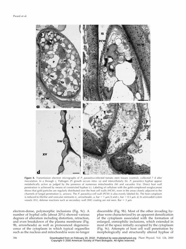
In the absence of oligandrin treatment, inoculationwith P. parasitica resulted in an intense fungal devel-opment in nearly all tissues except for the xylemvessels, which were seldom colonized (Fig. 8). Patho-gen growth was mainly intercellular (Fig. 8b) but itcould also occur intracellularly through direct hostwall penetration, which was achieved by means ofconstricted hyphae (Fig. 8c). It is surprising that sucha massive fungal invasion did not result in markedhost wall alterations as estimated by the pattern ofgold labeling following incubation of sections withthe gold-complexed exoglucanase (Fig. 8, b and c).Gold particles were regularly distributed over thehost cell walls, even in the areas closely adjacent tothe channels of fungal penetration (Fig. 8c, arrows).In contrast, the host cytoplasm was markedly alteredand usually reduced to fibrillar and vesicular rem-nants (Fig. 8c, arrowheads). Fungal cell walls werealso evenly labeled by the gold-complexed exoglu-
canase (Fig. 8c). In these control tomato plants,pathogen invasion failed to stimulate host reactionssuch as wall appositions, intracellular deposits, in-tercellular plugging, and xylem vessel occlusions(Fig. 8d).
Oligandrin-Treated Tomato Plants
In oligandrin-treated tomato plants, the pattern ofstem colonization by P. parasitica differed markedlyfrom that observed in control plants no matter whatthe timing of fungal inoculation (Fig. 9). Althoughextensive fungal multiplication was seen at the stemsurface (Fig. 9a), fungal growth in planta was mainlyrestricted to the outermost cell layers, including theepidermis and the outer cortex. Hyphae of the patho-gen were seldom seen in the inner tissues and theywere never detected in the endodermis or the vascu-lar stele. One of the most striking changes observedin oligandrin-treated plants as compared with con-trols was the obvious alteration of most fungal cells,the cytoplasm of which appeared either highly dis-organized (Fig. 9b) or aggregated and filled with
Figure 7. Light micrographs of stem samples from control (a) and oligandrin-treated tomato plants (b–f). a, Samples fromoligandrin-free (control) tomato stems, collected 7 d after inoculation with P. parasitica. Hyphae of the pathogen (P) multiplyabundantly in all tissues. Fungal growth occurs intra- and intercellularly (arrow). Pathogen invasion coincides with markedcytoplasm alterations (double arrows). IS, Intercellular space. Bar 5 10 mm. b through f, Samples from oligandrin-treatedtomato stems, collected 7 d after inoculation with P. parasitica. Fungal growth is mainly restricted to the outermost host celllayers and occurs only in some intercellular spaces. Invading hyphae appear severely damaged (b and d, arrows). Wallappositions (WA) are seen in the regions proximal to potential fungal penetration (e). An amorphous material (AM)accumulates in some infected cells. Hyphae of the pathogen (P), trapped in this material, are apparently immobilized (c).An intercellular space is plugged with a material that stains densely with toluidine blue (f).
Oligandrin-Mediated Induced Resistance in Tomato
Plant Physiol. Vol. 124, 2000 385 www.plantphysiol.orgon February 20, 2018 - Published by Downloaded from Copyright © 2000 American Society of Plant Biologists. All rights reserved.
electron-dense, polymorphic inclusions (Fig. 9c). Anumber of hyphal cells (about 20%) showed variousdegrees of alteration including distortion, retraction,and even breakdown of the plasma membrane (Fig.9b, arrowheads) as well as pronounced degeneres-cence of the cytoplasm in which typical organellessuch as the nucleus and mitochondria were no longer
discernible (Fig. 9b). Most of the other invading hy-phae were characterized by an apparent densificationof the cytoplasm associated with the formation ofenlarged, osmiophilic inclusions, which extended inmost of the space initially occupied by the cytoplasm(Fig. 9c). Attempts of host cell wall penetration bymorphologically and structurally altered hyphae of
Figure 8. Transmission electron micrographs of P. parasitica-infected tomato stem tissues (control), collected 7 d afterinoculation. In a through c, Pathogen (P) growth occurs intra- (a) and intercellularly (b). P. parasitica hyphae appearmetabolically active as judged by the presence of numerous mitochondria (M) and vacuoles (Va). Direct host wallpenetration is achieved by means of constricted hyphae (c). Labeling of cellulose with the gold-complexed exoglucanaseshows that gold particles are regularly distributed over the host cell walls (HCW), even in the areas closely adjacent to thechannels of fungal penetration (c, arrows). The P. parasitica cell wall (PCW) is also evenly labeled (b). The host cytoplasmis reduced to fibrillar and vesicular remnants (c, arrowheads). a, bar 5 1 mm; b and c, bar 5 0.5 mm. d, In uninvaded xylemvessels (XV), defense reactions such as secondary wall (SW) coating are not seen. Bar 5 3 mm.
Picard et al.
386 Plant Physiol. Vol. 124, 2000 www.plantphysiol.orgon February 20, 2018 - Published by Downloaded from Copyright © 2000 American Society of Plant Biologists. All rights reserved.
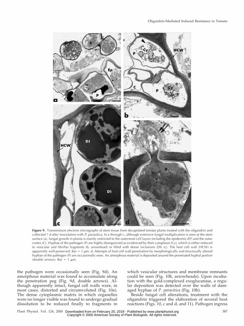
the pathogen were occasionally seen (Fig. 9d). Anamorphous material was found to accumulate alongthe penetration peg (Fig. 9d, double arrows). Al-though apparently intact, fungal cell walls were, inmost cases, distorted and circumvoluted (Fig. 10a).The dense cytoplasmic matrix in which organelleswere no longer visible was found to undergo gradualdissolution to be reduced finally to fragments in
which vesicular structures and membrane remnantscould be seen (Fig. 10b, arrowheads). Upon incuba-tion with the gold-complexed exoglucanase, a regu-lar deposition was detected over the walls of dam-aged hyphae of P. parasitica (Fig. 10b).
Beside fungal cell alterations, treatment with theoligandrin triggered the elaboration of several hostreactions (Figs. 10, c and d, and 11). Pathogen ingress
Figure 9. Transmission electron micrographs of stem tissue from decapitated tomato plants treated with the oligandrin andcollected 7 d after inoculation with P. parasitica. In a through c, although extensive fungal multiplication is seen at the stemsurface (a), fungal growth in planta is mainly restricted to the outermost cell layers including the epidermis (EP) and the outercortex (C). Hyphae of the pathogen (P) are highly disorganized as evidenced by their cytoplasm (Cy), which is either reducedto vesicular and fibrillar fragments (b, arrowhead) or filled with dense inclusions (DI) (c). The host cell wall (HCW) isapparently well preserved. Bar 5 1 mm. d, Attempts of host cell wall penetration by morphologically and structurally alteredhyphae of the pathogen (P) are occasionally seen. An amorphous material is deposited around the penetrated hyphal portion(double arrows). Bar 5 1 mm.
Oligandrin-Mediated Induced Resistance in Tomato
Plant Physiol. Vol. 124, 2000 387 www.plantphysiol.orgon February 20, 2018 - Published by Downloaded from Copyright © 2000 American Society of Plant Biologists. All rights reserved.
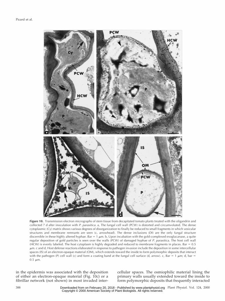
in the epidermis was associated with the depositionof either an electron-opaque material (Fig. 10c) or afibrillar network (not shown) in most invaded inter-
cellular spaces. The osmiophilic material lining theprimary walls usually extended toward the inside toform polymorphic deposits that frequently interacted
Figure 10. Transmission electron micrographs of stem tissue from decapitated tomato plants treated with the oligandrin andcollected 7 d after inoculation with P. parasitica. a, The fungal cell wall (PCW) is distorted and circumvoluted. The densecytoplasmic (Cy) matrix shows various degrees of disorganization to finally be reduced to small fragments in which vesicularstructures and membrane remnants are seen (c, arrowhead). The dense inclusions (DI) are the only fungal structurediscernible in these highly altered hyphae. Bar 5 1 mm. b, Upon incubation with the gold-complexed exoglucanase, a quiteregular deposition of gold particles is seen over the walls (PCW) of damaged hyphae of P. parasitica. The host cell wall(HCW) is evenly labeled. The host cytoplasm is highly degraded and reduced to membrane fragments in places. Bar 5 0.5mm. c and d, Host defense reactions elaborated in response to pathogen invasion include the deposition in some intercellularspaces (IS) of an electron-opaque material (OM), which extends toward the inside to form polymorphic deposits that interactwith the pathogen (P) cell wall (c) and form a coating band at the fungal cell surface (d, arrow). c, Bar 5 1 mm; d, bar 50.5 mm.
Picard et al.
388 Plant Physiol. Vol. 124, 2000 www.plantphysiol.orgon February 20, 2018 - Published by Downloaded from Copyright © 2000 American Society of Plant Biologists. All rights reserved.

with the wall of invading hyphae (Fig. 10c, arrow)and occasionally formed a coating band at the fungalcell surface (Fig. 10d, arrow). Another feature, occa-sionally seen in reacting host cells, was the formationof multitextured wall appositions at sites of potentialfungal penetration (Fig. 11a). These appositions,which could vary greatly in size, shape, and texture,were usually found to be made of an amorphousmatrix that was impregnated by osmiophilic sub-stances and was delimited by a loosely arrangedlayer of fine fibrillo-vesicular material (Fig. 11a). Thecore of wall appositions was frequently made of os-miophilic aggregates that formed short finger-likeprojections in the amorphous matrix (Fig. 11a, ar-row). The host cell wall itself displayed a higherelectron density than normal, thus indicating theprobable infiltration of structural molecules. Both theimpregnated host cell wall and the wall appositionswere efficient in preventing fungal ingress since suc-cessful hyphal penetration of these structures wasnot observed. It is interesting that host reactions weredetected in noninvaded xylem vessels. The host re-actions were mainly characterized by the coating ofsecondary walls with a band of osmiophilic material(Fig. 11b) or the deposition of an electron-opaque,fibrillar material in the vessel lumen (Fig. 11c). Con-trol tests, including pre-incubation of the exoglu-
canase-gold complex with b-1,4-glucans prior to sec-tion labeling, resulted in the absence of labeling overboth the cell walls and the wall appositions (notshown).
DISCUSSION
We recently reported that tomato plants, when pre-inoculated with the aggressive mycoparasite P. oli-gandrum, gained increased protection against crownand root rot caused by F. oxysporum f.sp. radicis-lycopersici (Benhamou et al., 1997). In direct line withthese earlier observations, attempts were made in thepresent study to delineate the mechanisms by whichP. oligandrum could exert its beneficial effects ontomato plants. Evidence is presented in this paperthat P. oligandrum secretes a proteinaceous metabolitethat displays the ability to operate as an elicitor ofresistance. Although the three-dimensional structureand the biophysical properties of this small, water-soluble molecule need to be further characterized, thepresent results support the view that this metabolite,termed oligandrin, shares some similarities with theelicitins identified in some Phytophthora spp. andPythium spp. (Huet et al., 1994, 1995; Panabieres etal., 1997; Ponchet et al., 1999). Among the criteria sofar identified for the assignment of a given protein to
Figure 11. Transmission electron micrographs of stem tissue from decapitated tomato plants treated with the oligandrin andcollected 7 d after inoculation with P. parasitica. a, A heterogeneous wall apposition (WA), made of an amorphous matrix(Ma) and containing a central multilobed core (Co) is formed at a site of potential pathogen (P) penetration. It is delimitedby a fine fibrillo-vesicular material (FVM). Bar 5 0.5 mm. b and c, Host reactions including the coating of secondary walls(SW) with a band of osmiophilic material (CM) (b) or deposition of an electron-opaque, fibrillar material (FM) (c) are seenin noninvaded xylem vessels (XV). b, Bar 5 1 mm; c, bar 5 2 mm.
Oligandrin-Mediated Induced Resistance in Tomato
Plant Physiol. Vol. 124, 2000 389 www.plantphysiol.orgon February 20, 2018 - Published by Downloaded from Copyright © 2000 American Society of Plant Biologists. All rights reserved.

the elicitin family is the amino acid composition ofthe N-terminal end. In that context, the absence ofTrp, His, and Arg residues and the relative abun-dance of Thr and Ser residues in the oligandrin ter-minal end are key characteristics that define an “elic-itin signature” (Ponchet et al., 1999). The lack of Trp,which was confirmed by the UV absorption spectrum(not shown), the low molecular mass (10 kD, about100 amino acids), and the migration profile withinthe plant also provide a helpful signature for includ-ing the oligandrin into the elicitin family. In spite ofthose similarities, significant differences characterizethe oligandrin with respect to its biological activity.The major difference probably relies on the findingthat oligandrin infiltration into tomato leaves failedto provoke the HR-associated necrotic response, areaction consistently found to occur in tobacco plantstreated with true elicitins (Ricci, 1997; Ponchet et al.,1999). By contrast, a high level of protection againstthe oomycete fungus, P. parasitica, was noticed, thussubstantiating the concept that the oligandrin can beconsidered as a resistance elicitor. Although the rea-sons why the oligandrin failed to mediate HR intomato plants are still unknown, one possibilitycould be the trapping of some oligandrin moleculesby components of the tomato cell wall matrix. In linewith this concept, recent investigations have dis-closed that effective binding of the elicitins fromPhytophthora species to membrane receptors requiredhigher concentrations of proteins in tomato than intobacco, mainly because a large number of moleculeswere trapped in the tomato cell wall, thus preventingsufficient access of the elicitins to their target recep-tors (Ponchet et al., 1999). In that context, one mightconsider that the tomato cell wall acts as a filter thatcontrols oligandrin diffusion in such a way thatmembrane-bound receptors are not fully saturated.Although it is clear that the early events involved inthe recognition of oligandrin molecules by tomatocells must be more fully investigated, the finding thatresistance was expressed upon oligandrin treatmentsuggests that the receptors involved in the specificoligandrin signaling pathway are functional.
Evidence is provided from the present ultrastruc-tural study that oligandrin-treated tomato plants af-ford increased protection against P. parasitica andthat this protection is at least partly associated with areduction in pathogen biomass and an increase inhyphal structural alterations. To our knowledge, thisis the first report about the effect of a protein offungal origin on the cytology of pathogen coloniza-tion in a host plant. A decrease in the amount offungal cell colonization and pathogen viability, asillustrated by the frequent occurrence of modifiedand/or highly damaged fungal cells, were typicalfeatures of reactions observed in oligandrin-treatedtomato plants only. Whether such alterations are at-tributable to the creation of a fungitoxic environmentassociated with the synthesis and accumulation of
antimicrobial compounds by the reacting host cells orsimply relate to a direct antifungal effect of the oli-gandrin in planta deserves to be biochemicallyinvestigated.
If one considers that the oligandrin is mobilewithin the plant, as evidenced by the migration pro-file, then it seems realistic to believe that a directfungitoxic effect might account for the observed fun-gal damage. However, the observation that exposureof P. parasitica hyphae to pure oligandrin did notlead, under our conditions, to structural alterationssimilar to those detected in planta (see Fig. 4) tendsto suggest that the oligandrin has no fungicidalactivity against P. parasitica. Such a conclusionshould, however, be viewed with caution since thepossibility that the molecule might have undergonestructural alterations once in the plant needs to beconsidered. Extraction and structural characteriza-tion of [I125]oligandrin from tomato leaves as well asbioassays would answer the question as to whatextent the oligandrin can be converted into a fungi-toxic compound that might operate against the in-vading hyphae in the tomato plant tissues.
The observation that invading fungal cells weremarkedly damaged at a time when the cellulosecomponent of their cell walls was quite well pre-served (Fig. 10b) favors the hypothesis of a specificplant defense reaction. Synthesis of phytoalexins asa site-specific response to fungal ingress is a well-documented response to elicitor treatment (Ham-merschmidt, 1999). Several lines of evidence haveshown that the fungitoxic effect of both phytoalexinsand preformed phenolics was related to their inter-action with membrane-bound lipids or phospholip-ids, resulting in an increase in fungal membranepermeability, pore formation, and leakage of cell con-tents (Weete, 1980). Phenolic-induced perturbationsin the permeability of the plasma membrane in P.parasitica cells might have promoted internal osmoticimbalances, leading to the observed disturbancessuch as plasmalemma retraction, cytoplasm aggrega-tion, condensation, and in some cases, complete lossof the protoplasm. In oligandrin-treated tomatoplants, phenolic compounds might be involved in atleast two key biological functions. First, the accumu-lation of phenolic compounds at sites of pathogenpenetration might cause inhibition of fungal growthas illustrated by the distorted and degenerative aspectof all fungal hyphae. Second, the impregnation ofphenolic compounds in the host cell walls (as indi-cated by the higher electron density of cell walls thannormal) and their accumulation in noninvaded xylemvessels as a coating along secondary walls might con-tribute to enhancing the mechanical strength of thesedefensive barriers.
Restriction of fungal growth to the outermost stemtissues was also found to correlate with the formationof heterogeneous wall appositions beyond the infec-tion sites. Reinforcing the host cell walls by either the
Picard et al.
390 Plant Physiol. Vol. 124, 2000 www.plantphysiol.orgon February 20, 2018 - Published by Downloaded from Copyright © 2000 American Society of Plant Biologists. All rights reserved.
impregnation of hydrophobic substances or by thedeposition of new wall-like polymers (Ride, 1983;Hahlbrock and Scheel, 1989) is an essential prerequi-site for preventing enzymatic degradation of the hostcell walls, a phenomenon that is considered to be oneof the most harmful events associated with the infec-tion process by pathogenic fungi (Collmer and Keen,1986). Support for the close association between oli-gandrin treatment and induced resistance also camefrom the observation that intercellular spaces, knownto be strategic sites for pathogen spread, were oftenfilled with an electron-dense material in which in-vading hyphae were trapped. Such host reactions,obviously designed to halt pathogen ingress, werenever seen in control plants where the pattern offungal colonization was similar in many respects tocolonization known to occur with necrotrophic fungi.Thus the present observations are of particular rele-vance since they bring further insight to the conceptthat oligandrin is capable of evoking biochemicalevents usually associated with the natural plant dis-ease resistance process.
Although wall appositions could be seen in theouter stem tissues, their extent never reached thatobserved in tomato plants treated with elicitors suchas chitosan (Benhamou et al., 1994; Benhamou andLafontaine, 1995) or with P. oligandrum itself (Ben-hamou et al., 1997). Although a clear explanation forsuch a difference in the rate and extent of this struc-tural response is still difficult to give, the possibilitythat the reduced number of wall appositions corre-lates with a reduced level of callose synthesis can beraised. Several lines of evidence have shown thatcallose formation is modulated directly or indirectlyby the intracellular concentration of free Ca21, whichis known to control the activity of one of the keystructural enzymes, b-1,3-glucan synthase (Kohle etal., 1985). Recent investigations of the earliest eventsleading to elicitin-mediated HR in tobacco haveshown that huge Ca21 uptake occurred within min-utes following elicitin application (Tavernier et al.,1995). Because the elicitin receptor is thought to be aligand-dependent calcium channel (Ponchet et al.,1999), it was suggested that Ca21 influx was ofligand-dependent type, thus indicating that satura-tion of the receptor by sufficient elicitin moleculeswas a prerequisite for optimal Ca21 uptake (Taver-nier et al., 1995; Keiser et al., 1998). In light of theseresults, one might speculate that the trapping of alarge number of oligandrin molecules in the tomatoplant cell wall might have hampered receptor satu-ration, leading to moderate Ca21 influx and conse-quently to a reduced activity of the callose synthesis-involved b-1,3-glucan synthase.
Even though the exact mechanisms by which oli-gandrin operates to trigger resistance in tomato arenot fully elucidated, the present results demonstratethat the beneficial effect exerted by this fungal pro-teinaceous molecule results from an integrated action
of biochemical and anatomical factors that develop atthe onset of pathogen penetration. The observationthat defense reactions were expressed in oligandrin-treated plants only upon challenge with P. parasiticasupports the hypothesis that a signal produced by thepathogen is essential for triggering synthesis and ac-cumulation of defense gene products. A similar con-clusion was reached in the case of chitosan-treatedtomato plants (Benhamou, 1996). It was shown thatdefense reactions accumulating in chitosan-coated to-mato roots infected with Fusarium oxysporum f.sp.radicis-lycopersici were seldom seen in noninfected,chitosan-treated tomato roots. Benhamou et al.(1996), similarly studying the protective effect of en-dophytic bacteria against fungal plant pathogens,found that extensive defense reactions occurred inbacterized plants only following pathogenic attack.These observations together with the present resultssuggest that biotic or abiotic agents sensitize theplant to respond more rapidly to microbial attackwithout causing accumulation of defense gene prod-ucts that would require extensive loss of energy.
In summary, evidence is provided in this studythat oligandrin, a small proteinaceous molecule pro-duced by the mycoparasite P. oligandrum, is anelicitin-like protein that displays the ability to triggera resistance response in tomato without inducingsymptoms of phytotoxicity such as those observedduring elicitin-mediated HR (Ricci et al., 1989).Among the resistance elicitors identified so far,fungal-derived proteinaceous molecules with highelicitor activity (Yu, 1995) are attracting a lot of at-tention not only because of their specific mechanismsof action on gene expression in plants (Ponchet et al.,1999) but also because their simple nature offers thebest prospects for the production of synthetic analogsthat can be introduced as a new biocontrol strategy inplant disease management.
MATERIALS AND METHODS
Plant Material
Tomato (Lycopersicon esculentum Mill. var. Prisca) seedswere sterilized by immersion in 7% (v/v) calcium hypo-chloride for 7 min and thoroughly rinsed in sterile distilledwater. Seeds were sown in sterilized vermiculite in a 37- 323-cm plastic tray. One-week-old tomato seedlings wereuprooted and transferred into pots (11 3 11 3 11 cm)containing peat (Vapogro, Griendtsveen, Netherlands) at adensity of one plantlet per pot. Plants were grown in agreenhouse at 22°C with a 14-h light period and werefertilized twice a week with a commercial plant nutrientsolution (Solufeed soluble fertilizer, ICI Agrochemicals,Paris). Experiments were performed with 2-month-oldplants harboring five to six fully expanded leaves.
Fungal Cultures
Pythium oligandrum Drechsler, strain 1010, was isolatedfrom pea roots in Denmark (provided by Dr J. Hockenhull,
Oligandrin-Mediated Induced Resistance in Tomato
Plant Physiol. Vol. 124, 2000 391 www.plantphysiol.orgon February 20, 2018 - Published by Downloaded from Copyright © 2000 American Society of Plant Biologists. All rights reserved.

The Royal Veterinary and Agricultural University, Copen-hagen). Phytophthora parasitica (isolate 149, highly virulenton tomato) was obtained from the collection maintained atInstitut National de la Recherche Agronomique (Antibes,France). Both fungi were cultivated on potato-dextroseagar medium (Difco, Detroit) at 25°C in the dark.
Production of the Culture Filtrates
Liquid cultures of P. oligandrum were obtained by grow-ing the fungus in a defined medium containing for 1,000mL of deionized water: 0.6 g of KH2PO4, 0.7 g of KNO3,0.25 g of MgSO4z7H2O, 0.125 g of K2HPO4z3H2O, 0.3 g of Ca(NO3)2, 1 mg of H3BO3, 1.5 mg of MnSO4zH2O, 4 mg ofZnSO4z7H2O, 0.1 mg of Na2MoO4z2H2O, 20 mg of KI, 20 mgof CuSO4z5H2O, 20 mg of CoCl2z6H2O, 8 mg of FeNa2
EDTA, 1 mg of nicotinic acid, 1 mg of pyridoxin, 1 mg ofcalcium panthotenate, 1 mg of thiamine hydrochloride, 1 gof AsnzH2O, and 20 g of Glc. This medium was chosen forits known potential of stimulating elicitin production (Bon-net et al., 1996). The flasks were incubated in the dark for8 d at 24°C. Culture filtrates of P. oligandrum were recov-ered after mycelium removal on a GF/C filter (Whatman,Clifton, NJ) under vacuum.
Protein Purification from the Culture Filtrates
Culture filtrates of P. oligandrum (5 L) were concentrated10-fold by evaporation under vacuum at 35°C and dialyzedagainst deionized water for 24 h at 4°C. Fifteen milliliters of0.34 m sodium-acetate was added to the concentrated fil-trate (495 mL) and the pH of the resulting solution wasadjusted to 3.5 with 10% (v/v) aqueous trifluoroacetic acid.The concentrated filtrate was loaded on a 20-mL cationicexchange Macroprep sulfopropyl High S column (Bio-Rad,Ivry sur Seine, France) previously equilibrated with 10 mmsodium-acetate (pH 3.5). The retained fraction was elutedwith 10 mm sodium-acetate containing 0.25 m NaCl (pH3.5) and adjusted to a pH of 7.0 before being subjected toreverse-phase liquid chromatography using a SynchroprepC4 column (30 mm, 300 Å, Synchrom Inc., distributed byEichrom Technologies, Paris) that was pre-equilibratedwith 10 mm sodium-acetate containing 0.25 m NaCl (pH7.0). Elution was carried out at room temperature using agradient of acetonitrile (CH3CN) (20%, 30%, 40%; v/v) in50 mm aqueous sodium-formate (HCOONa). A purifiedprotein, termed oligandrin, was recovered from the 40%CH3CN fraction.
Each chromatographic step was qualitatively assayed byHPLC. HPLC (625 LC system solvent delivery, Waters,Milford, MA) was performed by loading the active frac-tions on a Hema RP C18 column (10 mm, 150 3 4.6 mm i.d.,Interchim, Montlucon, France). Elution was carried outwith the following solvents: A [20% CH3CN, 10 mm(NH4)2SO4, and 20 mm HCOONa] and B (40% CH3CN and100 mm HCOONa) using a linear gradient: 100% A3 100%B (10 min), and 100% B hold for 2 min. The flow rate was1 mL/min. Elution was monitored with a Waters 996 diodearray detector (200–400 nm, resolution: 1.2 nm). Integration
at 280 nm, spectra, peak purity, and all calculations wereachieved with Millenium software (version 3.2, 1999, Wa-ters). For the last step, a peak was visualized from the 40%CH3CN fraction. After removal of CH3CN under vacuum,the pure protein was extensively dialyzed against ultra-pure water (Millipore, Bedford, MA) and freeze-dried. Pu-rity of the protein was further assessed by SDS-PAGE on15.4% (v/v) polyacrylamide-SDS gels (20 mA/gel in 0.25mm Tris [tris(hydroxymethyl)aminomethane], 1.92 m Gly,and 0.1% SDS) (Le Berre et al., 1994).
Protein Sequencing
The purified protein (1 mg) was reduced with dithio-threitol in 8 m urea and alkylated with iodoacetamide. Thereduced and alkylated protein was extensively dialyzedagainst ultrapure water and freeze-dried. N-terminal se-quencing of oligandrin was performed on both the nativeand the alkylated proteins. The two sequences were foundto be identical, with the exception of one Cys residuemissing in the native protein.
For sequence determination, the freeze-dried proteinwas resuspended in 1% (w/v) trifluoroacetic acid-20% ace-tonitrile solution at a final concentration of 2 mg/mL. Analiquot (0.5–1.5 nmol protein) was loaded on polybrene-treated glass fiber and N-terminal sequence determinationwas performed by automated Edman degradation using anApplied Biosystems (Foster City, CA) 470 A sequencer.Phenylthiohydantoin (PTH) amino acids were identifiedon-line with a 120 A Applied Biosystems PTH-Analyser byreverse-phase HPLC using a PTH-C18 cartridge (2.1 3 220mm, Brownlee, Applied Biosystems, Roissy, France). Allproducts, reagents, and programs used for sequencingwere from Applied Biosystems.
Multiple sequence alignment was performed using themethod of Thompson et al. (1994) with Clustal W (version2.0). Similarities between proteins were revealed usingthe WU-BLASTp in the Swall database at the EuropeanBioinformatics Institute (Cambridge, UK, http://www.ebi.ac.uk/).
Antifungal Potential of Oligandrin
Mycelial samples (1 mm3), collected from an activelygrowing colony of P. parasitica, were subjected to oligan-drin at a concentration ranging from 5 to 30 mg/mL indistilled water for 1, 2, 4, and 12 h at room temperature.Samples were rinsed thoroughly thereafter with distilledwater and processed for electron microscope investiga-tions. Controls included mycelial samples immersed insterile distilled water.
Radioiodination and Migration of Oligandrin
The protein was iodinated according to a previouslydescribed procedure (Wendehenne et al., 1995). The pro-tein (100 mg) was incubated in 100 mL of 50 mm phosphatebuffer (pH 7.4) with 1 mCi of Na-125I (Amersham, Buck-inghamshire, UK) and iodogen as the catalyst for 20 min at
Picard et al.
392 Plant Physiol. Vol. 124, 2000 www.plantphysiol.orgon February 20, 2018 - Published by Downloaded from Copyright © 2000 American Society of Plant Biologists. All rights reserved.

20°C. The [125I]oligandrin was purified by gel filtration ona G-25 Sephadex column (5 mL, Pharmacia, Uppsala) equil-ibrated with 50 mm Tris-HCl buffer (pH 7.4). Eluted frac-tions were collected and the radioactivity counted with a6000TA liquid scintillation analyzer (Beckman Instru-ments, Fullerton, CA). Fractions containing the radioactiveprotein were pooled and stored at 220°C.
Migration of the radiolabeled oligandrin in tomatoplants was determined by applying 3 nmol of [125I]oligan-drin (specific radioactivity was about 1.5 Ci/mmol) ontothe decapitated apex or onto the wounded petiole. Threehours after [125I]oligandrin treatment, migration of the pro-tein was monitored by a 10-min exposure of the tomatoplants with a PhosphorImager screen (Molecular Dynam-ics, Sunnyvale, CA).
Oligandrin Application and Challenge Inoculation
Tomato plants at the five- to six-leaf stage were decapi-tated above the fifth fully expanded leaf (Ricci et al., 1989)just prior to applying 30 mL of oligandrin (3 nmol/plant)onto the fresh wound. Control plants were decapitated andtreated with sterile water. At the same time or 24 h later,decapitated tomato plants were inoculated by placing aplug of actively growing mycelium of P. parasitica on thesection of the leaf petiole in position 2 (beneath). Controlplants were treated similarly but with fungus-free agarplugs. One week after fungal inoculation, the stem waslongitudinally sectioned, allowing visualization of patho-gen spread. Each plant was categorized according to thefollowing scale: I, near absence of stem lesions; II, smalllesions restricted to specific stem tissues including theepidermis and the cortex; and III, enlarged stem lesions.Stem invasion was estimated by the volume of discoloredtissues (in cm3) and percent of protection was computed asthe relative reduction of invasion compared with water-treated, inoculated control plants. The experiment was re-peated three times with 15 plants for each treatment.
The oligandrin, at concentrations ranging from 0.1 to 100nmol/20 mL, was directly infiltrated into tomato leavesthrough the abaxial epidermal layer to determine whetherHR (necrotic lesions) was expressed in tomato leaves.
Tissue Processing for Electron Microscope Studies
Mycelial samples and tomato stem samples (2 mm3),collected at or near the necrotic lesions 7 d after fungalinoculation, were fixed by immersion in 3% (v/v) gluta-raldehyde in 0.1 m sodium cacodylate buffer (pH 7.2) for2 h at room temperature and post-fixed with 1% (w/v)osmium tetroxide in the same buffer for 1 h at 4°C. Sampleswere dehydrated in a graded ethanol series and embeddedin Epon 812 (JBEM Chemicals Pointe-Claire, Quebec, Can-ada). Thin sections (0.7 mm), cut from the Epon-embeddedmaterial using glass knives, were mounted on glass slidesand stained with 1% (v/v) aqueous toluidine blue prior toexamination with an Axioscope microscope (Zeiss, Jena,Germany). Ultrathin sections (0.1 mm), collected on nickelgrids, were either contrasted with uranyl acetate and lead
citrate for immediate examination with a transmission elec-tron microscope (model 1200 EX, JEOL, Tokyo) operatingat 80 kV or further processed for cytochemical labeling. Foreach treatment, an average of five samples from threedifferent stems were investigated. For each sample, 10 to 15ultrathin sections were examined under the electronmicroscope.
Cytochemical Labeling
Colloidal gold, with particles averaging 12 nm in diam-eter, was prepared according to Frens (1973) using sodiumcitrate as a reducing agent. The b-1,4-exoglucanase-goldcomplex used for localization of cellulosic b-1,4-glucanswas prepared according to Benhamou et al. (1987) using ab-1,4-d-glucan cellobiohydrolase (EC 3.2.1.21) complexedto gold at pH 9.0.
Labeling with the gold-complexed exoglucanase wasperformed by first incubating the ultrathin tomato stemsections for 5 to 10 min on a drop of phosphate-bufferedsaline containing 0.02% (w/v) polyethylene glycol 20,000 atpH 6.0, and then transferring them to a drop of theenzyme-gold complex for 30 min at room temperature in amoist chamber. After careful washing with phosphate-buffered saline (pH 7.2) and rinsing with distilled water,sections were contrasted with uranyl acetate and lead ci-trate and observed with a JEOL 1200 EX transmissionelectron microscope operating at 80 kV.
Specificity of the labeling was assessed by the followingcontrol tests: (a) incubation of the probe on which waspreviously added b-1,4-glucans from barley, 1 mg/mL21;(b) incubation with the un-complexed protein followed byincubation with the gold complex; and (c) incubation withcolloidal gold alone.
ACKNOWLEDGMENTS
The authors wish to thank H. Osman (Institut Nationalde la Recherche Agronomique, Antibes, France), S. Vauth-rin (Institut National de la Recherche Agronomique, Dijon,France), and F. Belzile (Laval University) for their help inoligandrin purification and migration. Thanks are ex-tended to C. Garand and A. Goulet (Laval University) forexcellent technical assistance. The authors are grateful toD. Michaud (Laval University) and F. Panabieres (InstitutNational de la Recherche Agronomique, Antibes, France)for critical reading of the manuscript.
Received January 13, 2000; accepted May 28, 2000.
LITERATURE CITED
Benhamou N (1996) Elicitor-induced plant defense path-ways. Trends Plant Sci 1: 233–240
Benhamou N, Belanger RR, Paulitz T (1996) Ultrastruc-tural and cytochemical aspects of the interaction betweenPseudomonas fluorescens and Ri T-DNA transformed pearoots: host response to colonization by Pythium ultimumTrow. Planta 199: 105–117
Oligandrin-Mediated Induced Resistance in Tomato
Plant Physiol. Vol. 124, 2000 393 www.plantphysiol.orgon February 20, 2018 - Published by Downloaded from Copyright © 2000 American Society of Plant Biologists. All rights reserved.
Benhamou N, Chamberland H, Ouellette GB, Pauze FJ(1987) Ultrastructural localization of b-1,4-d-glucans intwo pathogenic fungi and in their host tissues by meansof an exoglucanase-gold complex. Can J Microbiol 33:405–417
Benhamou N, Lafontaine PJ (1995) Ultrastructural andcytochemical characterization of elicitor-induced re-sponses in tomato root tissues infected by Fusarium ox-ysporum f. sp. radicis-lycopersici. Planta 197: 89–102
Benhamou N, Lafontaine PJ, Nicole M (1994) Seed treat-ment with chitosan induces systemic resistance to Fusar-ium crown and root rot in tomato plants. Phytopathology84: 1432–1444
Benhamou N, Rey P, Cherif M, Hockenhull J, Tirilly Y(1997) Treatment with the mycoparasite, Pythium oligan-drum, triggers the induction of defense-related reactionsin tomato roots upon challenge with Fusarium oxysporumf. sp. radicis-lycopersici. Phytopathology 87: 108–122
Benhamou N, Rey P, Picard K, Tirilly Y (1999) Ultrastruc-tural and cytochemical aspects of the interaction betweenthe mycoparasite, Pythium oligandrum, and soilbornepathogens. Phytopathology 89: 506–517
Bonnet P, Bourdon E, Ponchet M, Blein JP, Ricci P (1996)Acquired resistance triggered by elicitins in tobacco andother plants. Eur J Plant Pathol 102: 181–192
Chet I (1993) Biotechnology in Plant Disease Control. JohnWiley & Sons, New York
Collmer A, Keen NT (1986) The role of pectic enzymes inplant pathogenesis. Annu Rev Phytopathol 24: 383–409
Cote F, Hahn MG (1994) Oligosaccharins: structures andsignal transduction. Plant Mol Biol 26: 1379–1411
Creamer JR, Bostock RM (1986) Characterization and bio-logical activity of Phytophthora infestans phospholipids inthe hypersensitive response of potato tuber. Physiol MolPlant Pathol 28: 215–225
Devergne JC, Bonnet P, Panabieres F, Blein JP, Ricci P(1992) Migration of the fungal protein cryptogein withintobacco plants. Plant Physiol 99: 843–847
De Wit PJGM, Spikman G (1982) Evidence for the occur-rence of race- and cultivar-specific elicitors of necrosis inintercellular fluids of compatible interaction betweenCladosporium fulvum and tomato. Physiol Plant Pathol 21:1–11
Ebel J, Scheel D (1992) Elicitor recognition and signaltransduction. In T Boller, F Meins Jr, eds, Genes Involvedin Plant Defense. Springer-Verlag, Vienna, pp 183–205
Frens G (1973) Controlled nucleation for the regulation ofthe particle size in monodisperse gold solution. Nat PhysSci 241: 20–22
Hahlbrock K, Scheel D (1989) Physiology and molecularbiology of phenylpropanoid metabolism. Annu RevPlant Physiol Plant Mol Biol 40: 347–364
Hammerschmidt R (1999) Phytoalexins: what have welearned after 60 years? Annu Rev Phytopathol 37:285–306
Huet JC, Le Caer JP, Nespoulous C, Pernollet JC (1995)The relationships between the toxicity and the primaryand secondary structures of elicitin-like protein elicitorssecreted by the phytopathogenic fungus Pythium vexans.Mol Plant-Microbe Interact 8: 302–310
Huet JC, Mansion M, Pernollet JC (1993) Amino acidsequence of the alpha-elicitin secreted by Phytophthoracactorum. Phytochemistry 34: 1261–1264
Huet JC, Nespoulous C, Pernollet JC (1992) Structures ofelicitin isoforms secreted by Phytophthora drechsleri. Phy-tochemistry 31: 1471–1476
Huet JC, Pernollet JC (1989) Amino acid sequence of cin-namomin, a new member of the elicitin family, and itscomparison to cryptogein and capsicein. FEBS Lett 257:302–306
Huet JC, Pernollet JC (1993) Sequences of acidic and basicelicitin isoforms secreted by Phytophthora megasperma me-gasperma. Phytochemistry 33: 797–805
Huet JC, Salle-Tourne M, Pernollet JC (1994) Amino acidsequence and toxicity of the alpha elicitin secreted withubiquitin by Phytophthora infestans. Mol Plant-MicrobeInteract 7: 302–304
Kamoun S, Young M, Glascock CB, Tyler BM (1993)Extracellular protein elicitors from Phytophthora: host-specificity and induction of resistance to bacterial andfungal pathogens. Mol Plant-Microbe Interact 6: 15–25
Keiser DW, Schuster B, Grant BR, Gayler KR (1998) In-teractions between elicitins and radish Raphanus sativus.Planta 204: 480–489
Kloepper JW (1993) Plant growth-promoting rhizobacteriaas biological control agents. In B Metting, ed, Soil Micro-bial Technologies. Marcel Dekker, New York, pp 255–274
Kohle H, Jeblick W, Poten F, Blaschek W, Kauss, H (1985)Chitosan-elicited callose synthesis in soybean cells as aCa21 dependent process. Plant Physiol 77: 544–551
Le Berre JY, Panabieres F, Ponchet M, Deneroy L, BonnetP, Marais A, Ricci P (1994) Occurrence of multiple formsof elicitins in Phytophthora cryptogea. Plant Physiol Bio-chem 32: 251–258
Lyon GD, Newton AC (1997) Do resistance elicitors offernew opportunities in integrated disease control strate-gies? Plant Pathol 46: 636–641
Lyon GD, Reglinski T, Newton AC (1995) Novel diseasecontrol compounds: the potential to immunize plantsagainst infection. Plant Pathol 44: 407–427
Martin FN, Loper JE (1998) Soilborne plant diseases causedby Pythium spp.: ecology, epidemiology and prospectsfor biological control. Crit Rev Plant Sci 18: 111–191
Mouton-Perronnet F, Bruneteau M, Denoroy L, BouliteauP, Ricci P, Bonnet P, Michel G (1995) Elicitin producedby an isolate of Phytophthora parasitica pathogenic to to-bacco. Phytochemistry 38: 41–44
Panabieres F, Ponchet M, Allasia V, Cardin L, Ricci P(1997) Characterization of border species among Pythia-ceae: several Pythium isolates produce elicitins, typicalproteins from Phytophthora spp. Mycol Res 101: 1450–1468
Perez V, Huet JC, O’Donohue M, Nespoulous C, Pernol-let JC (1999) A novel elicitin necrotic site revealed bya-cinnamomin sequence and site-directed mutagenesis.Phytochemistry 50: 961–966
Ponchet M, Panabieres F, Milat ML, Mikes V, MontilletJL, Suty L, Triantaphylides C, Tirilly Y, Blein JP (1999)Are elicitins cryptograms in plant-oomycete communi-cations? Cell Mol Life Sci 56: 1020–1047
Picard et al.
394 Plant Physiol. Vol. 124, 2000 www.plantphysiol.orgon February 20, 2018 - Published by Downloaded from Copyright © 2000 American Society of Plant Biologists. All rights reserved.

Rey P, Benhamou N, Wulff J, Tirilly Y (1998) Interactionsbetween tomato (Lycopersicon esculentum) root tissuesand the mycoparasite Pythium oligandrum. Physiol MolPlant Pathol 53: 105–122
Ricci P (1997) Induction of the hypersensitive response andsystemic acquired resistance by fungal proteins: the caseof elicitins. In G Stacey, NT Keen, eds, Plant-MicrobeInteractions, Vol 3. Chapman & Hall, New York, pp53–75
Ricci P, Bonnet P, Huet JC, Sallantin M, Beauvais-CanteF, Bruneteau M, Billard V, Michel G, Pernollet JC(1989) Structure and activity of proteins from pathogenicfungi Phytophthora eliciting necrosis and acquired resis-tance in tobacco. Eur J Biochem 183: 555–563
Ride JP (1983) Cell wall and other structural barriers indefense. In JA Callow, ed, Biochemical Plant Pathology.John Wiley & Sons, New York, pp 215–235
Tavernier E, Wendehenne D, Blein JP, Pugin A (1995)Involvement of free calcium in action of cryptogein, aproteinaceous elicitor of hypersensitive reaction in to-bacco cells. Plant Physiol 109: 1025–1031
Thompson JD, Higgins DG, Gibson TJ (1994) CLUSTALW: improving the sensitivity of progressive multiple se-
quence alignment through sequence weighting, positions-specific gap penalties and weight matrix choice. NucleicAcids Res 22: 4673–4680
Tuzun S, Kloepper JW (1995) Practical application andimplementation of induced resistance. In R Hammersh-chmidt, J Kuc, eds, Induced Resistance to Disease inPlants. Kluwer Academic Publishers, Dordrecht, TheNetherlands, pp 152–168
Weete ER (1980) Lipid Biochemistry of Fungi and OtherOrganisms. Plenum Press, New York
Wendehenne D, Binet MN, Blein JP, Ricci P, Pugin A(1995) Evidence for specific, high-affinity binding sitesfor a proteinaceous elicitor in tobacco plasma membrane.FEBS Lett 374: 203–207
Yedidia I, Benhamou N, Chet I (1999) Induction of defenseresponses in cucumber by the biocontrol agent Tricho-derma harzianum. Appl Environ Microbiol 65: 1061–1070
Yu LM (1995) Elicitins from Phytophthora and basic resis-tance in tobacco. Proc Natl Acad Sci USA 92: 4088–4094
Zanetti A, Beauvais F, Huet JC, Pernollet JC (1992) Move-ment of elicitins, necrosis-inducing proteins secreted byPhytophthora sp. in tobacco. Planta 187: 163–170
Oligandrin-Mediated Induced Resistance in Tomato
Plant Physiol. Vol. 124, 2000 395 www.plantphysiol.orgon February 20, 2018 - Published by Downloaded from Copyright © 2000 American Society of Plant Biologists. All rights reserved.