official newsletter of apsoprs 2018 volume 3 issue 2 & 3 · the woc also provided a forum for...
TRANSCRIPT

1 2018 Vol 3 Issue 2 & 3
President Message: Hunter Yuen Dear APSOPRS colleagues, Asia Pacific Society of Ophthalmic Plastic and Reconstructive Surgery (APSOPRS) was established since 2000, with the collaborative effort of all of us, we have gained more international recognition. APSOPRS is a member society for International Council of Ophthalomology (ICO) and Asia Pacific Academy of Ophthalmology (APAO), and APSOPRS is affiliated with American Society of Ophthalmic Plastic and Reconstructive surgery (ASOPRS) and European Society of Ophthalmic Plastic and Reconstructive surgery (ESOPRS). APSOPRS is constantly involved in numerous international meetings and conferences including World Ophthalmology Congress, American Academy of Ophthalmology (AAO) conference, APAO meeting, ASOPRS and ESOPRS meetings, International Society of Dacryology and Dry eye (ISDDE) meeting, APSOOP conference etc. At a regional level, APSOPRS also participates in numerous national oculoplastic meetings in our member countries. Through all these educational activities, we can educate the next generation and promote the development of specialty, strengthen the collaboration between our member countries and provide the best possible oculoplastic care for our patients. The number of new members has been going in the past several years, also, we will be following APAO and accept new members from the countries who has recently joined APAO (http://www.apaophth.org/members/national/). Regarding research and publication activities, other than active promotion at national and regional level, the council has launched a survey describing the Preferred practice patterns in endoscopic dacryocystorhinostomy among oculoplastic surgeons in Asia-Pacific region and this was published in Orbit (https://www.ncbi.nlm.nih.gov/pubmed/29039995). We have also built a Facebook page for paid members for case discussion and distribution of information related to our society. The coming APSOPRS 2018 meeting will be held in Hong Kong from 15-16 Dec 2018 (http://www.apsoprs-asmhk2018.com/) and
APSOPRS President Yuen Kwok Lai Hunter (Hong Kong, SAR)
Immediate Past President Hirohiko Kakizaki (Japan)
President-elect Raoul Paolo D. Henson (Philippines)
APSOPRS Vice-Presidents Kelvin Chong Kam Lung (Hong Kong, SAR) Gangadhara Sundar (Singapore)
Yasuhiko Takahashi (Japan)
Editor Audrey Looi (Singapore)
Editorial Board Ashok Grover (India)
Kelvin Chong (Hong Kong, SAR)
Yoon-Duck, Kim (South Korea)
Lily Li Dong Mei (China)
Raoul Henson (Philippines)
Sunny Shen (Singapore)
Official Newsletter of APSOPRS 2018 Volume 3 Issue 2 & 3
Asia-Pacific Society of Ophthalmic Plastic and Reconstructive Surgery

2 2018 Vol 3 Issue 2 & 3
I look forward to seeing you all. The APSOPRS AGM will be held on 16 Dec 2018 morning and the call for new council nomination will be sent out in Sept 2018. For those who are interested to be compete for new council posts, please be reminded to register for the election and meeting, as well as attend the AGM. I would like to thanks all of you especially my council members for help me in my term. Many thanks to all of you again! Warmest regards, YUEN Kwok Lai Hunter MBChB, FRCOphth, FRCS(Ed), FCOphthHK, FHKAM (Ophthalmology), DipClinDerm (London) Clinical Associate Professor (Honorary), Department of Ophthalmology and Visual Sciences, The Chinese University of Hong Kong (CUHK) Consultant, HKEH
Editorial Note Dear friends and colleagues,
Warm greetings to all members. We are delighted to present this issue of iPlastic which essentially includes contributions from the latter half of last year to this mid-year period. Due to a secretarial crunch, two issues have been combined into this current one. We look forward to one more issue in late November, just prior to our biennial meeting in Hong Kong at which point, we will see a new Editor elected to the post. Speaking of our APSOPRS meeting in Hong Kong in December, I would like to join our President, Prof Hunter Yuen, in encouraging all our members and their associates cum fellows to join us as we share our regional experiences with one another. Contributions to this issue reflect the diversity of our work and I am personally delighted to see the excellent clinical results achieved by our members, particularly by the leaders in our field. Truly, through sharing via our newsletter or during our meeting, we all gain greater expertise and understanding of what can be achieved for the better care of our patients. Additionally, I would like to extend a warm welcome to all members to join us in Singapore in February 2019 for the ITEDS meeting hosted by Prof Seah Lay Leng from the Singapore National Eye Centre as well as the ITEDS organizing committee.
Finally, I would like to take this opportunity to congratulate Professors Hunter Yuen and Kelvin Chong for their excellent organization of the recent APAO 2018 and WOC 2018 Oculoplastic Programmes and sterling leadership of our Society over the past 18 months.
Best wishes to all,
Adj Assoc Prof. Audrey Looi MBBS, M.Med (Ophth), FRCS (Ed), FAMS Senior Consultant Oculoplastic Department Editor, APSOPRS Singapore National Eye Centre

3 2018 Vol 3 Issue 2 & 3
CONTENT PAGE Page Highlights from WOC 2018 Barcelona By Gangadhara Sundar 4 Case Highlights Management of Extensive Epibulbar Choristoma With Microphthalmos: Experience in 2 Cases 4 By Zhijia Hou, Jingwen Ding and Dongmei Li Spontaneous Orbital Hematoma: A case report 6 By Muhammad Moin Surgical Outcome of Blepharophimosis Syndrome: Two stages vs single stage 9 By Syeed Mehbub Ul Kadir and Golam Haider
Acute Proptosis after ERCP 12 By Hunter Yuen Kwok-Lai Agricultural Pesticide Use and Childhood Orbital Malignancy 14 By Jayanta K Das, Satya B Paul, Isha Agarwalla, Harsha Bhattacharjee, Basant k Tiwari, Rajen Gogoi and Munlima Hazarika Announcement APSOPRS Hong Kong 2018 18 ITEDS Meeting 2019 20 ITEDS-SNEC Cadaveric Dissection Course 2019 22 Guidelines 23 iPlastic e-Newsletter Paper Contribution Guidelines

4 2018 Vol 3 Issue 2 & 3
WOC 2018, Barcelona, Spain
Gangadhara Sundar The World Ophthalmologic Congress 2018, Barcelona, Spain, organized by the International Council of Ophthalmology and hosted by the Spanish Society of Ophthalmology (along with SOE & SECOIR) ended with resounding success to the philosophy and concept of bringing ophthalmologists from around the globe together. Attended by over 8000 ophthalmologists and allied health care professionals from 146 nations and all continents, it brought together a good mix of conventional evidence-based ophthalmology to cutting edge techniques and technologies.
Our APSOPRS members participated and contributed with good zeal, enthusiasm and numbers adding to the scientific program with a good combination of Invited sessions like the trademark APSOPRS course on Updates in Lacrimal Surgery, Debates and controversies in Oculoplastic surgery, Socket and orbital reconstruction, Pediatric Ophthalmic Trauma and submitted sessions on Asian eyelid surgery etc. The WOC also provided a forum for informal exchanges between APSOPRS & ESOPRS, ASOPRS & SOPANOP with invited sessions and plans for future collaboration between these societies for future meetings.
Management of Extensive Epibulbar Choristoma With Microphthalmos: Experience in 2 Cases.
Zhijia Hou
Jingwen Ding Dongmei Li
ABSTRACT The simultaneous occurrence of extensive epibulbar choristomas in the setting of microphthalmia has rarely been reported. We herein report 2 cases and describe our experience in the diagnosis and management of this challenging condition. The Clinical features, surgical findings, histopathologic findings, and aethetic results of surgical treatment were recorded. Surgical management of extensive epibulbar choristoma with microphthalmos using a simplified method of partial tumor excision and skin grafting can result in satisfying cosmetic outcomes. INTRODUCTION Choristoma is a congenital overgrowth of histologically normal tissue in an abnormal location. Epibulbar choriostoma is rare, with an incidence of between 1/10,000 and 3/10,000, but is the most common type of epibulbar tumor in children. 1 Choristomas may occur as extensive lesions that interfere with normal ocular development but are fortunately rare. 2-4 We report 2 unusual cases of unilateral extensive epibulbar choristomas associated with microphthalmia and describe our surgical management.

5 2018 Vol 3 Issue 2 & 3
CASE 1 The first case was a 2-year-old girl who presented to the outpatient clinic with a large finger-like mass protruding from the right orbit at birth (Figure 1A). The child was the product of healthy, unrelated parents with no abnormalities during pregnancy. This lesion was associated with a coloboma of the right upper eyelid and erosion of the lower eyelid margin. No systemic abnormalities were found, nor was there any hypoplasia of the affected orbit. The left eye was normal by ophthalmic examination. The surgical management of this case required 3 separate operations to achieve functional and cosmetically acceptable outcome. During the first operation, a subtotal resection was performed to the level of the conjunctiva such that no residual tumor was extruding from the orbit. then the fornix was reconstructed with a piece of skin harvested from the anterior aspect of the lesion. A conformer was placed and held in position by a tarsorrhaphy. The coloboma was not addressed at this point. Six months after the initial surgery (Figure 1B), we opened the eyelids and found that the socket was contracted. During the second operation we reconstructed the socket again with a full thickness skin graft from the medial upper arm. The upper eyelid coloboma was addressed by resection and lid margin reconstruction. One year after the second operation (Figure 1C), frontalis suspension was used to correct the ptosis of upper eyelid. At two years of follow-up, there was improved function and cosmetic satisfaction by parents and surgeons (Figure 1D). Gross and pathologic examination showed the lesion was covered anteriorly by mature keratinized stratified squamous epithelium and dermis, abundant adipose tissue subcutaneously and a core of disorganized uveal tissue.
Fig 1. Case 1: (A), A 2-year-old girl with a large finger-like mass protruding from the right orbit. (B), Six months after the first operation, demonstrated the upper eyelid coloboma and tarsorrhaphy. (C), One year after the second operation, demonstrated the upper eyelid ptosis. (D), Three months after the third operation, an acceptable appearance was finally achieved.
CASE 2 A 1-month-old girl presented with a large mass extending from the right orbit (Figure 2C). The child was a product of non-consanguineous parents and was otherwise born healthy with an uneventful pregnancy and delivery. The lesion was present from birth and was well circumscribed and covered with skin. The lesion would partially move upon ocular ductions, presumably owing to some attachments to the extraocular muscles. Examination of the upper eyelid margin showed erosion. On external examination, no discernible ocular structures were identified on the right side. The fellow eye was normal by ophthalmic examination. Orbital CT scanning revealed a cystic lesion protruding from the right orbit with the rectus muscles and optic nerve attached to the posterior aspect of the mass (Figure2E). Comparison of the orbital volume of the two sides showed no significant bony hypoplasia. To discern the identity of the mass, excision and reconstruction was performed for both functional and aesthetic reasons. A subtotal resection was performed to the level of the conjunctiva such that no residual tumor was extruding from the orbit. On gross examination the lesion was covered by skin and deeper evaluation showed fibrofatty tissue and at the core, a small mass of dark, uvea-like tissue was noted (Figure 2A). The socket was reconstructed by utilizing the skin covering this benign choristoma to close the defect (Figure 2B). A conformer was placed and held in position by a tarsorrhaphy. One month after surgery, a customized prosthesis was fabricated with satisfaction by the parents and surgeons, although independent, objective documentation of the outcome was not obtained. Upon histopathologic examination, the lesion was shown to be covered anteriorly by mature keratinized stratified squamous epithelium and dermis. Deep to the dermis was adipose tissue and collagenous connective tissue surrounding an irregular cystoid structure. The wall of the small cyst was composed of a coarsely organized layer of collagenous fibers with no mature intraocular structure inside except for some neuroglial and uveal tissues (Figure 2F).

6 2018 Vol 3 Issue 2 & 3
Fig 2. Case 2. (A), The tumor was partially resected at the conjunctival level, on gross examination it was covered by skin and deeper evaluation showed abundant adipose tissue and at the core, a small mass of uvea-like tissue was noted (B), The socket was reconstructed by utilizing the skin covering this benign choristoma to close the defect (C), A 1-mongth-old girl with a large mass protruding from the right eyelid fissure. (D), Five year after operation, (E), Orbital CT scanning revealed a cystic lesion protruding from the right orbit with the rectus muscles and optic nerve attached to the posterior aspect of the mass. (F), Microscopically, the lesion was covered by keratinized, stratified squamous epithelium and fat-abounded stroma (hematoxylin-eosin×10). DISCUSSION The primary treatment goal of extensive epibulbar choristoma with microphthalmos should be focused on achieving cosmesis in these young patients since vision is hopeless. Traditionally, epibulbar choristomas have been treated by evisceration1, enucleation1,2, or even subtotal exenteration3. Huang and colleagues4 reported a modified method in which they debulked the external portion of the tumor and closed the wound at the conjunctival level, leaving the residual tumor in the orbit to stimulate bony orbit development. Their method was rationalized on the benign nature of choristomas. They opined that the mass should be excised at an early age when local mechanical erosion to the eyelid occurs and the appearance is disfiguring appearance. We did such two similar operations for our two cases. We debulked the anterior portion of the tumor but instead of direct closure, we opted to close the wound with a full thickness skin graft. In case 1, the skin was harvested from the surface of the tumor during the initial operation, and to reconstruct the socket for the second time, we used free graft from the upper arm. In case 2, we utilized the skin covering this benign choristoma to close the defect. In both cases, this full thickness skin graft provided a suitable fornix to allow placement and retention of an ocular conformer and ultimately a customized prosthesis. When possible, we elected to use the skin from the lesion to avoid donor
site morbidity but when necessary, the upper arm provides a suitable source of skin with minimal functional and cosmetic impairment. An additional theoretic consideration in patients with epibulbar choristoma with microphthalmos is the potential for sympathetic ophthalmia. When excising a cyst with potential uvea, care must be taken to completely remove all remnants of uveal tissues to minimize the risk to the fellow eye. In summary, we report our experience in the management of extensive epibulbar choristoma with microphthalmos, a rare entity with a benign nature. Using a simplified method of partial tumor excision and skin grafting can result in satisfying cosmetic outcomes as noted in our patients. REFERENCES: 1. Mansour AM, Barber JC, Reinecke RD, Wang FM. Ocular cho-
ristomas. Surv Ophthalmol. 1989;33:339-358. 2. Murata T, Ishibashi T, Ohnishi Y, Inomata H. Corneal
choristoma with microphthalmos. Arch Ophthalmol. 1991;109:1130-1133.
3. Casey RJ, Garner A. Epibulbar choristoma and microphthalmia: a report of two cases. Br J Ophthalmol. 1991;75:247-250.
4. Huang TY, Tsai YJ, Tan HY, Ma L. Managing epibulbar choristoma with microphthalmos. J Pediatr Ophthalmol Strabismus. 2008;45:172-173.
Spontaneous Orbital Hematoma: A case report.
Muhammad Moin
CASE SUMMARY To report the case and management of a 4 months old girl presenting with massive spontaneous proptosis, due to a large organized hematoma resulting from systemic vitamin K deficiency. Systemic features resolved with appropriate medical management, but proptosis was persistent and needed surgical intervention to prevent visual deterioration. Such a presentation of vitamin K deficiency has not been previously reported in the literature.

7 2018 Vol 3 Issue 2 & 3
INTRODUCTION Spontaneous intraorbital hematoma is a rare condition and is usually attributed to trauma, neoplasm, acute sinusitis or vascular malformations1. It was reported to occur spontaneously in very few cases2. Rarely, it presents as an early manifestation of hematological disorders.
Vitamin K deficiency can cause bleeding in an infant in the first weeks of life. This is known as Hemorrhagic Disease of the Newborn (HDN). HDN is divided into three categories: early, classic and late HDN. Early HDN occurs within 24 hours post-partum. Classic HDN occurs on days one to seven; common bleeding sites are gastrointestinal, cutaneous, nasal and from a circumcision. Late HDN occurs from week 2-12; the most common bleeding sites are intracranial, cutaneous, and gastrointestinal.3
Here we report the first case with intraorbital site of bleeding. CASE REPORT A 4 months old female, presented with severe, sudden, spontaneous, exophthalmos of the left eye, this was preceded with one day history of multiple bruises involving upper limbs, lower limbs and the back, followed by profuse conjunctival bleeding. This was associated with high grade fever relieved with antipyretics. However, there was no history of trauma, or previous bleeding diathesis, there was no concomitant ear, nose, or throat symptoms. and the child was not reluctant to feed. There was severe conjunctival chemosis, ecchymosis, conjunctival prolapse and profuse conjunctival bleeding (Fig. 1). Pupil was poorly reacting to light, proptosis was measured to be 28 mm and ocular motility was restricted in all directions of gaze. The eye was displaced superiorly and inward, liver was palpable 2 cm below right costal margin. There was no lymphadenopathy. Laboratory findings revealed deranged bleeding and clotting profile, both prothrombin time and activated partial thromboplastin time were prolonged to 120 seconds. Bleeding time was 3 mins and clotting time was 4:30 mins. Hemoglobin (Hb) was 7.8 mg/dl and blood smear showed microcytic hypochromic red blood cells with anisocytosis and the presence of elliptocytes. Leukocytes and platelets were normal in count and morphology. The rest of baseline investigations were within normal limits.
Figure 1. Initial Presentation Fresh frozen plasma (120 ml) was transfused followed by 1 pint of packed cell volume. Vitamin K injections were administered intravenously OD for 5 days. Injectable vancomycin was given along with oral antipyretics. Computed tomography revealed a well-defined soft tissue mass in the left orbit measuring 34 X 40 mm in dimensions with gross exophthalmos, (Fig. 2) Thinning of the lateral aspect of the left orbital roof. optic nerve and extraocular muscles couldn’t be identified. This suggested the diagnosis of a huge rhabdomyosarcoma involving the globe, optic nerve, and the extraocular muscles.
Figure 2: Axial CT scan showing soft tissue mass in Lt orbit

8 2018 Vol 3 Issue 2 & 3
As this did not correlate clinically magnetic resonance imaging of the orbit and brain was ordered. This revealed a large well circumscribed, ovoid lesion in the left orbit inferior-laterally occupying the whole orbital cavity. The lesion was predominantly isointense to brain parenchyma on T1W images. On T2W images the lesion shows heterogeneously hypointense contents and fluid signals from its least dependent part (Fig. 3). Gadolinium enhancement showed rim enhancement of the lesion.
Figure 3. Sagittal T2W MRI images showing heterogeneously hypointense contents.
Figure 4. Post Gadolinium images showing rim enhancement of the lesion.
These findings gave the impression of a large orbital hematoma. Bruises and fever were relieved but proptosis persisted and needed surgical drainage of hematoma. Surgical drainage was performed under general anesthesia as the patient’s bleeding and clotting profile stabilized after medical management, allowing for general anesthesia and surgical intervention. In the meantime, the patient was fortunate to have auto-decompression of the orbit due to gross anterior displacement of eyeball. Cornea remained protected from exposure under the cover of upper eyelid, due to the inferolateral location of the hematoma, which displaced the eyeball superior-medially. Infraciliary approach was used. A huge organized hematoma was seen after incising its containing sac. Its drainage was assisted by vacuum suction, and a temporary tarsorrhaphy was done afterwards. On the next week examination was performed under general anesthesia, tarsorrhaphy was opened, cornea was clear and proptosis was decreased to 19mm. optic disc, anterior and posterior segments were within normal limits. Retinoscopy revealed no error of refraction and intraocular pressure was 15mmHg. The child made a full recovery over the following 15 days (Fig. 5).
Figure 5. Post op picture after 2 weeks
DISCUSSION
Vitamin K deficiency bleeding (VKDB) in infancy is an acquired coagulopathy secondary to reduction of vitamin K (VK)-dependent coagulation factors (II, VII, IX,X), below hemostatic levels; 30-60% of cases are associated with intracranial haemorrhage7. In a bleeding infant a prolonged PT together with a normal fibrinogen level and platelet count is almost diagnostic of VKDB; rapid correction of the PT and/or cessation of bleeding after VK administration are confirmative4. A normal PT for age excludes the diagnosis of VKDB5.

9 2018 Vol 3 Issue 2 & 3
Our case seems to fall in the category of late VKDB. Late VKDB begins on or after day 8, most often between weeks 2 and 8 and rarely after 3 months6,7. In co-operative studies the upper age limit was set arbitrarily at the end of week 128; but infants presenting with VKDB between weeks 13 and 26 should also be reported9. Our case presented at 18 weeks of age. Late VKDB occurs almost exclusively in breast-fed infants, more often in boys than girls7; its incidence ranges widely; for reasons which may include racial variation, maternal diet, and different VK prophylaxis regimens and compliance. In early reports intracranial bleeding (ICH) was observed in 65-100%,10 but in more recent reports in 30-60%7. However, no cases of orbital bleeding are reported to occur as a manifestation of VKDB. Most of the cases of nontraumatic orbital hemorrhages are subperiosteal. A subperiosteal orbital hemorrhage is detected on computed tomography, it is usually superior in site, and results in no visual compromise. These features allow conservative management.11 Orbital hematomas rarely cause visual deterioration, but reportedly an irreversible one, unless treated with early surgical decompression.12 In the case of this report; Immediate medical management of bleeding diathesis was lifesaving. However, conservative management alone was found to be ineffective to treat the intraorbital, large, organized hematoma. Surgical intervention was required to restore visual function or prevent further deterioration.
CONCLUSION
Emphasis upon vitamin K prophylactic injections for every newborn should be increased.
REFERENCES:
1. Burkat CN, Lemke BN. Retrobulbar hemorrhage, inferolateral
anterior orbitotomy for emergent management. Arch ophthalmol.2005;123:9
2. Paramanthan V and Zolnourian A. spontaneous intraorbital hematoma case report. Clin ophthalmol.2011;5:1-2
3. Sayers SM. Indigenous newborn care. Pediatric Clinics of North America, 2009.
4. Sutor AH, Kries RV, Cornelissen M, McNinch AW, Andrew M. Vitamin K deficiency bleeding in infants. Thromb Haemost 1999; 81: 456–61.
5. Andrew M. The relevance of developmental hemostasis to hemorrhagic disorders of newborns. Sem Perinat 1997; 21: 70-85.
6. Chaou W-T, Chou M-L, Eitzman DV. Intracranial hemorrhage and vitamin K deficiency in early infancy. J Pediatr 1984; 105: 880-4.
7. Sutor AH, Dagres N, Niederhoff H. Late form of vitamin K deficiency bleeding in Germany. Klin Pädiatr 1995; 207: 89-97.
8. von Kries R, Hanawa Y. Neonatal vitamin K prophylaxis. Report of scientific and standardization subcommittee on perinatal hemostasis. Thromb Haemost 1993; 69: 293-5.
9. Tripp JH, Cornelissen M, Loughnan P, McNinch A, Schubiger G, von Kries R. Suggested protocol for the reporting of prospective studies of vitamin K deficiency bleeding (Previously called Hemorrhagic Disease of the Newborn). In: Sutor AH, Hathaway WE eds. Stuttgart, New York: Schattauer 1995; 395-9.
10. Isarangkura PB, Mahadandana C, Panstienkul B, Nakayama K, Tsukimoto I, Yamamoto Y, Yonekubo A. Vitamin K level in maternal breast milk of infants with acquired prothrombin complex deficiency syndrome. S East Asian J Tropical Med and Public Health 1983; 14: 275-6.
11. Atalla ML, McNab A, Sullivan TJ, Sloan B. Nontraumatic subperiosteal orbital hemorrhage. Ophthalmol2001; 108(1):183-189.
12. Kersten RC, Rice CD. Subperiosteal orbital hematoma. Ophthalmic surg 1987;18(6):423-7.
Surgical Outcome of Blepharophimosis Syndrome: Two stages vs single stage
Syeed Mehbub Ul Kadir Golam Haider
ABSTRACT Purpose: To assess the surgical outcome of Blepharophimosis syndrome (BPES). Methods: This interventional study has been evaluated pre and postoperatively of fifty patients in two tertiary eye hospitals in Bangladesh. All patients were divided into two groups like as group-A (35 patients) and group-B (15 patients). Telecanthus, Epicanthus and ptosis were corrected 3 months apart in group-A. All procedures were done in single setting in group-B. Results: Male patients were 21 (42%) and female patients were 29 (58%). In group-A, Satisfactory outcome of Y-V plasty was 89%, and 11% patient presented unsatisfactory correction of telecanthus. Under-correction after ptosis surgery was observed in 02 patients. In group-B, satisfactory outcome of C-V plasty was 93% and under-correction was observed in 4 patients followed by ptosis surgery. Conclusion: There is no significant difference between Y-V plasty and C-V plasty to correct telecanthus and epicanthus. Ptosis correction resulted well in separate sittings.
10 2018 Vol 3 Issue 2 & 3
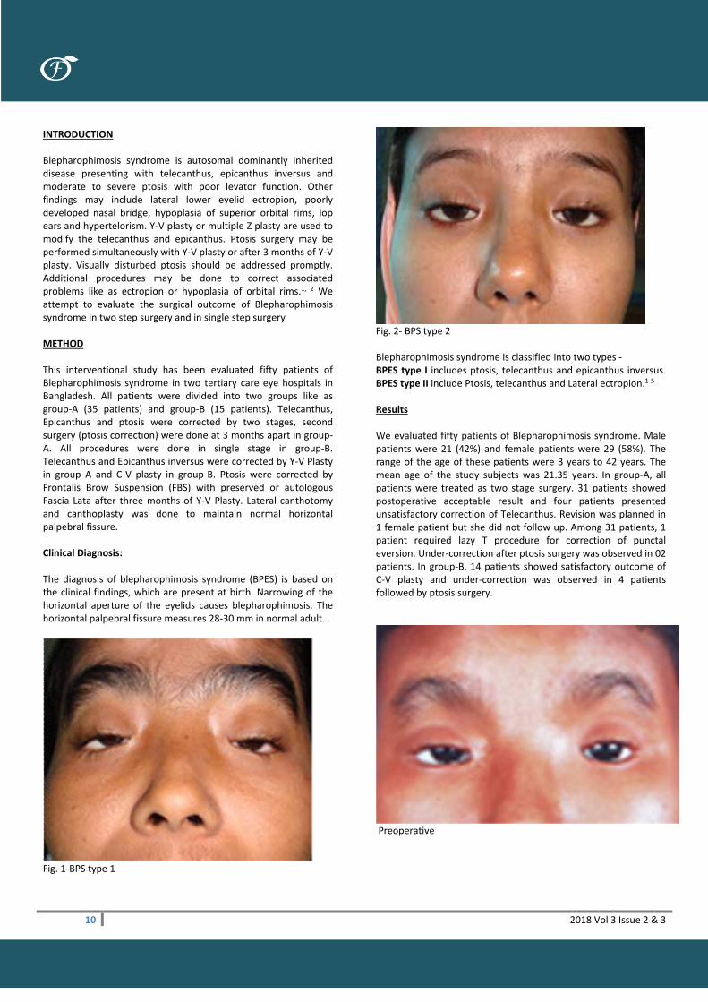
INTRODUCTION Blepharophimosis syndrome is autosomal dominantly inherited disease presenting with telecanthus, epicanthus inversus and moderate to severe ptosis with poor levator function. Other findings may include lateral lower eyelid ectropion, poorly developed nasal bridge, hypoplasia of superior orbital rims, lop ears and hypertelorism. Y-V plasty or multiple Z plasty are used to modify the telecanthus and epicanthus. Ptosis surgery may be performed simultaneously with Y-V plasty or after 3 months of Y-V plasty. Visually disturbed ptosis should be addressed promptly. Additional procedures may be done to correct associated problems like as ectropion or hypoplasia of orbital rims.1, 2 We attempt to evaluate the surgical outcome of Blepharophimosis syndrome in two step surgery and in single step surgery METHOD This interventional study has been evaluated fifty patients of Blepharophimosis syndrome in two tertiary care eye hospitals in Bangladesh. All patients were divided into two groups like as group-A (35 patients) and group-B (15 patients). Telecanthus, Epicanthus and ptosis were corrected by two stages, second surgery (ptosis correction) were done at 3 months apart in group-A. All procedures were done in single stage in group-B. Telecanthus and Epicanthus inversus were corrected by Y-V Plasty in group A and C-V plasty in group-B. Ptosis were corrected by Frontalis Brow Suspension (FBS) with preserved or autologous Fascia Lata after three months of Y-V Plasty. Lateral canthotomy and canthoplasty was done to maintain normal horizontal palpebral fissure. Clinical Diagnosis: The diagnosis of blepharophimosis syndrome (BPES) is based on the clinical findings, which are present at birth. Narrowing of the horizontal aperture of the eyelids causes blepharophimosis. The horizontal palpebral fissure measures 28-30 mm in normal adult.
Fig. 1-BPS type 1
Fig. 2- BPS type 2 Blepharophimosis syndrome is classified into two types - BPES type I includes ptosis, telecanthus and epicanthus inversus. BPES type II include Ptosis, telecanthus and Lateral ectropion.1-5 Results We evaluated fifty patients of Blepharophimosis syndrome. Male patients were 21 (42%) and female patients were 29 (58%). The range of the age of these patients were 3 years to 42 years. The mean age of the study subjects was 21.35 years. In group-A, all patients were treated as two stage surgery. 31 patients showed postoperative acceptable result and four patients presented unsatisfactory correction of Telecanthus. Revision was planned in 1 female patient but she did not follow up. Among 31 patients, 1 patient required lazy T procedure for correction of punctal eversion. Under-correction after ptosis surgery was observed in 02 patients. In group-B, 14 patients showed satisfactory outcome of C-V plasty and under-correction was observed in 4 patients followed by ptosis surgery.
Preoperative

11 2018 Vol 3 Issue 2 & 3
POD 7 V-Y Plasty (1st stage)
After 2nd stage op (FBS) Discussion We corrected telecanthus and epicanthus by Y-V plasty in 35 cases. Ptosis correction were done after three months of primary surgery. Lateral canthoplasty were corrected in some cases. New approach C-V plasty were done in fifteen cases to correct telecanthus and epicanthus. Lateral canthoplasty and frontalis brow suspension were done at same sitting. The result of Y-V plasty and C-V plasty were same but ptosis surgery was resulted better in separate approach after three months of first surgery. Under-correction was observed in ptosis surgery in six cases.
Pre-op of Y-V plasty
POD 7 Y-VPlasty (1st stage)
After 2nd stage FBS
BPES: Preoperative

12 2018 Vol 3 Issue 2 & 3
Single stage surgery: 6 wks of C-V plasty, and Frontalis brow suspension. The one-stage corrective procedure provided acceptable results both in function and cosmesis. 18 out of 23 (78%) patients underwent one-stage surgery before the age of 5 years. Only two patients had a blepharophimosis ratio greater than 1.5 as poor result. Two out of 18 (11%) patients with PFHs more than 2 mm needed a repeat operation, but all five (100%) patients with less than 2 mm (very severe ptosis) needed repeat operations.6 A total of 11 patients (8 males, 3 females) with a mean age of 9 years (range 6--22 years) were reviewed. The surgical outcome was assessed both functionally and cosmetically in single stage surgery. The mean preoperative visual acuity was 0.729 ± 0.316 SD and the mean postoperative visual acuity was 0.856 ± 0.277 SD (P <0.0428). There was a statistically significant improvement of telecanthus (P<0.0001) in terms of inner intercanthal distance, and horizontal palpebral fissure length (P=0.019). The mean preoperative and postoperative intracanthal distance was 3±0.33 SD and 2.418 ± 0.189 SD, respectively. There was also a significant postoperative improvement of ptosis (P< 0.01). All the patients had a stable functional and cosmetic result after a mean follow-up period of 3 years.7 Kuhn has suggested modifying the incision used in Y-V plasty to a C-shaped, so that it conforms the Langers skin fold and further reduces the scarring postoperatively. CONCLUSION There was no significant difference between Y-V plasty and C-V plasty to correct telecanthus and epicanthus. Ptosis correction resulted well in separate sittings. REFERENCES: 1. Allen CE, Rubin PA. Blepharophimosis -ptosis-epicanthus inversus syndrome (BPES): clinical manifestation and treatment. Int Ophthalmol Clin. 2008; 48(2): 15-23. 2. Mukherjee B, Alam MS. Double Jeopardy: Blepharophimosis Syndrome with Congenital Nasolacrimal Duct Obstruction in Twins. Orbit. 2013; 32 (5): 318-320 3. Beckingsale PS, Sullivan TJ, Wong VA, Oley C.
Blepharophimosis: a recommendation for early surgery in patients with severe ptosis. Clin Experiment Ophthalmol. 2003; 31: 138–42. 4. Choi KH, Kyung S, Oh SY. The factors influencing visual development in blepharophimosis-ptosis-epicanthus inversus syndrome. J Pediatr Ophthalmol Strabismus. 2006; 43: 285–8. 5. Dawson EL, Hardy TG, Collin JR, Lee JP. The incidence of strabismus and refractive error in patients with blepharophimosis, ptosis and epicanthus inversus syndrome (BPES). Strabismus. 2003; 11: 173–7. 6. S-Y Wu, L Ma, Y-J Tsai, J Z-C Kuo. One-stage correction for blepharophimosis syndrome. Eye. 2008; 22:378–88. 7. Huang WQ, Qiao Q, Zhao R, Wang XJ, Fang XQ authors. Surgical strategy for congenital blepherophimosis syndrome. Chin Med J. 2007; 120:1413–5.
Acute proptosis after ERCP
Dr Hunter Yuen Kwok-Lai CASE HISTORY A 41-year-old female underwent endoscopic retrograde cholangiopancreatogram (ERCP) for her abdomen problem. She was anxious and agitated during the procedure and the procedure was completed with some difficulties. She complained of acute onset of left eye pain, swelling, redness and diplopia after the procedure. Ophthalmic examination revealed left upper lid swelling and periorbital erythema, left eye non-axial proptosis with left eyeball displaced inferiorly. Her visual acuity, pupil, intraocular pressure and fundi were within normal limit. CT scan showed a 2.5 x 1 cm left superior extraconal soft tissue mass (Figure 1). This mass closely abutted the left superior rectus muscle, which was displaced inferiorly. MRI showed the lesion was hyperintense in T1 and T2 with a fluid level at the posterior border where it was slightly darker (Figure 2). This was more obvious with T2WI where it was hyperintense anteriorly and isointense posteriorly. A diagnosis of non-traumatic subperiosteal orbital hematoma was made. This was probably related to the increase in venous pressure during the struggling when performing ERCP. Transcutaneous needle aspiration was done and 2 cc blood stained fluid was aspirated (Figure 3). There was immediate improvement in proptosis and diplopia after the needle aspiration.

13 2018 Vol 3 Issue 2 & 3
Figure 1
Figure 2
Figure 3
DISCUSSION Orbital hematomas are categorized into intraorbital and subperiosteal types. The less common subperiosteal orbital hematoma is usually related to orbital trauma. Traumatic orbital subperiosteal hematoma can occur after facial and head trauma and due rupture of orbital subperiosteal vessels. Non-traumatic subperiosteal orbital hematoma is less commonly seen and are related sudden elevations of cranial venous pressure such as weight lifting, vomiting, coughing, scrub diving. In most cases patients present with proptosis, diplopia and painful eyeball movement. Nontraumatic subperiosteal orbital hematomas are more common in younger patients and orbital surface of the frontal bone is most commonly affected. Here, the periosteum is only loosely adherent to the underlying orbital bone and this uninterrupted boney surface in the orbit with lack of firm attachment making it the most susceptible to periosteal separation. In young adults with no calcifications, the periosteum attachment to the underlying bone is believed to be weaker as well. The diagnosis of subperiosteal hematoma can be confirmed with CT and MRI scan as illustrated. Conservative management can be used if patient is asymptomatic but the hematoma takes some time to resolve completely. Needle aspiration or orbitotomy with drainage is indicated if patient is having symptomatic diplopia, elevated intraocular pressures, vision loss due to eyeball compression of compressive optic neuropathy. In the acute setting of significant vision loss or severely elevated intraocular pressures, an emergent lateral canthotomy can be considered as a temporizing measure to relieve the orbital pressure. REFERENCES: 1. Atalla, M., McNab, A., Sullivan, T., and Sloan, B. Nontraumatic
subperiosteal orbital hemorrhage. Ophthalmology. 2001; 108: 183–189
2. Gomez-Ledesma, I., Mencia-Gutierrez, E., Gutierrez-Diaz, E., and Alonso-Santiago, M.A. Orbital subperiosteal hemorrhage while scuba diving. Orbit. 2006; 25: 19–22
3. Wolter, J.R. Subperiosteal hematomas of the orbit in young males: a serious complication of trauma or surgery in the eye region. Trans Am Ophthalmol Soc. 1979; 77: 104–120
4. Wolter, J.R., Leenhouts, J.A., and Coulthard, S.W. Clinical picture and management of subperiosteal hematoma of the orbit. J Pediatr Ophthalmol. 1976; 13: 136–138
5. Katz, B. and Carmody, R. Subperiosteal orbital hematoma induced by the valsalva maneuver. Am J Ophthalmol. 1985; 100: 617–618
6. Katz, R.S. and Abrams, G. Orbital subperiosteal hematoma (epidural hematoma of the orbit). J Clin Neuroophthalmol. 1981; 45: 45–52

14 2018 Vol 3 Issue 2 & 3
7. Boyer, M.M. and Lucarelli, M.J. Valsalva-induced subperiorbital hemorrhage during migraine. Arch Ophthalmol. 1998; 116: 106–107
8. Hunt, K.E. and Ross, J.J. Orbital hemorrhage in the nonoperated eye as a complication of general endotracheal anesthesia. Arch Ophthalmol. 1998; 116: 105–106
9. Kersten, R.C. and Rice, C.D. Subperiosteal orbital hematoma: visual recovery following delayed drainage. Ophthalmic Surg. 1987; 18: 423–427
10. Gillurn, W.N. and Anderson, R.L. Reversible visual loss in subperiosteal hematoma of the orbit. Ophthalmic Surg. 1981; 12: 203–209
Agricultural pesticide use and childhood orbital malignancy
Jayanta K Das Satya B Paul
Isha Agarwalla Harsha Bhattacharjee
Basant k Tiwari Rajen Gogoi
Munlima Hazarika
ABSTRACT Objective: The risk factor for most of paediatric orbital malignancy is to be identified. There are studies aimed at elucidating the epidemiological factors. Exposure to pesticides may be a possible contributing factor in the pathogenesis of childhood malignancy. Here we aim to investigate the role of agricultural pesticides in the aetiology of orbital childhood malignancy Methods: A prospective case control study done in North-eastern region of India from Jan 2005- Dec 2017. In the study group, a total of 33 patients of childhood orbital malignancy were included, controls were frequency matched with the cases on age, sex, geographical locality. Parental use of pesticides during pregnancy and childhood exposure to pesticides was investigated. Results: 33 cases were taken in study and control group. Nineteen cases (57.6 %) were male and fourteen cases (42.4%) were female in both the groups. The mean ages of study and control groups are 6.24 and 6.03 years respectively. The role of pesticide as a probable aetiology for paediatric orbital malignant tumour established by paired t- test analysis. The P value was found at 0.003 which is less than 0.05 and significant at 5 % level of significance. However, the disease process and its outcome were not influenced directly by the exposure to pesticide, but by the pathology and stage of the disease at which it was diagnosed. This was validated by the Pearson chi-square test. Conclusion: The study would put forward the possibility of exposure to pesticides as a major contributing factor of childhood
malignancy. Further studies are needed to establish the mechanisms also. Introduction Despite advances in treatment, cancer remains a leading cause of childhood mortality (Ries et al. 1999), and its etiology remains poorly understood (Chow et al. 1996). There are very few studies aimed at elucidating the epidemiological factors. Epidemiological data have shown that most cancers are due to environmental factors including life style practices and specific occupations. Experimental data confirm the role of environmental agents in multistep carcinogenesis, including genetics. The development of malignant phenotypes involves complex interactions among endogenous and external factors. Epidemiological research has been limited because of the difficulties in identifying a large study population. Moreover, the use of retrospectively ascertained childhood cancer cases in epidemiologic investigations has restricted the incorporation of biological and clinical parameters. (Leslie L Rabinson et al. 1995). Exposure to pesticides has been implicated as a possible contributing factor in the pathogenesis of childhood cancer. In two reviews (Daniels et al. 1997; Zahm and Ward 1998), parental pesticides use was fairly consistently associated with acute lymphocytic leukemia and CNS tumors, and less consistently with Wilms tumour, Ewing’s sarcoma, and soft-tissue sarcomas. Association between parental pesticide use and childhood cancer have been linked to either mother or father. Evidence from animal models suggests that exposure of the father during the preconception period may be especially important (Buckley et al. 1994). METHOD A case control study was carried out in eastern part of India involving four tertiary care hospitals over the period of four years in between April 2005- March 2009. In study group, 12 cases were of Rhabdomyosarcoma, 12 patients orbital manifestation of Acute Myeloid leukemia, four cases of neuroblastoma three patients of malignant histiocytosis and two cases of non-Hodgkin’s lymphoma. The demographical variables recorded were the medical record number, hospital-indoor registration number, date of first reporting/admission, name, age, sex, address, ethnicity, socioeconomic condition. All the cases were subjected to clinical history taking including history of family, consanguinity, exposure to any toxic substances (e.g. chemicals) and radiation hazard were thoroughly investigated, apart from standard clinical examination and investigation. Enrolment Criteria: Enrolment for study group The cases were identified directly by the investigators who reported four tertiary care hospitals in the above mention period.

15 2018 Vol 3 Issue 2 & 3
The all the cases of orbital malignancy were to have been newly diagnosed in between April 2005 to March 2017. Patients were also required to be less than 18 years of age. The reasons for non-eligibility were single parent, history of radiation and other known hazard which is carcinogenic except pesticide and habit of smoking of parents. The paternal age <40 years and maternal age <30 years at the time of pregnancy were excluded from study. Enrolment for control group Thirty-three children of age and sex matched included for control with the same geographical locality and demographic profile, fulfilling similar criteria with cases including same economic background. Data collection The interview and field visit were carried out within the one year period of first diagnosis. We asked the parents whether they have used insecticide at home and on pets. Participant gave oral informed consent to interview and field visit before the study. The method of interview and field visit was recorded either by video or photography. Additional details of pesticides used and work practice were obtained from agricultural field officer of that particular locality. General questioners include whether applicator personally mixed and applied pesticides, frequency of pesticide mixing and application (days/month). Applicator has been also be asked to indicate whether they generally used protective equipment, as per instruction by agriculture field worker. Stress was also made on elucidating the fact that whether the farmers used the pesticides in the recommended concentrations or in excess of that. We studied 33 cases with orbital malignant tumor and 33 age match controls of the similar locality and demographic profile. We made same questioners and analysis to the control group also. Data analysis To evaluate pesticide exposure, both maternal and paternal pesticide uses were included and two variables were also combined as follows: No pesticide use by parents. Considering small sample size pesticide used by any one parent was considered positive exposure and pesticide not used by parents were considered negative. RESULTS In the study group total of 33 patients of childhood orbital malignancy were included. 12 patients had Acute Myeloid leukemia, 12 cases of Rhabdomyosarcoma, four cases of neuroblastoma, three cases of orbital malignant Histiocytosis and two patients of non-hodgkins lymphoma were selected. Equal numbers of children and family were taken in control group. Most of the children’s age was between 2-14 years at the time of study enrollment. In the current study group 16 (48.5%) number of
parents were found exposed to pesticide compare to 11 (33.3%) number in control group.
Fig 1
Fig 2 Fig 3

16 2018 Vol 3 Issue 2 & 3
Fig 1. The figure shows a farmer spraying without protection. Fig 2. The children are exposed to pesticides every day. Fig 3 Common rack for cooking oil, hair oil and pesticides.
Types of Group Positive Exposure Negative Exposure
Case Group (n=33) 16 (48.48%) 17 (51.51%)
Control Group (n=33) 11 (33.33%) 22 (66.66%)
Table 10.1 showing no of patient with percentage in both control as well as study group in relation to exposure to agricultural pesticide (n=66)
The economic background of the 25 (76%) of the parents, were found poor and 8 (24%) from economically sound. In the study group five families have at least one member with stable job and hence economically sound, three families belong to business community and the rest are by profession farmer. In the control group also we choose the same socio- economic background of the families. In the both study and control group all of the applicators are farmer by profession. When protective measures used were examined, children of father of control group reported that they generally did not use any protective device during pesticide application, which is not seen in more than 50% of the control group. Moreover in 10 families none of the parents had completed high school level education in study group as compared to only 6 in control group. All of the 10 families belong to farmer by profession. Table 10.2 showing outcome of the patients of control group (n=33) in terms of exposure or non-exposure to agricultural pesticide:
Outcome of Disease Exposure Non Exposure
Success (Alive) 5 (15.15%) 8 (24.24%)
Failure (Expire) 11 (33.33%) 9 (27.27%)
The role of pesticide in the probable etiology among pediatric orbital malignant tumor in Pearson Chi-square test is found significant. Though outcome of the disease without pesticide exposure was slightly better than non-exposure patients, but not significant statistically, in the study group However, the disease process and its outcome influenced directly by the pathology and stage of the disease at which stage it was diagnosed. This was validated by the person chi-square test. DISCUSSION
Our study showed that most of the cancer were detected with pesticide user father’s group in comparison to control group. Our study, which is one of the several to suggest that children of parents who are occupationally exposed to pesticides incur an increased risk of childhood cancer. (Buckley et al. 1989; Daniels et al. 1997; Kristensen et al.1996; Shu et al. 1988; Zahm and Ward et al. 1998). In this study we examine the relationship between pesticide and risk of pediatric orbital malignancy. We cannot rule out the possibility that our observation occurred by chance alone, clearly, these findings need to be replicated. CONCLUSION In conclusion, the study findings strengthen the hypothesis that use of pesticides may play a role in the etiology of childhood orbital malignancies and especially the prenatal period may be a particularly vulnerable time window. The consistency of the findings with those of previous studies on acute leukemia in different populations, at different time periods, and using different designs, raises the question of the advisability of preventing pesticide use by pregnant women, even though a causal relationship still must be more fully documented. REFERENCES: 1. Anderson L, Diwan BA, Fear NT, Roman E, (2000): Critical
windows of exposure for children’s health: cancer in human epidemiological studies and neoplasm’s in experimental models. Environ Health Perspect’.108:573-94
2. Blair A, Axelson O, Paynter OE, Pearce N, Stevenson D, Trosko JE, Vainio H, Williams G, Woods J. Carcinogenic effects of pesticides. (1998): Advance in Modern Environmental Toxicology (Mehlman MA, ed). Vol XVIII
3. Camann DE, Carpet dust (1997): an indicator of exposure at home to pesticides, PAHs, and tobacco smokes. Research Triangle park, NC 1994. 141.
4. Chew W, Linet MS, Liff JM, Greenburg RS. (1996): Cancers in Children. In: Cancer Epidemology and prevention, New York: Oxford University Press. 1331-1363.
5. Daniels JI, Olshan AF, Savitz DA.(1997): Presticides and childhood cancers, Environ Health Perspect. 105:1068-1077
6. Kori B Flower, Jane A Hoppin, Chalres F Lynch, Aaron Blair, Charles Knott, David L Shore and Dale P Sandler. (2004): Cancer risk and parental pesticide application in children of Agriculture Health Study participants. Environ Health Perspect. 112(5): 631-635.
7. Leslie L. Robinson, Jonathan D. Buckley and Greta Bunin. (1995): Pesticide and childhood cancer, Environ Health Perspect. 103(Supp 6):111-16.
8. Moses M, Johnson ES, Anger WK Burse VW, Horstman SW, Jackson RJ, Lewis RG, Maddy KT, Mc Connell R, Maggs WJ. (1993): Environmental equity and pesticide exposure. Toxicol Ind Health. 9:913-959

17 2018 Vol 3 Issue 2 & 3
9. Olshan AF, Daniels JL. (2000): Invited commentary pesticides and childhood cancer. Am J Epidemiol. 151:647-649
10. Ries LAG, smith MA, Gurney JG, Linet M, Tamra T, Young JL, et al., eds (1999): Cancer incidence and Survival among Children and Adolescents: United States SEER Program 1975-1995.
11. Simcox NJ, Fenske RA, Wolz SA, Lee IC, Kalman DA. (1999): Pesticides in household dust and soil: exposure pathway for children of agricultural families. Environ Health Perspect. 103:1126;141.
12. Zahm SH, Blair A, and the farmwork Epidemiology Research Group. (1997):Cancer feasibility studies among migrant farmworks. Am J Ind Med. 32:301-302
13. Zahm SH, Ward MH. (1998): Pesticides and Childhood cancer. Environ Health Perspect 106:893-908.

18 2018 Vol 3 Issue 2 & 3
Announcement: APSOPRS-ASMHK 2018 Welcome Message
The biennial meeting of the Asia-Pacific Society of Ophthalmic Plastic and Reconstructive Surgery (APSOPRS), which will be held in conjunction with 30th Annual Scientific Meeting (ASM) jointly held by The College of Ophthalmologists of Hong Kong (COHK) and the Hong Kong Ophthalmological Society (HKOS) from December 15-16, 2018 at the Hong Kong Convention and Exhibition Centre. COHK and HKOS have jointly organized our Annual Scientific Meeting (ASM) since 1989. This annual conference is the most important platform for our ophthalmologists in Hong Kong to share our newest developments and research in ophthalmology. This is the first time for our annual scientific meeting held in conjunction with APSOPRS conference. The organizer is delighted to have Dr. Don Kikkawa and Prof. Geoffrey Rose as the Keynote speakers. We are expecting more than 80 regional experts to come and speak. It is guaranteed it would be an outstanding and comprehensive scientific program. Important Dates: Abstract Submission Deadline: Aug 30, 2018 Early Registration Deadline: Sept 30, 2018 Event website: http://www.apsoprs-asmhk2018.com/
Dr YUEN Kwok Lai Hunter • ASM Organizing Committee Chairman • President, Asia-Pacific Society of Ophthalmic
Plastic and Reconstructive Surgery (APSOPRS) • Past President, Hong Kong Society of Ophthalmic
Plastic and Reconstructive Surgery (HKSOPRS)

19 2018 Vol 3 Issue 2 & 3

20 2018 Vol 3 Issue 2 & 3
21 2018 Vol 3 Issue 2 & 3

22 2018 Vol 3 Issue 2 & 3

23 2018 Vol 3 Issue 2 & 3
Guidelines Below are the formats for the different categories of articles: Invited Articles (no more than 1600 words; include images where appropriate) Case Highlights This refers to the written presentation of an interesting or challenging case in the following format:
• History (no more than 100 words) • Examination (no more than 150 words; include clinical photos) • Investigations (include imaging where appropriate) • Management (no more than 100 words; include pathology images where appropriate) • Discussion (no more than 200 words; including challenges encountered in diagnosis or management)
Operative Pearls This refers purely to advice that allows the reader to improve on his or her intraoperative technique and can include immediate post-op advice (no more than 600 words; include images where appropriate) Meetings or Social Visits All meetings organized by APSOPRS members as well as social visits to each other’s' centers are eligible for inclusion in the newsletter (no more than 1000 words for the main APSOPRS biennial meeting and no more than 500 words for other meetings or visits). Philosophical Notes No more than 800 words; include images where appropriate