office medic user’s manual
TRANSCRIPT

Revised April 2021
Office Medic™ User’s Manual For use with: Orbit™ ● Universal SmartECG™

690002-00 Rev. K
Table of Contents
GENERAL CAUTIONS & WARNINGS ............................................................................ 4
GLOSSARY OF SYMBOLS 4 WARNINGS 5 CAUTIONS 6 ELECTRICAL SAFETY CLASSIFICATIONS 9
ACCESSORIES ........................................................................................................... 10
Spirometry Accessories 10 Electrocardiogram Accessories 11
OFFICE MEDIC BASICS .............................................................................................. 12
SYSTEM REQUIREMENTS 12 INSTALLATION 12 BACKING-UP AND RESTORING THE DATABASE 13 NAVIGATION 14 FILE MENU 15 TEST MENU 17 OPTIONS MENU 18 TOOLS MENU 20 HELP MENU 20
SPIROMETRY ............................................................................................................ 21
SPIROMETRY CAUTIONS & WARNINGS 21 SPIROMETRY GETTING STARTED 22 PROPER PATIENT PREPARATION 23 PROPER TESTING PROCEDURE 23 EFFORT QUALITY MESSAGES FOR ADULT SUBJECTS 24 TEST SESSION GRADES 25 UNACCEPTABLE SPIROMETRY TESTS 25 REPEATABILITY 25 PERFORMING A SPIROMETRY TEST 26 ABOUT THE SPIROMETRY TEST SESSION WINDOW 28 SPIROMETRY OPTIONS 29 SPIROMETRY TOOLS 34 PREDICTED VALUE EQUATIONS 36 LUNG AGE CALCULATION 47 SPIROMETRY INTERPRETATION 48

ELECTROCARDIOGRAPHY ......................................................................................... 52
ECG CAUTIONS, WARNINGS, AND OTHER INFORMATION 52 ECG GETTING STARTED 55 PERFORMING AN ECG TEST 56 ABOUT THE ACQUISITION WINDOW 57 ECG OPTIONS 59 REVIEWING AN ECG 62 PRINTING AN ECG 68 ECG DEVICE VERIFICATION 69 ECG ANALYSIS PROGRAM 69
TROUBLESHOOTING AND TIPS. ................................................................................ 70
SERVICE INFORMATION ........................................................................................... 74
DEVICE CARE & MAINTENANCE 74 SERVICE 75 LIMITED WARRANTY 75
GLOSSARY OF TERMS. ............................................................................................... 76
DEVICE SPECIFICATIONS ................................................................................................................ 79
UNIVERSAL SMARTECG SPECIFICATIONS 79 ORBIT PORTABLE SPIROMETER SPECIFICATIONS 80

General Cautions & Warnings 4
General Cautions & Warnings
Before conducting tests read the General Caution & Warnings and the specific Cautions & Warnings pertaining to your particular medical device. If you need further assistance see Service.
Glossary of Symbols
Attention Consult Accompanying Documents
Consult Instructions For Use Consult Accompanying Documents
Consult Instructions For Use Consult Accompanying Documents
Defibrillator proof type CF equipment Defibrillator proof type CF equipment complying with IEC Publication 60601.
CE Mark
Indicates this device is in compliance with MDD 93/42/ECC. 2797 is the Notified Body Number.
Do not reuse.
Class II, Electrical Equipment.
REF Catalogue or Model Number
S/N Serial Number
Manufacturer
Authorized representative in the European community.
Waste Electronic Electrical Equipment (WEEE). Separate collection for waste electrical and electronic equipment.
Rx only Federal (USA) law restricts this device to sale by or on the order of a physician.
General Cautions & Warnings 5
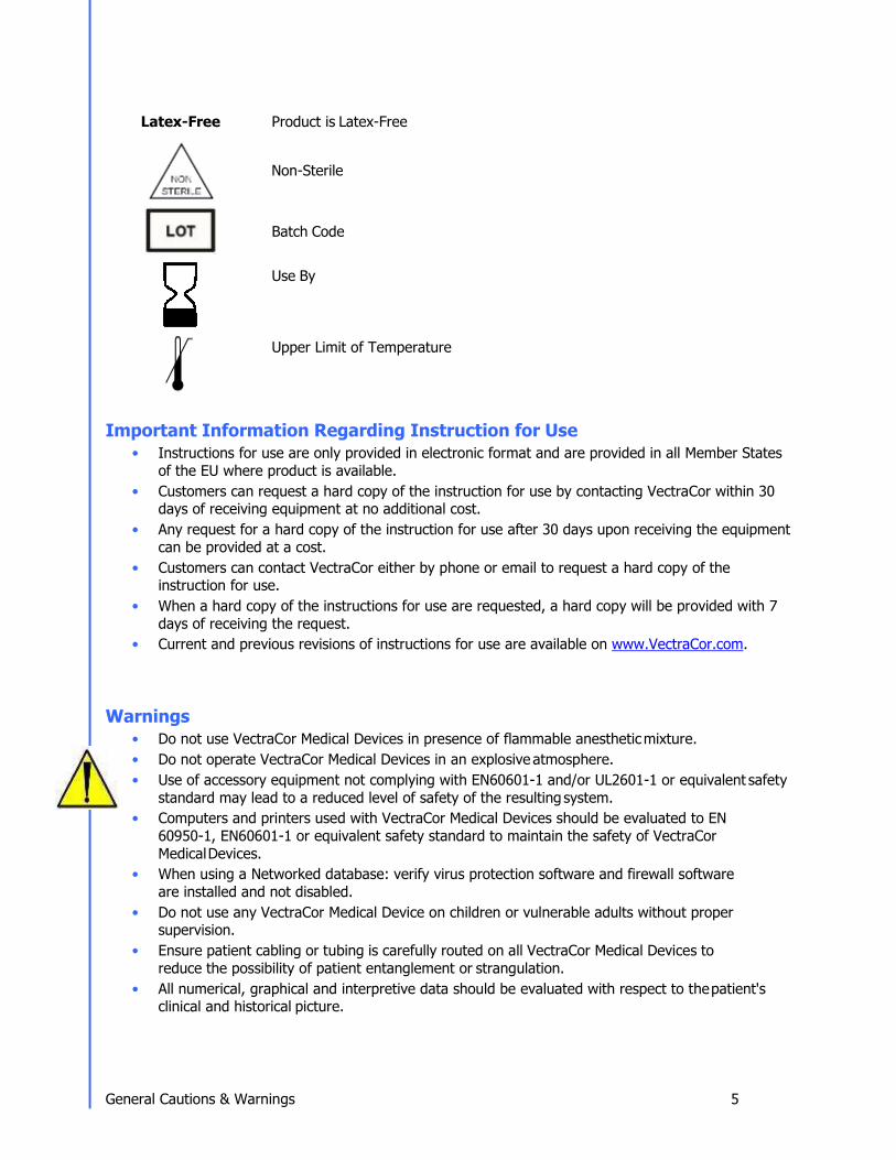
Latex-Free Product is Latex-Free
Non-Sterile
Batch Code
Use By
Upper Limit of Temperature Important Information Regarding Instruction for Use
• Instructions for use are only provided in electronic format and are provided in all Member States of the EU where product is available.
• Customers can request a hard copy of the instruction for use by contacting VectraCor within 30 days of receiving equipment at no additional cost.
• Any request for a hard copy of the instruction for use after 30 days upon receiving the equipment can be provided at a cost.
• Customers can contact VectraCor either by phone or email to request a hard copy of the instruction for use.
• When a hard copy of the instructions for use are requested, a hard copy will be provided with 7 days of receiving the request.
• Current and previous revisions of instructions for use are available on www.VectraCor.com. Warnings
• Do not use VectraCor Medical Devices in presence of flammable anesthetic mixture. • Do not operate VectraCor Medical Devices in an explosive atmosphere. • Use of accessory equipment not complying with EN60601-1 and/or UL2601-1 or equivalent safety
standard may lead to a reduced level of safety of the resulting system. • Computers and printers used with VectraCor Medical Devices should be evaluated to EN
60950-1, EN60601-1 or equivalent safety standard to maintain the safety of VectraCor Medical Devices.
• When using a Networked database: verify virus protection software and firewall software are installed and not disabled.
• Do not use any VectraCor Medical Device on children or vulnerable adults without proper supervision.
• Ensure patient cabling or tubing is carefully routed on all VectraCor Medical Devices to reduce the possibility of patient entanglement or strangulation.
• All numerical, graphical and interpretive data should be evaluated with respect to the patient's clinical and historical picture.

General Cautions & Warnings 6
• Do not attempt to insert any VectraCor Medical Device (including patient cables) directly into an electrical outlet.
• Restoring the database erases all of the data located in Office Medic and replaces it with the data contained in the back-up file. Data that was acquired after the date of the last back-up will be lost and cannot be recovered.
• Once deleted, data can only be recovered from the date of your last back-up. Maintain regular back-ups to ensure data is not lost.
• The computer regulates the battery and will provide a warning message to inform the user that the battery is low in order to prevent data loss.
• Do not load any other manufacturer’s SCP files. The Office Medic program is designed to work only with VectraCor SCP files.
• Do not use 3rd party applications to review or analyze VectraCor SCP files. • Use only VectraCor approved accessories with VectraCor devices.
Any serious incident that has occurred in relation to the device should be reported to the manufacturer and the competent authority of the Member State in which the user and/or patient is established.

General Cautions & Warnings 7
Cautions Disposal Instructions:
Due to the potential presence of hazardous substances in electrical or electronic equipment, DO NOT dispose of VectraCor medical devices with municipal waste. Improper disposal could have an adverse effect on the environment and human health.
For VectraCor products NOT marked with please contact your local municipal waste company for proper disposal instructions.
For VectraCor products MARKED with please contact your local sales representative (from whom you purchased the product) or your local municipal waste company for proper disposal instructions.
• Federal (USA) law restricts this device to sale by or on the order of a physician. • All VectraCorDevices are intended for use by a physician or by trained personnel under a
physician's supervision. Read all instructions for use and specifications provided prior to use.
Important! VectraCor medical devices are intended for use in the electromagnetic environment(s) specified below. Users of this equipment should ensure that it is used in such environment(s). Security precautions:
• User is responsible for protection of the login credentials/ access controls used to access the PC
hosting Office Medic and patient database. VectraCor will not be able to give access to any user who has lost their access to their host PC.
• User is responsible for equipping the PC hosting Office Medic and patient database with necessary protection against external attacks (virus, malware etc.) and protection configuration.
• VectraCor strongly recommends the user utilize credible antivirus, malware protection, firewall, etc. software on each piece of equipment where patient information is stored. Please keep in mind that VectraCor software will need to be given appropriate permissions to operate properly.
• User is responsible for creating/maintaining logs of login or Office Medic usage information. • User is responsible for patient database including patient demographic information and patient
medical data. VectraCor will not receive or maintain any patient identifiable data. • User is responsible for scheduled backup of the database to prevent data loss due to
unforeseen circumstances. Refer BACKING UP AND RESTORING DATABASE section for instructions.
• User is responsible for the integrity of Office Medic software and its components residing in the PC.
• VectraCor does not access, store or modify the patient demographic and medical data stored in the Office Medic database residing in the user’s PC.
• Use of other equipment that connects to the network may result in unidentified risks to patients, operators or third parties. The User is responsible for identifying, evaluating, and controlling these risks.
• Changes to the network (including, but no limited to changes in network configuration, connection of additional items, disconnection of items, update of equipment, or upgrade of equipment) may introduce new risks that requires additional analysis.

General Cautions & Warnings 8
Attention should be paid to the following EMC information prior to installing or using VectraCor medical devices.
• Portable and mobile Radio Frequency (RF) communication equipment may interfere with the
operation of VectraCor medical devices. RF equipment should only be used no closer than 30 cm (12 inches to any part of VectraCor medical devices.
• VectraCor medical devices have been tested and found to comply with IEC/EN 60601-1-2. • Computers, cables and accessories not tested to 60601-1-2 may result in increased emissions or
decreased immunity of VectraCor devices. • Verify normal operation if utilizing VectraCor medical devices adjacent to or stacked with other
electrical equipment.
Guidance and manufacturer's declaration - electromagnetic emissions and immunity Emissions Test Compliance Electromagnetic environment –
guidance RF emissions CISPR 11 Group 1 VectraCor equipment uses RF energy
only for its internal function. Therefore, its RF emissions are not likely to cause any interference in nearby electronic equipment.
RF emissions CISPR 11 Class B VectraCor medical devices are suitable for use in all establishments including domestic establishments and those directly connected to the public low-voltage power supplies buildings used for domestic purposes.
Harmonic emissions IEC 61000-3-2
Not applicable for VectraCor devices
Voltage Fluctuations/flicker emissions IEC 61000-3-3
Not applicable for VectraCor devices
Immunity Test IEC 60601 Test Level
Compliance Electromagnetic Environment Guidance Universal SmartECG Orbit Spirometer Electrostatic Discharge (ESD) IEC 61000-4-2
±8 kV contact ±2 kV, ±4 kV, ±8 kV, ±15 kV air
±2 kV, ±4 kV, ±6 kV contact ±2 kV, ±4 kV, ±8 kV air
Floors should be wood, concrete or ceramic tile. If floors are covered with synthetic material, the relative humidity should be at least 30%
Power frequency (50/60 Hz) magnetic field IEC 61000-4-8
30 A/m 3 A/m
Power frequency magnetic fields should be at levels characteristic of a typical location in a typical commercial or hospital environment.

General Cautions & Warnings 9
Radiated RF IEC 61000-4-3
3 V/m 80 MHz to 2.5 GHz
3 V/m 80 MHz to 2.5 GHz Portable and mobile RF Communications equipment should
be used no closer to any part of VectraCor medical devices, including cables, than the recommended separation distance calculated from the equation applicable to the frequency of the transmitter. Recommended separation distance: 𝑑𝑑 = 1.2√𝑃𝑃 80 MHz to 800 MHz 𝑑𝑑 = 2.3√𝑃𝑃 800 MHz to 2.5 GHz Where P is the maximum output power rating of the transmitter in watts (W) according to the transmitter manufacturer and d is the recommended separation distance in meters (m). Field strengths from fixed RF transmitters, as determined by an electromagnetic site surveya should be less than the compliance level in each frequency range. b
Interface may occur in the vicinity of equipment marked with the following symbol:

General Cautions & Warnings 10
NOTE: These guidelines may not apply in all situations. Electromagnetic propagation is affected by absorption and reflection from structures, objects and people.
a) Field strengths from fixed transmitters, such as base stations for radio (cellular/cordless) telephones and land mobile radios, amateur radio, AM and FM radio broadcast and TV broadcast cannot be predicted theoretically with accuracy. To assess the electromagnetic environment due to fixed RF transmitters, an electromagnetic site survey should be considered. If the measured field strength in the location in which VectraCor medical devices are used exceeds the applicable RF compliance level above, VectraCor medical devices should be observed to verify normal operation. If abnormal performance is observed, additional measures may be necessary, such as reorienting or relocating VectraCor medical devices. b) Over the frequency range 150 kHz to 80 MHz, field strengths should be less than 3 V/m.
Recommended separation distances between portable and mobile RF communications equipment and
VectraCor medical devices. VectraCor medical devices are intended for use in an electromagnetic environment in which radiated RF disturbances are controlled. The customer or the user of VectraCor medical devices can help prevent electromagnetic interference by maintaining a minimum distance between portable and mobile RF communications equipment (transmitters) and VectraCor medical devices as recommended below, according to the maximum output power of the communications equipment.
Rated maximum output power of transmitter
W
Separation distance according to frequency of transmitter (m) 150 kHz to 80 MHz 80 MHz to 800 MHz 800 MHz to 2.5 GHz
0.01 0.12 0.12 0.23 0.1 0.38 0.38 0.73 1 1.2 1.2 2.3 10 3.8 3.8 7.3 100 12 12 23
For transmitters rated at a maximum output power not listed above, the recommended separation distance d in meters (m) can be estimated using the equation applicable to the frequency of the transmitter, where P is the maximum output power rating of the transmitter in watts (W) according to the transmitter manufacturer. NOTE: These guidelines may not apply in all situations. Electromagnetic propagation is affected by absorption and reflection from structures, objects and people.

General Cautions & Warnings 11
Electrical Safety Classifications
Note: These classifications currently apply only to VectraCor Medical Devices.
• Class II Equipment • Type CF Equipment. Note: Universal SmartECG is Type CF with defibrillator-proof applied part. • IPXO - Ordinary Equipment. • Continuous Operation. • Not suitable for use in presence of flammable anesthetic mixture with air or with oxygen or
nitrous oxide.

Accessories 10
Accessories
Spirometry Accessories
Mouth Pieces (Z-5000-2608)
All QRS spirometers use pre-calibrated, disposable mouthpieces. These mouthpieces are individually calibrated during production using the same equipment LDS Hospital uses to validate spirometers against the American Thoracic Society's Recommendations for the Standardization of Spirometry. These pneumotachs are single patient use only. Pressure Tubes (Z-7000-2032)
Pressure tubes connect the pre-calibrated mouthpieces to the QRS spirometers. They are re-usable, but need to be replaced if kinked or if condensation forms inside the tube. Pressure tubes measure 48” long. Nose Clips (724050-00)
QRS, in line with ATS Guidelines, recommends the use of nose-clips when performing a Spirometry test with a QRS spirometer. Calibration Syringe (723000-00)

Accessories 11
QRS sells a three liter Volume Calibration Syringe designed to fit the QRS spirometers. Although QRS spirometers cannot be calibrated in the field, the syringe allows you to check your calibration. According to ATS Guidelines, the volume accuracy of the spirometer must be checked at least daily. If calibration is off, please contact QRS for recalibration of your spirometer.
Electrocardiogram Accessories

Office Medic Basics 12
Tab Electrodes
• Gel: Adhesive Gel • Chloride Content: 4% • Substrate: Synthetic Paper or Vinyl • Sensor: Ag/AgCl • Shape/Size: Rectangle, 1” x 7/8” • Adhesive Performance: Usable up to 1 hour • Shelf Life: 2 years, when stored between 10-32°C Wet Gel Electrodes
Superior Conductivity Up to 72 hours of continuous use Better Skin Contact Recommended for VectraplexECG System
Snap Electrode Adaptors
• Universal Adapter fits both snap and tab electrodes: Fits 3 mm to 4 mm pin leads • Latch Lock System: Securely locks on electrodes, leads won’t detach • Secure Connection: Reduces false alarms • Re-usable: Adapters can be cleaned/sterilized and re-used

Office Medic Basics 13
Office Medic Basics
System Requirements
Operating System: Microsoft® Windows®: 7 or 10 Free Disk Space: 600MB Internet Requirements: Internet Explorer 6.0 SP1 or later RAM: 4 GB or higher Processor: Dual-core 2 Ghz or higher Screen Resolution: 1024x768 (EKG Requirement) Interface: Available USB port Media: A CD/DVD drive or access to the internet to download the software.
Contact Customer care for download instructions and details. IT-Network information:
• Purpose of networking: Networked databases can be accessed by multiple computers and integrate Office Medic test sessions to an EMR/HER
• Required characteristics: Windows NT Client/Server domain. • Required configuration: Windows NT Active Directory • Technical specifications: Microsoft SQL trusted connect string • Intended information flow: The Office Medic software utilizes a Software Developer Kit (SDK) and
calls Office Medic to perform tests and return test data.
**Recommended system specifications: PC running Windows 7 or 10, Dual core CPU, 4 gigs of RAM, 300 gig HDD or better with an available USB port.

Office Medic Basics 14
Installation
Important! Do not connect the medical device to the PC prior to installing the software. The device drivers (step #9) must be installed prior to testing.
1. Ensure you are logged in with Administrator rights. 2. Remove all VectraCor devices from the computer. 3. Log out and close all programs. 4. Insert the Office Medic CD-ROM.
If the autorun feature on your computer is disabled go to the next instruction. If not follow the on screen prompts.
5. On the lower Windows toolbar select Start | Run or simultaneously press the Windows logo and R key. Type d:\setup.exe in the Open dialog box. Note: substitute the letter of your CD/DVD- ROM drive if it is different from d:.
6. Select Program(s) to be installed I. Office Medic
II. Office Medic & VectraplexECG 7. Select a language. ** Language package is available for Office Medic only. VectraplexECG is currently in English only.
Note: If you need to change the language, you will need to uninstall Office Medic. To do this go to your control panel, click on “Programs and Features” and then find the “Programs” and select “Uninstall a program. Find Office Medic on the list and uninstall. Finally, reinstall Office Medic using the setup program and select the correct language. Any data that was recorded will be preserved because uninstallation doesn’t delete data.
8. Follow the on-screen instructions.
Note: You will be given a choice to install a local or network database. The Network option requires an Office Medic Network Database formally called IDMS database. To learn more about obtaining a Network database, and networking Office Medic, contact Customer Care at VectraCor.
9. Once the installation is complete, connect the medical device to the PC with the CD-ROM still inserted. Follow the software prompts for installing the device driver.
An Office Medic shortcut will appear on your desktop when the installation is complete.

Office Medic Basics 15
Backing-up and Restoring the Database
Database Back-up Instructions Backing-up your database protects you from losing your patient data should a catastrophic event occur. Regular back-ups of the database should be maintained. Follow the steps below to back-up the database:
1. Close Office Medic. 2. Open folder: C:\Vectraplex\Database. 3. Copy the two files VectraplexECG.MDF and VectraplexECG_Log.LDF to a secure location. This is
the back-up copy of your Office Medic database. Copy these files as often as needed to maintain a current back-up file.
Database Restore Instructions
Warning! Restoring the database erases all of the data located in Office Medic and replaces it with the data contained in the back-up file. Data that was acquired after the date of the last back-up will be lost and cannot be recovered.
Follow the steps below to restore the database: 1. Close Office Medic. 2. Copy and paste the two back-up files into the following location: C:\Vectraplex\Database. 3. Open Office Medic.
The database should look exactly as it did on the date of the last back-up.

Office Medic Basics 16
Navigation
Select the Office Medic icon to open the software. The initial screen displays the directory of patients, sessions and tests. Contact VectraCor Technical Support for instructions on how to hide patient names.
Note: ECG tests with an asterisk symbol are acquired by VectraplexECG. ECG’s with an asterisk symbol will be displayed in VectraplexECG when opened.
Note: VectraplexECG only requires account number and gender to create a new patient. Some functions in Office Medic require more information.
Note: If an ECG was first acquired using Office Medic and the user changed the default ECG program from Office Medic to VectraplexECG, the ECG will be displayed in VectraplexECG when opened. Upon closing the ECG in VectraplexECG, the user will be prompted to save the ECG. If the user clicks yes to save the ECG, an ECG test will be created in the Office Medic patient tree under that specific patient. The original ECG from Office Medic will remain. If you do not save, a new file will not be created. When viewing these cases in the future open the appropriate file according to your default ECG program.

Office Medic Basics 17
File Menu
New (Ctrl+N) Opens the Patient Information window. Required fields are highlighted by an asterisk.
Note: Smoking-Pack Years is calculated by multiplying the number of cigarette packs smoked per day by
the number of years the patient has smoked.

Office Medic Basics 18
Open (Ctrl+O) Select a patient, session or test and then select Open to view the selected data.
Delete (Ctrl+D) Select a patient, session or test and then select Delete to delete the selected data.
Delete All The Delete All option deletes the entire database.
Warning! Once deleted, data can only be recovered from the date of your last back-up. Maintain regular back-ups to ensure data is not lost.
Print to File Creates an image file (either JPEG, TIFF, or PDF) of an Office Medic report. Highlight the session or test in the patient tree and select this option.
Note: The default location for image files is My Documents\Diagnostic Test Data\Image Files.
Batch Print The Batch Print option allows for the printing of multiple patient reports.
Print Preview Reports can be previewed by selecting the desired session or test and then select File | Print Preview.
Print (Ctrl+P) Select a patient, session or test and select File | Print to print a report.
Refresh Patient Tree (F5) Select to refresh the patient database.
Database Connection... Select to switch between local and network databases.
Exit Exits the Office Medic program.

Office Medic Basics 19
Test Menu Select a patient and then select the desired test from the Test menu to begin testing.
For details on spirometry testing see Performing a Spirometry Test For details on ECG testing see Performing an ECG Test

Office Medic Basics 20
Options Menu Select Options to change program settings.
General Options

Office Medic Basics 21
Default ECG programs Select Office Medic or VectraplexECG. Units Select Imperial or Metric.
Export File Creates tab delimited ASCII text files: Session.txt, SpTest.txt, SpCalibr.txt, OxiSess.txt and OxiTest.txt. The Export Flow/Volume Points feature creates two files called SpGraph.txt and SpCalGr.txt.
Image File Directory:
Select the browse button to change the default path where image files are saved.
Allow remote handhelds to initiate unattended synchronization sessions: MedicSync will launch automatically on the host computer and execute the existing handheld synchronization profile automatically within 30 seconds. If a conflict occurs, the handheld database will always overwrite the host computer database as no manual conflict resolution is available when using unattended synchronization.
Note: If the host computer is set to delete data from the remote, then data will be deleted from the remote during an automatic synchronization.
For details on changing the spirometry options see Spirometry Options For details on changing the ECG options see ECG Options

Office Medic Basics 22
Tools Menu
General Tools
For details on the spirometry tools see Spirometry Tools Help Menu
User’s Manual Opens the Office Medic User Manual.
ECG Physician’s Guide Opens the Physician’s Guide for the ECG interpretation algorithm.
About VectraCor-QRS Provides information for contacting VectraCor, Inc.
About Office Medic Displays the version of Office Medic and statistics about any connected device.

Spirometry 21
Spirometry
Note: The information in this chapter applies to spirometry tests acquired using an Orbit Portable Spirometer
Spirometry Cautions & Warnings
Warnings • Use only VectraCor mouthpieces manufactured to meet calibration requirements for the QRS
Orbit Portable Spirometer.
• Mouthpieces are single patient use only and MUST be replaced for each patient. • Exercise caution when performing spirometry testing on patients with a history of COPD. • Do not use mouthpieces on a patient with an injured mouth. • Do not obstruct the opening at the end of the mouthpiece. Obstruction may result in erroneous
results. • FVC and MVV testing can cause fatigue and some patients may be at risk for vertigo, arrhythmia
or syncope. • Patients should open, handle and dispose of his/her own mouthpiece to reduce the risk of cross
contamination. • If condensation forms inside the pressure tube or the pressure tube becomes visibly kinked it
must be replaced.
Warning! The ATS/ERS Task Force: Standardisation of Lung Function Testing recommends daily calibration checks.
Cautions Physicians must properly train individuals, under their care, in the use of this product. All tests must be evaluated by a qualified physician.
Indications for Use: Diagnostic Spirometry Patient Population: Male/Female, Pediatric to Adult Device Functionality: Diagnostic Spirometry Spirometric Parameters: FVC, MVV, SVC, and FEF Environment of Use: Hospital, Clinical and Home Use

Spirometry 22
Spirometry Getting Started
For the Orbit Portable Spirometer 1. Insert the USB cable into an available USB Port on your PC. 2. Connect the pressure tube to the Luer fitting. Ensure the pressure tube is not kinked or restricted
in any way. 3. Connect the other end of the pressure tube to the disposable mouthpiece.
Warning! Ensure pressure tube is properly connected. If condensation forms inside the pressure tube or the pressure tube becomes visibly kinked it must be replaced.

Spirometry 23
Proper Patient Preparation To obtain diagnostically reliable results:
• Loosen tight clothing (ties, belts, bras). • Remove patient's dentures. • Explain the procedure thoroughly, including demonstrating it yourself with your own mouthpiece. • Have the patient sit or stand in an upright position during the test. When standing, place a chair
behind them in case they become dizzy. • Before beginning the test have the patient take several slow, deep inhalations/exhalations to feel
comfortable.
Proper Testing Procedure To obtain diagnostically reliable results proper testing procedures must be followed:
• When the equipment is zeroing (two circles flashing) have the patient keep the mouthpiece away from their mouth.
• When testing ensure the patient has a tight seal with their lips around the mouthpiece. The patient should not bite the tube or have pursed lips.
• Place a disposable nose clip securely on the patient's nose or instruct the patient not to exhale through the nose.
• Verbally instruct the patient on properly performing the procedure:
– FVC – instruct the patient to take the largest possible inhalation, insert the mouthpiece into their mouth and exhale forcefully and completely. If a Flow/Volume Loop is desired, verbally instruct the patient to inhale after completely exhaling.
– SVC - instruct the patient to take the largest possible inhalation, insert the mouthpiece into their mouth, and exhale slowly and completely.
– MVV - instruct the patient to breathe as deeply and rapidly for 12 to 15 seconds into the mouthpiece. This test is often difficult to perform for many patients.
Important! Ensure the patient has a tight seal around the mouthpiece and is not covering or obstructing the fabric at the end of the mouthpiece with their hand.
• Encourage the patient to keep exhaling as long as possible. It is helpful to coach the patient with verbal commands and physical gestures. A proper expiration should last at least six seconds.
• Once finished, have the patient remove the mouthpiece and breathe normally until they have recovered.
Important! Using the mouthpiece more than 20 times, or for more than 10 consecutive days, may generate inaccurate results. Use a new mouthpiece after 20 attempts and/or 10 days to get the most accurate results.

Spirometry 24
Effort Quality Messages for Adult Subjects
Warning Message Criteria “Don’t hesitate.” BEV (Ext. Vol) > 150 mL or 5% of the FVC “Blast out faster.” PEFT > 120 msec “Blow out longer.” FET < 6.0 s for subjects aged 10 years and older or FET < 3 s for subjects
aged less than 10 years, and EOTV > 40 mL “Blast out harder.” PEF values do not match within 1.0 L/s “Deeper breath.” FEV6 values do not match within 150 mL Warning message does not appear.
Effort meets above criteria.
“Good test session.” Two acceptable efforts meet the repeatability requirements.

Spirometry 25
Test Session Grades Each test session is given a grade which indicates the degree of confidence in the results.
Grade Criteria
A At least 2 maneuvers with the largest two FEV1 values matching within 100mL and the largest two FEV6 values matching better than 100mL.
B At least 2 maneuvers with FEV1 values matching between 101 and 150 mL. C At least 2 maneuvers with FEV1 values matching between 151 and 200 mL. D Only one maneuver, or more than one, but the FEV1 values match > 200mL.
Unacceptable Spirometry Tests A spirometry test is considered unacceptable when:
• Insufficient initial inhalation (lungs not completely filled before the test). • Slow or hesitant start of expiration. • Leakage around the mouthpiece or nose clip. • Mouthpiece obstruction by teeth, tongues, or lips. • Coughing during the test. • Large variation of FVC or FEV1 between tests. • Other problems as indicated by test evaluation messages displayed by the software. • Mouthpiece was obstructed during test. Obstruction can cause the volume to be unusually high.
Repeatability You will be informed when the patient has met the ATS/ERS 2005 repeatability criteria when:
• Three maneuvers have been accepted and • The two highest FVC values from any of the maneuvers are within 150ml and the two highest
FEV1 values from any of the maneuvers are within 150ml. For tests with an FVC of ≤ 100ml both of these values are 100ml.
An ATS/ERS 2005 warning will be displayed if more than 8 maneuvers are performed on a patient. You will be informed when the patient has met the BTS-NICE (2004-05) repeatability criteria when:
• Three maneuvers have been accepted and • The two highest FVC values from any of the maneuvers are within 100ml (or 5%) and the two
highest FEV1 values from any of the maneuvers are within 100ml (or 5%).

Spirometry 26
Performing a Spirometry Test 1. Prepare the patient as described in the Proper Patient Preparation section.
2. Select the patient and then select Test | Spirometry or the icon
The Spirometry Test Session screen will appear. Select one of the test buttons to conduct a maneuver.
Important! Ensure correct patient is selected.
If a patient was created in VectraplexECG and only the account number and gender was entered, the user will receive a pop up message (as shown above) when a spirometry test is initiated. The user will be required to enter the missing information in VectraplexECG before starting a spirometry test. In order to obtain spirometry predictor values the user will also need to input height, race and birthdate.
3. Enter the Mouthpiece Number.
Enter the number on the mouthpiece label following the # sign.

Spirometry 27
4. Perform the Maneuver. After the mouthpiece number is entered, select OK when ready to test. Two circles will flash red and yellow. When both circles become green instruct the patient to begin the maneuver. Ensure proper testing procedures are being followed as described in the Proper Testing Procedure section.
Important! Ensure the patient does not cover the fabric at the end of the mouthpiece.
5. Select YES to save the test and display the results. Select NO to delete the test and return to Spirometry Test Session window.

Spirometry 28
Select another test button to perform additional maneuver. Select Session Comments to enter text relevant to the session.

Spirometry 29
About the Spirometry Test Session Window
Interp Button Provides an interpretation for the test visible in the test session window. For additional information see the Spirometry Interpretation section.
Print Button Prints the individual test visible in the test session window.
Delete Button Deletes the individual test visible in the test session window.
Calibration Check Tab Checks the calibration of the Spirometer and appends the results to the patient’s spirometry report. For instructions on performing a calibration check see the Spirometry Calibration Check section.
Session Demographics Tab Select Session Demographics to update patient information. This will affect current and future tests only.
When the session is complete, select OK to save the session and return to the patient database.

Spirometry 30
Spirometry Options Select Options | Spirometry from the menu bar.
General Tab Select General to change the graphical incentive displayed.
Interpretation Turn the Narrative Interpretation and Lung Age options ON and OFF. For details on the interpretation criteria see the Spirometry Interpretation section. For details on the Lung Age calculation see the Lung Age Calculation section.
Spirometry Standard Select between the ATS/ERS (2005) and the BTS-NICE (2004-05) standard.
Units Select to have results displayed in Liters per second (L/sec) or Liters per minute (L/min).

Spirometry 31
Environmental Tab Select Environmental to adjust environmental conditions such as temperature, elevation and barometric pressure.
• Elevation: Elevation is your altitude above sea level. Use this option if you do not have a barometer.
• Elevation with Relative Barometric Pressure: The relative barometric pressure is the measured air pressure in your area and varies from day to day.
• Absolute Barometric Pressure: Absolute barometric pressure is the true barometric pressure observed at a specific elevation and not corrected for altitude above mean sea level.
Select Barometric Pressure Units Select the units of barometric pressure in either inches of Mercury ("Hg), millimeters of Mercury (mmHg) or millibars hPa (mb).
Current Settings Select the Update button to change temperature, barometric pressure and elevation data.
BTPS Options Use BTPS Correction should be turned on when testing patients. For calibration testing BTPS is automatically turned off and Room Temperature cannot be adjusted.

Spirometry 32
Printing Tab Select Printing to change or activate printing options:
Print Full Page Graphs Prints two additional pages, containing full page F(V) and V(T) graphs, in the report.
Overlay Pre Tests Overlays the best three Pretests in Color or Black & White.
Note: When a Post test is performed the report will overlay the best Pre and best Post test. Once a Post test is performed, the best three Pretests will not overlay on the report.
Custom Report Header Select Edit Report Header to create or edit a custom header. Select the Custom Report Header checkbox to activate the custom report header.
Note: Report headers contain patient demographics.
FVC Reports Prints the F(V) and/or V(T) graphs at the bottom of the report. Select the Graph Predicteds options to have the predicted values plot on the F(V) report.
Note: Predicteds will not plot on V(T) graphs.

Spirometry 33
Predictors Tab Select Predictors to change or activate the Predictor options.
Predictors A first and second Predictor choice is allowed. Should a patient fall out of the age or height range of the first choice predictor, the second predictor will be used. If the patient falls out of range of both predictors, no predicted data will be shown. See the Predicted Value Equations section for equation parameters.
Settings Sets a race correction for Blacks and Asians. The correction is applied to the predicted value and predicted value LLN. The software default is 12% for Blacks and 6% for Asians. Enter 0% if you do not want to correct for race.

Spirometry 34
Optional Parameters Tab Select Optional Parameters to set the parameters displayed on reports.

Spirometry 35
Spirometry Tools
Spirometry Calibration Check There are two methods for accessing and storing the Calibration test:
1. Select Tools ׀ Spirometry ׀ Perform Calibration Check. This method stores the calibration report chronologically under Calibration Data in the Patient Directory window.
2. Select Calibration Check within a test session window. This method appends the calibration
results to the patient’s spirometry test report.
There are two methods of calibration: • Standard - A single volumetric test. • ATS - ATS 3-speed flow and volume test.
Note: The Spirometer does not require a calibration check in order to operate.

Spirometry 36
To check calibration: Orbit Portable Spirometer 1. Insert the USB cable into the USB port. 2. Connect the pressure tube to the Luer fitting. 3. Connect the pressure tube to the mouthpiece.
4. Connect a syringe to the mouthpiece (recommended 3-liter syringe).
Note: The calibration syringe must form a tight seal around the mouthpiece. If you are unable to get a tight seal contact Technical Support for more information.
5. Select the desired calibration check:
– For standard calibration select Begin Stnd, enter the mouthpiece number and the syringe volume (1 to 10 liters) and select OK.
– For ATS/ERS 2005 calibration select ATS and enter the mouthpiece number and select OK. A 3-liter syringe must be used.
6. When both circles stop flashing and turn green push the syringe in fully.
Note: The calibration check is for verification only. If the spirometer is found to be out of calibration, repeat with a different mouthpiece. If the problem persists, see Service.
Spirometry 37
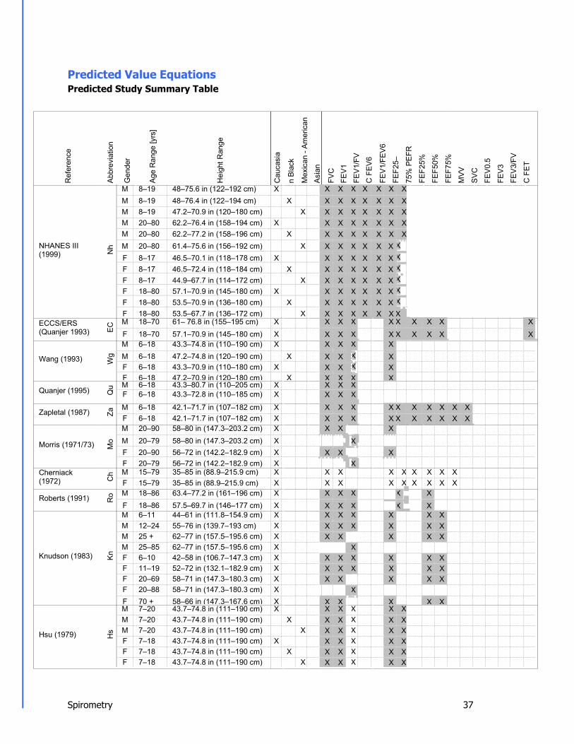
Predicted Value Equations Predicted Study Summary Table
NHANES III (1999)
ECCS/ERS (Quanjer 1993)
Wang (1993)
Quanjer (1995)
Zapletal (1987)
Morris (1971/73)
Cherniack (1972)
Roberts (1991)
Knudson (1983)
Hsu (1979)
Ref
eren
ce
Hs
Kn
Ro
Ch
Mo
Za
Qu
Wg
EC
Nh
Abbr
evia
tion
Gen
der
Age
Ran
ge [y
rs]
Hei
ght R
ange
Cau
casi
a
n Bl
ack
Mex
ican
- Am
eric
an
Asia
n
FVC
FE
V1
FEV1
/FV
C F
EV6
FEV1
/FEV
6
FEF2
5–
75%
PEF
R
FEF2
5%
FEF5
0%
FEF7
5%
MVV
SVC
FEV0
.5
FEV3
FEV3
/FV
C F
ET
M 8–19 48–75.6 in (122–192 cm) X X X X X X X X M 8–19 48–76.4 in (122–194 cm) X X X X X X X X M 8–19 47.2–70.9 in (120–180 cm) X X X X X X X X M 20–80 62.2–76.4 in (158–194 cm) X X X X X X X X M 20–80 62.2–77.2 in (158–196 cm) X X X X X X X X M 20–80 61.4–75.6 in (156–192 cm) X X X X X X X X F 8–17 46.5–70.1 in (118–178 cm) X X X X X X X X F 8–17 46.5–72.4 in (118–184 cm) X X X X X X X X F 8–17 44.9–67.7 in (114–172 cm) X X X X X X X X F 18–80 57.1–70.9 in (145–180 cm) X X X X X X X X F 18–80 53.5–70.9 in (136–180 cm) X X X X X X X X F 18–80 53.5–67.7 in (136–172 cm) X X X X X X X X M 18–70 61– 76.8 in (155–195 cm) X X X X X X X X X X F 18–70 57.1–70.9 in (145–180 cm) X X X X X X X X X X M 6–18 43.3–74.8 in (110–190 cm) X X X X X M 6–18 47.2–74.8 in (120–190 cm) X X X X X F 6–18 43.3–70.9 in (110–180 cm) X X X X X F 6–18 47.2–70.9 in (120–180 cm) X X X X X M 6–18 43.3–80.7 in (110–205 cm) X X X X F 6–18 43.3–72.8 in (110–185 cm) X X X X M 6–18 42.1–71.7 in (107–182 cm) X X X X X X X X X X X F 6–18 42.1–71.7 in (107–182 cm) X X X X X X X X X X X M 20–90 58–80 in (147.3–203.2 cm) X X X X M 20–79 58–80 in (147.3–203.2 cm) X X F 20–90 56–72 in (142.2–182.9 cm) X X X X F 20–79 56–72 in (142.2–182.9 cm) X X M 15–79 35–85 in (88.9–215.9 cm) X X X X X X X X X F 15–79 35–85 in (88.9–215.9 cm) X X X X X X X X X M 18–86 63.4–77.2 in (161–196 cm) X X X X X X F 18–86 57.5–69.7 in (146–177 cm) X X X X X X M 6–11 44–61 in (111.8–154.9 cm) X X X X X X X M 12–24 55–76 in (139.7–193 cm) X X X X X X X M 25 + 62–77 in (157.5–195.6 cm) X X X X X X M 25–85 62–77 in (157.5–195.6 cm) X X F 6–10 42–58 in (106.7–147.3 cm) X X X X X X X F 11–19 52–72 in (132.1–182.9 cm) X X X X X X X F 20–69 58–71 in (147.3–180.3 cm) X X X X X X F 20–88 58–71 in (147.3–180.3 cm) X X F 70 + 58–66 in (147.3–167.6 cm) X X X X X X M 7–20 43.7–74.8 in (111–190 cm) X X X X X X M 7–20 43.7–74.8 in (111–190 cm) X X X X X X M 7–20 43.7–74.8 in (111–190 cm) X X X X X X F 7–18 43.7–74.8 in (111–190 cm) X X X X X X F 7–18 43.7–74.8 in (111–190 cm) X X X X X X F 7–18 43.7–74.8 in (111–190 cm) X X X X X X

Spirometry 38
Po
Wa
Cr M 15–91 61.8–76.4 in (157–194 cm) X X X X X X X Crapo (1981) F 17–84 57.5–70.1 in (146–178 cm) X X X X X X X
M < 18 35.4–74 in (90–188 cm) X X X X X X X X Warwick (1977) F < 18 35.4–70.1 in (90–178 cm) X X X X X X X X Polgar (1971)
M 4–17 43.3–67 in (110–170 cm) X X X X X X X F 4–17 43.3–67 in (110–170 cm) X X X X X X X
Shaded = LLN available
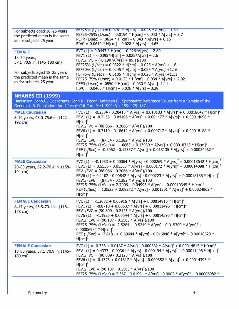
MALE 20–90 years, 58–80 in. (147.3–203.2 cm)
FEMALE 20–90 years, 56–72 in. (142.2–182.9 cm)
FVC (L) = 0.148 * H[in] - 0.025 * A[yrs] - 4.241 FEV1 (L) = 0.092 * H[in] - 0.032 * A[yrs] - 1.26 FEF25–75% (L/sec) = 0.047 * H[in] - 0.045 * A[yrs] + 2.513
MALE, 20–79 years FEV1/FVC (L/sec) = (-0.3118 * H[in] - 0.2422 * A[yrs] + 107.12)/100 FVC = 0.115 * H[in] - 0.024 * A[yrs] - 2.852 FEV1 = 0.089 * H[in] - 0.025 * A[yrs] - 1.932 FEF25–75% = 0.06 * H[in] - 0.03 * A[yrs] + 0.551
FEMALE, 20–79 years FEV1/FVC (L/sec) = (-0.0679 * H[in] - 0.1815 * A[yrs] + 88.7)/100
MALE 15–79 years, 35–85 in. (88.9–215.9 cm)
FEMALE 15–79 years, 35–85 in. (88.9–215.9 cm)
FVC (L) = 0.12102 * H[in] - 0.01357 * A[yrs] - 3.18373 FEV1 (L) = 0.09107 * H[in] - 0.0232 * A[yrs] - 1.50723 FEF25% (L/sec) = 0.0903 * H[in] - 0.01987 * A[yrs] + 2.72554 FEF50% (L/sec) = 0.06526 * H[in] - 0.03049 * A[yrs] + 2.40337 FEF75% (L/sec) = 0.03583 * H[in] - 0.04142 * A[yrs] + 1.98361 FEF25–75% (L/sec) = 0.05948 * H[in] - 0.037 * A[yrs] + 2.61187 PEFR = 0.14393 * H[in] - 0.02403 * A[yrs] + 0.22544 MVV = 3.02915 * H[in] - 0.81621 * A[yrs] - 37.94893 FVC (L) = 0.07833 * H[in] - 0.01539 * A[yrs] - 1.04912 FEV1 (L) = 0.06029 * H[in] - 0.01936 * A[yrs] - 0.18693 FEF25% (L/sec) = 0.06876 * H[in] - 0.01926 * A[yrs] + 2.14653 FEF50% (L/sec) = 0.0622 * H[in] - 0.02344 * A[yrs] + 1.4264 FEF75% (L/sec) = 0.02334 * H[in] - 0.0345 * A[yrs] + 2.21596 FEF25–75% (L/sec) = 0.04931 * H[in] - 0.0312 * A[yrs] + 2.2561 PEFR = 0.0913 * H[in] - 0.01776 * A[yrs] + 1.1316 MVV = 2.13844 * H[in] - 0.68503 * A[yrs] - 4.86957
MORRIS (1971/1973) Morris, James F., et. Al.: Spirometric Standards for Healthy Non-smoking Adults. American Review of Respiratory Disease 1971; vol 103(1): 57–67. Morris, James F, et al.: Normal values for the ratio of one-second forced expiratory volume to forced vital capacity. American Review of Respiratory Disease 1973 Vol 108: 1000–1003.
CHERNIACK (1972) Cherniack, RM and Raber, MB: Normal Standards for Ventilatory Function Using an Automatic Wedge Spirometer American Review of Respiratory Disease 1972; Vol 106(1), p38–46.
ROBERTS (1991) Roberts, Michael C. et. al: Reference values and prediction equations for normal lung function in non-smoking white

Spirometry 39
MALE 18–86 years, 63.4–77.2 in. (161–196 cm)
FEMALE 18–86 years, 57.5–69.7 in. (146–177 cm)
FVC (L) = 0.06628 * H[cm] - 0.028 * A[yrs] - 5.377 FEV1 (L) = 0.03961 * H[cm] - 0.033 * A[yrs] - 1.558 FEV1/FVC = (-0.21476 * H[cm] - 0.242 * A[yrs] + 126.252)/100 PEFR = 0.05317 * H[cm] - 0.062 * A[yrs] + 3.884 FEF50% (L/sec) = -0.044 * A[yrs] + 6.456 FVC (L) = 0.04321 * H[cm] - 0.023 * A[yrs] - 2.379 FEV1 (L) = 0.03321 * H[cm] - 0.025 * A[yrs] - 1.394 FEV1/FVC = (-0.172 * A[yrs] + 88.134)/100 PEFR = 0.04087 * H[cm] - 0.05 * A[yrs] + 2.945 FEF50% (L/sec) = -0.038 * A[yrs] + 5.556
MALE 6–11 years, 44–61 in. (111.8–154.9 cm)
MALE 12–24 years, 55–76 in. (139.7–193.0 cm)
MALE 25+ years, 62–77 in. (157.5–195.6 cm)
FEMALE 6–10 years, 42–58 in. (106.7–147.3 cm)
FEMALE 11–19 years, 52–72 in. (132.1–182.9 cm)
FEMALE 20–69 years, 58–71 in. (147.3–180.3 cm)
FVC (L) = 0.0409 * H[cm] - 3.3756 FEV1 (L) = 0.0348 * H[cm] - 2.8142 FEF50% (L/sec) = 0.0378 * H[cm] - 2.5454 FEF75% (L/sec) = 0.0171 * H[cm] - 1.0149 FEF25–75% (L/sec) = 0.0338 * H[cm] - 2.3197 FEV1/FVC = 100.4389 – 0.0813 * H[cm] FVC (L) = 0.059 * H[cm] + 0.0739 * A[yrs] - 6.8865 FEV1 (L) = 0.0519 * H[cm] + 0.0636 * A[yrs] - 6.1181 FEF50% (L/sec) = 0.0543 * H[cm] + 0.115 * A[yrs]-6.3851 FEF75% (L/sec) = 0.0397 * H[cm] - 0.0057 * A[yrs] - 4.2421 FEF25–75% L/sec) = 0.0539 * H[cm] + 0.0749 * A[yrs] - 6.199 FEV1/FVC = 100.4389 – 0.0813 * H[cm] FVC (L) = 0.0844 * H[cm] - 0.0298 * A[yrs] - 8.7818 FEV1 (L) = 0.0665 * H[cm] - 0.0292 * A[yrs] - 6.5147 FEF50% (L/sec) = 0.0684 * H[cm] - 0.0366 * A[yrs] - 5.5409 FEF75% (L/sec) = 0.031 * H[cm] - 0.023 * A[yrs] - 2.4827 FEF25–75% (L/sec) = 0.0579 * H[cm] - 0.0363 * A[yrs] - 4.5175 MALE ≥ 25 and < 85 years FEV1/FVC = 86.6862 – 0.105 * A[yrs] FVC (L) = 0.043 * H[cm] - 3.7486 FEV1 (L) = 0.0336 * H[cm] - 2.7578 FEF50% (L/sec) = 0.1846 * A[yrs] + 0.7362 FEF75% (L/sec) = 0.0109 * H[cm] - 0.1657 FEF25–75% (L/sec) = 0.022 * H[cm] - 0.8119 FEV1/FVC = 109.9739 – 0.1909 * H[cm] + 0.6655 * A[yrs] FVC (L) = 0.0416 * H[cm] + 0.0699 * A[yrs] - 4.447 FEV1 (L) = 0.0351 * H[cm] + 0.0694 * A[yrs] - 3.7622 FEF50% (L/sec) = 0.0288 * H[cm] + 0.1111 * A[yrs] - 2.304 FEF75% (L/sec) = 0.0243 * H[cm] + 0.2923 * A[yrs] - 4.4009 - 0.0075 * A[yrs]2
FEF25–75% (L/sec) = 0.0279 * H[cm] + 0.1275 * A[yrs] - 2.8007 FEV1/FVC = 109.9739 – 0.1909 * H[cm] + 0.6655 * A[yrs] FVC (L) = 0.0444 * H[cm] - 0.0169 * A[yrs] - 3.1947 FEV1 (L) = 0.0332 * H[cm] - 0.019 * A[yrs] - 1.821 FEF50% (L/sec) = 0.0321 * H[cm] - 0.024 * A[yrs] - 0.4371
urban population. Thorax 1991; 46: 643–650
KNUDSON (1983) Knudson, Ronald J., et. al: Change in the Normal Maximum Expiratory Flow-Volume Curve with Growth and Aging. American Review of Respiratory Disease 1983; 127(5–6): 725–734.

Spirometry 40
FEMALE 70+ years, 58–66 in. (147.3–167.6 cm)
FEF75% (L/sec) = 0.0174 * H[cm] - 0.0254 * A[yrs] - 0.1822 FEF25–75% (L/sec) = 0.03 * H[cm] - 0.0309 * A[yrs] - 0.4057 FEMALE ≥ 20 and < 88 years FEV1/FVC = 121.6777 – 0.1852 * H[cm] – 0.1896 * A[yrs] FVC (L) = 0.0313 * H[cm] - 0.0296 * A[yrs] - 0.1889 FEV1 (L) = 0.0143 * H[cm] - 0.0397 * A[yrs] + 2.6539 FEF50% (L/sec) = 0.0118 * H[cm] - 0.0755 * A[yrs] + 6.2402 FEF75% (L/sec) = -0.0172 * A[yrs] + 1.8894 FEF25–75% (L/sec) = -0.0615 * A[yrs] + 6.3706
To determine the Predicted FEV1/FVC value for this predicted set QRS software uses: Pred FEV1/Pred FVC MALE, White 7–20 years, 43.7–74.8 in. (111–190 cm)
MALE, Black 7–20 years, 43.7–74.8 in. (111–190 cm)
MALE, Mexican-American 7–20 years, 43.7–74.8 in. (111–190 cm)
FEMALE, White 7–18 years, 43.7–74.8 in. (111–190 cm)
FEMALE, Black 7–18 years, 43.7–74.8 in. (111–190 cm)
FEMALE, Mexican-American 7–18 years, 43.7–74.8 in. (111–190 cm)
FVC [L] = (0.000358 * H[cm]3.18)/1000 FEV1 [L] = (0.000774 * H[cm]3)/1000 PEFR [L/min] = 0.000335 * H[cm]2.79 FEF25–75% [L/min] = 0.000798 * H[cm]2.46
FVC [L] = (0.00107 * H[cm]2.93)/1000 FEV1 [L] = (0.00103 * H[cm]2.92)/1000 PEFR [L/min] = 0.000174 * H[cm]2.92 FEF25–75% [L/min] = 0.000361 * H[cm]2.60
FVC [L] = (0.00106 * H[cm]2.97)/1000 FEV1 [L] = (0.00173 * H[cm]2.85)/1000 PEFR [L/min] = 0.000769 * H[cm]2.63 FEF25–75% [L/min] = 0.000913 * H[cm]2.45
FVC [L] = (0.00257 * H[cm]2.78)/1000 FEV1 [L] = (0.00379 * H[cm]2.68)/1000 PEFR [L/min] = 0.00258 * H[cm]2.37
FEF25–75% [L/min] = 0.00379 * H[cm]2.16
FVC [L] = (0.000834 * H[cm]2.98)/1000 FEV1 [L] = (0.00114 * H[cm]2.89)/1000 PEFR [L/min] = 0.000551 * H[cm]2.68
FEF25–75% [L/min] = 0.00145 * H[cm]2.34
FVC [L] = (0.00125 * H[cm]2.92)/1000 FEV1 [L] = (0.00161 * H[cm]2.85)/1000 PEFR [L/min] = 0.000697 * H[cm]2.64
FEF25–75% [L/min] = 0.00120 * H[cm]2.40
MALE 15–91 years, 61.8–76.4 in. (157–194 cm)
FVC (L) = 0.06 * H[cm] - 0.0214 * A[yrs] - 4.65 FEV05 (L) = 0.0327 * H[cm] - 0.0152 * A[yrs] - 1.914 FEV1 (L) = 0.0414 * H[cm] - 0.0244 * A[yrs] - 2.19 FEV3 (L) = 0.0535 * H[cm] - 0.0271 * A[yrs] - 3.512
FEF25–75% (L/sec) = 0.0204 * H[cm] - 0.038 * A[yrs] + 2.133
HSU (1979) Hsu, Katharine, et. al.: Ventilatory Functions of Normal Children and Young Adults – Mexican American, White and Black. J Pediatr 1979; 95: 14–23.
CRAPO (1981) Crapo, et. al: Reference Spirometric Values using Techniques and Equipment that Meet ATS Recommendations. American Review of Respiratory Disease 1981; 123: 659–664.

Spirometry 40
FEMALE 17–84 years, 57.5–70.1 in. (146–178 cm)
FEV1/FVC = (-0.13 * H[cm] - 0.152 * A[yrs] + 110.49)/100 FEV3/FVC = (-0.0627 * H[cm] - 0.145 * A[yrs] + 112.09)/100 FVC (L) = 0.0491 * H[cm] - 0.0216 * A[yrs] - 3.59 FEV05 (L) = 0.0238 * H[cm] - 0.0185 * A[yrs] - 0.809 FEV1 (L) = 0.0342 * H[cm] - 0.0255 * A[yrs] - 1.578 FEV3 (L) = 0.0442 * H[cm] - 0.0257 * A[yrs] - 2.745 FEF25–75% = 0.0154 * H[cm] - 0.046 * A[yrs] + 2.683 FEV1/FVC = (-0.202 * H[cm] - 0.252 * A[yrs] + 126.58)/100 FEV3/FVC = (-0.0937 * H[cm] - 0.163 * A[yrs] + 118.16)/100
MALE < 18 YEARS, 35.4–74 in. (90–188 cm)
FEMALE < 18 YEARS, 35.4–70.1 in. (90–178 cm)
LnFVC (L) = 3.0131 * ln(H[cm]) - 14.0535 LnFEV1 (L) = 2.7572 * ln(H[cm]) - 12.9007 LnFEV1/FVC = -0.2679 * ln(H[cm]) + 1.2137 LnFEF50% (L/sec) = 2.1326 * ln(H[cm]) - 9.3589 LnFEF75% (L/sec) = 2.1534 * ln(H[cm]) - 10.2213 LnPEFR (L/sec)= 2.4991 * ln(H[cm]) - 10.7785 LnFET (s) = 1.6208 * ln(H[cm]) – 7.2327 LnFVC (L) = 2.9446 * ln(H[cm]) - 13.8007 LnFEV1 (L) = 2.7522 * ln(H[cm]) - 12.921 LnFEV1/FVC = -0.2126 * ln(H[cm]) + 0.9719 LnFEF50% (L/sec) = 2.1958 * ln(H[cm]) - 9.6458 LnFEF75% (L/sec) = 2.2961 * ln(H[cm]) - 10.8666 LnPEFR (L/sec) = 2.4369 * ln(H[cm]) - 10.535 LnFET (s) = 1.2423 * ln(H[cm] – 5.3288
To determine the Predicted FEV1/FVC value for this predicted set QRS software uses: Pred FEV1/Pred FVC MALE 4–17 years, 43.3–67 in. (110–170 cm)
FEMALE 4–17 years, 43.3–67 in. (110–170 cm)
FVC (L) = 0.0000044 * H[cm]2.67
FEV1 (L) = 0.0000021 * H[cm]2.8
FEF25–75% (L/min) = -207.70 + 2.621 * H[cm] PEFR (L/min)= -425.5714 + 5.2428 * H[cm] MVV = 1.276 * H[cm] - 99.507 FVC (L) = 0.0000033 * H[cm]2.72
FEV1 (L) = 0.0000021 * H[cm]2.8
FEF25–75% (L/min) = -207.70 + 2.621 * H[cm] PEFR (L/min)= -425.5714 + 5.2428 * H[cm] MVV = 1.276 * H[cm] - 99.507
MALE 18–70 years, 61–76.8 in. (155–195 cm)
FVC (L) = 0.0576 * H[cm] – 0.026*A[yrs] – 4.34 FEV1 (L) = 0.0430*H[cm] – 0.029*A[yrs] – 2.49 FEV1/FVC = (-0.180*A[yrs] + 87.21)/100 FEF25% (L/sec) = 0.0546 * H[cm] – 0.029 * A[yrs] – 0.47
FEF50% (L/sec) = 0.0379*H[cm] – 0.031 * A[yrs] – 0.35
WARWICK (1977/80) Warwick, WJ: Pulmonary Function in Healthy Minnesota Children. Minnesota Medicine 1977; Supplement 60: 435–440. Warwick, WJ: Pulmonary Function in Healthy Minnesota Children. Minnesota Medicine March 1980; 191–195.
POLGAR (1971) Polgar and Promadhat: Pulmonary Function Testing in Children: Techniques and Standards 1971.
ECCS/ERS (Quanjer 1993) Quanjer, Ph.H, et. al: Lung Volumes and Ventilatory Flows: Official Statement of the European Respiratory Society. European Respiratory Journal 1992–1993; Supplement 15–16: 5–40.
Spirometry 41
For subjects aged 18–25 years the predicted mean is the same as for subjects 25 year.
FEMALE 18–70 years, 57.1–70.9 in. (145–180 cm)
For subjects aged 18–25 years the predicted mean is the same as for subjects 25 year.
FEF75% (L/sec) = 0.0261 * H[cm] – 0.026 * A[yrs] – 1.34 FEF25–75% (L/sec) = 0.0194 * H[cm] – 0.043 * A[yrs] + 2.7 PEFR (L/sec) = .0614 * H[cm] – 0.043 * A[yrs] + 0.15 FIVC = 0.0610 * H[cm] – 0.028 * A[yrs] – 4.65 FVC (L) = 0.0443 * H[cm] – 0.026*A[yrs] – 2.89 FEV1 (L) = 0.0395*H[cm] – 0.025*A[yrs] – 2.6 FEV1/FVC = (-0.190*A[yrs] + 89.1)/100 FEF25% (L/sec) = 0.0322 * H[cm] – 0.025 * A[yrs] + 1.6 FEF50% (L/sec) = 0.0245 * H[cm] – 0.025 * A[yrs] +1.16 FEF75% (L/sec) = 0.0105 * H[cm] – 0.025 * A[yrs] +1.11 FEF25–75% (L/sec) = 0.0125 * H[cm] – 0.034 * A[yrs] + 2.92 PEFR (L/sec) = .0550 * H[cm] – 0.030 * A[yrs] -1.11 FIVC = 0.0466 * H[cm] – 0.026 * A[yrs] – 3.28
MALE Caucasian 8–19 years, 48.0–75.6 in. (122– 192 cm)
MALE Caucasian 20–80 years, 62.2–76.4 in. (158– 194 cm)
FEMALE Caucasian 8–17 years, 46.5–70.1 in. (118– 178 cm)
FEMALE Caucasian 18–80 years, 57.1–70.9 in. (145– 180 cm)
FVC (L) = -0.2584 - 0.20415 * A[yrs] + 0.010133 * A[yrs]2 + 0.00018642 * H[cm]2
FEV1 (L) = -0.7453 - 0.04106 * A[yrs] + 0.004477 * A[yrs]2 + 0.00014098 * H[cm]2 FEV1/FVC = (88.066 - 0.2066 * A[yrs])/100 FEV6 (L) = -0.3119 - 0.18612 * A[yrs] + 0.009717 * A[yrs]2 + 0.00018188 * H[cm]2
FEV1/FEV6 = (87.34 - 0.1382 * A[yrs])/100 FEF25–75% (L/Sec) = - 1.0863 + 0.13939 * A[yrs] + 0.00010345 * H[cm]2
PEF (L/Sec) = -0.5962 - 0.12357 * A[yrs] + 0.013135 * A[yrs]2 + 0.00024962 * H[cm]2
FVC (L) = -0.1933 + 0.00064 * A[yrs] - 0.000269 * A[yrs]2 + 0.00018642 * H[cm]2
FEV1 (L) = 0.5536 - 0.01303 * A[yrs] - 0.000172 * A[yrs]2 + 0.00014098 * H[cm]2
FEV1/FVC = (88.066 - 0.2066 * A[yrs])/100 FEV6 (L) = 0.1102 - 0.00842 * A[yrs] - 0.000223 * A[yrs]2 + 0.00018188 * H[cm]2
FEV1/FEV6 = (87.34 - 0.1382 * A[yrs])/100 FEF25–75% (L/Sec) = 2.7006 – 0.04995 * A[yrs] + 0.00010345 * H[cm]2
PEF (L/Sec) = 1.0523 + 0.08272 * A[yrs] - 0.001301 * A[yrs]2 + 0.00024962 * H[cm]2
FVC (L) = -1.2082 + 0.05916 * A[yrs] + 0.00014815 * H[cm]2
FEV1 (L) = -0.8710 + 0.06537 * A[yrs] + 0.00011496 * H[cm]2
FEV1/FVC = (90.809 - 0.2125 * A[yrs])/100 FEV6 (L) = -1.1925 + 0.06544 * A[yrs] + 0.00014395 * H[cm]2
FEV1/FEV6 = (90.107 - 0.1563 * A[yrs])/100 FEF25–75% (L/Sec) = -2.5284 + 0.5249 * A[yrs] - 0.015309 * A[yrs]2 + 0.00006982 * H[cm]2 PEF (L/Sec) = -3.6181 + 0.60644 * A[yrs] - 0.016846 * A[yrs]2 + 0.00018623 * H[cm]2
FVC (L) = -0.356 + 0.0187 * A[yrs] - 0.000382 * A[yrs]2 + 0.00014815 * H[cm]2
FEV1 (L) = 0.4333 - 0.00361 * A[yrs] - 0.000194 * A[yrs]2 + 0.00011496 * H[cm]2
FEV1/FVC = (90.809 - 0.2125 * A[yrs])/100 FEV6 (L) = -0.1373 + 0.01317 * A[yrs] - 0.000352 * A[yrs]2 + 0.00014395 * H[cm]2 FEV1/FEV6 = (90.107 - 0.1563 * A[yrs])/100 FEF25–75% (L/Sec) = 2.367 - 0.01904 * A[yrs] - 0.0002 * A[yrs]2 + 0.00006982 *
NHANES III (1999) Hankinson, John L., Odencrantz, John R., Fedan, Kathleen B.. Spirometric Reference Values from a Sample of the General U.S. Population. Am J Respir Crit Care Med 1999; Vol 159: 179–187.
Spirometry 42
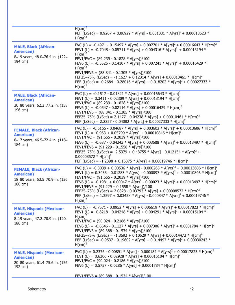
MALE, Black (African- American) 8–19 years, 48.0–76.4 in. (122– 194 cm)
MALE, Black (African- American) 20–80 years, 62.2–77.2 in. (158– 196 cm)
FEMALE, Black (African- American) 8–17 years, 46.5–72.4 in. (118– 184 cm)
FEMALE, Black (African- American) 18–80 years, 53.5–70.9 in. (136– 180 cm)
MALE, Hispanic (Mexican- American) 8–19 years, 47.2–70.9 in. (120– 180 cm)
MALE, Hispanic (Mexican- American) 20–80 years, 61.4–75.6 in. (156– 192 cm)
H[cm]2
PEF (L/Sec) = 0.9267 + 0.06929 * A[yrs] - 0.001031 * A[yrs]2 + 0.00018623 * H[cm]2
FVC (L) = -0.4971 - 0.15497 * A[yrs] + 0.007701 * A[yrs]2 + 0.00016643 * H[cm]2
FEV1 (L) = -0.7048 - 0.05711 * A[yrs] + 0.004316 * A[yrs]2 + 0.00013194 * H[cm]2 FEV1/FVC = (89.239 - 0.1828 * A[yrs])/100 FEV6 (L) = -0.5525 - 0.14107 * A[yrs] + 0.007241 * A[yrs]2 + 0.00016429 * H[cm]2
FEV1/FEV6 = (88.841 - 0.1305 * A[yrs])/100 FEF25–75% (L/Sec) = -1.1627 + 0.12314 * A[yrs] + 0.00010461 * H[cm]2
PEF (L/Sec) = -0.2684 - 0.28016 * A[yrs] + 0.018202 * A[yrs]2 + 0.00027333 * H[cm]2
FVC (L) = -0.1517 - 0.01821 * A[yrs] + 0.00016643 * H[cm]2
FEV1 (L) = 0.3411 - 0.02309 * A[yrs] + 0.00013194 * H[cm]2
FEV1/FVC = (89.239 - 0.1828 * A[yrs])/100 FEV6 (L) = -0.0547 - 0.02114 * A[yrs] + 0.00016429 * H[cm]2
FEV1/FEV6 = (88.841 - 0.1305 * A[yrs])/100 FEF25–75% (L/Sec) = 2.1477 - 0.04238 * A[yrs] + 0.00010461 * H[cm]2
PEF (L/Sec) = 2.2257 - 0.04082 * A[yrs] + 0.00027333 * H[cm]2
FVC (L) = -0.6166 - 0.04687 * A[yrs] + 0.003602 * A[yrs]2 + 0.00013606 * H[cm]2
FEV1 (L) = -0.963 + 0.05799 * A[yrs] + 0.00010846 * H[cm]2 FEV1/FVC = (91.655 - 0.2039 * A[yrs])/100 FEV6 (L) = -0.637 - 0.04243 * A[yrs] + 0.003508 * A[yrs]2 + 0.00013497 * H[cm]2
FEV1/FEV6 = (91.229 - 0.1558 * A[yrs])/100 FEF25–75% (L/Sec) = -2.5379 + 0.43755 * A[yrs] - 0.012154 * A[yrs]2 + 0.00008572 * H[cm]2
PEF (L/Sec) = -1.2398 + 0.16375 * A[yrs] + 0.00019746 * H[cm]2
FVC (L) = -0.3039 + 0.00536 * A[yrs] - 0.000265 * A[yrs]2 + 0.00013606 * H[cm]2
FEV1 (L) = 0.3433 - 0.01283 * A[yrs] - 0.000097 * A[yrs]2 + 0.00010846 * H[cm]2
FEV1/FVC = (91.655 - 0.2039 * A[yrs])/100 FEV6 (L) = -0.1981 + 0.00047 * A[yrs] - 0.00023 * A[yrs]2 + 0.00013497 * H[cm]2
FEV1/FEV6 = (91.229 – 0.1558 * A[yrs])/100 FEF25–75% (L/Sec) = 2.0828 - 0.03793 * A[yrs] + 0.00008572 * H[cm]2
PEF (L/Sec) = 1.3597 + 0.03458 * A[yrs] - 0.000847 * A[yrs]2 + 0.00019746 * H[cm]2
FVC (L) = -0.7571 - 0.0952 * A[yrs] + 0.006619 * A[yrs]2 + 0.00017823 * H[cm]2
FEV1 (L) = -0.8218 - 0.04248 * A[yrs] + 0.004291 * A[yrs]2 + 0.00015104 * H[cm]2 FEV1/FVC = (90.024 - 0.2186 * A[yrs])/100 FEV6 (L) = -0.6646 - 0.1127 * A[yrs] + 0.007306 * A[yrs]2 + 0.0001784 * H[cm]2
FEV1/FEV6 = (89.388 - 0.1534 * A[yrs])/100 FEF25–75% (L/Sec) = -1.3592 + 0.10529 * A[yrs] + 0.00014473 * H[cm]2
PEF (L/Sec) = -0.9537 - 0.19602 * A[yrs] + 0.014497 * A[yrs]2 + 0.00030243 * H[cm]2
FVC (L) = 0.2376 - 0.00891 * A[yrs] - 0.000182 * A[yrs]2 + 0.00017823 * H[cm]2
FEV1 (L) = 0.6306 - 0.02928 * A[yrs] + 0.00015104 * H[cm]2 FEV1/FVC = (90.024 - 0.2186 * A[yrs])/100 FEV6 (L) = 0.5757 - 0.0286 * A[yrs] + 0.0001784 * H[cm]2
FEV1/FEV6 = (89.388 - 0.1534 * A[yrs])/100

Spirometry 43
FEMALE, Hispanic (Mexican- American) 8–17 years, 44.9–67.7 in. (114– 172 cm)
FEMALE, Hispanic (Mexican- American) 18–80 years, 53.5–67.7 in. (136– 172 cm)
FEF25–75% (L/Sec) = 1.7503 - 0.05018 * A[yrs] + 0.00014473 * H[cm]2
PEF (L/Sec) = 0.087 + 0.0658 * A[yrs] - 0.001195 * A[yrs]2 + 0.00030243 * H[cm]2
FVC (L) = -1.2507 + 0.07501 * A[yrs] + 0.00014246 * H[cm]2
FEV1 (L) = -0.9641 + 0.0649 * A[yrs] + 0.00012154 * H[cm]2
FEV1/FVC = (92.360 - 0.2248 * A[yrs])/100 FEV6 (L) = -1.241 + 0.07625 * A[yrs] + 0.00014106 * H[cm]2
FEV1/FEV6 = (91.644 - 0.1670 * A[yrs])/100 FEF25–75% (L/Sec) = -2.1825 + 0.42451 * A[yrs] - 0.012415 * A[yrs]2 + 0.0000961 * H[cm]2 PEF (L/Sec) = -3.2549 + 0.47495 * A[yrs] - 0.013193 * A[yrs]2 + 0.00022203 * H[cm]2
FVC (L) = 0.121 + 0.00307 * A[yrs] - 0.000237 * A[yrs]2 + 0.00014246 * H[cm]2
FEV1 (L) = 0.4529 - 0.01178 * A[yrs] - 0.000113 * A[yrs]2 + 0.00012154 * H[cm]2
FEV1/FVC = (92.36 - 0.2248 * A[yrs])/100 FEV6 (L) = 0.2033 + 0.0002 * A[yrs] - 0.000232 * A[yrs]2 + 0.00014106 * H[cm]2
FEV1/FEV6 = (91.664 - 0.167 * A[yrs])/100 FEF25–75% (L/Sec) = 1.7456 - 0.01195 * A[yrs] - 0.000291 * A[yrs]2 + 0.0000961 * H[cm]2
PEF (L/Sec) = 0.2401 + 0.06174 * A[yrs] - 0.001023 * A[yrs]2 + 0.00022203 * H[cm]2
MALE 6–18 years, 42.1–71.7 in. (107–182 cm)
FEMALE 6–18 years, 42.1–71.7 in. (107–182 cm)
FVC (L) = 10 (-2.9236 + 2.936 * log(H[cm])) / 1000 FEV1 (L) =10 (-2.8652 + 2.8729 * log(H[cm])) / 1000 FEV1/FVC = (90.6043 - 0.04104 * H[cm])/100 FEF25% (L/Sec) = 10 (-4.0164 + 2.1541 * log(H[cm]))
FEF50% (L/Sec) = 10 (-4.2168 + 2.1771 * log(H[cm]))
FEF75% (L/Sec) =10 (-4.5808 + 2.2116 * log(H[cm]))
FEF25–75% (L/Sec) = 10 (-4.6651 + 2.3588 * log(H[cm]))
PEFR (L/Sec) = 10 (-4.3722 + 2.3422 * log(H[cm]))
SVC (L) = 10 (-2.5768 + 2.7799 * log(H[cm])) / 1000 MVV (L/Min) = 10 (-1.9178 + 3.0388 * log(H[cm])) / 1000
FVC (L) = 10 (-2.704 + 2.8181 * log(H[cm])) / 1000 FEV1 (L) =10 (-2.6056 + 2.7413 * log(H[cm])) / 1000 FEV1/FVC = (90.6043 - 0.04104 * H[cm])/100 FEF25% (L/Sec) = 10 (-4.0164 + 2.1541 * log(H[cm]))
FEF50% (L/Sec) = 10 (-4.2168 + 2.1771 * log(H[cm]))
FEF75% (L/Sec) =10 (-4.5808 + 2.2116 * log(H[cm]))
FEF25–75% (L/Sec) = 10 (-4.6651 + 2.3588 * log(H[cm]))
PEFR (L/Sec) = 10 (-4.3722 + 2.3422 * log(H[cm]))
SVC (L) = 10 (-2.297 + 2.6361 * log(H[cm])) / 1000 MVV (L/Min) = 10 (-1.9178 + 3.0388 * log(H[cm])) / 1000
MALE 6–18 years,
LnFVC [l] = -1.2782 + [1.3731 + 0.0164 * A[yrs]] * H[m] LnFEV1 [l] = -1.2933 + [1.2669 + 0.0174 * A[yrs]] * H[m]
ZAPLETAL (1987) Zapletal, A.: Lung Function in Children and Adolescents. Methods, Reference Values. Progress in Respiration Research Vol 22 (1987)
QUANJER (1995) Quanjer, PhH, et. al.: Spirometric Values for White European Children and Adolescents: Polgar Revisited, Pediatric Pulmonology 1995, 19: 135–142.

Spirometry 44
43.3–80.7 in. (110–205 cm) FEV1/FVC = 86.2 FEMALE 6–18 years, 43.3–72.8 in. (110–185 cm)
LnFVC [l] = -1.4507 + [1.4800 + 0.0127 * A[yrs]] * H[m] LnFEV1 [l] = -1.5974 + [1.5016 + 0.0119 * A[yrs]] * H[m] FEV1/FVC = 88.9
MALE, White 6–18 years, 43.3–74.8 in. (110–190 cm) MALE, Black 6–18 years, 47.2–74.8 in. (120–190 cm) FEMALE, White 6–18 years, 43.3–70.9 in. (110–180 cm) FEMALE, Black 6–18 years, 47.2–70.9 in. (120–180 cm)
LnFVC(L) = α + β*lnHt[m] LnFEV1(L) = α + β*lnHt[m] LnFEV1/FVC(L) = α + β*lnHt[m] LnFEF25–75%(L/s) = α + β*lnHt[m]
Refer to the Wang look-up tables for α and β.
Wang look-up tables:
MALE, White, 6–18 years
WANG (1993) Wang, Xiaobin, et.al,: Pulmonary Function Between 6 and 18 Years of Age. Pediatric Pulmonology 1993; 15: 75–88.
Age [years] FVC FEV1 FEV1/FVC FEF25–75% α β α β α β α β 6 -0.024 2.470 -0.109 2.252 -0.078 -0.248 - - 7 -0.018 2.489 -0.104 2.270 -0.086 -0.220 - - 8 0.005 2.443 -0.089 2.257 -0.091 -0.199 0.264 1.505 9 0.017 2.426 -0.063 2.197 -0.086 -0.206 0.308 1.443 10 0.030 2.407 -0.057 2.212 -0.081 -0.209 0.290 1.557 11 0.009 2.468 -0.093 2.324 -0.101 -0.147 0.242 1.738 12 -0.061 2.649 -0.161 2.512 -0.101 -0.133 0.165 1.982 13 -0.175 2.924 -0.292 2.843 -0.116 -0.085 0.007 2.396 14 -0.219 3.060 -0.329 2.983 -0.106 -0.087 0.014 2.483 15 -0.079 2.859 -0.141 2.709 -0.060 -0.155 0.241 2.163 16 0.104 2.591 0.062 2.409 -0.045 -0.178 0.503 1.764 17 0.253 2.374 0.262 2.099 0.008 -0.272 0.762 1.368 18 0.296 2.316 0.251 2.129 -0.054 -0.170 0.678 1.528

Spirometry 45
MALE, Black, 6–18 years Age [years] FVC FEV1 FEV1/FVC FEF25–75%
α β α β α β α β 6 -0.088 1.961 -0.166 1.723 -0.091 -0.152 - - 7 -0.040 2.040 -0.122 1.846 -0.091 -0.153 - - 8 -0.094 2.323 -0.225 2.271 -0.118 -0.104 0.097 1.544 9 -0.074 2.308 -0.142 2.059 -0.079 -0.218 0.255 1.248 10 -0.110 2.417 -0.157 2.117 -0.047 -0.303 0.230 1.428 11 -0.138 2.453 -0.176 2.166 -0.048 -0.263 0.256 1.438 12 -0.224 2.710 -0.307 2.548 -0.084 -0.162 0.085 1.936 13 -0.342 2.975 -0.486 2.962 -0.141 -0.018 -0.121 2.476 14 -0.337 3.035 -0.472 3.010 -0.123 -0.050 -0.115 2.536 15 -0.226 2.889 -0.318 2.789 -0.070 -0.140 0.170 2.120 16 0.058 2.425 0.074 2.140 0.018 -0.289 0.663 1.299 17 0.148 2.310 0.053 2.223 -0.095 -0.087 0.505 1.618 18 0.152 2.341 0.130 2.121 -0.041 -0.190 0.859 1.053
FEMALE, White, 6–18 years
Age [years] FVC FEV1 FEV1/FVC FEF25–75% α β α β α β α β 6 -0.013 2.007 -0.109 1.949 -0.097 -0.055 - - 7 0.062 2.385 -0.144 2.243 -0.084 -0.132 - - 8 -0.055 2.381 -0.137 2.239 -0.079 -0.152 0.247 1.668 9 -0.039 2.351 -0.123 2.222 -0.084 -0.128 0.254 1.710 10 -0.068 2.458 -0.161 2.364 -0.092 -0.097 0.195 1.933 11 -0.120 2.617 -0.223 2.558 -0.102 -0.061 0.161 2.091 12 -0.174 2.776 -0.264 2.709 -0.090 -0.067 0.185 2.120 13 -0.061 2.576 -0.153 2.535 -0.093 -0.040 0.294 1.976 14 0.139 2.208 0.046 2.178 -0.096 -0.026 0.450 1.711 15 0.210 2.099 0.148 2.008 -0.062 -0.093 0.581 1.486 16 0.226 2.097 0.181 1.972 -0.048 -0.120 0.654 1.366 17 0.214 2.146 0.176 1.992 -0.038 -0.154 0.688 1.290 18 0.195 2.179 0.152 2.031 -0.069 -0.096 0.520 1.622

Spirometry 46
FEMALE, Black, 6–18 years Age [years] FVC FEV1 FEV1/FVC FEF25–75%
α β α β α β α β 6 -0.172 2.117 -0.288 2.182 -0.109 0.059 - - 7 -0.135 2.132 -0.250 2.158 -0.104 -0.030 - - 8 -0.176 2.362 -0.276 2.295 -0.103 -0.066 -0.283 2.990 9 -0.200 2.452 -0.294 2.330 -0.097 -0.104 0.025 2.062 10 -0.230 2.571 -0.344 2.507 -0.120 -0.043 0.051 2.028 11 -0.204 2.526 -0.308 2.460 -0.089 -0.105 0.078 2.006 12 -0.107 2.342 -0.219 2.312 -0.115 -0.021 0.225 1.804 13 -0.042 2.294 -0.117 2.196 -0.051 -0.148 0.418 1.504 14 0.105 2.021 0.041 1.920 -0.063 -0.103 0.574 1.257 15 0.253 1.787 0.203 1.662 -0.043 -0.139 0.599 1.281 16 0.111 2.098 0.129 1.824 -0.022 -0.188 0.653 1.175 17 0.205 1.930 0.273 1.547 0.048 -0.342 0.713 1.067 18 -0.042 2.423 -0.084 2.259 -0.197 0.145 -0.209 2.896
The Global Lungs Initiative (GLI) equations are the first global multi-ethnic reference equations for spirometry that span all-ages. These are the result of unprecedented, unselfish and professional international cooperation endorsed by five international societies. Briefly, data from 74,187 healthy non-smokers (57.1% females) aged 3-95 years were used to derive multi-ethnic reference equations using modern statistical methods, including development of age dependent lower limits of normal.
*Note: When Asian is selected under the Race demographic dropdown in Office Medic, the reference values will be based on the SE Asian GLI dataset, as this set provided a larger patient population.
For more information on the Global Lung Initiative and the specific reference equations, please visit http://www.ers-education.org/guidelines/global-lung-function-initiative.aspx
GLI (2012) Multi-ethnic reference values for spirometry for the 3-95 year age range: the global lung function 2012 equations.
Quanjer, P.H., Stanojevic, S., Cole, T.J., Baur, X., Hall, G.L., Enright, P.L., Hankinson, J.L., Ip, M.S., Zheng, J., Stocks, J. on behalf of the ERS Global Lung Function Init iative. (2012), European Respiratory Journal, 2012; 40: 1324–1343.

Spirometry 47
Lung Age Calculation Lung age is calculated for patients 20-84 years old. *Lung age is equal to the predicted FEV1 that matches the patient’s actual FEV1.
For example: Predicted equation: Crapo Patient demographics: Height: 5ft 10in
Age: 46 years Gender: Male Race: Caucasian Actual FEV1: 4.49L Predicted FEV1: 4.05L
Patient’s Lung Age: 28 years
Based on Crapo’s predicted equation, the patient’s actual FEV1 (4.49L) is equal to the predicted FEV1 of a 28 year old. Therefore, the patient’s lung age is 28 years old.
Note: Lung age may differ based on the predicted equation selected.
* Morris JF, Temple W.; Spirometric “lung age” estimation for motivating smoking cessation. Prev Med. 1985 Sep: 14)5):655-62.
Note: “Lung age not available” dialog box may appear when certain predictors and ages are selected because they are not supported for this function.

Spirometry 48
Spirometry Interpretation
Note: A disclaimer is provided on all spirometry reports: “All test results should be evaluated by a qualified physician.”
Enright (1997) Office Spirometry: A Practical Guide to the Selection and Use of Spirometers by Paul L. Enright, M.D. Robert E. Hyatt M.D. 1987
FEV1/FVC < LLN?
Yes
FEV1 >= LLN?
No
FEV1 >= 65% of predicted?
Yes
Yes
Borderline Obstruction
Mild Obstruction
No
FEV1 >= 50%
No
Moderate Obstruction
Severe Obstruction
FVC < LLN?
Yes
and low vital capacity; cannot rule out superimposed restriction.
FVC < LLN? No Normal Spirometry
Yes
FVC >= 65% of predicted?
No
Mild Restriction
FVC >= 50% of predicted? Yes Moderate Restriction
Sever Restiction
of predicted? Yes
No
No
Yes

Spirometry 49
BTS-NICE (2004-05) The British Thoracic Society (BTS) COPD Consortium: Spirometry in Practice: A Practical Guide to Using Spirometry in Primary Care. Second Edition. April 2005.
National Institute for Clinical Excellence (NICE): Chronic obstructive pulmonary disease: Management of chronic obstructive pulmonary disease in adults in primary and secondary care. Clinical Guideline 12. February 2004. Developed by the National Collaborating Centre for Chronic Conditions.
FEV1/FVC < 0.7 (70%)
Yes
FEV1 < 80% of predicted? No Normal Spirometry
Yes
No FEV1 < 50% of predicted? No Mild Obstruction
Yes
FEV1 < 30% of predicted? No Moderate Obstruction
Severe Obstruction
FEV1 and FVC < 80% of predicted?
Yes
Restrictive Disorder
No
Normal Spirometry
Yes
Spirometry 50
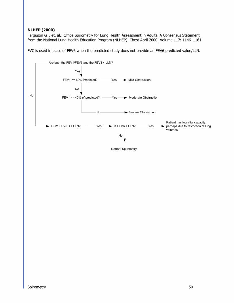
NLHEP (2000) Ferguson GT, et. al.: Office Spirometry for Lung Health Assessment in Adults. A Consensus Statement from the National Lung Health Education Program (NLHEP). Chest April 2000; Volume 117: 1146–1161.
FVC is used in place of FEV6 when the predicted study does not provide an FEV6 predicted value/LLN.
Are both the FEV1/FEV6 and the FEV1 < LLN?
Yes
FEV1 >= 60% Predicted?
No
No FEV1 >= 40% of predicted?
Yes
Yes
Mild Obstruction
Moderate Obstruction
Severe Obstruction
FEV1/FEV6 >= LLN?
Yes
Is FEV6 < LLN?
No
Yes
Patient has low vital capacity, perhaps due to restriction of lung volumes.
Normal Spirometry
No

Spirometry 51
FEV1/FVC < LLN?
Yes FEV1 >/= 100% of predicted? Yes
No FEV1 >/= 70% of predicted? Yes
No FEV1 >/= 60% of predicted? Yes
No FEV1 >/= 50% of predicted? Yes
No FEV1 >/= 35% of predicted? Yes
No
FVC < LLN? No
ATS/ERS (2005) ATS/ERS Task Force: Interpretive strategies for lung function tests. Standardisation of spirometry. Eur. Respir. J., Nov 2005; 26: 948-968.
May be a physiological variant
Mild Obstruction
Moderate Obstruction
No Moderately Severe Obstruction
Severe Obstruction
Very Severe Obstruction
FVC < LLN? Yes
Normal Spirometry
And low vital capacity, cannot rule out superimposed restriction.
Yes
FVC >/= 70% Predicted? Yes Mild Restriction
No
FVC >/= 60% Predicted?
Yes
Moderate Restriction
No
FVC >/= 50% Predicted?
No
FVC >/= 35% Predicted?
Yes
Yes
Moderately Severe Restriction
Severe Restriction
Very Severe Restriction No

Electrocardiography 52
Electrocardiography
ECG Cautions, Warnings, and Other Information Warnings
• The computerized interpretation is only valid when used in conjunction with clinical findings. All computer generated tracings and interpretations must be confirmed by a qualified physician. Test interpretations are intended for the physician's use only. All ECG numerical and graphical data should be evaluated with respect to the patient's clinical and historical picture.
• No specific filter settings were used to pass the distortion test. • Isoelectric segments within the VectraCor are excluded from the VectraCor waves. • Use printout for final diagnosis. • The ECG Device is not intended for use in a sterile environment. • Do not use for direct cardiac application. • The ECG device is reusable. • The ECG is protected against malfunction caused by electrosurgery. • The ECG does not incorporate means to protect the patient against burns when used during
electrosurgery • To prevent burns from ECG electrodes during monopolar electrosurgery:
• Make sure that the patient return pad or large-surface neutral electrode is properly applied.
• Position ECG electrodes away from the electrosurgery site and current pathway through the body.
• Loss of ECG signal may occur during high frequency electrosurgery. Signal should re-establish within 10 seconds of removal of the high-frequency source. No other conditions of use must be met to operate the device in a high – frequency electrosurgery environment. Indications for use are the same as stated above.
• If signal quality is lost due to electromagnetic disturbances, the ECG signal may be affected which can lead to incorrect diagnosis.
• The use of cardiac pacemakers or other electrical stimulators may lead to incorrect interpretations and diagnosis.
• Do not attempt to insert the ECG device (including patient cables) into an electrical outlet.
• Avoid patient movement to reduce artifact. The ECG Device is for acquiring resting ECGs only. The device should not be used for stress testing.
• Though false positive errors will intentionally outnumber false negative errors, both will occur, thus the necessity for over reading by a qualified physician of any computer-interpreted ECG. The computer interpretation does not produce a definitive diagnosis.
• Ensure electrodes are connected only to patient. • Conductive parts of electrodes and connectors, including neutral electrode, should not
contact other conductive parts including earth. • Select a three lead view during defibrillation to ensure signals are clearly separated
following electrode polarization. • Defibrillator warnings:
– Do not touch the patient during defibrillation.

Electrocardiography 53
– Do not touch the defibrillator’s paddle-electrode surface when discharging the defibrillator.
– Keep defibrillation electrodes well clear of other electrodes or metal parts in contact with the patient.
– Do not touch the patient, bed, or any conductive material in contact with the patient during defibrillation.
– When defibrillation discharge is applied, the ECG may be overloaded with voltage, but will recover normal function within 5s.
– Defibrillation use requires use of manufacturer specified electrodes

Electrocardiography 54
Cautions • For diagnostic ECG according to the requirements of the AAMI EC11:1991 standard, use
factory default settings. ECG diagnosis should be based on a printed 3x4 report with software filters off, and using a 1:1 scale 300dpi printer.
• The Universal SmartECG is designed for use with electrodes that comply with AAMI EC12:2000.
• Reseal electrode pouch after opening to prevent dehydrating. • Suggested maximum electrode duration is 8 hours. • Leakage current of device is increased when several items of the device are interconnected.
However, leakage current is tested to be within acceptable levels. • Do not clean the case with alcohol. • Do not saturate or immerse the case with liquid during cleaning. • Do not sterilize ECG device.
ECG Indications for Use: Receipt, Storage, Viewing, Printing and Interpretive Analysis of 12 channel simultaneous ECGs
• The system can be used within electrosurgical environment. Application Specification:
Medical Indication: Diagnosis of cardiovascular conditions/diseases User Profile: Trained professional Patient Population: Adult Male/Female (18+) Environment of Use: Hospital and Clinical Use and HF Surgical environments Tissue Type of Device Interaction: Patient Skin Conditions of Use: Single patient, non-sterile, single-use electrodes, non-invasive,
resting ECGs, continuous use, portable Operating Principle: [Physical Method] Electrodes are attached to the patient to
acquire the ECG (electrocardiogram) with software application. [Mechanism] The attached electrodes records the ECG signal. The software application starts and stops the recording of the ECG. Then, the software can display or print a hard copy of the ECG report.

Electrocardiography 55
ECG Getting Started Connecting the 12 Channel ECG device to your PC:
• Connect the USB port of the ECG device to the USB port on your PC. • The LED will light up to indicate when powered.
Note: The LED on the ECG will not only indicate when the device is powered (solid green) and ready for acquisition, but also when it is acquiring data (flashing green). If the LED is turned off, the device is inoperable.

Electrocardiography 56
Performing an ECG Test: Quick Start Guide 1. Connect the ECG device to the PC. 2. Select a patient from the Patient Directory.
• If patient is not in the directory, create the patient. 3. Prep Patient & Connect the ECG device
• Shave electrode sites if necessary. Thoroughly clean the area and let dry. • Prep skin by briskly rubbing with gauze, being careful not to break or damage the skin. • Remove electrodes from backing. • Apply each electrode, adhesive side down to desired site. • For positive electrode contact, start from outer edge and run your finger around the
electrode several times, working toward the center. • Connect the lead wires to the patient ensuring correct lead placement. Excess
movement can cause artifact. Patient should be stable.
4. Click the ECG icon on the Toolbar, or select Test | ECG.
The acquisition window will appear on the screen with the patients real-time ECG displayed.
Warning: Avoid patient movement to reduce artifact. The ECG Device is for acquiring resting ECGs only. The device should not be used for stress testing.
5. After the ECG data is captured by Office Medic, select Save and Review. 6. After review, select Print or Print to File.
V2/C2 V1/C1 V3/C3
V4/C4 V5/C5
V6/C6

Electrocardiography 57
About the Acquisition Window
View Select Scrolls through the different leads when you view the 3 or 6 lead sets.
Lead
Identifiers
Connection Status
Identifies each of the 12 leads. If a lead is disconnected (leads
off) then a red circle with a diagonal line is placed over the lead identifier. Displays the status of the connected ECG cable.
Heart Rate (bpm)
Displays the active Heart Rate of the patient.

Electrocardiography 58
20mm/mV.
50mm/s. Leads View 3, 6 or 12 leads, or 3x4 view. The ability to select between
the Limb leads and the Chest leads is also available when viewing 3 or 6 leads at a time. A Custom Lead group can be defined in the ECG Options.
Power Filter Turns the Main filter on and off. Note: the default frequency of the Main filter is set in the ECG Options.
Muscle Filter Turns the Muscle filter on and off.
Stop Stops the real time recording to view the previous 15 minutes of ECG. The user can select the desired 10 seconds of ECG and select Save to save and exit the test.
Record Resumes recording data. Once selected, all paused data will no longer be available.
Print Allows you to print all or a selected portion of the stopped ECG. This printed report is not intended for diagnostic use, or as a patient record - for that purpose print from the review window, or use "Save and Print."
Elapsed Time Minutes and seconds of current ECG acquisition.
Status Bar Represents whether 10 seconds of valid ECG data has been received. When the status bar is full, the Save button is activated and data can then be saved.
Audible Indication
Audibly indicates a “leads off” status or QRS detection as selected in “options.”
Save and Print Saves the test and automatically prints a report or
PDF (optional). Cancel Closes the test without saving.
Note: Users can acquire ECG either from Office Medic or VectraplexECG. Please see VectraplexECG user manual for additional information. To acquire ECG with VectraplexECG, set the default ECG program to VectraplexECG by going to Options > General. Please call VectraCor or visit www.VectraCor.com for more information about VectraplexECG.
Pulse Provides a visual indication of the combined sensitivity (1mV vertical height) and speed (100ms horizontal width).
Sensitivity Changes the number of millimeters that represent one millivolt. The available options are 5mm/mV, 10mm/mV, and
Save Saves the test and closes the window.
Save and Review
Saves the test and launches a review of the results.
Speed Changes the number of millimeters that are passed in one second. The available options are 12.5mm/s, 25mm/s and

Electrocardiography 59
ECG Options Select Options | ECG.
General Tab Select General to set or change general ECG Options.

Electrocardiography 60
Analysis Tab Select Analysis to set or change the ECG analysis options.
Acquisition Settings Tab Select Acquisition Settings to set or change options available for the acquisition window.

Electrocardiography 61
Review Window Tab Select Review Window to set or change options available for the ECG Review Window.
Edit Lead Order Tab Select Edit Lead Order to change the lead order. Note, the setting applies to both the acquisition and review windows.

Electrocardiography 62
Reviewing an ECG Reviewing an ECG within the ECG Review Window
Note: ECG tests with an asterisk symbol are acquired by VectraplexECG. VectraplexECG acquired ECGs must be viewed in VectraplexECG.

Electrocardiography 63
File Menu
Menu Item Icon Function Save NA Saves changes.
Printer Setup… NA Opens the print setup window for the default printer.
Print… Prints the ECG test.
Print Preview… NA Previews the hardcopy report. Print to File… NA Creates an image file (JPEG, TIFF, or PDF) of the hardcopy report. Close NA Closes open tests without closing the review window. Exit NA Closes open tests and exits the review window.

Electrocardiography 64
N
View Menu
12 Lead Selects the 12 lead view of the ECG. See 12 Lead View for an example.
Strips Selects the three lead strip view of the ECG. See ECG Strips View for an example.
Zoom Selects the zoom view of the ECG. See Zoom View for an example.
Details Displays the interpretation, comments, and detailed measurements. See Details View for an example.
Rhythm Lead Position Toggles the Rhythm Lead to the top or the bottom of the
screen (12 Lead view only). Zoom In Enlarges the ECG.
Next Lead/ Previous Lead
Scrolls through recorded leads.
Show Averaged Complexes
Switches between averaged complexes and 10 second lead strip displays.
Show Toolbar NA Displays or removes the toolbar. Show Measurements Panel NA Displays or removes the summary measurements panel.
Show Control Panel NA Displays or removes the Control Panel.
Zoom Out Reduces the ECG.
Show Measurements Turns the averaged complex’s measurements On and OFF.
Show Grid Turns the grid lines on and off.
Menu Item Icon Function

Electrocardiography 65
Filter Muscle Filter
ECG Options Menu
Cursor or Toggles the cursor between the Zoom tool used to increase or decrease the ECG display or the Measure tool used for on-screen calipers.
Measurement Units NA Selects Millimeters or MicroVolts
Activates the Muscle Filter.
Power Filter Activates the Mains Filter. The Hz are set in the ECG Options.
Edit Lead Order… NA Select Edit Lead Order to change the lead order.
Remove Analysis NA Removes the detailed measurements. The narrative interpretation and comments remain unchanged.
Re-analyze NA Resets the interpretation to its original state and removes all changes made by the user to the narrative interpretation.
Menu Item Icon Function
Speed Sensitivity
Changes the number of millimeters that are passed in one second. The available options are 12.5mm/s, 25mm/s and 50mm/s. Changes the number of millimeters that represent one millivolt. The available options are 5mm/mV, 10mm/mV, and 20mm/mV.

Electrocardiography 66
Views
12 Lead 12 channels of averaged complexes and a rhythm strip.
Strips Displays ECG strips. Use the Leads Shown option to change the number of visible leads.
Zoom Displays a single lead strip or an averaged complex.

Electrocardiography 67
Details View
The details view shows the interpretation, comments, detailed measurements and patient details of the ECG.
Interpretation and Comments
Measurements
Patient and Recording Details
Warning! The computerized interpretation provided by the Office Medic software is only valid when used in conjunction with clinical findings. All computer generated tracings and interpretations must be confirmed by a qualified physician.

Electrocardiography 68
Printing an ECG Select File | Print or the print icon.
Single Lead Report Prints a single strip or averaged complex with scale options: 1x, 2x, 4x, 8, 16x).
3X4 Simultaneous Prints 2.5 second segments of all twelve channels displayed at the same point in time along with a 10 second single channel rhythm strip.
3X4 Sequential Report Prints 2.5 second segments of all twelve channels displayed at the same point in time progressing in four sequential columns along with a 10 second single channel rhythm strip.
Average Complexes Print an average QRS complex for all 12 channels along with a 10 second single channel rhythm strip.
6X1 Format, 2 Page Prints a ten second trace of each channel (2 page report). 6X2 Format, 1 Page Prints a five second trace of each channel (1 page report). Measurements table Prints a chart with amplitude, slope and duration data for all twelve channels. Include Allows you to select whether to include Measurements, Interpretation, and/or
Comments in the report(s). Speed and Sensitivity Allows you to select the Speed (12.5, 25, or 50mm/s) and Sensitivity (5, 10, or
20 mm/mV) of the ECG reports.
Minor grid Allows you to select the minor grid: Lines, Dots, or None.
Note: when printing to a low resolution printer select Dots or None for the minor grid.
Print Options Description

Electrocardiography 69
ECG Device Verification A periodic check of the ECG system with an ECG simulator is recommended. Intervals for these checks can be set at the discretion of your Medical Director. There are commercially available ECG simulators which may be used for this purpose, refer to the accompanying information for instructions on the use of these.
For further information on device verification, contact VectraCor at www.VectraCor.com.
ECG Analysis Program Office Medic provides analysis and interpretation of 12 channel ECGs. This is based on algorithms developed by Cardionics S.A. For further information consult the ECG Physician's Guide.
What to expect from the analysis program The ECG Analysis Program provides an analysis of the amplitudes, duration, and morphologies of the ECG waveform. The analysis is based upon standards of interpretation of these parameters and calculations of the electrical axis and relationship between leads.
The interpreted ECG is a tool to assist the physician in making a clinical diagnosis, and is not a substitute for the physician's knowledge, the patient's history, results of the physical exam, the ECG tracing or other findings.

Troubleshooting 70
Troubleshooting and Tips Office Medic shows “Error connecting to Database” message Error Description: Invalid connection string attribute, Error number: 2147467259
If Office Medic had been working before the error, perform the following steps to correct the issue: 1. Open command prompt as administrator. 2. Enter following command in command prompt.
SC QUERY “MSSQL$OFFICEMEDIC” If the STATE is RUNNING, skip to step 5.
3. Enter following command to start SQL Server database service. SC START “MSSQL$OFFICEMEDIC”
4. Enter the following command to delay the SQL service at boot time. SC CONFIG “MSSQL$OFFICEMEDIC” start=delayed-auto
5. Enter the following command to restart service if SQL service fails to boot. SC FAILURE “MSSQL$OFFICEMEDIC” actions=restart/180000/restart/180000/restart/180000 reset=86400
6. Right Click on Office Medic and choose “Run as administrator” to reset the user permission to access the database.
If the error occurs after installation and during first use, perform the following steps to correct the issue: 1. Reboot the computer. 2. After reboot, click on Start button. Search for and/or run “Windows update”. 3. Apply all Windows updates marked as important. 4. Reboot the computer and re-run “Windows update”. 5. Repeat Steps 1 to 4 until all updates are run. 6. Re-install Office Medic.
How to hide patient names from the patient tree in Office Medic? User intention: To hide the patient from the Patient tree or to search for patient rather than utilizing patient tree in Office Medic.
It is possible to customize Office Medic in search mode where only certain patients will appear on Patient tree. The following steps should be performed to achieve this functionality: 1. Right click on the Office Medic shortcut icon. 2. Place your cursor at the end of the Target field. Insert a space and enter the term ‘-query’.
3. Select OK. 4. Open Office Medic. A patient query screen will appear. Enter as much patient information as
possible and select OK.

Troubleshooting 71
Instructions for using Patient Query: • More specific patient information input results in loading fewer patients. • In the Patient Query window, clicking OK after leaving blank fields will lead to loading of entire
database. • If Cancel is selected, Office Medic loads with no patients. Click F5 or Refresh Patient Tree to search
for a patient.
How to reduce artifacts in an ECG tracing? Artifacts can occur for different reasons:
1. Incorrect lead placement. 2. Failure to properly prep the patient like not using enough conductive materials (alcohol, gel etc.). 3. Artifact created by excessive muscle movement or electrical interference. 4. Faulty lead wires. By promptly following the instructions for Performing an ECG as mentioned in Quick start guide, the user should be able to eliminate or reduce the artifact. If not, contact Technical support to receive an RMA#. Device should be inspected to determine if the lead(s) is faulty.
How to troubleshoot Leads Off Indication? Error Description: A leads off condition is characterized by a drop in signal on one or more ECG leads. The Software notifies the user that a lead is off by 1. Placing a red circle with a slash on the affected marker (visible on left side of the
screen). 2. Sounding an alarm (optional).
To determine which lead(s) is down, refer to the table below: Electrode down AHA (IEC)
Affected lead(s) as displayed by Office Medic
RL (N) All Leads RA (R) All Leads LA (L) All but Lead II (L) LL (F) All Leads V1 (C1) V1 (C1) V2 (C2) V2 (C2) V3 (C3) V3 (C3) V4 (C4) V4 (C4) V5 (C5) V5 (C5) V6 (C6) V6 (C6)
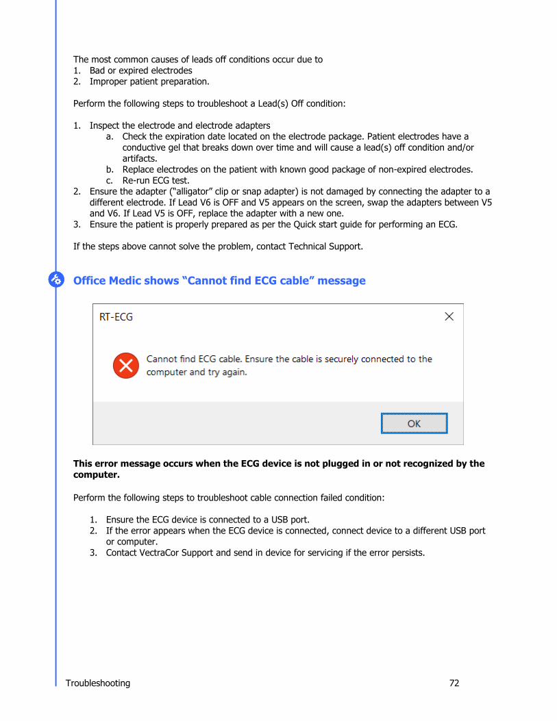
Troubleshooting 72
The most common causes of leads off conditions occur due to 1. Bad or expired electrodes 2. Improper patient preparation.
Perform the following steps to troubleshoot a Lead(s) Off condition: 1. Inspect the electrode and electrode adapters
a. Check the expiration date located on the electrode package. Patient electrodes have a conductive gel that breaks down over time and will cause a lead(s) off condition and/or artifacts.
b. Replace electrodes on the patient with known good package of non-expired electrodes. c. Re-run ECG test.
2. Ensure the adapter (“alligator” clip or snap adapter) is not damaged by connecting the adapter to a different electrode. If Lead V6 is OFF and V5 appears on the screen, swap the adapters between V5 and V6. If Lead V5 is OFF, replace the adapter with a new one.
3. Ensure the patient is properly prepared as per the Quick start guide for performing an ECG.
If the steps above cannot solve the problem, contact Technical Support.
Office Medic shows “Cannot find ECG cable” message
This error message occurs when the ECG device is not plugged in or not recognized by the computer.
Perform the following steps to troubleshoot cable connection failed condition:
1. Ensure the ECG device is connected to a USB port. 2. If the error appears when the ECG device is connected, connect device to a different USB port
or computer. 3. Contact VectraCor Support and send in device for servicing if the error persists.

Troubleshooting 73
Other Office Medic messages to users
Office Medic will output messages to the user in response to different user actions usually to help the user complete an action. Office Medic Message: Reason for Message: Correction:
Office Medic will show this message if a patient has not been selected for a test
Highlight a patient before initializing the test
A calibration, session, or test has not been selected when user tries to print
Select a calibration, session, or test prior to printing
User tries to create a patient without filling the required fields
Fill in the required fields before creating the patient
Spirometer is not connected when trying to do a spirometry test
Connect spirometer to PC
User hit Interp or Print buttons when no test is performed for the test session
Perform a spirometer test for the session

Service and Information 74
Service Information Device Care & Maintenance Cleaning
To clean the spirometer, clean surfaces with a damp cloth using water only. DRY THOROUGHLY. AVOID CLEANING AROUND CONNECTORS. Excess moisture in or on the case, cables or air fittings could affect operation.
To clean the ECG device, wipe the surfaces of the case, the leadwires, and connectors with a clean cloth moistened in water only. To disinfect the ECG device wipe the case, the leadwires, and connectors with an alcohol-free hospital grade disinfectant. DRY THOROUGHLY.
Cautions: • Do not clean the case with alcohol. • Do not saturate or immerse the case with liquid during cleaning. • Do not sterilize ECG device.
Calibration
Spirometer: Follow directions in the Spirometry Tools section ECG Device: User calibration of device is not required. However, a periodic check of the ECG system with an ECG simulator is recommended. Intervals for these checks can be set at the discretion of your Medical Director. There are commercially available ECG simulators which may be used for this purpose; refer to its accompanying information for instruction on the use of these. If calibration of the device is necessary (e.g. because of physical shock) and an ECG simulator is not available, contact the VectraCor Customer Care/Service Department.
Handling
Do not insert a dirty USB cable into the USB port. Spirometer: Avoid contaminating the luer (connector to the pressure tube).
Storage
Store the device in a dry place. Avoid sudden changes in temperature.
Physical Shock Avoid physical shock; a device that has been dropped should have the calibration verified before use on a patient.
Inspection
Inspect device for damage initially and before each use. Do not use devices that show visual signs of damage. Contact the VectraCor Service department with questions related to device damage and repair.
Training
Contact VectraCor for training on Office Medic and its devices; VectraCor will provide materials and training for the system.

Service and Information 75
Service Contact the VectraCor service department: VectraCor, Inc. 785 Totowa Road Suite 100 Totowa, NJ 07512 USA Monday through Friday 8am to 6pm EST Phone: 973-904-0444 www.vectracor.com A Return Merchandise Authorization (RMA) number will be issued for repairs THE INSTRUMENT MUST BE RETURNED FOR REPAIRS AT THE EXPENSE OF THE PURCHASER. IN-WARRANTY REPAIRED UNITS ARE RETURNED AT THE EXPENSE OF QRS OR ITS AUTHORIZED AGENT. FOR OUT OF WARRANTY WORK THE CUSTOMER IS RESPONSIBLE FOR ALL FREIGHT CHARGES. Limited Warranty • All instruments sold and supplied by VectraCor are guaranteed to be free from defects in material and workmanship for a period of 3 year from date of purchase. All supplies and accessories carry a 90-day limited warranty. This includes oximetry sensors. If in the judgment of VectraCor the instrument is proven to be defective during the warranty period it will be repaired or replaced with no charge for parts or labor. • This warranty does not cover any instrument that has been damaged by accident, misuse, abuse or has been altered or repaired by anyone other than an authorized VectraCor agent. This warranty also does not cover any unit that has had the serial number removed, defaced or rendered illegible. • THIS WARRANTY IS IN LIEU OF ALL OTHER WARRANTIES EXPRESSED OR IMPLIED, INCLUDING WARRANTIES OF MERCHANTABILITY AND FITNESS AND IS HEREBY LIMITED TO REPAIR OR REPLACEMENT OF INSTRUMENTS FOUND DEFECTIVE DURING THE WARRANTY PERIOD. AN AUTHORIZED VECTRACOR AGENT, MUST MAKE ALL REPAIRS. INSTRUMENTS SENT BY MAIL OR COMMON CARRIER SHOULD BE INSURED AGAINST LOSS OR DAMAGES, AS THEY ARE NOT COVERED BY THIS WARRANTY. • Technical support on software is under warranty for 1-year. This includes ECG lead wires. A software support package is available after 1-year at an additional cost.

Glossary of Terms 76
Glossary of Terms
%PRED Ratio of patient’s actual results compared to predicted normal values, expressed as a percentage. Abnormality is defined by using one standard deviation for each variable rather than any specific percentage below the predicted value. Results above 100% are above average.
ATS American Thoracic Society, a scientific medical organization active in pulmonary research and care of patients with lung diseases. The ATS has recommended standards for spirometers.
BF Equipment Degree of protection against electrical shock.
Bronchodilator A type of drug (i.e. Albuterol), usually administered in an aerosol spray, that is used to dilate air passages to reduce any restrictions to airflow.
BTPS Body Temperature and Pressure, Saturated: A number, which uniformly expresses all Spirometry results at body temperature and pressure, fully saturated with water.
Calibration Syringe A large syringe which injects a measured amount of air into the mouthpiece. Many syringes have a stop ring on the plunger, which allows injecting various calibrated amounts of air.
Class II Equipment Double insulated equipment.
COPD Chronic Obstructive Pulmonary Disease.
EOTV End-of-test volume.
ERS European Respiratory Society.
EX TIME Expiratory Time, expressed in seconds - time elapsed between the beginning and completion of expiration.
FEF 25-75% Forced expiratory flow during the middle half (25-75%) of the FVC (formerly
called the maximum middle expiratory flow rate), expressed in liters per second. This is the most sensitive measure of small airways obstruction (typically seen in smokers).
FEFxx% Forced Expiratory Flow at xx% point of the FVC, expressed in liters per second.
FET Forced Expiratory Time.
FEV1/FEV6 Ratio of FEV6 exhaled in one second. May be used as a surrogate for FEV1/FVC.
FEV6 (L) forced expiratory volume
Measured six seconds after commencement of expiration. May be used as a surrogate for FVC.
FEVx/FVC% The percentage ratio of Forced Expiratory Volume (timed) to Forced Expiratory Vital Capacity, expressed as a percentage.
FIF.2-1.2 Forced Inspiratory Flow between 200ml. and 1200ml. Flow of inspired
air measured after the first 200ml. And during the next 1000ml.

Glossary of Terms 77
FIF 25-75% Forced Inspiratory flow during the middle half (25-75%) of the FIVC expressed in liters per second.
FIFxx% Forced Inspiratory Flow at xx% point of the FIVC, expressed in liters per second.
FIVx/FIC% The percentage ratio of Forced Inspiratory Volume (timed) to Forced Inspiratory Vital capacity, expressed as a percentage.
Flow vs. Volume Curve
Forced Expiratory Flow
Forced Expiratory Volume (timed), (FEV(t))
Forced Inspiratory Vital Capacity, (FIVC)
Graph obtained by forced exhalation test, Flow is plotted on the vertical axis and volume on the horizontal axis.
It is the rate of flow, expressed in liters per second, at various points in the volumetric flow, i.e. FEF25%, FEF50%, FEF75%.
Maximal volume of air, expressed in liters, which can be expelled in specific time in a forced capacity test.
Total volume of air, expressed in liters, which can be inhaled during a rapid forced inhalation after a maximal expiration.
Forced Inspiratory Flow
Forced Vital Capacity (FVC)
Inspiratory rate of flow, expressed in liters per second, at various points in the volumetric flow, i.e. FIF25%, FIF50%, FIF75%.
Total volume of air, expressed in liters, which can be exhaled during a rapid forced exhalation after a maximal inspiration.
LLN Lower limit of normal. Maximum Voluntary Ventilation (MVV)
The maximum volume of air that can be inhaled and exhaled repeatedly through the lungs over a period of time (usually 12 seconds) and extrapolated to one minute.
Obstruction Limitation of airflow. It is shown by the FVC test. Low FEV1/FVC% ratio is the main indication of airways obstruction. Reductions in FEV3/FVC% and FEF25– 75% best demonstrate obstruction of small airways.
PC Card Also known as a PCMCIA card. A standard 68-pin computer card designed to add modular hardware to computers.
Perfusion Display indicating if the pulse waveform signal is of good quality and the SpO2 data is accurate.
Peak Expiratory Flow Rate (PEFR)
Maximum instantaneous flow in the FVC test.
PFT Pulmonary Function Test. PEFT Peak Expiratory Flow Time. PIFR Peak Inspiratory Flow Rate, expressed in liters per second. Predictor Predicted value according to the "normal" equations used. Pulmonary Functions Tests
see PFT

Glossary of Terms 78
Pulse Rate Heart rate measured in beats per minute (bpm). RR Respiratory Rate: the average number of inhalations/exhalations per minute
performed during a test.
Signal Intensity Indication displaying the patient’s pulse. Slow Vital Capacity (SVC)
Total volume of air, expressed in liters, which can be exhaled during a slow exhalation after a maximal inspiration. Amount may be decreased because of disorders that cause volume restriction in the lung.
SpO2 Approximate percentage of oxygen saturation in hemoglobin.

Device Specifications 79
Universal SmartECG Specifications Hub Weight 280 - 335 grams (0.62 – 0.66 lb) Hub Dimensions 85mm x 91mm x 20mm (3.3” x 3.6” x 0.8”)
Patient Leads Length 1.0 meter (3.3 ft) for limb leads 0.6 meter (2.0 ft) for chest leads
PC Connection Length 1.8 meter (5.9 ft) USB cable
Patient Leads 12 Lead Cable (10 patient leads)
Case Material ABS Plastic Electrode Connections 4 mm Banana plug with "tab" or "snap" connectors
Electrode Labeling Abbreviations and colors to comply with either IEC or AAMI (AHA) standards
Display and Operating Console
Dependent on PC (supplied by user)
Gain/Sensitivity 5, 10, 20 mm/mV Input Range ±5 mV
Acquisition sample rate 1000 samples per second (compressed to 500Hz with peak picking and averaging algorithm)
Heart Rate Range 30 bpm - 200 bpm
Frequency Response 0.05 to 175Hz ± 3dB Defibrillator Protection Patient leads are isolated from system and operator, with 5 kV protection
Common Mode Rejection -60dB (minimum) Safety Standards Complies with IEC 60601-1, IEC 60601-1-2, and IEC 60601-2-25
Accuracy Accurate to IEC 60601-2-25 essential performance requirements. Leads Off Indicators Connection status for each lead is shown on Acquisition screen
Power Source Powered by PC via USB interface
Supply Voltage 5V DC
Supply Current <17mA DC
Permanent Filters High Pass: 0.05Hz 2nd order Low Pass: 175Hz 2nd order Baseline Wander: Baseline reset by adaptive zeroing algorithm
Notch filter (Mains Noise Rejection)
Low pass (Muscle Artifact Filter)
50Hz 4th order Butterworth 60Hz 4th order Butterworth 35Hz 4th order
Report Capabilities User selectable Report formats Environmental Conditions Operating Temperature: 10 to 35° C (50 to 95° F)
Storage Temperature: -20 to 70° C (-4 to 158° F) Operating Relative Humidity: 20 to 85% (non-condensing) Storage Relative Humidity: 10 to 85% (non-condensing) Atmospheric Pressure: 70 kPA – 106 kPa

Device Specifications 80
Weight 226.8 grams (0.5 lb.) Dimensions 109.2 mm x 94.0 mm x 43.2 mm (4.3” x 3.7” x 1.7”)
Communication Port USB Software Compatibility Office Medic Version 5.5 (or later)
Storage Conditions Temperature: -15 to 50° C (5 to 122° F) Relative Humidity: < 90% (non-condensing) Atmospheric Pressure: 700 to 1060 hPa
Power Supply 5 Vdc ±5% 100 mA or less from the host PC USB Port Operating Conditions Temperature: 15 to 40° C (59 to 104° F)
Relative Humidity: 10 to 90% (non-condensing) Atmospheric Pressure: 700-1060 hPa
Spirometry Measurement Principle
The pressure is converted to flow. Volume measurement by flow integration.
Measurement Time FVC – 60 sec.; SVC – 60 sec.; MVV – 15 sec. Sampling Rate 125 Hz
Range (BTPS) FLOW: ±14 liters/second VOLUME: 0.5 – 8.0 liters
Accuracy (BTPS) FLOW: • FEF 25-75: ±5% of indication or ±200 ml/sec, whichever is greater • PEF: ±10% of indication or ±300 ml/sec, whichever is greater
VOLUME: ±3% of indication or ±50 ml, whichever is greater for FVC and FEV1 • FVC and FEV1: ±3% of indication or ±50 ml, whichever is greater • MVV: ±10% of indication or ±15 L/min, whichever is greater
Precision (BTPS) FLOW: PEF: ±5% of indication or 150 ml/sec, whichever is greater VOLUME: FVC and FEV1: ±3% or 50 ml, whichever is greater
Minimum Tracing Size FLOW VOLUME: Flow (vertical): 5 mm/L/S; Volume (horizontal): 10 mm/L VOLUME TIME: Volume (vertical): 10 mm/L; Time (horizontal): 20 mm/S
Calibration ATS 3-speed or standard calibration check Predicted Normals ADULT FVC: Crapo (1981), Cherniack (1972), Morris (1971/73), Knudson (1983),
Roberts (1991), ECCS/ERS/Quanjer (1993), NHANES III (1999), GLI (2012) PEDIATRIC FVC: Hsu (1979), Knudson (1983), Polgar (1971), Warwick (1977), NHANES III (1999), Zapletal (1987), Wang (1993), Quanjer (1995), GLI (2012) ADULT MVV: Cherniack (1972) PEDIATRIC MVV: Polgar (1971), Zapletal (1987)
Interpretation ATS/ERS 2005, BTS-NICE 2004-2005, NLHEP 2000, Enright 1987
Report Format Pre-test overlay with full page graphs Pre/Post test overlay with full page graphs
Parameters Measured FVC, FEV0.5, FEV1, FEV6, FEV1/FEV6, FEV3, FEV1/FVC, FEV3/FVC, PEFR, PEFT, FEF25%, FEF50%, FEF75%, FEF25-75%, FIVC, FIV0.5, FIV1, FIV3, FIV1/FIVC, FIV3/FIVC, PIFR, FIF50%, FIF 25-75%, FIF.2-1.2, FVC/FIVC, Extrapolated Volume (Ext. Vol. BEV), EOTV, FET, MVV, RR, MTV, SVC
Orbit Portable Spirometer Specifications