ofatumumab in cll: evaluation of in-vitro response mechanisms
TRANSCRIPT

UNIVERSITÄTSKLINIKUM ULM, ZENTRUM FÜR INNERE MEDIZIN, KLINIK FÜR INNERE
MEDIZIN III, ÄRZTLICHER DIREKTOR: PROF. DR. H. DÖHNER
Ofatumumab in CLL:
Evaluation of in-vitro response
mechanisms
Dissertation zur Erlangung
des Doktorgrades der Medizin
der Medizinischen Fakultät der
Universität UIm
Matthias Volden, Sindelfingen
2013

II
Amtierender Dekan: Prof. Dr. Thomas Wirth
1. Berichterstatter: Prof. Dr. Thorsten Zenz
2. Berichterstatter: Prof. Dr. Thomas Barth
Tag der Promotion: 11.07.2014

III
Dedication
To my parents

IV
Contents
List of abbreviations....……………………………………………………......………- v -
1 Introduction ................................................................................................. - 1 -
2 Materials and Methods ................................................................................ - 4 -
2.1 Patient samples ..................................................................................... - 4 -
2.2 Data analyses ....................................................................................... - 4 -
2.3 Chemoluminescence based viability assay ........................................... - 5 -
2.4 Flow cytometry based cytotoxicity assay assay .................................... - 6 -
2.5 Whole blood assay (incubation) ............................................................ - 8 -
2.6 Microscopic readout to assess homotypic adhesion ............................. - 9 -
2.7 Flow cytometric readout (True Count method) .................................... - 10 -
3 Results ...................................................................................................... - 17 -
3.1 Chemoluminescence based viability assay, Assay No. 1 .................... - 17 -
3.2 Chemoluminescence based viability assay, Assay No. 2 .................... - 20 -
3.3 Chemoluminescence based viability assay, Assay No. 3 .................... - 22 -
3.4 Chemoluminescence based viability assay, Assay No. 4 .................... - 23 -
3.5 Chemoluminescence based viability assay, Assay No. 5 .................... - 26 -
3.6 Chemoluminescence based viability assay, Assay No. 6 .................... - 28 -
3.7 Flow cytometry based cytotoxicity assay, Assay No. 7 ....................... - 31 -
3.8 Flow cytometry based cytotoxicity assay, Assay No. 8 ....................... - 32 -
3.9 Whole blood assay, microscopic readout, Assay No. 9 ....................... - 34 -
3.10 Whole blood assay, flow cytometric readout, Assay No. 10 ............. - 35 -
4 Discussion ................................................................................................. - 37 -
5 Summary ................................................................................................... - 41 -
6 References ................................................................................................ - 43 -
7 Curriculum Vitae ........................................................................................ - 49 -

V
List of abbreviations
13 q del deletion of the long arm of chromosome 13, detected by FISH,
prognostic marker for CLL, associated with long survival
11q del deletion of the long arm of chromosome 11, detected by FISH,
prognostic marker for CLL
17p del deletion of the short arm of chromosome 17, detected by FISH,
prognostic marker for CLL, associated with short survival
12+ trisomy of chromosome 12, detected by FISH, prognostic
marker for CLL
7-AAD 7-Amino-Acridine, fluorescent dye used for e.g. flow cytometry, late
cell death marker used for flow cytometry
ADCC antibody dependent cytotoxicity
AKT Protein Kinase B, plays a key role in multiple cellular processes, e.g.
apoptosis, cell proliferation
Annexin-V cellular protein, located on the inner surface of the cell membrane,
positive staining of Annexin-V indicates beginning cell death (flow
cytometry)
BCL-2 B-cell lymphoma 2, apoptosis regulator protein
BD Becton Dickinson (company, global player in biotechnology)
BR Bendamustine (chemotherapeutic agent) + Rituximab
Ca Calcium
CD cluster of differentiation, cell surface antigen
CDC complement dependent cytotoxicity
CLL chronic lymphocytic leukemia
DMSO Dimethyl sulfoxide, polar solvent that dissolves both polar and
nonpolar compounds
F Fludarabine, chemotherapeutic agent
FCR Fludarabine + Cyclophosphamide (both chemotherapeutic agents) +
Rituximab
FCS bovine (fetal) calf serum
FACS flow cytometry (fluorescence activated cell sorting)
FFP fresh frozen plasma
FISH fluorescence in-situ hybridization, specific technique to stain parts of
the DNA with a fluorescent dye, read-out e.g. microscopy
FITC fluorescein isothiocyanate, chemical compound, fluorescent dye
used for fluorescent staining of cells before e.g. cytometric read-out
HS5-cells cell line derived from bone marrow stroma cells
Ig Immune globuline
IgHV Immune globuline heavy (chain) variable part:

VI
IgHV m mutated IgHV (positive prognostic marker for CLL)
IgHV um unmutated IgHV (negative prognostic marker for CLL)
kDa kiloDalton (unit for molecular mass of a protein)
mAb monoclonal antibody
Mg Magnesium
p53 tumor suppressor protein, regulates cell cycle, encoded by TP53
gene, guardian of the genome (p53 runs as 53 kDa protein on SDS-
Page)
PBS phosphate buffered saline
PCD programmed cell death
PE Phycoerythrin, fluorescent dye
Raji-cells cells derived of a Burkitt-lymphoma derived cell line
RPMI 1640 cell culture medium
SDS-Page sodium dodecyl sulfate polyacrylamide gel electrophoresis; to
separate proteins according to their electrophoretic mobility
SSC sideward scatter, flow cytometric read-out mode, measures
granularity of cells
TP53 gene encodes for p53
ZAP-70 Zeta-chain-associated protein kinase 70 (70kDa), prognostic marker
in CLL

- 1 -
1 Introduction
CLL is the most common adult leukemia in the western world. It is characterized
by an expansion of monoclonal, mature B cells, which accumulate in different body
compartments. The clinical definition of the disease is based on the characteristic
expression of epitopes (positivity for both CD19 and CD5) and a blood count of
over 5000 of these malignant B cells. CLL can be further distinguished by dividing
the disease into different subgroups. Prognostic factors such as IGHV mutational
status, β2-microglobulin, ZAP-70 expression, CD38 expression and clinical stage
are used to predict disease course. In addition, there are a number of recurrent
genomic aberrations occurring in CLL cells, which is relevant for the choice of
treatment strategy. CLL cells usually proliferate in lymph nodes and not in the
blood compartment, where they mainly accumulate and remain inactive.
Low or intermediate risk CLL patients (patients without a 17p-deletion, without
TP53 mutation, and not refractory to purine analogues) can be effectively treated
with chemoimmunotherapy such as FCR (Fludarabine + Cyclophosphamide +
Rituximab) or BR (Bendamustine + Rituximab) (Hallek M, 2009, Fischer et al.,
2011, Stilgenbauer and Zenz, 2010). It should be noted, that elderly patients, who
constitute the majority of CLL patients, are often not regarded as suitable for such
aggressive treatment due to comorbidity(Eichhorst et al., 2009). Apart from that,
chemotherapeutic agents cause side effects or secondary tumors after
years(Morrison et al., 2002). High-risk-patients (defined by p53 mutation) are un-
likely to respond to purine analogues such as Fludarabine and F-based therapies.
This is because chemotherapeutic approaches often require a functional DNA
damage/p53 pathway, which is absent in this patient group (Zenz et al.,2008).
There is a need for novel, effective, and well-tolerated treatment options and
treatment strategies. A number of substances have been shown to act inde-
pendently of a functional p53 pathway and these offer theoretic advantages in this
patient population, which ideally should be demonstrated in randomized trials.
Among those are monoclonal antibodies.
Three different therapeutic antibodies are currently approved and recommended
for the therapy of CLL: The CD20-antibodies Rituximab and Ofatumumab as well
as the CD52-antibody Alemtuzumab. In Europe, Rituximab is mainly used in com-
- 2 -
binations and rarely as a single agent in CLL. Alemtuzumab is mainly used as a
single-agent in Fludarabine-refractory cases and for high-risk CLL patients who
are not eligible for other therapeutic options, such as stem cell transplantation.
Ofatumumab has been approved for double-refractory CLL or CLL with so-called
bulky disease.
CD20 is a 33–37 kDa, non-glycosylated phosphoprotein expressed on the surface
of almost all normal and malignant B cells. CD20 has no known natural ligand and
CD20 knockout mice display an almost normal phenotype. It probably functions as
a store-operated calcium channel following ligation of the B cell receptor for anti-
gen (Walshe et al., 2008, Cragg, 2005). Rituximab and Ofatumumab can be de-
scribed as type-I-CD20-antibodies. Ofatumumab, Rituximab, and Alemtuzumab
are potent inducers of CDC, though each to a different extent and more important-
ly, depending on the disease and antigen-expression. CLL is known for rather low
CD20 expression and even though an antibody such as Rituximab seems highly
effective in inducing certain effector mechanisms in other B-cell malignancies, this
does not necessarily mean that this accounts for CLL too. We observed a dramatic
difference of Rituximab-induced CDC in our control cell-line, Burkitt-lymphoma-
derived Raji-cells, which were very susceptible towards it and CLL-patient sam-
ples, which didn’t seem to be susceptible at all. Other mechanisms such as ADCC,
PCD, inducing of a vaccine-like effect or chemosensitization have been described
to contribute to the over-all activity of the antibodies in vivo. As multiple factors can
influence these mechanisms, it is important to identify the principal one or ones for
a certain disease. Combinations of new and different targeted therapeutic options
should yield major efficiency and elicit least-possible side effects. Some patients
seem to benefit more of antibodies than others. This hints, that intrinsic resistance
mechanisms need to be identified. It remains a challenging topic to develop strat-
egies to enhance antibody therapy in CLL.
We tested and compared Rituximab, Ofatumumab, and Alemtuzumab in vitro to
assess their biological activity with special regard to possible enhancement of their
use in future CLL therapy. Most of the in-vitro antibody assays are usually per-
formed under normal culture conditions. Regarding CLL the disease extent in dif-
ferent body compartments and different disease stages can influence the efficacy
of a therapy. We used different culture conditions to mimic the in vivo situation and

- 3 -
to detect possible limitations of the antibodies. We hypothesized different suscep-
tibility to antibody treatment of different CLL subgroups and subdivided our CLL-
patient samples according to risk-stratification and antibody-pretreatment. In con-
trast to Rituximab, relatively little is known about the benefit of Ofatumumab used
in combination with other drugs. We tried to discover new strategies to achieve
both targeted use of antibodies and potential enhancement of their antitumor activ-
ity by combining them with other drugs. We admit that due to the in-vitro character
of our assays we cannot guarantee that our results can be fully reproduced in vivo
and clinical trials would be needed to determine their significance.

- 4 -
2 Materials and Methods
2.1 Patient samples
CLL cells were isolated from blood taken of CLL patients with high tumor burden in
the blood compartment and who have been previously diagnosed according to
standard criteria (Hallek et al., 2008). Ficoll gradient centrifugation was used for
isolation. Cells were then washed, cryo-preserved in FCS with 5% DMSO for sub-
sequent analysis. Experiments were performed after informed written consent had
been obtained from all patients. Age, gender, pretreatment, and risk stratification
(interphase-FISH for the detection of chromosomal abnormalities, TP53-mutation-
analysis, IgHV-mutation-status, ZAP-70-expression) of the CLL patients are sum-
marized in tables I to V. (end of “Materials and Methods”)
2.2 Data analyses
Error bars represent the SD of experiments which were performed in triplicates.
GraphPad Prism 4 was used to perform unpaired t-tests and non-linear regres-
sion. Data from successful and valid experiments are summarized.

- 5 -
Assays
2.3 Chemoluminescence based viability assay
CLL-cells were incubated at increasing concentrations of monoclonal antibodies
(and partially in combination with either Fludarabine, Alemtuzumab, or AKT-
inhibitor) in complete medium with 30 % fresh frozen serum at a cell concentration
of 1000 cells /µl. Raji-cells, which showed to be very sensitive to complement de-
pendent cytotoxicity, were used as an internal control. Isotype IgG-antibody and
complete medium were used as untreated negative controls. Viability was as-
sessed with Promega Cell Titer Glo Luminescent cell viability assay according to
manufacturer’s protocol. Read-out was performed after 3, 24, and/or 48 hours. For
combination assays Fludarabine and Alemtuzumab, were used at fixed concentra-
tions: 1µmol Fludarabine and 1µg/ml Alemtuzumab. Pan-AKT-Inhibitor GSK
690693 was provided by GlaxoSmithKline and used at different concentrations
(maximum 10µmol).
Materials:
Cell titer Glo luminescent viability as-say
Promega GmbH, Mannheim
RPMI 1640 Medium Biochrom AG, Berlin
BD Pharmigen Purified Mouse IgG1 K isotype control
Becton Dickinson GmbH, Heidelberg
Rituximab Roche, Hoffmann, La Roche, Grenzach-Whylen
Alemtuzumab Bayer Schering Pharma AG, Berlin und Genzyme GmbH, Neu-Isenburg
Sera Plus special processed FCS PAN Biotech GmbH, Aidenbach
Human Serum FFP / off the clot Sanquin Plasma Products, Amsterdam, The Netherlands
PBS Dulbecco w/o Ca, Mg Biochrom AG, Berlin
Ofatumumab Genmab, Utrecht, The Netherlands
Fludarabine Hospital pharmacy, University of Ulm, Ulm
AKT-Inhibitor GSK 690693 GSK, London, England

- 6 -
2.4 Flow cytometry based cytotoxicity assay
CLL-cells were incubated with therapeutic antibody (10µg/ml) in complete medium
with 30 % fresh frozen serum at a cell concentration of 10000 cells /µl. Assay was
performed in duplicates. Isotype IgG-antibody and complete medium were used as
untreated negative controls. This implies that the amount of apoptotic cells of neg-
ative control samples were regarded as spontaneously apoptotic and therefore
subtracted of the amount of apoptotic cells of therapeutically treated samples.
CLL-cells were stained with both 7-AAD and Annexin-V-PE prior to flow-cytometric
measurement. Flow cytometric data were acquired with a FACS Calibur cytometer
(BD Biosciences), operated by CellQuest software and 10000 events were collect-
ed per sample. Staining with FITC-labelled Anti-CD45-antibody (BD Biosciences)
was performed to identify CLL-cells in co-incubation assays with HS5-cells. For
these assays, HS5-cells were harvested and redistributed to 48-well-plates 24
hours prior to antibody-incubation at a density of 200cells/µl. Ratio of HS5-cells to
CLL-cells was approximately 1 to 5.
Figure 1 A-C:
Readout after 3 hours, example of cytometry dot plots. A: Alemtuzumab, B: Ofatumumab, C: iso-
type-antibody: There are three representative dot blots. The x-axis describes the extent of Annexin-
V-PE (early cell death marker)-staining in a logarithmic scale, the y-Axis describes the extent of 7-
AAD (7-Amino-Acridine, fluorescent dye, late cell death marker) -staining in a logarithmic scale –
the dense clouds of white dots represent cells. A: In the case of Alemtuzumab a lot of cells are both
positive for Annexin-V-PE and 7-AAD, B: in the case of Ofatumumab there are two major fractions:
in the upper right square there is a cloud of cells which are positive for both Annexin-V-PE and 7-
AAD and a cloud of cells in the lower left square, which are negative for both Annexin-V-PE and 7-
Annexin-V-PE
7-AAD
A B C

- 7 -
AAD, which means that there are both apoptotic and viable cells. C: without therapeutic antibody
there is only a cloud in the lower left square detectable.)
Materials:
RPMI 1640 Medium Biochrom AG, Berlin
Human Serum FFP / off the clot Sanquin Plasma Products, Amsterdam, The Netherlands
Sera Plus special processed FCS PAN Biotech GmbH, Aidenbach
PBS Dulbeco w/o Ca, Mg Biochrom AG, Berlin
Annexin V-PE Becton Dickinson Biosciences, Heidel-berg
7-AminoactinomycinD Sigma, Saint Louis, Missouri, USA
Rituximab Roche, Hoffmann, La Roche, Grenzach-Whylen
BD Pharmigen Purified Mouse IgG1 K isotype control
Beckton Dickinson GmbH, Heidelberg
Annexin-binding-buffer Self-production
HEPES Buffer Solution PAA, Pasching, Austria
Calciumchlorid Serva Feinbiochemica GmbH & Co., Heidelberg
Natriumchlorid AnalaR Normapur VWR International Ltd., Leicestershire, England
Alemtuzumab Bayer Schering Pharma AG, Berlin und Genzyme GmbH, Neu-Isenburg
Ofatumumab Genmab, Utrecht, The Netherlands
HS5-cells DSMZ, Braunschweig

- 8 -
2.5 Whole blood assay (incubation)
Fresh blood from patients with high tumor load (mean CLL-cell concentration, as-
sessed by routine diagnostic, at least 56.89 Giga/L (CD19+/CD5+), mean leuko-
cyte count 73.68 Giga/L) in the blood compartment was directly incubated at room
temperature with monoclonal antibodies at a concentration of 10µg/ml on a rotat-
ing table.
Materials:
PBS Dulbecco w/o Ca, Mg Biochrom AG, Berlin
BD Facs Lysing Solution Becton Dickinson GmbH, Heidelberg
BD CD19 FITC (4G7) Becton Dickinson GmbH, Heidelberg
BD Pharmigen Purified Mouse IgG1 K isotype control
Becton Dickinson GmbH, Heidelberg
Aqua Delta Select Spüllösung/ Irrigati-on Solution
Delta Select GmbH, München
Rituximab Roche, Hoffmann, La Roche, Grenzach-Whylen
Alemtuzumab Bayer Schering Pharma AG, Berlin und Genzyme GmbH, Neu-Isenburg
Ofatumumab Genmab, Utrecht, The Netherlands
ACD (citrate buffer) Self-production
Blue Heparinate standard syringe blue Sarstedt , Nümbrecht
Lepirudine Refludan Celgene, München

- 9 -
2.6 Microscopic readout to assess homotypic adhesion
After 1 hour incubation on a rotating table, blood samples (n=10) were delivered to
the diagnostic laboratory of the hematologic department and blood smears were
made according to clinical routine procedures. After Giemsa-staining, blood
smears were semi-quantitatively assessed for homotypic adhesion. Five randomly
picked high power fields were considered and evaluated in a blinded fashion.
Figure 2:
Microscopic readout after 1 hour, 4 different representative high-power fields of the same CLL
(chronic lymphocytic leukemia) patient (A-D) are shown: blue cells are mainly CLL cells. The rest of
the cells are erythrocytes. No aggregation of CLL cells can be detected in any of the investigated
antibodies.

- 10 -
2.7 Flow cytometric readout (True Count method)
This assays had the goal to assess the impact of high cell count on effector con-
sumption as well as the potential contribution of e.g. ADCC under in vivo condi-
tions with special regard to CLL cell lysis. Therefore we chose the BD Biosciences
TrueCount cytometric method. After 3 hours of incubation, CD19-FITC stained
cells were counted using a bead-based FACS-measurement and cell to bead ratio
was compared with 6 negative controls. The assay was performed in triplicates
and according to manufacturer’s protocol (BD Biosciences) except for the amount
of blood used for measurement: in order to obtain a comparable B-cell to bead
ratio, a maximum of 10 µl of blood were used instead of 50 µl due to high B-cell-
number in CLL patient blood. In order to eliminate possible systematic errors
caused by the use of a specific anticoagulant (Heparine interacts with several se-
rum-components including complement factors), whole blood assays were per-
formed with three different anticoagulants: Heparine [n=6], Lepirudine [n=3], and
Citrate [n=13]. The incubations of the assays with Lepirudine-anticoagulation were
made at the same culture conditions as the other assays (37°C, humidified, 5%-
CO2 incubator).
Materials:
BD TruCount Tubes Becton Dickinson GmbH, Heidelberg
ART Aerosol Resistant Tips Thermo Fisher Scientific-Molecular Bio Products, San Diego, USA
48 Well Cell Culture Plate Cellstar Greiner Bio-One GmbH, Frickenhausen
Derma Clean Latex gloves Ansell GmbH, München
Falcon tubes Falcon, Becton Dickinson, Franklin La-kes, USA
Falcon Serological Pipet Becton Dickinson GmbH, Heidelberg
Combitips plus/ Spitze Multistep-Pipette Eppendorf Deutschland GmbH, Wes-seling-Berzdorf
Devices:
BD FACSCalibur/ flow cytometer Becton Dickinson Bioscience, Heidel-berg
Reax 2000 Heidolph Instruments GmbH & Co.KG, Schwabach
GFL-3005 vortex machine GFL Gesellschaft für Labortechnik GmbH, Burgwedel
Multipette stream Eppendorf Deutschland GmbH, Wes-seling-Berzdorf
SterilGard3 Advance Baker Company, Sanford, Maine, USA
Julabo SW 20 heater Julabo Labortechnik GmbH, Seelbach

- 11 -
Coulter Ac T 5diff AL Beckman Coulter GmbH, Krefeld
Pipetboy acu INTEGRA Biosciences GmbH, Fern-wald
Eppendorf Research Pipettes Eppendorf Deutschland GmbH, Wes-seling-Berzdorf
Gilson Pipettes Gilson Inc., Middleton, USA
Figure 3:
Readout after 3 hours to assess both the potential cell lysis and the impact of high cell density on
CDC (complement dependent cytotoxicity). This CD19-FITC (B-cell-marker, x-axis) / SSC (side-
ward scatter, y-axis) dot-plot shows an untreated control: red dots represent beads, lilac dots rep-
resent CD19-negative cells (monocytes, T-lymphocytes), green dots represent C19-positive cells
(CLL-cells and B-cells), yellow dots represent CD19-negative cells with increased side scattering
due to intracellular granula (granulocytes).

- 12 -
Patient samples
Table 1:
CLL (chronic lymphocytic leukemia) samples (taken of CLL patients treated in Ulm during the peri-
od of 2000 and 2010, certain prognostic parameters of the CLL cells and clinical data of the pa-
tients) used for: Chemo-luminescence-based viability assay, read-out after 3 and 48 hours: The list
contains 10 high-risk CLL-cases (they are specified by at least one of the three following character-
istics: 17p (short arm of chromosome 17)-deletion, TP53 (encodes for p53)-mutation, Fludarabine-
refractory). The TP53 (tumor suppressor protein 53) -status is a prognostic marker for survival and
response to conventional chemotherapy schemes of CLL, the ZAP-70-protein is associated with
either long or short survival of CLL patients. FISH (fluorescence in-situ hybridization) is used to
further distinguish between CLL subgroups. The IGHV (variable part of the immune globuline
heavy chain)-status is another marker, which is associated with survival and response to treatment.
no. gender FISH TP53 IGHV ZAP-70 pre-
treatment age
1 F 13q- WT M Negative yes 64
2 M 11q-, 12q+, t14q32 WT UM Positive no 65
3 F 17p-, 12q+ WT UM n/d yes 87
4 F 17p-14q- c.584T>C UM Positive yes 65
5 M 11q-, 13q- c.919-7A>C UM n/d yes 77
6 M 17p, 12q+ WT n/d n/d yes 82
7 M 13q- WT M Negative no 50
8 M 17p-, 11q-, 13q- c.499C>T, c.830G>T
UM n/d yes 72
9 M 13q-, 11q- WT M Negative no 49
10 F 13q- c.847C>T M Negative no 73
11 M 13q-, 11q- WT UM n/d no 69
12 M 13q-, 11q- WT UM Negative no 37
13 F 13q- WT M Negative yes 64
14 F normal WT M Negative no 65
15 M 13q- WT M n/d no 72
16 M 13q- WT UM Positive no 70
17 M 13q- WT UM Positive no 69
18 M normal WT UM Positive no 71
19 M 13q bidel WT M Negative no 61
20 M 17p-, 13q- 753-759del7bp UM n/d yes 58
21 M 11q- WT UM Positive yes 69
22 M 17p-, 12q+, 13q- WT UM n/d no 68
23 F normal n/d M n/d yes 65
24 F normal WT UM Positive yes 74
25 F 17p-,13q-,12q+ c.809T>G UM n/d no 73
26 M 17p-, 11q-, 13q- c.329G>T UM Negative no 69
27 F normal WT UM Positive no 38
10 fema-le
17 male 8 with 17p del
7 with TP53-mutation
17 with un- mutated
IGHV-status
11 with prior treatment
65,78

- 13 -
Table 2:
CLL (chronic lymphocytic leukemia) samples (taken of CLL patients treated in Ulm during the peri-
od of 2000 and 2010, certain prognostic parameters of the CLL cells and clinical data of the pa-
tients) used for Chemo-luminescence-based viability assay, read-out after 48 hours, combination
with Fludarabine: The list contains 11 high-risk CLL-cases (they are specified by at least one of the
three following characteristics: 17p (short arm of chromosome 17)-deletion, TP53 (encodes for
p53)-mutation, Fludarabine-refractory). The TP53 (tumor suppressor protein 53) -status is a prog-
nostic marker for survival and response to conventional chemotherapy schemes of CLL, the ZAP-
70-protein is associated with either long or short survival of CLL patients. FISH (fluorescence in-
situ hybridization) is used to further distinguish between CLL subgroups. The IGHV (variable part of
the immune globuline heavy chain)-status is another marker, which is associated with survival and
response to treatment.
no. gender FISH TP53 IGHV ZAP-70 pre-
treatment age
6 M 17p, 12q+ WT n/d n/d yes 82
7 M 13q- WT M Negative no 50
8 M 17p-, 11q-, 13q- c.499C>T, c.830G>T UM n/d yes 72
9 M 13q-, 11q- WT M Negative no 49
10 F 13q- c.847C>T M Negative no 73
11 M 13q-, 11q- WT UM n/d no 69
12 M 13q-, 11q- WT UM Negative no 37
13 F 13q- WT M Negative yes 64
14 F normal WT M Negative no 65
15 M 13q- WT M n/d no 72
16 M 13q- WT UM Positive no 70
17 M 13q- WT UM Positive no 69
18 M normal WT UM Positive no 71
28 M 12q+, t(14:18) WT M Negative no 81
29 F 17p c.842A>G UM Positive yes 42
30 M 17p c.602-603Ins4bp UM Negative yo 69
31 M 17p WT UM n/d no 73
32 F normal c.484A>T UM Positive yes 64
33 M 13q WT UM n/d no 66
34 M 13q WT UM Negative yes 77
35 M normal WT UM Negative yes 72
36 F Tris.12, 17p c.673-2A>T M Negative yes 73
37 M 17p, 13q c.413C>T M Negative yes 72
38 F 13q n/d M n/d no 59
39 F 17p, 13q C.733G>T UM Negative yes 76
8 female 17 male
8 with 17p del 8 with TP53-mutation 14 with un-
mutated IGHV-status
10 with prior treatment
66,68

- 14 -
Table 3:
This table summarizes the CLL (chronic lymphocytic leukemia) samples (taken of CLL patients
treated in Ulm during the period of 2000 and 2010, certain prognostic parameters of the CLL cells
and clinical data of the patients) used for: Chemo-luminescence-based viability assay, read-out
after 48 hours, combination with either only the AKT (Protein Kinase B)-inhibitor or with both the
AKT-inhibitor and Alemtuzumab (these patient sample numbers have a footnote “*”):
The list contains 11 high-risk CLL-cases (they are specified by at least one of the three following
characteristics: 17p (short arm of chromosome 17)-deletion, TP53 (encodes for p53)-mutation,
Fludarabine-refractory). The Alemtuzumab-cohort consists of 7 high risk CLL-cases and 5 low-risk
CLL cases, patient characteristics of patient no.15 is not on the list, as the sample taken from that
patient hasn’t been investigated in the AKT-cohort , the last row summarizes the table columns.
The TP53 (tumor suppressor protein 53) -status is a prognostic marker for survival and response to
conventional chemotherapy schemes of CLL, the ZAP-70-protein is associated with either long or
short survival of CLL patients. FISH (fluorescence in-situ hybridization) is used to further distinguish
between CLL subgroups. The IGHV (variable part of the immune globuline heavy chain)-status is
another marker, which is associated with survival and response to treatment.
no. gender FISH TP53 IGHV ZAP-70 pre-
treatment age
6* M 17p, 12q+ WT UM n/d yes 82
7* M 13q- WT M Negative no 50
25 F 17p-,13q-,12q+ c.809T>G UM n/d no 73
27 F normal WT UM Positive no 38
26 M 17p-, 11q-,
13q- c.329G>T UM Negative no 69
28 M 12q+, t(14:18) WT M Negative no 81
29* F 17p c.842A>G UM Positive yes 42
30* M 17p c.602-
603Ins4bp UM Negative no 69
31* M 17p WT UM n/d no 73
32* F normal c.484A>T UM Positive yes 64
33* M 13q WT UM n/d no 66
34* M 13q WT UM Negative yes 77
35 M normal WT UM Negative yes 72
36 F Tris.12, 17p c.673-2A>T
M Negative yes 73
37* M 17p, 13q c.413C>T M Negative yes 72
38* F 13q n/d M n/d no 59
39* F 17p, 13q C.733G>T UM Negative yes 76
40 M 17p, 13q, 6q n/d UM n/d yes 66
7 female 11 male
10 with 17p del 8 with TP53-
mutation
12 with un- mutated
IGHV-status
9 with prior treatment
66,24

- 15 -
Table 4:
CLL (chronic lymphocytic leukemia) samples (taken of CLL patients treated in Ulm during the peri-
od of 2000 and 2010, certain prognostic parameters of the CLL cells and clinical data of the pa-
tients) used for: Whole blood assay, microscopic read-out after 1 h: only in 10 randomly picked
patients of the following list we did microscopic assessment of homotypic adhesion, cytometric
read-out after 3 and 8 hours. The list contains 6 high-risk CLL-cases (they are specified by at least
one of the three following characteristics: 17p (short arm of chromosome 17)-deletion, TP53 (en-
codes for p53)-mutation, Fludarabine-refractory). The TP53 (tumor suppressor protein 53) -status
is a prognostic marker for survival and response to conventional chemotherapy schemes of CLL,
the ZAP-70-protein is associated with either long or short survival of CLL patients. FISH (fluores-
cence in-situ hybridization) is used to further distinguish between CLL subgroups. The IGHV (vari-
able part of the immune globuline heavy chain)-status is another marker, which is associated with
survival and response to treatment.
no. gender FISH TP53 IGHV ZAP-70
pre-trea-tmen
t
leu-kocyte count
age
1 F 13q- WT M Negative yes 239,8 64
4 F 17p-14q- c.584T>
C UM Positive yes 132,7 65
7 M 13q- WT M Negative no 102,5 49
11 M 13q-, 11q- WT UM n/d no 71,9 69
12 M 13q-, 11q- WT UM Negative no 105,7 36
14 F normal WT M Negative no 27,6 65
18 M normal WT UM Positive no 95,1 48
20 M 17p- 753-759 del7bp
UM Positive yes 46,2 59
27 F normal WT UM Positive no 98,8 37
31 M 17p- WT UM Negative no 47,6 68
34 M 13q WT UM n/d no 111,4 67
37 F Tris.12, 17p- c.673-2A>T
M Negative yes 28,4 73
40 F 17p, 13q n/d UM n/d yes 74,2 76
42 M normal WT M n/d no 47,8 39
43 M t14q32 WT UM Negative yes 59,8 72
44 M 13q- WT UM n/d yes 61,8 67
45 M 11q-, 13q-,
14q- WT UM Negative no 141,3 73
46 M 12p+11, t(14;19)
WT UM Positive yes 26,6 49
47 M 13q- c:716A>
6 M n/d yes 11,7 76
48 M 13q WT M n/d no 84,2 57
49 M 11q23 WT UM Negative yes 10,8 58
6 female 15 male
5 with 17p del
4 with TP53-
mutation
14 with unmutated IgHV-status
10 with prior treat-ment
77,42 60,3

- 16 -
Table 5:
This table summarizes the CLL (chronic lymphocytic leukemia) samples (taken of CLL patients
treated in Ulm during the period of 2000 and 2010, certain prognostic parameters of the CLL cells
and clinical data of the patients) used for: Cytometry-based Apoptosis assay / HS5-coculture-assay
(*), read-out after 3 hour incubation. The list contains 2 high-risk CLL-cases (they are specified by
at least one of the three following characteristics: 17p (short arm of chromosome 17)-deletion,
TP53 (encodes for p53)-mutation, Fludarabine-refractory). The TP53 (tumor suppressor protein 53)
-status is a prognostic marker for survival and response to conventional chemotherapy schemes of
CLL, the ZAP-70-protein is associated with either long or short survival of CLL patients. FISH (fluo-
rescence in-situ hybridization) is used to further distinguish between CLL subgroups. The IGHV
(variable part of the immune globuline heavy chain)-status is another marker, which is associated
with survival and response to treatment.
no. Gender FISH TP53 IGHV ZAP-70 pre-
treatment age
7* M 13q- WT M Negative no 50
13* F 13q- WT M Negative yes 64
14* F Normal WT M Negative no 65
16* M 13q- WT UM Positive no 70
17* M 13q- WT UM Positive no 69
18* M Normal WT UM Positive no 71
20 M 17p-, 13q- 753-759del7bp UM n/d yes 58
21 M 11q- WT UM Positive yes 69
35 M Norm WT UM Negative yes 72
39 F 17p, 13q C.733G>T UM Negative yes 76
41 F Norm WT M n/d no 75
4 female 7 male
2 with 17p del
2 with TP53mutation
7 with unmutated IgHV-status
5 with prior treatment
67,18

- 17 -
3 Results
3.1 Assay No. 1: Comparison of CLL-cell-viability after 3 and 48 hour mAb-treatment
Figure 4 A – D:
Comparison of CLL (chronic lymphocytic leukemia) cell viability after a 3 and a 48 hour treatment.
A: viability of CLL cells after a 3 hour incubation with either Ofatumumab (green), Alemtuzumab
(blue), Rituximab (red), or isotype antibody (grey) at different concentrations. B: viability of Raji
cells after a 3 hour incubation with either Alemtuzumab (first bar), Rituximab (second bar), or iso-
type antibody (right bar). C: viability of CLL cells after a 48 hour incubation with either Ofatumumab
(green), Alemtuzumab (blue), Rituximab (red), or isotype antibody (grey) at different concentra-
tions. D: viability of Raji cells after a 48 hour incubation with either Alemtuzumab (first bar), Rituxi-
mab (second bar), or isotype antibody (right bar).

- 18 -
Figure 5:
viability bars of therapeutic antibodies at different concentrations and after different incubation
times (3 and 48 hours).
Assay No. 1: Viability measured after 3 and 48 hour incubation represents the
means and SD of 27 and 26 experiments, respectively (figure 4 A, C). Both Ofa-
tumumab and Alemtuzumab, but not Rituximab deplete CLL-cells after 3 hours of
incubation in the presence of 30% fresh frozen serum (figure 4 A). Mean viability
of CLL cells for Ofatumumab is 64.11 % (range: 6.46% - 96.5%) at 10µg/ml and
44.7% (range: 3.83% - 87.3%) at 100µg/ml concentration. Alemtuzumab reduces
viability to 26.99% (range: 4.18% - 74.24%) at 10µg/ml. Both Ofatumumab and
Rituximab nearly completely deplete Raji-cells after 3 hours of incubation (figure 4
B). Raji-cell-depletion is used to guarantee serum-quality qualitatively (figure 4
B/D). After 48 hours of incubation Ofatumumab, Alemtuzumab, and Rituximab
show activity to a different extent. Rituximab leads to a reduction of viability to
72.97% (range: 12.76% – 103.9%) and 63.14% (range: 3.885% - 110.9%) at 10
and 100µg/ml, respectively. Ofatumumab reduces viability to 57.06% (range:

- 19 -
7.79% - 87.82%) and 39.36% (range: 5.28% - 76.5%) at 10 and 100µg/ml viability.
Alemtuzumab induces a decrease of viability to 30.65% (range: 4.34% - 77.45%)
at 10µg/ml. Comparing viability after 3 and 48 hour incubation shows a delayed
effect of the activity of Rituximab in relation to Ofatumumab and Alemtuzumab.
Both Rituximab and Ofatumumab show greater reduction of viability after 48 hour
incubation compared to 3 hour incubation. (figure 5) Alemtuzumab doesn’t show
an increased effect after prolonged incubation.

- 20 -
3.2 Assay No. 2: Comparison of CLL-cell-viability after 3 and 48 hour mab treatment with special regard to genetic CLL-subgroups (high-risk CLL vs. low-risk CLL)
Figures 6 A – D:
A: viability of CLL (chronic lymphocytic leukemia) cells after a 3 hour incubation with either Ofatu-
mumab (green), Alemtuzumab (blue), Rituximab (red), or isotype antibody (grey) at different con-
centrations. B: viability of Raji cells after a 3 incubation with either Alemtuzumab (first bar), Rituxi-
mab (second bar), or isotype antibody (right bar). C: viability of CLL cells after a 48 hour incubation
with either Ofatumumab (green), Alemtuzumab (blue), Rituximab (red), or isotype antibody (grey)
at different concentrations. D: viability of Raji cells after a 48 hour incubation with either
Alemtuzumab (first bar), Rituximab (second bar), or isotype antibody (right bar).

- 21 -
Assay No. 2: Dividing results into subgroups according to clinically relevant risk
stratification (see methods) reveals no significant difference of the cytotoxic activity
between 10 high-risk and 16 low-risk CLL cases for neither Ofatumumab (p-value:
0.79), nor Rituximab (p-value: 0.97), nor Alemtuzumab (p-value: 0.44) after 48
hours. (figure 6 A-D). CLL cells seem to respond similarly to each of the investi-
gated antibodies, irrespective of genetic background/responsiveness to Fludara-
bine, which was used to subdivide our samples in accordance with up to date clin-
ical guidelines into different risk-stratified groups.

- 22 -
3.3 Assay No. 3: Comparison of CLL-cell-viability after 3 and 48 hour mab-treatment with special regard to prior Rituximab-treatment of CLL pa-tients (Rituximab-pretreated CLL vs. Rituximab-untreated CLL)
Figure 7 A – D:
A: viability of CLL (chronic lymphocytic leukemia) cells after a 3 hour incubation with either Ofatu-
mumab (green), Alemtuzumab (blue), Rituximab (red), or isotype antibody (grey) at different con-
centrations. B: viability of Raji cells after a 3 hour incubation with either Alemtuzumab (first bar),
Rituximab (second bar), and isotype antibody (right bar). C: viability of CLL cells after a 48 hour
incubation with either Ofatumumab (green), Alemtuzumab (blue), Rituximab (red), and isotype an-
tibody (grey) at different concentrations. D: viability of Raji cells after a 48 hour incubation with ei-
ther Alemtuzumab (first bar), Rituximab (second bar), or isotype antibody (right bar).
Assay no. 3: 4 of the 27 CLL patient samples used were obtained from Rituximab-
treated patients. Comparing the viability of this subgroup with the viability of CLL
samples, which were obtained of patients who were previously not treated with
Rituximab, no different activity of Ofatumumab, Rituximab, and Alemtuzumab can
be observed after 48 hours of incubation. However, the group of Rituximab-
pretreated CLL samples is too small to obtain definite conclusions.

- 23 -
3.4 Assay No. 4: Comparison of CLL-cell-viability after 48 hour mAb-only, Fludarabine-only, and combined mAb/Fludarabine (high-risk CLL vs. low-risk CLL)
combination with Fludarabine, n=25
Ofa
tum
umab
2,5
µg/m
l
Ofa
tum
umab
2,5
µg/m
l + F
ludar
abin
e 1µ
M
Ritu
xim
ab 1
0µg/m
l
Ritu
xim
ab 1
0µg/m
l + F
ludar
abin
e 1µ
M
isoty
pe-an
tibody
Fludar
abin
e 1µ
M
0
20
40
60
80
100
120
drug (/-combination)
% v
iab
ilit
y n
orm
alized
to t
rip
licate
co
ntr
ol
Figure 8:
viability of CLL (chronic lymphocytic leukemia) cells after a 48 hour incubation with Ofatumumab
(1st bar), Ofatumumab and Fludarabine (2nd bar), Rituximab (3rd bar), Rituximab and Fludarabine
(4th bar), isotype-antibody (5th bar) and Fludarabine (6th bar).
Assay No.4: To assess potential synergistic or additive effects, CLL cells were
treated with single-antibody at a low concentration, Fludarabine (1µM), or with a
combination of both the therapeutic antibody and Fludarabine (figure 8). As men-
tioned above, Ofatumumab outperforms Rituximab as a single agent by eliciting
more immediate cell death (probably via CDC). Four times lower dosed Ofatu-
mumab (mean viability: 47.99%, range: 11.86% - 102.7%, n=25) in combination
with 1µM Fludarabine shows equal activity (p-value: 0.84) as the Rituximab-
Fludarabine combination (mean: 46.4%, range: 15.13% - 116.0%, n=25). We ad-
mit that our assay is a highly simplified in vitro model for the assessment of com-
bined activity.

- 24 -
combination with Fludarabine high risk, n=10
Ofa
tum
umab
10µ
g/ml
Ofa
tum
umab
2,5
µg/m
l
Ofa
tum
umab
2,5
µg/m
l + F
ludar
abin
e 1µ
M
Ritu
xim
ab 1
0µg/m
l
Ritu
xim
ab 1
0µg/m
l + F
ludar
abin
e 1µ
M
isoty
pe-an
tibody
Fludar
abin
e 1µ
M
0
20
40
60
80
100
120
drug (/-combination)
% v
iab
ilit
y n
orm
alized
to t
rip
licate
co
ntr
ol
Figure 9:
viability of CLL (chronic lymphocytic leukemia) cells after a 48 hour incubation with Ofatumumab
(1st and 2
nd bar), Ofatumumab and Fludarabine (3
rd bar), Rituximab (4
th bar), Rituximab and
Fludarabine (5th bar), isotype-antibody (6
th bar) and Fludarabine (7
th bar).
combination with Fludarabine low risk, n=15
Ofa
tum
umab
10µ
g/ml
Ofa
tum
umab
2,5
µg/m
l
Ofa
tum
umab
2,5
µg/m
l + F
ludar
abin
e 1µ
M
Ritu
xim
ab 1
0µg/m
l
Ritu
xim
ab 1
0µg/m
l + F
ludar
abin
e 1µ
M
isoty
pe-an
tibody
Fludar
abin
e 1µ
M
0
20
40
60
80
100
120
drug (/-combination)
% v
iab
ilit
y n
orm
alized
to t
rip
licate
co
ntr
ol
Figure 10:
viability of CLL (chronic lymphocytic leukemia) cells after a 48 hour incubation with Ofatumumab
(1st and 2
nd bar), Ofatumumab and Fludarabine (3
rd bar), Rituximab (4
th bar), Rituximab and
Fludarabine (5th bar), isotype-antibody (6
th bar) and Fludarabine (7
th bar).

- 25 -
As expected Fludarabine showed almost no activity in high-risk CLL cases and
potentially additive or synergistic activity with either Ofatumumab or Rituximab was
therefore absent. Single Ofatumumab at a concentration of 10µg/ml was used as
an internal control to guarantee the validity of the assay. (figure 9)
Regarding low-risk CLL cases only, the observed antibody-activity was compara-
ble to the observations without clinical risk-stratification and 1µM Fludarabine lead
to a considerable reduction of CLL cells (mean viability: 36.1%, range: 13.67% -
64.83%). Four times lower dosed Ofatumumab in combination with 1µM Fludara-
bine showed comparable activity (p-value: 0.9739) as Rituximab in combination
with 1 µM Fludarabine after 48 hour incubation (mean viability Ofa.-F: 29.42%,
range 11.86% – 50.83% and Rit.-F: 29.26%, range 15.13% – 57.03%, n=15). (fig-
ure 10)

- 26 -
3.5 Assay No. 5: Comparison of CLL-cell-viability after 48 hour Ofatu-mumab, AKT-inhibitor GSK 690693, and combined Ofatumumab/AKT-inhibitor GSK 690693-treatment
Ofatumumab + AKT-inhibitor GSK 690693, n=18
Ofa
tum
umab
2,5
µg/ml
GSK
690
693
2,5µ
M
GSK
690
693
10µM
Ofa
tum
umab
2,5
µg/ml +
2,5
GSK
690
693
Ofa
tum
umab
2,5
µg/ml +
10
GSK
690
693
isoty
pe-an
tibody
0
20
40
60
80
100
120
% v
iab
ility
no
rmaliz
ed
to t
rip
licate
co
ntr
ol
Figure 11:
viability of CLL (chronic lymphocytic leukemia) cells after a 48 hour incubation with Ofatumumab
(1st bar), AKT (Protein Kinase B)-inhibitor (2
nd and 3
rd bar), Ofatumumab and AKT-inhibitor (4
th and
5th bar), and isotype-antibody (6
th bar).
Assay No. 5: To assess potential synergistic or additive effects, CLL cells were
treated with single-Ofatumumab, single-AKT-inhibitor GSK 690693, and with a
combination of the two. (figure 11)

- 27 -
Ofatumumab + AKT-inhibitor GSK 690693, n=11
Ofa
tum
umab
2,5
µg/m
l
GSK 6
9069
3 2,
5µM
GSK 6
9069
3 10
µM
Ofa
tum
umab
2,5
µg/m
l + 2
,5 G
SK 6
9069
3
Ofa
tum
umab
2,5
µg/m
l + 1
0 GSK
690
693
isoty
pe-an
tibody
0
20
40
60
80
100
120
% v
iab
ilit
y n
orm
alized
to t
rip
licate
co
ntr
ol
Ofatumumab + AKT-inhibitor GSK 690693, n=7
Ofa
tum
umab
2,5
µg/m
l
GSK 6
9069
3 2,
5µM
GSK 6
9069
3 10
µM
Ofa
tum
umab
2,5
µg/m
l + 2
,5 G
SK 6
9069
3
Ofa
tum
umab
2,5
µg/m
l + 1
0 GSK
690
693
isoty
pe-an
tibody
0
20
40
60
80
100
120
% v
iab
ilit
y n
orm
alized
to t
rip
licate
co
ntr
ol
high-risk CLL (n=11) low-risk CLL (n=7)
Figure 12:
viability of high- and low-risk CLL (chronic lymphocytic leukemia) cells after a 48 hour incubation
with Ofatumumab (1st bar), AKT (Protein Kinase B)-inhibitor (2
nd and 3
rd bar), Ofatumumab and
AKT-inhibitor (4th and 5
th bar), and isotype-antibody (6
th bar).
Viability reduction by the combination of Ofatumumab and AKT-inhibitor GSK
690693 was highest after 48 hours (mean viability: 71.75%, range: 45.99% –
88.72%, n=18). The two drugs seem to have at least an additive effect. To further
examine the susceptibility of different CLL risk groups, we subdivided results again
according to genetic risk stratification. The Ofatumumab/AKT-inhibitor-combination
reduced the viability to a higher extent, though not statistically significant, within
the high-risk CLL-cohort (p-value: 0.14) compared to the low-risk CLL cohort. (fig-
ure 12)

- 28 -
3.6 Assay No. 6: Comparison of CLL-cell-viability after 48 hour incubation with either CD20-antibody-only, Alemtuzumab-only, or combined CD20-antibody/Alemtuzumab treatment (high-risk CLL vs. low-risk CLL)
combination with Alemtuzumab, n=12
Ofa
tum
umab
2,5
µg/m
l
Ofa
tum
umab
2,5
µg/m
l + A
lem
tuzu
mab
1µg
/ml
Ritu
xim
ab 1
0µg/m
l
Ritu
xim
ab 1
0µg/m
l + A
lem
tuzu
mab
1µg
/ml
isoty
pe-an
tibody
Ale
mtu
zum
ab 1
µg/m
l
0
20
40
60
80
100
120
drug (/-combination)
% v
iab
ilit
y n
orm
alized
to t
rip
licate
co
ntr
ol
Figure 13:
viability of CLL (chronic lymphocytic leukemia) cells after a 48 hour incubation with Ofatumumab
(1st bar), Ofatumumab and Alemtuzumab (2
nd bar), Rituximab (3
rd bar), Rituximab and
Alemtuzumab (4th bar), isotype antibody (5
th bar) and Alemtuzumab (6
th bar).
Assay No. 6: This analysis had the goal to measure potentially additive effects be-
tween CD20-antibodies and Alemtuzumab. As Rituximab didn’t show its maximum
activity until after an incubation of 48 hours, we chose this time-point for meas-
urement. Again, the in vitro assay can only partially mimic the in vivo situation as
several effector mechanisms, which could play a relevant role in vivo, are neglect-
ed in our model. For example, with regard to pharmakokinetics, much higher anti-
body concentrations of CD20-antibodies can be achieved and well-tolerated in
vivo. CLL samples were treated with either Ofatumumab (2.5µg/m), with Rituximab

- 29 -
(10µg/ml), with single Alemtuzumab (2.5µg/ml), or with a combination of each of
the CD20 antibodies and Alemtuzumab, respectively (figure 13). A minor reduction
of viability was assessed for single Ofatumumab (2.5µg/ml) compared to single
Rituximab (10µg/ml) in this cohort (mean viability Ofatumumab: 80.84%, range:
68.56% - 110.3%; mean viability Rituximab: 69.08%, range: 42.27% - 105.8%).
However, Ofatumumab (2.5µg/ml) combined with Alemtuzumab (2.5µg/ml)
showed equal mean viability after 48 hour incubation (p-value: 0.8655) compared
to Rituximab (10µg/ml) combined with Alemtuzumab (2.5µg/ml).
combination with Alemtuzumab, high risk, n=7
Ofa
tum
umab
2,5
µg/m
l
Ofa
tum
umab
2,5
µg/m
l + A
lem
tuzu
mab
1µg
/ml
Ritu
xim
ab 1
0µg/m
l
Ritu
xim
ab 1
0µg/m
l + A
lem
tuzu
mab
1µg
/ml
isoty
pe-an
tibody
Ale
mtu
zum
ab 1
µg/m
l
0
20
40
60
80
100
120
drug (/-combination)
% v
iab
ilit
y n
orm
alized
to t
rip
licate
co
ntr
ol
Figure 14:
viability of CLL (chronic lymphocytic leukemia) cells after a 48 hour incubation with Ofatumumab
(1st bar), Ofatumumab and Alemtuzumab (2
nd bar), Rituximab (3
rd bar), Rituximab and
Alemtuzumab (4th bar), isotype antibody (5
th bar) and Alemtuzumab (6
th bar) in a cohort of seven
high-risk CLL cases.
Comparing mean viability after dividing samples according to genetic CLL sub-
groups, no significant difference between high and low risk CLL cohorts can be
observed between each of the CD20-antibody/Alemtuzumab-combinations (Ofa-
- 30 -
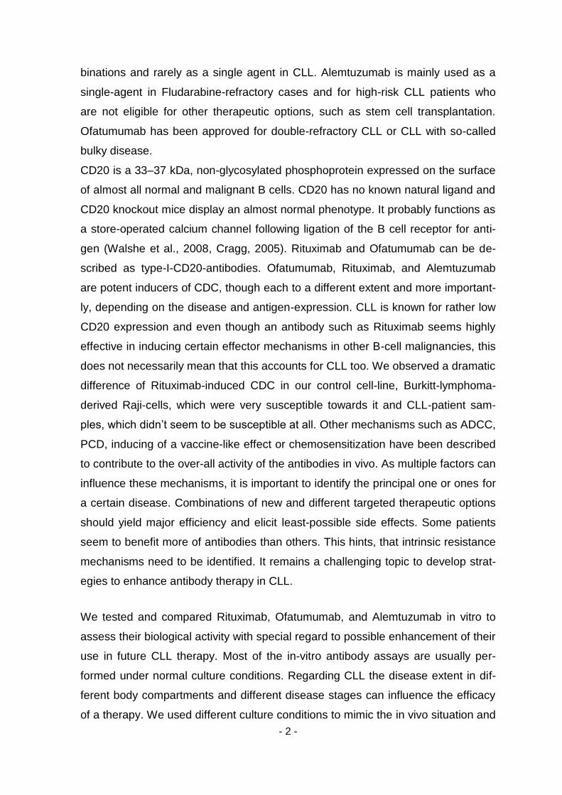
tumumab/Alemtuzumab mean viability high risk: 40.35%, range: 0.4809% -
87.97%, low risk: 30.00%, range: 2.299% - 58.64%, p-value = 0.5783; Rituxi-
mab/Alemtuzumab mean viability high risk: 41.83%, range: 0.6237% - 84.68%, low
risk: 32.91%, range: 9.9% - 62.21%, p-value = 0.62).
combination with Alemtuzumab, low risk, n=5
Ofa
tum
umab
2,5
µg/m
l
Ofa
tum
umab
2,5
µg/m
l + A
lem
tuzu
mab
1µg
/ml
Ritu
xim
ab 1
0µg/m
l
Ritu
xim
ab 1
0µg/m
l + A
lem
tuzu
mab
1µg
/ml
isoty
pe-an
tibody
Ale
mtu
zum
ab 1
µg/m
l
0
20
40
60
80
100
120
drug (/-combination)
% v
iab
ilit
y n
orm
alized
to t
rip
licate
co
ntr
ol
Figure 15:
viability of CLL (chronic lymphocytic leukemia) cells after a 48 hour incubation with Ofatumumab
(1st bar), Ofatumumab and Alemtuzumab (2
nd bar), Rituximab (3
rd bar), Rituximab and
Alemtuzumab (4th bar), isotype antibody (5
th bar) and Alemtuzumab (6
th bar) in a cohort of five low-
risk CLL cases.
- 31 -
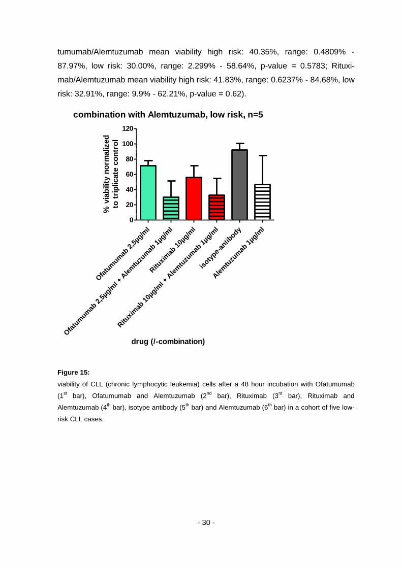
3.7 Assay No. 7: Flow cytometry based cytotoxicity assay
Annexin/7AAD-assay (3h), n=10
Ofa
tum
umab
Ale
mtu
zum
ab
Ritu
xim
ab
isoty
pe-an
tibody
0
20
40
60
80
100
monoclonal antibody 10 µg/ml
% A
nn
exin
-V/
7-A
AD
po
sit
ive c
ells
Figure 16:
percentage of “Apoptosis-marker-stained” CLL (chronic lympocytic leukemia) cells in relation to
untreated CLL cells after a short incubation time with either therapeutic or isotype-antibody. Spon-
taneous background apoptosis has already been subtracted.
Assay No.7: Early (Annexin-V-PE) and late (7-AAD) Apoptosis markers were used
for the identification of antibody induced killing mechanism. After a 3 hour lasting
incubation at a cell density of 10000 cells/µl and in the presence of 30% human
serum, both Ofatumumab (mean of Annexin-V-PE/7-AAD-positive cells: 24.37%,
range: 5.38% – 69.49%, n=11) and Alemtuzumab (mean of Annexin-V-PE/7-AAD-
positive cells: 57.97%, range: 15.36% - 83.46%, n=10) display rapid cell death in-
duction, while Rituximab (mean of Annexin-V-PE/7-AAD-positive cells: 2.31,
range: -1.46% - 10.03%, n=11) doesn’t. This is in accordance with the results in
the assays described above. However, cell death elicited by Ofatumumab and
Alemtuzumab was less when compared with the viability assays no.1 to 3, which
were performed at a concentration of 1000cells/µl. Another possibility for slightly
reduced activity compared to the viability assays could be cell lysis, which wouldn’t
be detected by this readout.

- 32 -
3.8 Assay No. 8: Flow cytometry based cytotoxicity assay after coincuba-tion of cells derived of the fibroblastoid bone marrow derived cell line HS5 with CLL cells
Monoclonal antibody
Figure 17:
percentage of apoptotic CLL (chronic lymphocytic leukemia) cells after 48 hours of incubation. The
left bar represents the coincubated CLL cells, the right bar represents the CLL cells, which were
not coincubated. Cells were treated with either therapeutic antibodies (first four columns:
Alemtuzumab, next four columns Ofatumumab, next four columns Rituximab) at different antibody
concentrations (1st concentration 1µg/ml, 2
nd concentration 10µg/ml) or isotype-antibody (last 2
columns). Spontaneous background apoptosis has already been subtracted. An additional staining
(CD45) was used to differ between HS5 (fibroblastoid bone marrow stroma) cells and CLL cells.
Assay No.8: Comparing antibody-mediated cell death in the presence or absence
of fibroblastoid bone marrow stroma cell line HS5 (CLL coculture model) reveals a
significant reduction of induced cell death by the antibody Alemtuzumab (Mean
- 33 -
apoptotic cells in the absence of HS5-cells: 71.33%, range: 46.7% - 83.46%, mean
apoptotic cells in the presence of HS5-cells: 37.71%, range: 26.35% - 47.71%, p-
value: 0.0022), and only a slight reduction of cytotoxicity of Ofatumumab (Mean
apoptotic cells in the absence of HS5-cells: 28.66%, range: 2.26% - 69.49%, mean
apoptotic cells in the presence of HS5-cells: 18.10%, range: -0.8% - 67.07%, p-
value: 0.5282).
As bone-marrow stroma cells seem to provide protection of CLL-cells against anti-
body-induced apoptotic-like cell death, further research to determine the underly-
ing mechanisms of protection are required to enhance antibody activity (Kurtova et
al., 2009). It is known, that SYK-inhibition can reduce resistance to chemotherapy
of CLL-cells, which are co-incubated with nurse-like cells (Buchner et al., 2010).
Apart from that, bone marrow derived fibroblastoid cells induce expression of PI3-
Kinase, AKT, NF-KB-pathway genes and a pro-angiogenic phenotype (Edelmann
et al., 2008). Drugs targeting the SYK, IP3, AKT, NF-kB pathway or drugs as Le-
nalidomide may act synergistic to antibodies by possibly decreasing the protective
effect of cells like HS5-cells.

- 34 -
3.9 Assay No. 9: Whole blood assay, microscopic read-out to assess ho-motypic adhesion
semiquantitative assessmentof homotypic adhesion
Ofa
tum
umab
Ritu
xim
ab
Ale
mtu
zum
ab
untrea
ted c
ontrol
0.0
0.5
1.0
1.5
2.0
2.5
3.0
antibody [10µg/ml]
sem
iqu
an
tita
tive s
cale
[0..
.3]
Figure 18:
extent of homotypic adhesion of CLL (chronic lymphocytic leukemia) cells caused by the different
antibodies (1st bar: Ofatumumab, 2
nd bar: Rituximab, 3
rd bar: Alemtuzumab, each at 10µg/ml) and a
negative (untreated) control after 1 hour incubation at room temperature.
Assay No.9: Inspection of five randomly picked high power fields after 1 hour
whole-blood-incubation at room-temperature with therapeutic antibody (10µg/ml)
or RPMI medium as negative (untreated) control displayed normal CLL blood
smear appearance. No effector cells could be detected in close proximity to CLL
cells. The Type-I-CD20 mAb-property (absent induction of homotypic adhesion) of
both Ofatumumab and Rituximab could be confirmed in 10 out of 10 tested patient
samples. (figure 18)

- 35 -
3.10 Assay No. 10: Whole blood assay, cytometric read-out, assessment of the impact of CLL-typical blood cell count on antibody-induced anti-CLL effect
whole blood assay, medium leukocyte count:Citrate 67,7 G/L; Heparine 115,6 G/L; Lepirudine 68,8 G/L
Ofa
tum
umab
Ritu
xim
ab
Ale
mtu
zum
ab
0
25
50
75
100
125
150
Citrate [n=13]
Heparine [n=6]
Lepirudine [n=3]
antibody [10µg/ml]
rem
ain
ing
cells n
orm
alized
to u
ntr
eate
d c
on
tro
l aft
er
3h
Figure 19:
relative numbers of leukocytes after a 3 hour incubation with either Ofatumumab, (first three bars),
Rituximab (next three bars), or Alemtuzumab (last three bars) using different anticoagulants (left
bar: Citrate, middle bar Heparine, right bar: Lepirudine) at a constant antibody concentration of 10
µg/ml.
Assay No. 10: This assay had the goal to assess the impact of high cell count on
effector consumption under in vivo conditions as well as to assess other possible
antibody induced mechanisms which were neglected in the above assays such as
ADCC. We chose the BD Biosciences True Count cytometric method to exclude
the possibility of undetected cell lysis and stained CLL cells with CD19-FITC.
Three different anticoagulation methods had no impact on the results. Results
were assembled according to used anticoagulation: Heparine [n=6], Lepirudine
[n=3], and Citrate [n=13]. Almost no antibody activity was detectable for all anti-
bodies at high cell concentrations. (figure 19)
- 36 -
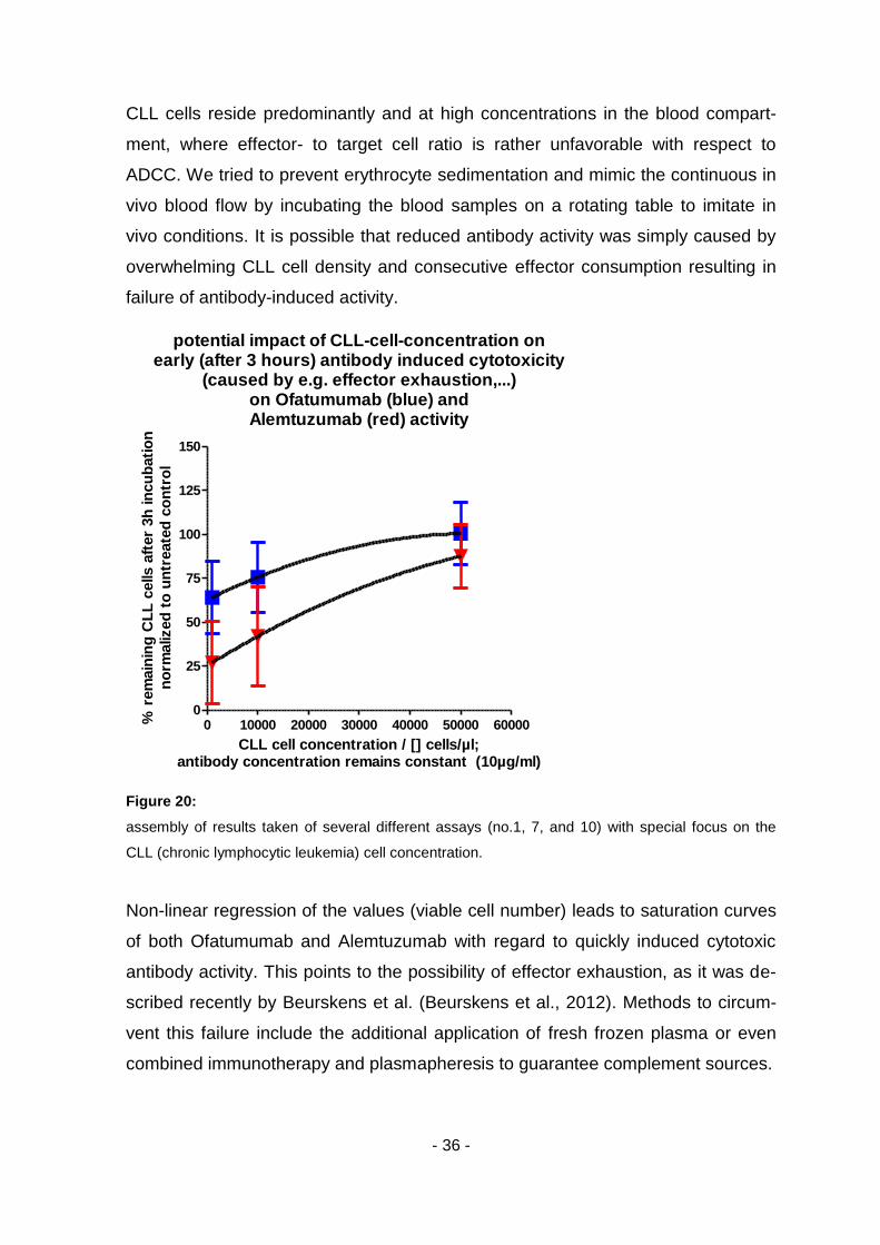
CLL cells reside predominantly and at high concentrations in the blood compart-
ment, where effector- to target cell ratio is rather unfavorable with respect to
ADCC. We tried to prevent erythrocyte sedimentation and mimic the continuous in
vivo blood flow by incubating the blood samples on a rotating table to imitate in
vivo conditions. It is possible that reduced antibody activity was simply caused by
overwhelming CLL cell density and consecutive effector consumption resulting in
failure of antibody-induced activity.
potential impact of CLL-cell-concentration onearly (after 3 hours) antibody induced cytotoxicity
(caused by e.g. effector exhaustion,...)on Ofatumumab (blue) andAlemtuzumab (red) activity
0 10000 20000 30000 40000 50000 600000
25
50
75
100
125
150
CLL cell concentration / [] cells/µl;antibody concentration remains constant (10µg/ml)
% r
em
ain
ing
CL
L c
ells a
fter
3h
in
cu
bati
on
no
rmalized
to
un
treate
d c
on
tro
l
Figure 20:
assembly of results taken of several different assays (no.1, 7, and 10) with special focus on the
CLL (chronic lymphocytic leukemia) cell concentration.
Non-linear regression of the values (viable cell number) leads to saturation curves
of both Ofatumumab and Alemtuzumab with regard to quickly induced cytotoxic
antibody activity. This points to the possibility of effector exhaustion, as it was de-
scribed recently by Beurskens et al. (Beurskens et al., 2012). Methods to circum-
vent this failure include the additional application of fresh frozen plasma or even
combined immunotherapy and plasmapheresis to guarantee complement sources.
- 37 -
4 Discussion
Ofatumumab seems an active CD-20-antibody, which could enhance CLL-therapy
in various ways. Ofatumumab seems effective in its immediate depleting activity in
low- and high-risk CLL and induces complement dependent cell death. Comparing
the single-agent activity of the three monoclonal antibodies at a concentration of
10µg/ml after 48 hours of incubation, Alemtuzumab has a higher activity than Ofa-
tumumab, while Ofatumumab has a higher activity than Rituximab. However, it
should be noted, that much higher doses of CD20-antibodies can be administered
in vivo compared to Alemtuzumab. Alemtuzumab has a wide spectrum of severe
side effects (Lundin et al., 2002). Comparing the viability after 3 and 48 hours of
antibody-incubation, we detected a difference especially in Rituximab-treated CLL-
cells, but only a minor difference in the Ofatumumab-treated CLL-cells: Rituximab
showed almost no effect after 3 hours and considerably more activity after 48
hours, suggesting a different mode of action. Ofatumumab-treated CLL cells were
depleted mostly after 3 hours by complement-dependent cytotoxicity and a small
additional depleted fraction could be detected after 48 hours of incubation. The
activity of Alemtuzumab remained constant comparing 3 and 48 hour results. (fig-
ure 1E) We were especially interested in the activity of all antibodies in high-risk
CLL and Rituximab-pretreated CLL. We defined high-risk CLL as the presence of
at least one of the following: 17p-deletion detected by FISH -, TP53-mutation de-
tected by wave and confirmed by direct sequencing, or Fludarabine-refractory dis-
ease (Stilgenbauer and Zenz, 2010) . No difference in their activity was observed
for all three antibodies if results were divided into subgroups neither according to
genetic risk-stratification nor Rituximab pretreatment. All antibodies seem to act
independently of genetic aberrations in CLL.
Ofatumumab seems to possess at least equal potential and intrinsic activity for
immunochemotherapeutic combination as Rituximab (antibody + Fludarabine).
This option seems suitable especially for low-risk patients.
In order to assess potential chemo-sensitizing activity and potential use in immu-
nochemotherapeutic combinations, we used a lower Ofatumumab-concentration to
prevent rapid cell depletion. Regarding Fludarabine combinations, comparable
depletion was induced by the two CD20-antibodies. The combined activity of the
CD20-antibodies and Fludarabine could only be observed in low-risk CLL. As ex-

- 38 -
pected, high-risk CLL did not respond to Fludarabine, which is in accordance with
previous findings (Zenz et al., 2008) .If Rituximab can sensitize lymphoid cells to
Fludarabine treatment by down-regulating BCL-2, the same mechanism could be
induced by Ofatumumab.
Ofatumumab can be effectively combined with Alemtuzumab or the AKT-inhibitor
GSK 690693 and possibly other/similar-acting small-molecule inhibitors, which
target the same pro-apoptotic pathway. This seems to be an option rather for high-
risk patients.
Both Ofatumumab and Alemtuzumab seem to elicit their activity in a similarly fast
and complement-dependent way. The results showed a greater CLL cell depletion
not only for the combination of Alemtuzumab and Ofatumumab, but as well for the
combination of Alemtuzumab and Rituximab. However, as the time course of cell
death induction is rather different between the latter ones (Alemtuzumab and
Rituximab) it is debatable if a synergistic effect at least on an immunologic basis
(CDC) takes place in vivo. Apart from that it needs to be noted that not only for the
Rituximab but as well for the Ofatumumab combination, our in vitro models cannot
reflect the in vivo situation. It is possible that complement components, especially
in compartments, where CLL cells are proliferating and residing in high concentra-
tion, become rapidly consumed and this consumption could be even accelerated if
antibodies are used in combination. However, the solution of this possible problem
could simply be the substitution of complement components by intravenous appli-
cation of fresh frozen plasma as described previously (Klepfish et al., 2009).
Type-I-antibodies (such as Rituximab) can inhibit the constitutively activated
PI3Kinase/AKT pathway (Baritaki et al., 2011, Suzuki et al., 2007, Bonavida,
2007).We used a 4-fold lower antibody-concentration of Ofatumumab compared to
Rituximab for the combination not only with Fludarabine or Alemtuzumab (see
above), but also for the combination with the AKT-inhibitor GSK 690693. Interest-
ingly we could detect a slightly, though statistically not significant, higher activity of
this combination in high-risk CLL compared to the activity in low-risk CLL. Taken
together Ofatumumab seems a more potent antibody than Rituximab. There seem
to be several combination options for Ofatumumab and especially high-risk pa-
tients might benefit of those. This is in accordance with recently published findings
(Bologna et al., 2013).
- 39 -
Ofatumumab elicits its activity through so-far not fully understood mechanisms. In
contrast to Rituximab, Ofatumumab is able to induce a quick cell death induction in
CLL, which is complement-dependent. However, the observed saturation of its fast
activity points to complement consumption (Boross et al., 2011). As the ATP-
dependent luciferase-based assay, which we used in the assays no. 1-6 is re-
stricted to low cell concentrations (maximum: 50000 cells/well), we used a cy-
tometric read-out for assays with higher cell concentrations (assay no. 7 – 10). We
used double-staining with 7-AAD (late-apoptosis marker) and AnnexinV-PE (early
apoptosis marker) to differentiate between apoptotic and viable cells. The cells
were incubated at a concentration of 10000cells/µl in this assay, which reflects the
upper boarder of physiological leukocyte counts in healthy humans. Ofatumumab-
induced CDC was decreased by approximately one third compared to the assays
performed at a concentration of 1000cells/µl. It should be noted that a high fre-
quency of complement deficiency in CLL has been described (Klepfish et al.,
2009). Taken together with our results, this points again to the potential benefit of
the substitution of complement components.
Ofatumumab seems to possess higher activitiy in assays with low leukocyte con-
centrations. This observation is in accordance with recently published results of
Beurskens et al.(Beurskens et al., 2012) and could be due to the dependency of
Ofatumumab on effector mechanisms which are not regenerating, refreshing, that
is to say consumed during in vitro incubation or due to unknown reasons. Internal-
ization and shaving of CD20/antibody complexes lead to CD20-loss on the surface
of CLL cells. It has been recently published, that shaving/trogocytosis happens
even faster than internalization and could constitute a major obstacle of antibody-
mediated therapeutic effects (Beum et al., 2011, Pedersen et al., 2011). In theory
shaving should happen less if the antibody molecules are bound by complement,
which would point again to the combined application of ofatumumab and fresh fro-
zen plasma.
To better mimic the in vivo microenvironment of CLL cells and create survival-
inducing culture-conditions, we co-incubated them with HS5 cells (Schulz et al.,
2011). Under these circumstances, we could detect a significantly reduced activity
of Alemtuzumab and a slight reduction of Ofatumumab-induced activity.

- 40 -
A new treatment strategy could be to combine Alemtuzumab and Ofatumumab
with fresh frozen plasma in order to tackle high-risk CLL with an extended thera-
peutic arsenal. This has been recently proposed by Baig et al. (Baig et al., 2012).
The results of the currently recruiting study which investigates the combination of
Alemtuzumab, Ofatumumab, and high-dose glucocorticoids will hopefully provide
an insight whether or not at least the combination of the two antibodies might be
an effective new therapeutic option (Jennifer R. Brown, 2011 - present).
Overlapping effector mechanisms elicited by a single antibody have been de-
scribed previously (Zent et al., 2008, Boross et al., 2011). We varied the culture
conditions to assess relative contribution of different mechanisms of action. Our
whole blood assay was designed to mimic the in vivo situation in the blood com-
partment. A significant contribution to antibody activity by effector cells couldn’t be
detected. This, however, might not reflect the in vivo situation where blood CLL
cells are often in close contact to cells of the reticulo-endothelial system: Effector
cells, which often remain rather stationary in liver, spleen, lymphatic nodes and so
on, and which can influence antibody activity (Gong et al., 2005) via ADCC, ADCP
(phagocytosis), or reduce antigen expression via shaving are probably much high-
er concentrated and active under in vivo circumstances than in our obtained blood
samples (Beum et al., 2006). Development of SIRS-like immune cascades cannot
be entirely mimicked on incubation plates. In-vitro assays can’t detect the long-
lasting antitumor protection by anti-CD20 antibody through cellular immune re-
sponse as described by Abès et al. (Abes et al., 2010). Finally, blood samples with
very high leukocyte counts were used for the whole blood assays. Again, effector
consumption (this time, consumption of effector cells) could be a reason for low
antibody activity. Taken together, we couldn’t detect a significant contribution to
antibody activity of effector cells in whole blood assays. It remains an enigma
whether Rituximab uses effector cells and ADCC to elicit its activity.

- 41 -
5 Summary
Introduction:
The application of monoclonal antibodies has become standard of care in the
treatment of Chronic lymphocytic leukemia (CLL). We compared the single-agent-
activity of three currently approved therapeutic antibodies for CLL, namely Rituxi-
mab, Alemtuzumab, and Ofatumumab.
Question:
Our goal was to identify underlying mechanisms of action of monoclonal antibod-
ies in CLL in order to recognize potential limitations of their use and to develop
strategies to overcome these limitations or at least to use the antibodies in the
most beneficial way. We had our major focus on the impact of genetic subgroups
of CLL and on Ofatumumab, a relatively new CD20-antibody in comparison with
Rituximab and Alemtuzumab.
Method:
Patient samples were analysed by flow cytometry, fluorescence in-situ hybrid-
iziation, liquid chromatography, and classic sequencing. Antibody-treatment was
performed in both cell culture and whole blood assays. We assessed CLL-cell-
viability, homotypic adhesion, potential synergism with other substances, and the
impact of the cellular micro-environment by co-culturing CLL cells with fibroblas-
toid stroma cells. In parallel, CLL-cell-concentration, and effector consumption
were assessed. Read out methods comprised microscopy, multi-color flow cytom-
etry and luminometry,
Results:
Rituximab, Alemtuzumab, and Ofatumumab showed varying activity and different
mechanisms of action. Regarding each antibody individually, no considerable dif-
ference could be detected among different risk-stratified CLL subgroups. In a sub-
group of patients, we mimicked in vivo micro-environment by coincubation of CLL
cells with HS5-cells and discovered a decrease of Alemtuzumab-activity but not
Ofatumumab-activity. Rituximab is most beneficial if used in combination with oth-
er agents such as Fludarabine, Cyclophospamide (FCR) or Bendamustine (BR).
We hypothesized a potential synergistic activity of Ofatumumab: submaximal con-
centrations of Ofatumumab combined with each Fludarabine, Alemtuzumab, and

- 42 -
the nanomolecular AKT-inhibitor GSK 690693, respectively, lead to promising re-
sults.
Summary:
Ofatumumab seems highly suitable for various combinations and high-risk CLL
patients might benefit of those. Based on our in vitro findings, we suggest several
strategies to enhance in vivo Ofatumumab activity.

- 43 -
6 References
1. ABES, R., GELIZE, E., FRIDMAN, W. H. & TEILLAUD, J. L. Long-lasting antitumor protection by anti-CD20 antibody through cellular immune re-sponse. Blood, Volume 116 Issue 6, p. 926-934. (2010)
2. BAIG, N. A., TAYLOR, R. P., LINDORFER, M. A., CHURCH, A. K., LAPLANT, B. R., PAVEY, E. S., NOWAKOWSKI, G. S. & ZENT, C. S. Complement dependent cytotoxicity in chronic lymphocytic leukemia: ofa-tumumab enhances alemtuzumab complement dependent cytotoxicity and reveals cells resistant to activated complement. Leuk Lymphoma, Volume 53 Issue 11, p. 2218-2227. (2012)
3. BARITAKI, S., MILITELLO, L., MALAPONTE, G., SPANDIDOS, D. A., SALCEDO, M. & BONAVIDA, B. The anti-CD20 mAb LFB-R603 interrupts the dysregulated NF-kappaB/Snail/RKIP/PTEN resistance loop in B-NHL cells: role in sensitization to TRAIL apoptosis. Int J Oncol, Volume 38, Issue 6, p. 1683-1694. (2011)
4. BEUM, P. V., KENNEDY, A. D., WILLIAMS, M. E., LINDORFER, M. A. & TAYLOR, R. P. The shaving reaction: rituximab/CD20 complexes are re-moved from mantle cell lymphoma and chronic lymphocytic leukemia cells by THP-1 monocytes. J Immunol, Volume 176, Issue 4, p. 2600-2609. (2006)
5. BEUM, P. V., PEEK, E. M., LINDORFER, M. A., BEURSKENS, F. J., EN-GELBERTS, P. J., PARREN, P. W., VAN DE WINKEL, J. G. & TAYLOR, R. P. Loss of CD20 and bound CD20 antibody from opsonized B cells occurs more rapidly because of trogocytosis mediated by Fc receptor-expressing effector cells than direct internalization by the B cells. J Immunol, Volume 187, Issue 6, p. 3438-3447. (2011)
6. BEURSKENS, F. J., LINDORFER, M. A., FAROOQUI, M., BEUM, P. V., ENGELBERTS, P., MACKUS, W. J., PARREN, P. W., WIESTNER, A. & TAYLOR, R. P. Exhaustion of cytotoxic effector systems may limit mono-clonal antibody-based immunotherapy in cancer patients. J Immunol, Vol-ume 188, Issue 7, p. 3532-3541. (2012)
7. BOLOGNA, L., GOTTI, E., DA ROIT, F., INTERMESOLI, T., RAMBALDI, A., INTRONA, M. & GOLAY, J. Ofatumumab is more efficient than rituximab in lysing B chronic lymphocytic leukemia cells in whole blood and in combi-nation with chemotherapy. J Immunol, Volume 190, Issue 1, p. 231-239. (2013)
8. BONAVIDA, B. Rituximab-induced inhibition of antiapoptotic cell survival pathways: implications in chemo/immunoresistance, rituximab unrespon-siveness, prognostic and novel therapeutic interventions. Oncogene, Vol-ume 26, Issue 25, p. 3629-3636. (2007)
9. BOROSS, P., JANSEN, J. H., DE HAIJ, S., BEURSKENS, F. J., VAN DER POEL, C. E., BEVAART, L., NEDEREND, M., GOLAY, J., VAN DE WIN-KEL, J. G., PARREN, P. W. & LEUSEN, J. H. The in vivo mechanism of ac-tion of CD20 monoclonal antibodies depends on local tumor burden. Hae-matologica, Volume 96, Issue 12, p. 1822-1830. (2011)
10. BUCHNER, M., BAER, C., PRINZ, G., DIERKS, C., BURGER, M., ZENZ, T., STILGENBAUER, S., JUMAA, H., VEELKEN, H. & ZIRLIK, K. Spleen ty-rosine kinase inhibition prevents chemokine- and integrin-mediated stromal

- 44 -
protective effects in chronic lymphocytic leukemia. Blood, Volume 115, Is-sue 22, p. 4497-4506. (2010)
11. CRAGG, M. S., WALSHE, C. A., IVANOV, A. O., GLENNIE, M. J. The Biol-ogy of CD20 and Its Potential as a Target for mAb Therapy. Curr Dir Auto-immun., Volume 8, p. 140–174. (2005)
12. EDELMANN, J., KLEIN-HITPASS, L., CARPINTEIRO, A., FÜHRER, A., SELLMANN, L., STILGENBAUER, S., DÜHRSEN, U. & DÜRIG, J. Bone marrow fibroblasts induce expression of PI3K/NF-κB pathway genes and a pro-angiogenic phenotype in CLL cells. Leukemia research, Volume 32, Is-sue 10, p. 1565-1572. (2008)
13. EICHHORST, B., GOEDE, V., HALLEK, M. Treatment of elderly patients with chronic lymphocytic leukemia. Leuk Lymphoma, Volume 50, Issue 2, p. 171-178 (2009)
14. FISCHER, K., CRAMER, P., BUSCH, R., STILGENBAUER, S., BAHLO, J., SCHWEIGHOFER, C. D., BOTTCHER, S., STAIB, P., KIEHL, M., ECKART, M. J., KRANZ, G., GOEDE, V., ELTER, T., BUHLER, A., WINKLER, D., KNEBA, M., DOHNER, H., EICHHORST, B. F., HALLEK, M. & WENDT-NER, C. M. Bendamustine combined with rituximab in patients with re-lapsed and/or refractory chronic lymphocytic leukemia: a multicenter phase II trial of the German Chronic Lymphocytic Leukemia Study Group. J Clin Oncol, Volume 29, Issue 26, p. 3559-3566. (2011)
15. GONG, Q., OU, Q., YE, S., LEE, W., CORNELIUS, J., DIEHL,L., LIN, W., HU, Z., LU, Y., CHEN, Y., WU, Y., MENG, Y., GRIBLING, P., LIN, Z., NGUYEN, K., TRAN, T., ZHANG, Y., ROSEN, H., MARTIN, F., CHAN, A. Importance of cellular microenvironment and circulatory dynamics in B cell immunotherapy. J Immunol, Volume 174, Issue 2, p. 817-826. (2005)
16. HALLEK, M., CHESON, B. D., CATOVSKY, D., CALIGARIS-CAPPIO, F., DIGHIERO, G., DOHNER, H., HILLMEN, P., KEATING, M. J., MONTSER-RAT, E., RAI, K. R. & KIPPS, T. J. Guidelines for the diagnosis and treat-ment of chronic lymphocytic leukemia: a report from the International Work-shop on Chronic Lymphocytic Leukemia updating the National Cancer Insti-tute-Working Group 1996 guidelines. Blood, Volume 111, Issue 12, p. 5446-5456. (2008)
17. HALLEK M, FINGERLE-ROWSON G, FINK AM, BUSCH R, MAYER J, HENSEL M, HOPFINGER G, HESS G, VON GRUENHAGEN U, BERG-MANN M, CATALANO J, ZINZANO P, CAPPIO F, SEYMOUR J, BERREBI A, JAEGER U, CAZIN B, TRNENY M, WESTERMANN A, WENDTNER C, EICHHORST B, STAIB P, BOETTCHER S, RITGEN M, MENDILA M, KNEBA M, DOEHNER H, STILGENBAUER S, FISCHER K. 2009. First-line treatment with fludarabine (F), cyclophosphamide (C), and rituximab (R) (FCR) improves overall survival (OS) in previously untreated patients (pts) with advanced chronic lymphocyticleukemia (CLL): Results of a randomized phase III trial on behalf of an international group of investigators and the German CLL Study Group. ASH congress: Abstract 535, https://ash.confex.com/ash/2009/webprogram/Paper16896.html, Download 12/2012
18. JENNIFER R. BROWN, M., PHD http://clinicaltrials.gov/ct2/show/NCT01465334. Dana-Farber Cancer Insti-tute. 2011 - present, Download 12/2012

- 45 -
19. KLEPFISH, A., GILLES, L., IOANNIS, K., RACHMILEWITZ, E. A. & SCHATTNER, A. Enhancing the action of rituximab in chronic lymphocytic leukemia by adding fresh frozen plasma: complement/rituximab interactions & clinical results in refractory CLL. Ann N Y Acad Sci, Volume 1173, p. 865-873. (2009)
20. KURTOVA, A. V., BALAKRISHNAN, K., CHEN, R., DING, W., SCHNABL, S., QUIROGA, M. P., SIVINA, M., WIERDA, W. G., ESTROV, Z., KEATING, M. J., SHEHATA, M., JAGER, U., GANDHI, V., KAY, N. E., PLUNKETT, W. & BURGER, J. A. Diverse marrow stromal cells protect CLL cells from spontaneous and drug-induced apoptosis: development of a reliable and reproducible system to assess stromal cell adhesion-mediated drug re-sistance. Blood, Volume 114, Issue 20, p. 4441-4450. (2009)
21. LUNDIN, J., KIMBY, E., BJORKHOLM, M., BROLIDEN, P. A., CELSING, F., HJALMAR, V., MOLLGARD, L., REBELLO, P., HALE, G., WALDMANN, H., MELLSTEDT, H. & OSTERBORG, A. Phase II trial of subcutaneous an-ti-CD52 monoclonal antibody alemtuzumab (Campath-1H) as first-line treatment for patients with B-cell chronic lymphocytic leukemia (B-CLL). Blood, Volume 100, Issue 3, p. 768-773. (2002)
22. MORRISON, V. A., RAI, K. R., PETERSON, B. L., KOLITZ, J. E., ELIAS, L., APPELBAUM, F. R., HINES, J. D., SHEPHERD, L., LARSON, R. A. & SCHIFFER, C. A. Therapy-related myeloid leukemias are observed in pa-tients with chronic lymphocytic leukemia after treatment with fludarabine and chlorambucil: results of an intergroup study, cancer and leukemia group B 9011. J Clin Oncol, Volume 20, Issue 18, p. 3878-3884. (2002)
23. PEDERSEN, A. E., JUNGERSEN, M. B. & PEDERSEN, C. D. Monocytes mediate shaving of B-cell-bound anti-CD20 antibodies. Immunology, Vol-ume 133, Issue 2, p. 239-245. (2011)
24. SCHULZ, A., TOEDT, G., ZENZ, T., STILGENBAUER, S., LICHTER, P. & SEIFFERT, M. Inflammatory cytokines and signaling pathways are associ-ated with survival of primary chronic lymphocytic leukemia cells in vitro: a dominant role of CCL2. Haematologica, Volume 96, Issue 3, p. 408-416. (2011)
25. STILGENBAUER, S. & ZENZ, T. Understanding and managing ultra high-risk chronic lymphocytic leukemia. Hematology Am Soc Hematol Educ Pro-gram, Volume 2010, p. 481-488. (2010)
26. SUZUKI, E., UMEZAWA, K. & BONAVIDA, B. Rituximab inhibits the consti-tutively activated PI3K-Akt pathway in B-NHL cell lines: involvement in chemosensitization to drug-induced apoptosis. Oncogene, Volume 26, Is-sue 42, p. 6184-6193. (2007)
27. WALSHE, C. A., BEERS, S. A., FRENCH, R. R., CHAN, C. H., JOHNSON, P. W., PACKHAM, G. K., GLENNIE, M. J. & CRAGG, M. S. Induction of cy-tosolic calcium flux by CD20 is dependent upon B Cell antigen receptor sig-naling. J Biol Chem, Volume 283, Issue 25, p. 16971-16984. (2008)
28. ZENT, C. S., SECRETO, C. R., LAPLANT, B. R., BONE, N. D., CALL, T. G., SHANAFELT, T. D., JELINEK, D. F., TSCHUMPER, R. C. & KAY, N. E. Direct and complement dependent cytotoxicity in CLL cells from patients with high-risk early-intermediate stage chronic lymphocytic leukemia (CLL) treated with alemtuzumab and rituximab. Leuk Res, Volume 32, Issue 12, p. 1849-1856. (2008)

- 46 -
29. ZENZ, T., BENNER, A., DOHNER, H. & STILGENBAUER, S. Chronic lym-phocytic leukemia and treatment resistance in cancer: the role of the p53 pathway. Cell Cycle, Volume 7, Issue 24, p. 3810-3814. (2008)
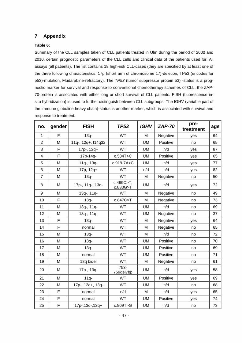
- 47 -
7 Appendix
Table 6:
Summary of the CLL samples taken of CLL patients treated in Ulm during the period of 2000 and
2010, certain prognostic parameters of the CLL cells and clinical data of the patients used for: All
assays (all patients). The list contains 18 high-risk CLL-cases (they are specified by at least one of
the three following characteristics: 17p (short arm of chromosome 17)-deletion, TP53 (encodes for
p53)-mutation, Fludarabine-refractory). The TP53 (tumor suppressor protein 53) -status is a prog-
nostic marker for survival and response to conventional chemotherapy schemes of CLL, the ZAP-
70-protein is associated with either long or short survival of CLL patients. FISH (fluorescence in-
situ hybridization) is used to further distinguish between CLL subgroups. The IGHV (variable part of
the immune globuline heavy chain)-status is another marker, which is associated with survival and
response to treatment.
no. gender FISH TP53 IGHV ZAP-70 pre-
treatment age
1 F 13q- WT M Negative yes 64
2 M 11q-, 12q+, t14q32 WT UM Positive no 65
3 F 17p-, 12q+ WT UM n/d yes 87
4 F 17p-14q- c.584T>C UM Positive yes 65
5 M 11q-, 13q- c.919-7A>C UM n/d yes 77
6 M 17p, 12q+ WT n/d n/d yes 82
7 M 13q- WT M Negative no 50
8 M 17p-, 11q-, 13q- c.499C>T, c.830G>T
UM n/d yes 72
9 M 13q-, 11q- WT M Negative no 49
10 F 13q- c.847C>T M Negative no 73
11 M 13q-, 11q- WT UM n/d no 69
12 M 13q-, 11q- WT UM Negative no 37
13 F 13q- WT M Negative yes 64
14 F normal WT M Negative no 65
15 M 13q- WT M n/d no 72
16 M 13q- WT UM Positive no 70
17 M 13q- WT UM Positive no 69
18 M normal WT UM Positive no 71
19 M 13q bidel WT M Negative no 61
20 M 17p-, 13q- 753-
759del7bp UM n/d yes 58
21 M 11q- WT UM Positive yes 69
22 M 17p-, 12q+, 13q- WT UM n/d no 68
23 F normal n/d M n/d yes 65
24 F normal WT UM Positive yes 74
25 F 17p-,13q-,12q+ c.809T>G UM n/d no 73

- 48 -
26 M 17p-, 11q-, 13q- c.329G>T UM Negative no 69
27 F normal WT UM Positive no 38
28 M 12q+, t(14:18) WT M Negative no 81
29 F 17p c.842A>G UM Positive yes 42
30 M 17p c.602-
603Ins4bp UM Negative no 69
31 M 17p WT UM n/d no 73
32 F Normal c.484A>T UM Positive yes 64
33 M 13q WT UM n/d no 66
34 M 13q WT UM Negative yes 77
35 M Norm WT UM Negative yes 72
36 F Tris.12, 17p c.673-2A>T M Negative yes 73
37 M 17p, 13q c.413C>T M Negative yes 72
38 F 13q n/d M n/d no 59
39 F 17p, 13q C.733G>T UM Negative yes 76
40 M 17p, 13q, 6q n/d UM n/d yes 66
41 F Norm WT M n/d no 75
42 M normal WT M n/d no 39
43 M t14q32 WT UM Negative yes 72
44 M 13q- WT UM n/d yes 67
45 M 11q-, 13q-, 14q- WT UM Negative no 73
46 M 12p+11, t(14;19) WT UM Positive yes 49
47 M 13q- c:716A>6 M n/d yes 76
48 M 13q WT M n/d no 57
49 M 11q23 WT UM Negative yes 58

- 49 -
8 Curriculum Vitae
Name: Matthias Volden
Born in: Sindelfingen
Year: 1986
Education:
06/2012 Approbation als Arzt, german MD licence
05/2012 Staatsexamen 2. Teil, second (final) part of graduation (grade: 3)
2010 – 2011 one year internship, rotation through 3 different departments:
01 – 08/2011 second and third part at the University of Oslo, Norway:
Kirurgisk avdeling, SSHF Arendal (surgery)
Medisinsk avdeling, SSHF Arendal (internal medicine)
09 – 12/2010 first part at the department of pathology, University of Ulm, Germany
2007 – 2010 clinic studies, University of Ulm, Germany,
09/2007 Staatsexamen 1. Teil, first part of graduation (grade: 2.5)
2005 – 2007 preclinic studies, University of Ulm, Germany
1996 – 2005 school: Goldberg-Gymnasium Sindelfingen, Baden-Württemberg,
Germany (grade: 1.5)