of lplucmu is mediated by the a protein altered at its ... · gene, 71 (1988) 177-186 elsevier 177...
TRANSCRIPT
Gene, 71 (1988) 177-186
Elsevier
177
GEN 02680
Transposition of lplucMu is mediated by the A protein altered at its carboxy-terminal end
(Mu transposase; ZacZ fusions; bacteriophages E. and Mu; transducing phages; recombinant DNA)
Erhard BremeP, Thomas .J. Siihavy b and George M. Weinstock’
U Department of Biology, University of Konstanz, D-7750 Konstanz (F.R.G.); ’ Department of Molecular Biology. Princeton University, Princeton, NJ 08544 (U.S.A.) Tel. (609)452-5889, and ’ Department of Biochemistry and Molecular Biology, University qf Texas Medical School, Houston, TX 77225 (U.S.A.) Tel. (713)792-5600
Received 27 May 1988
Accepted 22 June 1988
Received by publisher 4 August 1988
SUMMARY
dplacMu phages are derivatives of bacteriophage J that use the transposition machinery of phage Mu to insert into chromosomal and cloned genes. When inserted in the proper fashion, these phages yield stable fusions to the Escherichia coli lac operon in a single step. We have determined the amount of DNA from the c end of phage Mu present in one of these phages, IplacMu3, and have shown that this phage carries a 3 i37-bp fragment of Mu DNA. This DNA segment carries the Mu c-end attachment site and encodes the Mu genes cts62, ner + , and gene A lacking 179 bp at its 3’ end (A ‘). The product of this t~ncated gene A’ retains transposase activity and is sufficient for the transposition of IpfacMu. This was demonstrated by showing that /IpZacMu derivatives carrying the A am 1093 mutation in the A ’ gene are unable to transpose by themselves in a Su - strain, but their transposition can be triggered by coinfection with ApMu507(A ‘B +). We have constructed several new AplacMu phages that carry the A’am1093 gene and the kan gene, which confers resistance to kanamycin. Chromosomal insertions of these new phages are even more stable than those of the previously reported dplacMu phages, which makes them useful tools for genetic analysis.
INTRODU~ION
Transposition is an integral part of the life cycle of the temperate bacteriophage Mu (for reviews see Toussaint and Resibois, 1983; Mizuuchi and
-..-- Correspondence ro; Dr. E. Bremer, Department of Biology, Uni-
versity of Konstanz, P.O. Box 5560, D-7750 Konstanz (F.R.G.)
Tel. (49)753 I-882042.
Abbreviations: A ‘, the 3’ truncated gene A of phage Mu; aa,
amino acid(s); bp, base pair(s); kb, 1000 bp; Km, kanamycin;
LB, Luria broth; pfu. pIaque-forming units; R, resistance; XGal,
5-bromo-4-chloro-indolyI-~-D-gaIactop~anoside; ::, novel joint.
Craigie, 1986). Both iytic and lysogenic pathways require the obligatory inte~ation of the Mu DNA into the host chromosome. This integration occurs by a highly efficient transposition process with little site-specificity. Mu transposition requires both the S and c attachment sites located, respectively, at the right and left ends of the phage genome, and it is mediated by the Mu-encoded A and B proteins. The A protein, which is regarded as a transposase, is essential for transposition, whereas the B protein significantly (approx. IO&fold) enhances transposi- tion efhciency but is not essential.
The ability of Mu to transpose into many genes
037%1119,@8!$03.50 0 1988 Elsevier Science Publishers B.V. (Biomedical Division)

178
(Taylor, 1963) and into many locations within a
singlegene (Bukhari and Zipser, 1972; Daniel1 et al.,
1972) has been exploited to create IRK fusions in vivo
(Casadaban, 1975; 1976; Casadaban and Cohen,
1979; Casadaban and Chou,1984; Castilho et al.,
1984). Such fusions have wide-ranging applications
in studies of gene expression and function (for
reviews see Weinstock et al., 1983; Silhavy and
Beckwith, 1985). We previously described the con-
struction of several /~-MU hybrid phages (~p~ff~Mu~
in which bacteriophage R is endowed with the trans-
position properties of phage Mu (Bremer et al.,
1984; 1985). These tlpIacMu phages are plaque-
forming derivatives of phage I that lack the phage
sequences (at@) necessary for the efficient site-
specific integration into the E. coli chromosome at
attB. They carry both of the Mu attachment sites (S
and c) and an appropriately constructed lac operon
that can form stable protein (AplacMu9) or operon
(~p~~cMu53) fusions in a single step when they are
inserted into a chromosomal or cloned target gene.
The structure of a ~p~acMu phage and the proposed
scheme of /ZplacMu transposition is shown in Fig. 1.
Like phage Mu, these phages create a 5-bp dupli-
cation at the point of insertion (Nag and Berg, 1987).
The /ZplacMu phages also permit isolation of /1 spe-
cialized transducing phages carrying various seg-
ments of the E. cob chromosome (Bremer et al.,
1984; 1985). Such transducing phages have various
applications in genetic analysis (Silhavy et al., 1984)
and facilitate cloning of the fat fusion as well as
intact genes adjacent to the ApiaeMu insertion
(Bremer et al., 1984; Trun and Siihavy, 1987). The
use of the Ap/acMu system was recently extended
from its natural host E. co/i to Salmonella
typhimurium (Harkki et al., 1987).
The Mu c region (including the Mu c-end attach-
ment site) present on the JplacMu phages was
derived from Rpl(209) by a series of in vivo
homologous recombination events (Bremer et al.,
1984; 1985; Casadaban, 1976). Its size was deter-
mined by electron microscopy to be about 2.8 kb
(Leathers et aI., 1979). Since 3316 bp are required to
encode the Mu c-end attachment site and a complete
A gene (Priess et al., 1987), a substantial segment of
the 3’ end of A should be missing from the Mu c
AR
(a> hplacMu 9 Inlrn J
i-7 km N
(b)
lac i AR
_c N
Fig. 1. Transposition of IPIQCMU. The proposed scheme of
transposition for the phage IpfucMu9, which forms IncZ protein
fusions, is shown. Upon infection into E. cd, the circularized
DNA of IplucMu9 (a) transposes into a target gene X (b) by
means ofthe Mu attachment sites Sand c to form a Lac + protein
fusion between gene 2’ and 1acZ (c). The bacterial DNA (wavy
line) present between the Mu ends (a) is lost during this process
and the Mu S and c attachment sites are joined to sequences
from gene X whose integrity is disrupted into X’ and ‘X. The
formation of a Lac ’ fusion requires the integration of the trans-
posable phage into the target gene in the proper orientation and
reading frame with respect to the fused gene. Expression of the
hybrid gene depends on the target gene’s transcriptional and
translational initiation signals (P) and leads to the formation of
a hybrid protein whose N terminus (N) is encoded by the target
gene. This N-terminal region is attached to a large C-terminal
segment (C) of’/&galactosidase. The thin line represents 2DNA.
The positions of several genes from phage ,?(.I. A, R, N) and its
immunity (imm) region are also indicated. The ‘IucZ and /WY”
genes are shown by the cross-hatched areas, and the Mu material
is symbolized by the blackened boxes. The km gene, which
confers Km resistance, is represented by the stippled box.
region. It was expected that a A ‘B + helper phage
supplying the A and B proteins in tram would be
required for the transposition of JptacMu phages;
however, these phages can transpose in the absence
of such a helper phage, although at a lOO-fold re-
duced level (Bremer et al., 1984; 1985). We show
here that the A gene present in JplacMu has a dele-
tion of 179 bp at its 3’ end (A ‘). However, the
encoded A’ protein still has transposase activity and
is responsible for the heIper-independent transposi-
tion of i,pf~cMu. In addition, we report the isolation
of several new transposition-defective J_plrrrMu
phages that yield chromosomal insertions with fur-
ther enhanced genetic stability.
MATERIALS AND METHODS
(a) Bacterial strains, bacteriophages and plasmids
The bacterial strains, phages and plasmids used
are listed in Table I.
(b) Media, chemicals and enzymes
LB, M63, MacConkey, and tetrazolium media
were prepared as previously described (Miller, 1972;
TABLE 1
Bacteria, bacteriophages and plasmids
179
Silhavy et al., 1984). Liquid minimal medium
containing M63 salts was supplemented with
glycerol at 0.4%. When indicated, 0.1 ml of a
10 mg/ml solution of XGal in dimethlyformamide
was spread on agar plates or added to L soft agar to
detect colonies and phage plaques showing a LacZ +
phenotype. Ampicillin, Km and tetracycline were
added to media at 125 pg/ml, 30 gg/ml and
25 Lig/ml, respectively. Deoxy- and dideoxyribo-
nucleoside triphosphates, PolIk, and sequencing
primer were from Bethesda Research Laboratories
~ -- Strain Description 3 __.._ -_._
Bacteria derived from E. coli K-12
Orjgin/reference -
MC4100
SE5000
MBM7007
MBM7014
MH219
TST3
BRE1047
BREI 167
BREi297
BRE1299
BRE1223b
BRE2003”
BRE2004”
Bacteriophages
ilplacMu507
IRZ2
dplacMul
AplacMu3
IplacMuS
LplrcMul3
LplacMulS
dplacMu50
AplacMu52
,Ipi0cMuS4
AplacMu55
Plasmids
pGEl72
pGE178
pFRV1 _______-
F araD139 d(argF-lac) U169 rpsf.150 relA deoC1 ptsF2S rbsRflbRS301 MC4100 recA 56 F araCam araD d(arXI;-luc)U169 rrpam mulEam rpsL relA thi
MBM7007 supF
mel pro supF (Mucts62Aam1093)
MC4100 malT::TnlO
MC4100 4(malK-‘lacZ)hybl002 (LplucMul) malT::TnlO
MC4100 +(malK-IucZ’ )I I13 (/ZplucMu50) maiT::TnlO MC4100 #ma/K-‘lacZ)hybl238 (iplucMu5) malT::TniO
MC4100 ~(mafK-i~eZ-‘)l263 (,IplacMuSZj maiT::TnlO
MBM7014 (Mucts6~anllOP3)
~fBM7007 red srl::TnfO
MBM70~4 recA sri::TnlO
Mucts62A +B+ Aclts857 Sam7
cI+ att+ +(tyrT-IacZ + ) kun Mucts62 ner + A’ ’ ura’ MuS’ ‘1acZ lacy” 1acA’ immi, lplacMu1 imm21 lpfacMu1 A’am1093 imml
IplacMu5 imm21
IpfacMu5 kan imml Mucts62 ner+ A’ ’ uvrD’ Mu&” ‘trp’ lacZ’ lacy’ lacA’ immd ~p~ueMu50 A’am1093 immR IplacMu52 imm21
IplacMu52 kan imml
pBR327:: IpIacMu3 Bremer et al. (1984)
pGEl72d(SmaI-SmaI) This work
pBR322 (Mu ner+ A’) This work
Casadaban (1976)
Silhavy et al. (1984)
Berman and Beckwith (1979)
Berman and Beckwith (1979)
M. Howe
T.J. Silhavy
Bremer et al. (1984)
Bremer et al. (1985)
This work
This work
This work
This work
This work
Magazin et al. (1977)
R. Zagursky
Bremer et al. (1984)
Bremer et al. (1984)
This work
This work
This work
Bremer et al. (1985)
This work
This work
This work
“ The symbol #indicates the presence of a 1ucZ fusion, and the abbreviation hyb indicates that the gene fusion encodes a hybrid protein.
The symbols lacZ + and ‘lucZ denote 1ucZ genes with or without translational initiation signals, respectively. Genes marked with a prime
ate truncated.
h This strain is unable to grow on glycerol M63 mjnimal plates but can grow on glucose M63 plates, indicating that it carries a Mu
insertion in a gene involved in glycerol transport or catabohsm. Multiple Mucts624 am 1093 prophages might be present in this strain,
f The TnlO insertion present in this strain is located at an undetermined position within the gene cluster encoding proteins for sorbitol
(srl) transport and metabolism.

180
(BRL) and Boehringer (~annheim); T4 hgase and
restriction enzymes were purchased from several
commercial sources.
(c) methods used with nucleic acids
Isolation and tr~sfo~ation of plasmid DNA,
digestion conditions for restriction enzymes, DNA
ligation with T4 ligase and recovery of restriction
fragments from agarose gels were all performed as
previously described (Maniatis et al., 1982; Silhavy
et al., 1984).
(d) Construction of plasmid pFRV1 and nucleatide
sequence analysis of the A’-.% joint
Plasmid pEGI (Bremer et al., 1984) contains
the hybrid ;IplacMu3 phage transposed into plasmid
pBR327 (Soberon et al., 1980). Most of the ;1 mate-
rial was deleted from pEGI by SmaI digestion
followed by religation of the plasmid at two SmaI
restriction sites occuring at bp 19399 and 3 1619 of
phage n (Sanger et al., 1982; Daniels et al., 1983).
From the resulting plasmid, pEG178, a 4.74-kb
HindIII-SmaI fragment was excised and then in-
serted into the Hind111 and PvuII sites of pBR322
(Bolivar et al., 1977), yielding plasmid pFRV 1
(Fig. 3). A 1.44-kb PstI-XmnI restriction fragment
(Fig, 3) was then isolated from pFRV1 and cloned
into the phage vector M13mp19 (Norrander et al.,
1983), which had been cleaved with PstI and HindII.
Analysis of the hybrid M 13mpl9 phages followed
the procedures described in the BRL cloning and
sequencing manual and those of Messing (1983).
Nucleotide sequence determination was carried out
using a modified version of the dideoxynucleotide
chain-termination method of Sanger et ai. (1977) as
described in the BRL manual. Sequencing samples
were run on a 14-cm 87; po~yacryl~ide gel with
8 M urea using the ‘direct blotting’ method and
apparatus described by Beck and Pohl(1984). After
the run the blotting membrane was exposed to
Kodak XAR film at room temperature for 24 h.
(e) Genetic procedures
duction with phage Plvir {Miller, 1972; Silhavy
et al., 1984). Lysogens of phage Mu were isolated as
described (Bukhari and Ljungquist, 1977). The
imm21 region and the kan gene, which confers re-
sistance to Km, were crossed into the IplacMu
phages as described (Austin and Abeles, 1983;
Bremer et al., 1985). Strains were made recA by Pl
transduction using a recA .wt: : Tnlt’? donor strain.
Chromosomal insertions of AptucMu phages were
isolated in strain MC4100 after coinfection with 1
~~~507 and subsequent selection for Lac + colonies
on lactose M63 minimal agar or for KmR colonies on
LB agar containing Km as described (Bremer et al.,
1985). 1ucZ fusions to the malK gene were identified
according to Bremer et al. (1984; 1985). To ensure
that only one RpfacMu insertion was present in the
strain &ally used, the rn~t~-t~cZ fusions were trans-
duced with phage Ptvir into strain MC4100.
(f) Isolation of ApfucMu phages carrying the
MuAam1093 mutation
The JplacMul lysogens in strain BRE1223
(MuAam1093) were isolated by exploiting the Mu c
homology present on the Mu prophage and
~ptffcMu1 (Fig. 2a,b). homologous recombination
between these sequences forms a ~pfffcMul-
MuA am 1093 double lysogen in which the ~pfa~ Mu 1
prophage is flanked by Mu c homology (Fig. 2~).
Recombination between these flanking sequences
leads to spontaneous excision of the AptacMu 1 pro-
phage at various positions within the Mu c end. The
excised ;IpfacMu prophage will carry either the
original A’ gene or an A’ gene harboring the
Aam mutation (Fig. 2,c,d). The AptncMu
phages carrying this mutation were detected by their
inabiIity to form Lac -+ lysogens with strain BRE2003
(Su- d(lac) recA) (Bremer et al., 1984). These
phages could form Lac + lysogens by themselves
with strain BRE2004 (supFd(lac) recA) and formed
Lac + lysogens on both strains when coinfected with
the RpMu507(,4 + B + ) helper. The same strategy was
used to isolate a derivative of ~pt~cMu50 carrying
the Aam mutation.
Standard techniques were used for the growth of
bacteria and bacteriophages, titer&g bacteriophages,
lysogen formation of phage ;1, and generalized trans-
181
(4
(b)
lac
I
$?‘A’ N ,,,,,,, R/, lac
J Y Z CI ner Aom
(c) n S ‘am’ - - -
f t
(d) hplach5
Y z s lac
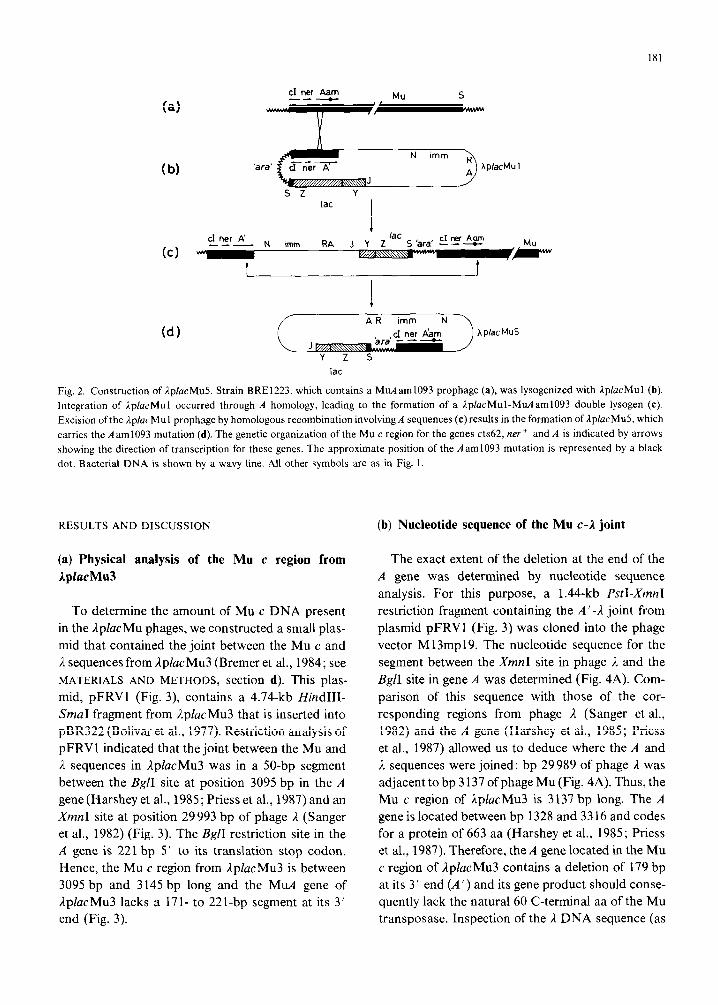
Fig. 2. Construction of IplucMu5. Strain BRE1223, which contains a MuAam1093 prophage (a), was lysogenized with lplucMu1 (b).
Integration of lplacMu1 occurred through A homology, leading to the formation of a lplacMul-MuAam1093 double lysogen (c).
Excision ofthe iplucMu1 prophage by homologous recombination involving A sequences (c)results in the formation of IplacMu5, which
carries the A am1093 mutation (d). The genetic organization of the Mu c region for the genes cts62, ner + and A is indicated by arrows
showing the direction of transcription for these genes. The approximate position of the Aaml093 mutation is represented by a black
dot. Bacterial DNA is shown by a wavy line. All other symbols are as in Fig. 1.
RESULTS AND DISCUSSION (b) Nucleotide sequence of the Mu c-1 joint
(a) Physical analysis of the Mu c region from
IplacMu3
To determine the amount of Mu c DNA present
in the LplucMu phages, we constructed a small plas-
mid that contained the joint between the Mu c and
L sequences from 1pZucMu3 (Bremer et al., 1984; see
MATERIALS AND METHODS, section d). This plas-
mid, pFRV1 (Fig. 3), contains a 4.74-kb HindHI-
SmaI fragment from AplucMu3 that is inserted into
pBR322 (Bolivar et al., 1977). Restriction analysis of
pFRV1 indicated that the joint between the Mu and
A sequences in AplacMu3 was in a 50-bp segment
between the BglI site at position 3095 bp in the A
gene (Harshey et al., 1985; Priess et al., 1987) and an
XmnI site at position 29993 bp of phage il (Sanger
et al., 1982) (Fig. 3). The BgjI restriction site in the
A gene is 221 bp 5’ to its translation stop codon.
Hence, the Mu c region from ApZacMu3 is between
3095 bp and 3145 bp long and the MLL~ gene of
;ipZacMu3 lacks a 171- to 221-bp segment at its 3’
end (Fig. 3).
The exact extent of the deletion at the end of the
A gene was determined by nucleotide sequence
analysis. For this purpose, a 1.44-kb PstI-XmnI
restriction fragment containing the A’-,? joint from
plasmid pFRV1 (Fig. 3) was cloned into the phage
vector M 13mp19. The nucleotide sequence for the
segment between the XmnI site in phage 1 and the
BgZI site in gene A was determined (Fig. 4A). Com-
parison of this sequence with those of the cor-
responding regions from phage 2 (Sanger et al.,
1982) and the A gene (Harshey et al., 1985; Priess
et al., 1987) allowed us to deduce where the A and
2 sequences were joined: bp 29989 of phage ,J was
adjacent to bp 3 137 ofphage Mu (Fig. 4A). Thus, the
Mu c region of AplacMu3 is 3 137 bp long. The A
gene is located between bp 1328 and 33 16 and codes
for a protein of 663 aa (Harshey et al., 1985; Priess
et al., 1987). Therefore, the A gene located in the Mu
c region of ilplucMu3 contains a deletion of 179 bp
at its 3’ end (A’) and its gene product should conse-
quently lack the natural 60 C-terminal aa of the Mu
transposase. Inspection of the L DNA sequence (as

P XHN
i 1 11
P
I X I ,
B 7 t MU cl ni A B ’
‘i’ P
BX C 151
ii pFRV1
“er A
Fig. 3. Physical and genetic organization of the Mu c-I joint in
pFRV I, Restriction maps of segments from phage 1. and Mu are
compared with the cloned Mu c-l fragment present in pFRV1.
The position of the restriction sites shown for i and Mu were
compiled from the literature (Sanger et al., 1982; Priess et al.,
1987). Those present in pFRVl were determined by suitable
single and multiple digests. The location and direction of trans-
cription of the Mu genes cts62, ner, A and E (Priess et al,, 1987)
are shown by horizontal arrows. The Mu material to the left of
the unique Hind111 site in the Mu c region is also present in
IplacMu3 but was not cloned. The SmaI restriction site (position
3 1619 bp in phage 2) used for the cloning ofthe Mu c-2 fragment
was destroyed during the btunt-end ligation into the Pvulf site of
pBR322 and is therefore shown in parentheses. DNA from phage
1 is indicated by a thin line; that of phage Mu by a thick line. B,
ⅈ C, ClaI; H, HindIII; N, N&I; P,PsrI; S, SmcrI; X. XmnI.
deduced from the published nucleotide sequence of
phage A; Sanger et al., 1982) at the fusion joint
revealed a short open reading frame for 59 aa
adjacent to and in frame with A’ (Fig. 4A). There is
no apparent homology between these amino acids
and the natural C-terminal end of the A protein (not
shown).
It has been reported that ;ip1(209), from which the
Mu c region of LplucMu3 was derived (Bremer et al.,
1984), was generated by an illegitimate recombina-
tion event between a ,I prophage and an adjacent Mu
prophage (Casadaban, 1976). The Mu c-2 joint of
IphcMu3 contains a 6-bp segment (Fig. 48) present
both in phage /z (Sanger at al., 1982) and phage Mu
(Priess et al., 1987). This is probably the site of the
reconlbination event that generated npl(209). DNA
sequences have become available from two other
specialized transducing phages (iplac5 and 1guf8)
which have been reported to result from illegitimate
recombination events (Shpakovski and Berlin, 1984;
Debouck et al., 1985). Homologous stretches of 19
and 9 bp, respectively, were at the junction of the
A
B
Fig. 4. Nucleotide sequences at the A’-2 joint in pFRV1. (A) The
nucleotide sequence at the A ‘-1 joint in pFRV I, originating from
iplarMu3, is shown. The nucleotide sequence between the ,YrhnI
and BglI sites was determined; the sequence of DNA 3’ to the
Xmn site was taken from the literature (Sanger et al., 1082).
Numbering of gene A sequence and the indicated reading frame
of the A protein is according to Priess et al. (1987); /( DNA
shown in bold print is numbered according to Sanger et al.
(1982). (B) ‘The nucleotide sequence at the A’-,2 joint from
IpiacMu? is compared to the corresponding regions of phages
Mu and i. The sequence shared with phage Mu is underlined.
and that shared with phage /, is overlined. The 6-bp homology
(boxed) is present in all three sequences. The nucieotide sc-
quences are written in a 5’-to-3’ direction,
bacterial and phage /z DNA. These results, and the
data presented here, suggest that such short segments
of homology might be of general importance for the
generation of specialized transducing phages.
The physical analysis of the Mu c region reported
here involved a cloned segment derived from
ApfcrcMu3. However, the conclusions drawn about
the size of its Mu c material are not limited to this
particular phage, since all transposable LplucMu
phages isolated so far (Bremer et al., 1984; 1985)
were derived by an identical series of in vivo
homologous recombination events. This 3 137-bp

183
Mu c segment includes the Mu c-end attachment site
and encodes the Mu genes cts62, lzer + and a partially
deleted A gene (A ’ ).
(c) Genetic analysis of the A’ gene
Because the A protein is absolutely required for
Mu transposition (O’Day et al., 1978), a functional
form of this transposase is apparently encoded by
the A’ gene of lpZucMu phages. To rule out the
possibility that other factors might be responsible for
,IplacMu transposition in the absence of the
ApMu507(A + B + ) helper phage, we introduced an
amber mutation into the A’ gene. We then tested
whether the resultant phages could transpose and
give rise to Lac + lysogens in the absence or presence
of a supF allele. Strain BRE1223 carries a Mu
prophage with the well-defined A am1093 mutation
that is located approximately in the middle of the A
gene (Magazin et al., 1977; 1978; Toussaint et al.,
1987). This mutation was crossed into ;IplucMul
(Bremer et al., 1984) (see MATERIALS AND
METHODS, section f; Fig. 2). When the resulting
phage (AplucMu5) was spotted onto strain
MBM7007 (SIX), no Lac’ lysogens were found,
whereas Lac + colonies did appear when the supF
strain MBM7014 was employed for this test (Fig. 5).
The number of Lac + lysogens formed by IplacMu5
in strain MBM7014 was reduced, however, com-
pared to IpZacMul. Thus, either the efficiency of
suppression was limiting or the incorporated amino
acid (tyrosine) did not fully restore transposase
activity. The ability to transpose and give Lac+
fusions was not impaired in this test when either
MBM7007 or MBM7014 was coinfected with
IplacMu5 and the A ‘B + helper phage ApMu507
(Fig. 5). Thus, ApZucMul derivatives carrying the
A am1093 mutation in the A’ gene undergo supF-
dependent transposition.
We also isolated a transposition-defective deriva-
tive of phage IpZucMu50 (which is used to obtain
ZucZ operon fusions; Bremer et al., 1985). Like
;IpZucMu5, the derivative of AplacMu50 carrying the
Aam mutation within the A’ gene (1plucMu52)
is unable to transpose by itself unless the amber
mutation is suppressed (data not shown). Taken
together, these data conclusively show that an A
protein lacking its natural 60 aa at the C-terminal
end still has transposase activity.
(d) Genetic stability of ,I.placMu insertions carrying
the A ’ am1093 allele
Fusions to the 1acZ gene can be used to isolate
mutations affecting expression of the fused gene by
selecting or screening for an altered Lac phenotype
hp!aCMu 1 h I/ hpMu 507
xh”‘M”,“$J71/ P
i A”
hpMu 507 ‘AplacMu 5
Fig. 5. Transposition of AplacMul and r2plucMu5. Approximately 1O’pfu of IplucMul(A’) and lpIucMu5(A’am1093) were spotted onto
lawns of strains MBM7007 (Su- ) (left plate) and MBM7014 (supF) (right plate) on lactose MacConkey agar. When strains were
coinfected with lpMu507, approx. 10’ pfu of lpMu507 were added. The plates were incubated for two days at 37°C. The Lac * lysogens
(red colonies) appear on the photograph as shaded areas against a white (Lac-) background.

184
in lac fusion strains (Weinstock et al., 1983; Silhavy
and Beckwith, 1985). Such applications can be com-
plicated if the Iac genes transpose at high frequency
from their original position within a fused gene to
create additional IacZ fusions. We have previously
shown that 1placMu insertions in the E. colichromo-
some are very stable and rarely give rise to secondary
transposition events, although these phages can
transpose at an appreciable frequency upon infection
into their host (Bremer et al., 1984; 1985).
The finding that AplacMu phages carrying the
MuA’am1093 allele no longer transpose by them-
selves upon infection in a Su _ strain suggested that
insertions of these phages should exhibit even greater
genetic stability. We tested this by measuring the
frequency of Lac+ mutants arising from Lac
strains harboring malK-1acZ fusions. First, Lac +
insertions of AplacMu5 (protein fusion) and
ApfacMu52 (operon fusion) were isolated in the
malK gene in a Su- host using the ApMu507 helper
phage. Expression of the fused genes in these strains
was prevented by introducing with Plvir a
maIT: : TnlO insertion; malT is the positive regula-
tory gene of the maltose regulon (Schwartz, 1987).
As expected, the resulting Lac - strains gave rise to
Lac + mutants at a lower frequency than their parent
A’ phages /2placMul and AplacMu50 (Table II). We
suspect that the Lac + mutants found with the
operon fusion phage in strain BRE1299 are not
caused by transposition events but arise from other
types of mutation. The higher frequency of Lac +
mutants in the operon fusion strain is expected, since
a larger category of events can lead to expression of
TABLE II
the fused lac genes. The enhanced stability of
Ap/acMu insertions containing the A ’ am 1093 muta-
tion should make them even more suitable than
insertions of the parental phages for the isolation of
mutants affecting the expression of the fused gene.
(e) Derivatives of IplucMu5 and IplucMu52 carry-
ing a gene for kanamycin resistance
Since the iplacMu phages carrying the A ‘am 1093
allele yield very stable 1acZ fusions to chromosomal
genes, they should be valuable genetic tools. To
facilitate the isolation of AplacMu insertions, we
introduced agene (kan) which confers Km resistance
into AplacMu5 and ;IplacMu52 (see MATERIALS
AND METHODS, section e). This construction yielded
;iplacMu 15 and AplacMu55, respectively. Both
phages are unable to transpose by themselves in a
Su host but can insert into the E. coli chromosome
upon coinfection with the ApMu507(A + B + ) helper
phage. When KmR insertions of these new ApfacMu
phages were isolated in strain MC4100, AplacMulS
gave Lac + colonies with a frequency of 10-l 5 %,
while approx. 50% Lac ’ colonies were found with
AplacMu55. Thus, as expected, a higher frequency of
operon fusions than of protein fusions was found.
These frequencies probably overestimate the true
frequency of Lac + fusions since multiple insertions
can occur under these conditions (Bremer et al.,
1984; 1985). The different levels of fat expression
found among fusion strains when they were streaked
on several lactose indicator media strongly suggest
that the lac operon was fused to different genes.
Stability of lplucMu prophages carrying the A’ or the A’am1093 genes
Strain Prophage a Frequency of Lac’
colonies h
BRE1047 $(malK-‘lacZ)hyb1002 (ipfacMu1) malT::TnlO
BRE1297 4(malK-‘lacZ)hyb1283 (J.plucMuS) malT::TnlU
BREI 167 $(malK-IacZ+)1113 (IplacMuSO) malT::TnlU
BRE12YY @(ma/K-lacZ +)1263 (A^pfacMu52) malT::TnIO
3.5 x IO_ H
3.2 x IO- ‘”
1.7 x 10 ’
5.6 x lo-’
I’ The ip/acMul and A-placMuS0 prophages carry the A’ gene, while the IplucMu5 and IplacMu52 prophages encode the A’amlOY3
allele. These lysogens were originally isolated as Lac + protein (BRE1047, BRE1297) or operon (BREl167, BRE129Y) fusion strains.
Expression of the lac fusions in all strains is prevented by a TnlO insertion in the positive regulatory gene (m&T) of the maltose regulon.
h Strains carrying Lac lplacMu insertions were grown in 0.4:” glycerol-minimal medium overnight at 37°C. These cultures were
titered on M63 glycerol plates for colony-forming units and on M63 lactose plates for iac + derivatives. The plates were incubated for
two days at 37°C. The values given are the mean values from two independent experiments.

185
Indeed, we have already used AplucMu55 to isolate
tsx-lucZ+ operon fusions (Bremer et al., 1988).
(f) Conclusions
Transposition of Mu DNA to random chromoso-
ma1 sites is a multistep process that is dependent on
the activity of the 663-aa A transposase. The data
presented here show that the transposase function is
retained by a mutant A protein (A’) missing its
natural 60 C-terminal aa. Initiation of the trans-
position process requires the binding of the A protein
to specific sequences in the S- and c-end attachment
sites (Mizuuchi and Craigie, 1986), and the DNA
binding domain has been localized to the N-terminal
end of A (Nakayama et al., 1987; Betermier et al.,
1987). Obviously, DNA binding activity is not
abolished in the A’ protein but we do not know
whether the overall activity of the A’ transposase is
reduced since no lplacMu phage carrying an intact
A gene is available. However, an analysis of the
properties of several A proteins with deletions at
their C-terminal ends has shown that a deletion of
47 aa reduces Mu transposition about ten-to 20-fold
compared to wild-type A protein (Harshey and
Cuneo, 1986). In the same study it was also shown
that transposase activity was abolished by the
removal of approx. 75 aa but not of 55 aa. Thus the
lplucMu phages contain the shortest functional A
segment yet reported. As previously mentioned, the
A ’ gene is fused to a DNA segment from phage J.
containing an open reading frame for 59 aa. These aa
are not homologous to the natural C-terminal end of
the Mu transposase but possibly could contribute to
the stability of the A’ protein.
When theAam mutation was crossed into the
A’ gene a series of new AplacMu phages was obtained
that cannot transpose by themselves in a Su- strain;
however, efficient transposition of these phages can
be conveniently triggered by coinfection with the
A+B+ lpMu507 helper phage. The enhanced
genetic stability of lac fusions isolated with these new
AplacMu phages is particularly important when
selections are applied to isolate mutants with altered
regulation of the fused gene.
ACKNOWLEDGEMENTS
We thank M. Berman, M. Howe and R. Zagursky
for providing strains and bacteriophages and S.
Beck for advice on DNA sequencing. We are grateful
to D. Kamp for the communication of data on the
Mu4 sequence prior to publication. We thank M.
Manson and G. Sweet for critical reading of the
manuscript, V. Koogle for help in preparing the
manuscript, and A. Middendorf for technical assist-
ance. E.B. thanks W. Boos for his support. This
work was supported in part by the National Cancer
Institute, Department of Health and Human Ser-
vices, under Contract No. NOl-CO-23909 with
Litton Bionetics, and in part by the Deutsche
Forschungsgemeinschaft through SFB 156. E.B.
was the recipient of a fellowship from the Deutsche
Akademische Austauschdienst.
REFERENCES
Austin, S. and Abeles, A.: Partion of unit-copy mini plasmids to
daughter cells. J. Mol. Biol. 163 (1983) 353-372.
Beck, S. and Pohl, F.M.: DNA sequencing with direct blotting
electrophoresis. EMBO J. 3 (1984) 2905-2909.
Berman, M.L. and Beckwith, J.: Fusions of the lac operon to the
transfer RNA gene tyrT of Escherichia coli. J. Mol. Biol. 130
(1979) 285-301.
Betermier, M., Alazard, R., Ragueh, F., Roulet, E., Toussaint, A.
and Chandler, M.: Phage Mu transposase: deletion of the
carboxy-terminal end does not abolish DNA-binding activity.
Mol. Gen. Genet 210 (1987) 77-85.
Bremer, E., Silhavy, T.J., Weisemann, J.M. and Weinstock,
G.M.: IplacMu: a transposable derivative of phage lambda
for creating [acZ protein fusions in a single step. J. Bacterial.
158 (1984) 1084-1093.
Bremer, E., Silhavy, T.J. and Weinstock, G.M.: Transposable
lplacMu phages for creating IacZ operon fusions and
kanamycin-resistance insertions in Escherichia cofi. J. Bac-
teriol. 162 (1985) 1092-1099.
Bremer, E., Gerlach, P. and Middendorf, A.: Double negative
and positive control of tsx expression in Escherichia coli. J.
Bacterial. 170 (1988) 108-116.
Bolivar, F., Rodriguez, R.L., Green, P.J., Betlach, M., Heyneker,
H.L., Boyer, H.W., Crosa, J.H. and Falkow, S.: Construction
and characterization of new cloning vehicles, II. A multi-
purpose cloning system. Gene 2 (1977) 95-l 13.
Bukhari, A.1. and Ljungquist, E.: Bacteriophage Mu: methods
for cultivation and use. In Bukhari, AI., Shapiro, J.A. and
186
Adhya S.L. (Eds.), DNA insertion Elements, Plasmids, and
Episomes. Cold Spring Harbor Laboratory, Cold Spring
Harbor, NY, 1911, pp. 149-156.
Bukhari, AI. and Zipser, D.: Random insertion of Mu-l DNA
within a single gene. Nature New Biol. 136 (1972) 240-243.
Casadaban, M.J.: Fusion of the Escheriehiu coli lac genes to the
aru promoter: a general technique using bacteriophage Mu. 1
insertions. Proc. Natl. Acad. Sci. USA 72 (1975) 809-813.
Casadaban, M.J.: Transposition and fusion of the Iuc genes to
selected promoters in Escherichiu coli using bacteriophage
lambda and Mu. J. Mol. Biol IO4 (1976) 541-555.
Casadaban, M.J. and Chou, J.: In vivo formation ofgene fusions
encoding hybrid ~-gaIactosidase proteins in one step with
transposable MU-/UC transducing phage. Proc. Nat]. Acad.
Sci. USA 81 (1984) 535-539.
Casadaban, M.J. and Cohen, S.N.: Lactose genes fused to exo-
genous promoters in one step using a Mu-he bacteriophage:
in vivo probe for transcriptional control sequences. Proc.
Natl. Acad. Sci USA 76 (1979) 4530-4533.
Castilho, B.A., Olfson, A. and Casadaban, M.J.: Plasmid inser-
tion mutagenesis and iac gene fusion with mini-Mu bacterio-
phage transposons. J. Bacterial 158 (1984) 488-495.
Daniell, E., Roberts, R. and Abelson, J.: Mutations in the lactose
operon caused by bacteriophage Mu. J. Mol. Biol. 69 (1972)
l-8.
Daniels, D.L., Schroeder, J.L., Szybalski, W., Sanger, S.,
Coulson, A.R., Hong, G.F., Hill, D.F., Petersen, G.B. and
Blattner, F.R.: Complete annotated lambda sequence. In
Hendrix, R.W., Roberts, J.W.. Stahl, F.W. and Weisberg,
R.A. (Eds.), Lambda II. Cold Spring Harbor Laboratory,
Cold Spring Harbor, NY, 1983, pp. 519-576.
Debouck, C., Riccio, A., Schumperli, D., McKenny, K.. Jeffers,
J., Hughes, C., Rosenberg, M., Heusterspreute, M.. Brunei,
F. and Davison J.: Structure of the galactokinase gene of
E.wherichia co/i, the last (?) gene of the galoperon. Nucl. Acids
Res. I3 (1985) 1841-1853.
Harkki, H., Karkku, H. and Palva, E.T.: Use of A- vehicles to
isolate uinpC-1acZ gene fusions in ~ai~~o~te~la t~~~i~uriu~ LT2. Mol. Gen. Genet. 209 (1987) 607-611.
Harshey, R.M. and Cuneo, S.D.: Carboxyl-terminal mutants of
phage Mu transposase. J. Genet. 65 (1986) 159-174.
Harshey, R.M., Getzoff, E.D., Baldwin, D.L., Miller, J.L. and
Chaconas. G.: Primary structure of phage Mu transposase:
homology to Mu repressor. Proc. Natl. Acad. Sci. USA 82
(1985) 7676-7680.
Leathers, T.D., Noti. J. and Umbarger, HE.: Physical charac-
terization ofih4ac fusions. J. Bacterial. 140 (1979) 251-260.
Magazin, M., Howe, M. and Allet, B. Partial correlation of the
genetic and physical maps of bacteriophage Mu. Virology 77
(1977) 677-688.
Magazin, M., Reeve, J.N., Maynard-Smith, S. and Symonds, N.
Bacteriophage Mu encoded polypeptides synthesised in
infected minicells. FEMS Microbial. Lett. 4 (1978) 5-9.
Maniatis, T., Fritsch, E.F. and Sambrook, J.: Molecular Cloning.
A Laboratory Manual. Cold Spring Harbor Laboratory. Cold
Spring Harbor, NY, 1982.
Messing, J.: New M 13 vectors for cloning. Methods Enzymol.
101 (1983) 20-78.
Miller, J.H.: Experiments in Molecular Genetics. Cold Spring
Harbor Laboratory, Cold Spring Harbor, NY, 1972.
Mizuuchi, K. and Craigie, R.: Mechanisms of bacteriophage Mu
transposition. Annu. Rev. Genet. 20 (1986) 385-429.
Nag, D.K. and Berg, D.E.: Specificity of bacteriophage Mu
excision. Mol. Gen. Genet. 207 (1987) 395-401.
Nakayama, C., Teplow, D.B. and Harshcy. R.M.: Structural
domains in phage Mu transposase: ide~lti~catioll of the site-
specific DNA-binding domain. Proc. Natl. Acad. Sci. USA 84
(1987) 1809-1813.
Norrander, J., Kempe, T. and Messing, J.: Construction of
improved M I3 vectors using oligodeoxynucleotide-directed
mutagenesis. Gene 26 (1983) 101-106.
O’Day, K.J., Schultz, D.W. and Howe, M.M.: Search for inte-
gration-deficient mutants of bacteriophage Mu. In Schlessin-
ger, D. (Ed.) Microbioiogy-1978. American Society for
Microbiology, Washington, DC, 1978, pp. 48-5 I.
Priess, H., Kamp, D., Kahmann, R., Brauer, B. and Delious, H.:
Nucleotide sequence of the immunity region of bacteriophage
Mu. Mol. Gen. Genet. 186 (1982) 315-321.
Priess, H., Brluer, B., Schmidt, C. and Kamp, D.: Sequence of
the left end of Mu. In Symonds, N., Toussaint, A., Van de
Putte, P. and Howe, M. (Eds.), Phage Mu. Cold Spring
Harbor Laboratory, Cold Spring Harbor, NY, 1987, pp.
277-296.
Sanger, F., Nicklen, S. and Coulson, A.R.: DNA sequencing with
chain-terminating inhibitors. Proc. Natl. Acad. Sci. USA 74
(1977) 5463-5467.
Sanger, F., Couison A.R., Hong, G.F., Hill, D.F. and Petersen,
G.B.: Nucleotide sequence of bacteriophage EDNA. J. Mol.
Biol. 162 (1982) 729-773.
Schwartz, M.: The maltose regulon. In Neidhardt, F.C. (Ed.),
~~~~eriehi~ colt’ and Sal~o~e~lu l~p~ti~u~iu~. American
Society for Microbiology, Washington, DC, 1987. pp.
1482-1502.
Shpakovski, G.V. and Berlin, Y.: Site-specificity of abnormal
excision: the mechanism of formation of a specialized trans-
ducing bacteriophage Ipluc5. Nucleic Acids Res. I2 (1984)
6779-6795.
Silhavy, T.J. and Beckwith, J.R.: Uses oflc2c fusions for the study
of biological problems. Microbial. Rev. 49 (1985) 398-4 18.
Silhavy, T.J., Berman, M.L. and Enquist, L.W.: Experiments
with Gene Fusions. Cold Spring Harbor Laboratory, Cold
Spring Harbor, NY, 1984.
Sober&, X., Covarrubias, L. and Bolivar, F.: Construction and
characterization of new cloning vehicles. Gene 9 (1980)
287-305.
Taylor, A.L.: Bacteriophage-induced mutation in E. coli. Proc.
Natl. Acad. Sci. USA 50 (1963) 1043-1051.
Toussaint, A. and Resibois, A.: Phage Mu: transposition as a life
style. In Shapiro, A. (Ed.), Mobile Genetic Elements.
Academic Press, New York, 1983, pp. 105-158.
Toussaint, A., Desmet, L., Faelen, M., Alazard, R., Chandler, M.
and Pato, M.: In vivo mutagenesis of bacteriophage Mu
transposase. J. Bacterial. 169 (1987) 5700-5707.
Trun, N.J. and Silhavy. T.J.: Characterization and in vivo cloning
of prlC, a suppressor of signal sequence mutations in
~sche~chiff co/i K12. Genetics 116 (1987) 513-521.
Weinstock, G.M.. Berman, M.L. and Silhavy, T.J.: Chimeric
genetics with /I-galactosidase. In: Papas. T.S., Rosenberg, M.
and Chirikjian (Eds.), Gene Amplification and Analysis, Vol.
3. Elsevier, New York, 1983, pp. 23-64.
Communicated by J. Davison