occupational dermatoses learning objectives...occupational dermatoses basic course in occupational...
TRANSCRIPT
American Osteopathic College of Occupational and Preventive MedicineBasic Course in Occupational MedicineSan Diego, California; October 7, 2012
G-1
OCCUPATIONAL DERMATOSES
Basic Course in Occupational Medicine
Part II
Liz Clark, D.O., MPH & TM, FAOCOPM
American Osteopathic College ofOccupational and Preventive Medicine
Learning Objectives:
• To better understand the epidemiology andeconomic impact of Occupational Dermatoses
• To review medical definitions and terminology orderto better describe Occupational Dermatoses
• To review the proper way to examine the skin inorder to more effectively assess OccupationalDermatoses
Learning Objectives Cont’d
• To review effective history taking techniquesin order to more effectively diagnose
Occupational Dermatoses.
• To review the common clinical morphologicpatterns of Occupational Skin Disease andtheir etiologic causes
• To review preventive strategies to preventOccupational Skin disorders
Introduction
• Skin is the most prominent interfacebetween the worker and the environment
• Largest organ of the body - 15% of the totalbody weight
• Marked size and exposure causes increasevulnerability to occupational injury anddiseases
Definitions
• Occupational Skin Disease - any abnormality ofthe skin induced or aggravated by the workenvironment
• Dermatitis - describing a skin disease as havingan inflammatory component involved in itspathogenesis
• Dermatosis - describes a skin disease from anycause including inflammatory and non-inflammatory causes
Epidemiology
• Occupational skin diseases and disorders are themost common non-trauma occupational illness
• Skin diseases account for approximately 30-45% ofall occupational illnesses
• Bureau of Labor Statistics (BLS) data 2009 estimatedthat greater than 15% of occupational injury/illnesswas due to skin diseases (estimated that totalnumber of occupational skin disorders may be 10-50 times greater than what BLS is able to estimate)
American Osteopathic College of Occupational and Preventive MedicineBasic Course in Occupational MedicineSan Diego, California; October 7, 2012
G-2
Epidemiology “Cont’d”
Affects approximately one worker per thousand inprivate sector. Annual incidence rate in 1993 bythe BLS was 76 cases per 100,000 workers
Greatest number of cases of occupational skindisease are seen in manufacturing industry butthe highest incidence rate is seen in theagriculture/forestry/fishing industry
BLS data reveals that approximately 20% of alloccupational skin disease result in days away fromwork with a median work absence of 3 days
Epidemiology Cont’d”
• Economic cost is a result of:
– medical costs
– lost work time
– rehabilitation cost
– worker’s compensation litigation
– re-hiring and training new workers
Epidemiology “Cont’d”
• Only 1/3 of U.S. workforce is employed in large placefacilities which are more likely to have acomprehensive occupational health program
• 2/3 of U.S. workers employed by small companiesemploying less than 500 workers. Incidence rates ofoccupational diseases are higher in these facilitiesusually due to lack of comprehensive healthprograms.
Evaluation ofOccupational Dermatoses
• Physicians engaged in evaluating occupationaldermatoses should be familiar with:
• importance of their appearance
• causes
• methods of evaluation
• diagnosis
• treatment
• prevention
Evaluation of OccupationalDermatoses Cont’d”
• It is crucial to evaluate a potentialoccupational skin disorder thoroughly– enables the worker to benefit from appropriate
diagnosis and treatment
• Proper diagnosis is essential– may be difficult to change later
– Wrong diagnosis may make the workerineligible for many other positions in the samecompany or other industries
Types of Skin Lesions
PRIMARY LESIONS
how skin diseases begin
key to accurate description and interpretation
SECONDARY LESIONS
develop during the evolutionary process of theskin disease
may be created by scratching or infection
American Osteopathic College of Occupational and Preventive MedicineBasic Course in Occupational MedicineSan Diego, California; October 7, 2012
G-3
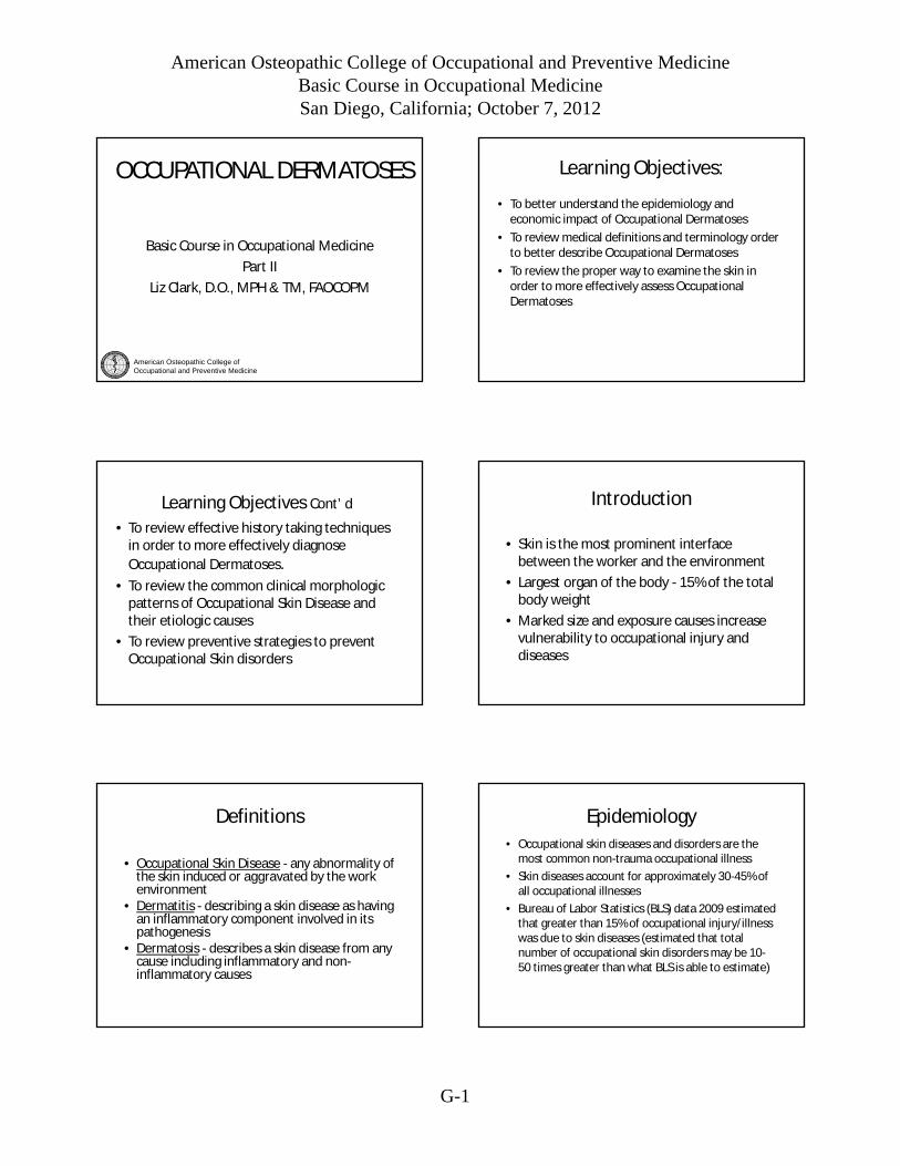
Primary vs Secondary
• Primary Lesions– Macule– Papule– Nodule– Vesicle– Petechiae– Patch– Plaque– Tumor– Bulla– Ecchymosis– Wheal– Pustule
Secondary Lesions Atrophy Crust Erosion Excoriation Fissure Lichenification Scale Scar TelangestasiaUlcer
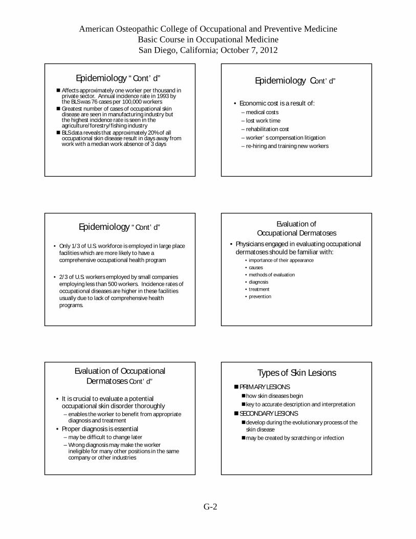
Primary Lesions
Macule - up to 1 cm in size, circumscribed,flat discolorations of the skin.Examples: freckles, flat nevi.
Primary Lesions
Patch - larger than 1 cm, circumscribed, flatdiscolorations of the skin.Examples: vitiligo, senile freckles, measles rash.
Primary Lesions
Papule - up to 1 cm in size, circumscribed,elevated, superficial, solid lesions.
Examples: elevated nevi, warts, lichen planus
A wheal is a type of papule that is edematous andtransitory.
Examples: hives, insect bites, (Fire Ant bite)
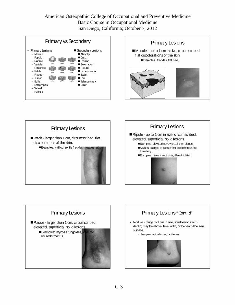
Primary Lesions
Plaque - larger than 1 cm, circumscribed,elevated, superficial, solid lesions.
Examples: mycosis fungoides, localizedneurodermatitis.
Primary Lesions “Cont’d”
• Nodule - range to 1 cm in size, solid lesions withdepth; may be above, level with, or beneath the skinsurface.
• Examples: epitheliomas, xanthomas
American Osteopathic College of Occupational and Preventive MedicineBasic Course in Occupational MedicineSan Diego, California; October 7, 2012
G-4
Primary Lesions “Cont’d”
• Tumor - larger than 1 cm, solid lesions withdepth; may be above level with, or beneaththe skin surface.
• Examples: tumor stage of mycosis fungoids,larger epitheliomas
Primary Lesions Cont’d”
Vesicle - range to 1 cm in size and arecircumscribed elevations of the skincontaining serous fluid.
Examples: early chickenpox, zoster,contact dermatitis.
Primary Lesions
• Bullae - larger than 1 cm, circumscribedelevations of the skin containing
serous fluid
• Examples: pemphigus,
second-degree burns
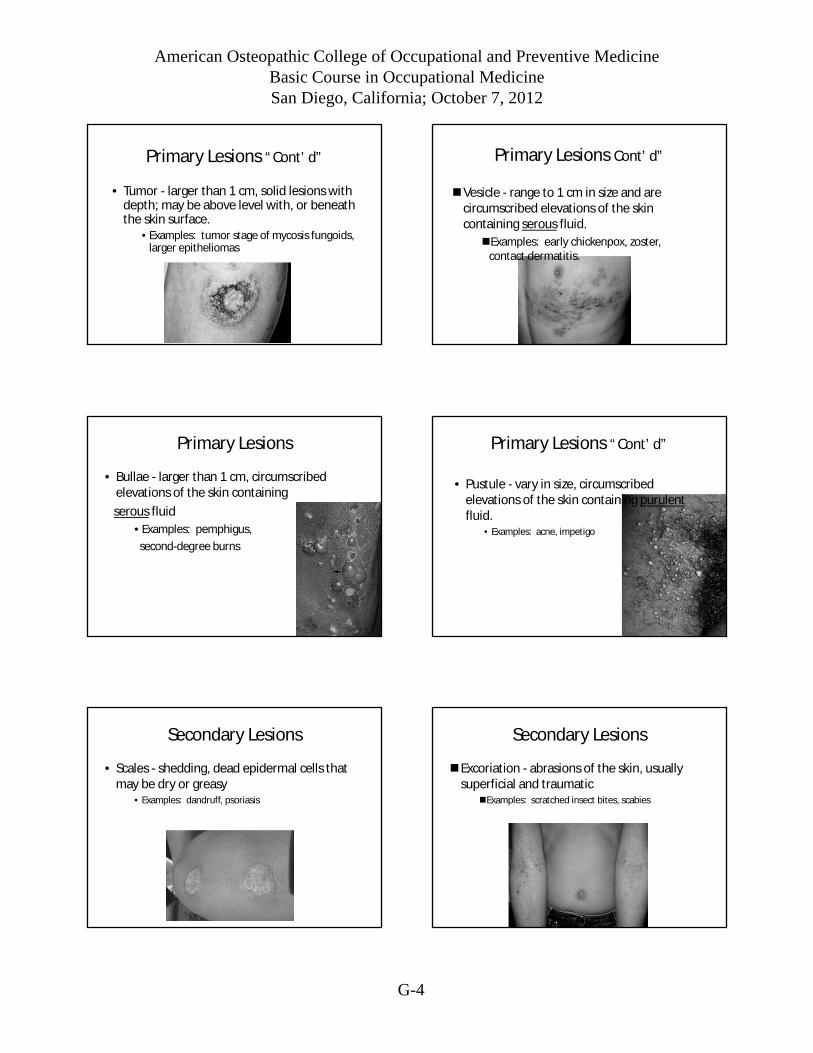
Primary Lesions “Cont’d”
• Pustule - vary in size, circumscribedelevations of the skin containing purulentfluid.
• Examples: acne, impetigo
Secondary Lesions
• Scales - shedding, dead epidermal cells thatmay be dry or greasy
• Examples: dandruff, psoriasis
Secondary Lesions
Excoriation - abrasions of the skin, usuallysuperficial and traumatic
Examples: scratched insect bites, scabies
American Osteopathic College of Occupational and Preventive MedicineBasic Course in Occupational MedicineSan Diego, California; October 7, 2012
G-5
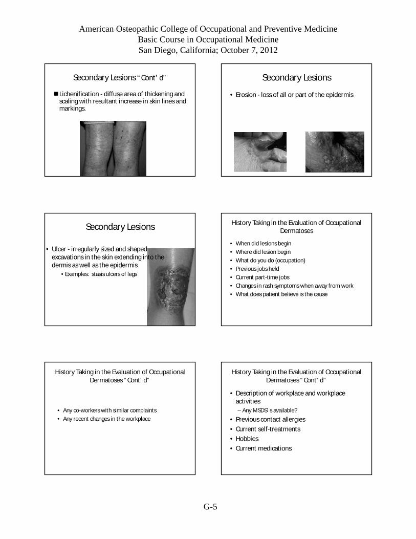
Secondary Lesions “Cont’d”
Lichenification - diffuse area of thickening andscaling with resultant increase in skin lines andmarkings.
Secondary Lesions
• Erosion - loss of all or part of the epidermis
Secondary Lesions
• Ulcer - irregularly sized and shapedexcavations in the skin extending into thedermis as well as the epidermis
• Examples: stasis ulcers of legs
History Taking in the Evaluation of OccupationalDermatoses
• When did lesions begin
• Where did lesion begin
• What do you do (occupation)
• Previous jobs held
• Current part-time jobs
• Changes in rash symptoms when away from work
• What does patient believe is the cause
History Taking in the Evaluation of OccupationalDermatoses “Cont’d”
• Any co-workers with similar complaints
• Any recent changes in the workplace
History Taking in the Evaluation of OccupationalDermatoses “Cont’d”
• Description of workplace and workplaceactivities
– Any MSDS’s available?
• Previous contact allergies
• Current self-treatments
• Hobbies
• Current medications

American Osteopathic College of Occupational and Preventive MedicineBasic Course in Occupational MedicineSan Diego, California; October 7, 2012
G-6
Skin Examination
• Well-lighted room
• In order to understand the effects ofvarious occupational agents on the skin, aconcept of its barrier function is needed.
• The skin consists of three major units:• Epidermis
• Dermis: includes follicle, sweat gland
• Subcutaneous fat
Skin Examination
• Examine all of the skin surface
• Regional inspection
– Begin at a distance
• Distribution
• Grouping
• Stage of the lesions
• Lesion inspection
Clinical MorphologicPatterns of Skin Disease
and Their
Occupational Causes
Acute Contact Dermatitis
Contact Dermatitis
• Irritant or allergic contact dermatitis
– Acute - blistering reactions
• e.g. hydrofluoric acid (HF), ethylene oxide
– Chronic - rough scaling and thickened skin
• e.g. chronic turpentine exposure or solventexposure, rubber compounds
Routes of Entry
American Osteopathic College of Occupational and Preventive MedicineBasic Course in Occupational MedicineSan Diego, California; October 7, 2012
G-7
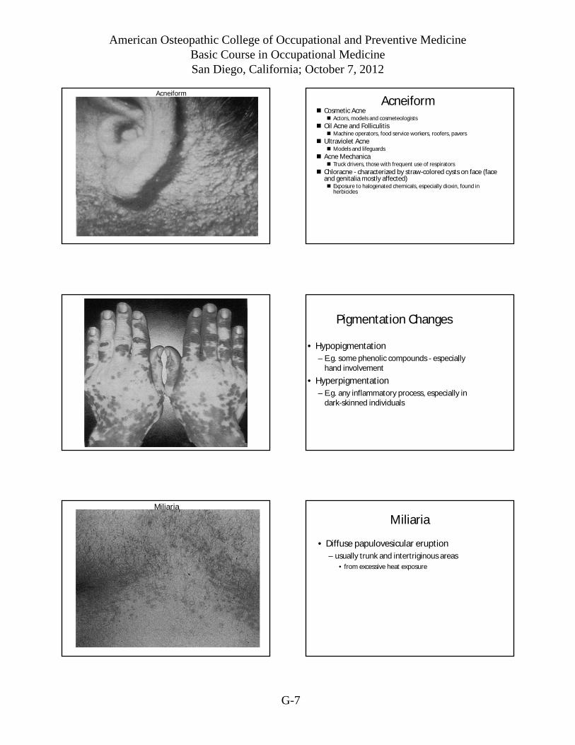
Acneiform
Acneiform Cosmetic Acne
Actors, models and cosmeteologists
Oil Acne and Folliculitis Machine operators, food service workers, roofers, pavers
Ultraviolet Acne Models and lifeguards
Acne Mechanica Truck drivers, those with frequent use of respirators
Chloracne - characterized by straw-colored cysts on face (faceand genitalia mostly affected) Exposure to halogenated chemicals, especially dioxin, found in
herbicides
Pigmentation Changes
• Hypopigmentation
– E.g. some phenolic compounds - especiallyhand involvement
• Hyperpigmentation
– E.g. any inflammatory process, especially indark-skinned individuals
Miliaria
Miliaria
• Diffuse papulovesicular eruption
– usually trunk and intertriginous areas
• from excessive heat exposure
American Osteopathic College of Occupational and Preventive MedicineBasic Course in Occupational MedicineSan Diego, California; October 7, 2012
G-8
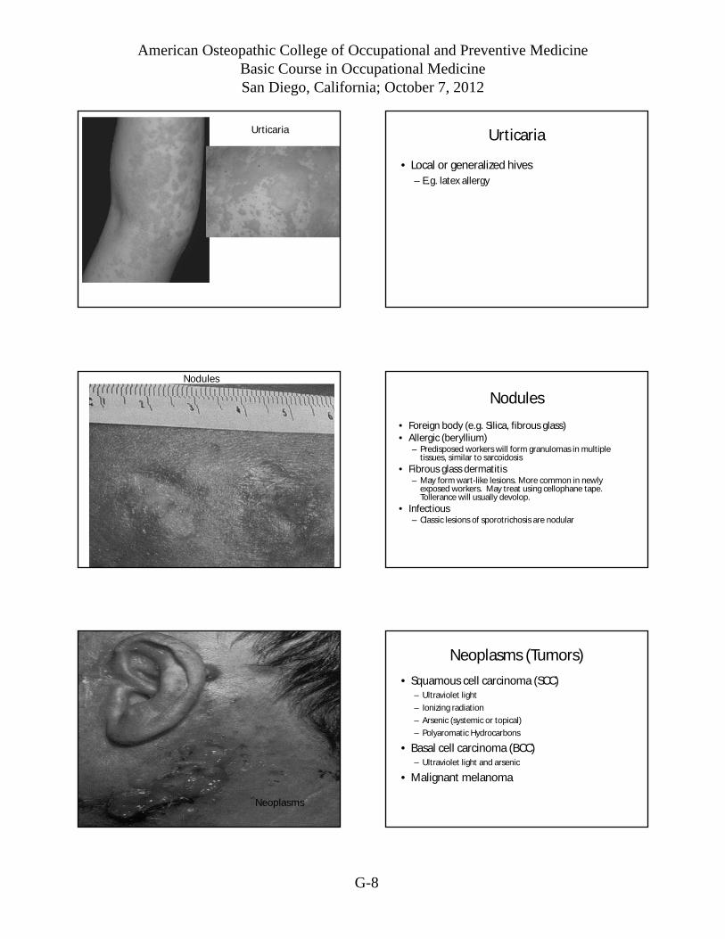
Urticaria Urticaria
• Local or generalized hives
– E.g. latex allergy
Nodules
Nodules
• Foreign body (e.g. Silica, fibrous glass)• Allergic (beryllium)
– Predisposed workers will form granulomas in multipletissues, similar to sarcoidosis
• Fibrous glass dermatitis– May form wart-like lesions. More common in newly
exposed workers. May treat using cellophane tape.Tollerance will usually devolop.
• Infectious– Classic lesions of sporotrichosis are nodular
Neoplasms
Neoplasms (Tumors)
• Squamous cell carcinoma (SCC)– Ultraviolet light
– Ionizing radiation
– Arsenic (systemic or topical)
– Polyaromatic Hydrocarbons
• Basal cell carcinoma (BCC)– Ultraviolet light and arsenic
• Malignant melanoma

American Osteopathic College of Occupational and Preventive MedicineBasic Course in Occupational MedicineSan Diego, California; October 7, 2012
G-9
Ulcerations
• Chromium (metal)
– Exposure to fumes happens in the production ofstainless steel
– Chronic exposure may cause erosion of the nasalseptum
– Chrome holes are painless erosive ulcerationsusually found on the fingers, knuckles andforearms
Direct Causes of Occupational Dermatoses
• 4 direct causes of occupational dermatoses, inorder of frequency;
– Chemical
– Mechanical
– Physical
– Biological
Irritant Contact Dermatitis
• 80% of all occupationally caused contactdermatitis
• Caused by substances that damage the skin atthe site of contact by non-immunologicmechanisms
• Many factors contribute to irritant reactions:
Irritant Contact Dermatitis“Cont’d”
– Potential Irritant(s):• Chemical properties• Physical properties
– Quantitative Aspects of Exposure• Concentration• Duration of exposure• Frequency and number of exposures
– Qualitative Aspects of Exposure• Occlusion of substance against skin• Temperature of substance on skin surface• Pre-existing skin damage to prevent skin barrier• Anatomic skin site
Irritant Contact Dermatitis“Cont’d”
– Host susceptibility• Atopic disease• Race (?)• Sex (?)• Age (?)• Allergies• Cleanliness• Season
• Most common predisposing factors in developmentof irritant dermatitis are atopy, dry skin, andadvancing age.

American Osteopathic College of Occupational and Preventive MedicineBasic Course in Occupational MedicineSan Diego, California; October 7, 2012
G-10
Irritant Contact Dermatitis“Cont’d”
• Irritant Dermatitis is divided into two types:
– Immediate (absolute)
– Delayed
Immediate Irritant Dermatitis
• Single contact with a strong chemical substancecauses acute, toxic reaction similar to burn.
• Erythema, blistering, and ulceration occur at sitealmost immediately after contact.
• Examples– Strong alkalis
– Acids
– Certain metallic substances and their salts
– Many organic compounds
Immediate Irritant Dermatitis“Cont’d”
• Chief Determinants
– Intrinsic nature of chemical
– Concentration of the chemical
– Duration of contact
• Almost everyone will respond the same way tothese substances
Delayed Irritant Dermatitis
• Repeated or prolonged chemical contacts
• Clinical findings of erythema, increasingdryness and thickening, patchyhyperkeratosis with pruritus, and painfulfissuring are characteristic
American Osteopathic College of Occupational and Preventive MedicineBasic Course in Occupational MedicineSan Diego, California; October 7, 2012
G-11
Delayed Irritant Dermatitis“Cont’d”
• Most Common Causes:– Soaps– Detergents– Mild acids and alkalis
• Most Common Contributing Factors:– Friction– Occlusion– Minor lacerations– Excessive environmental heat or cold– Low relative humidity
• Often confused with allergic contact dermatitis
Allergic Contact Dermatitis
Less frequent than irritant dermatitis
Greater importance to diagnose becauseordinary protective measures usually areineffective, and many patients must changejobs or learn a new trade.
Is an immunologic reaction classified as a TypeIV, delayed or cell mediated hypersensitivity
Allergic Contact Dermatitis“Cont’d”
• Sensitization is variable among individuals andalso dependent on numerous factors
• Allergic contact dermatitis must bedifferentiated from atopic dermatitis,psoriasis, Herpes Simplex & Zoster, idiopathicvesicular reactions to Trichophyton infectionsof feet, dyshidrotic eczema, and drugeruptions.
Example of Delayed or Cell Mediated HypersensitivityReaction
• The allergen in poison ivy or oak will sensitize nearly70% of exposed persons where p-phenylenediamine,allergen in permanent hair dyes, sensitizes a smallnumber of people with repeated exposure.
– Sensitization usually requires relatively short durationof exposure to develop though many workers mayhave repeated contact with an allergen in theirworkplace for months, and even years, beforedeveloping sensitivity.
– Once allergic sensitization occurs, the dermatitisbegins quickly (24 to 48 hours) after contact.
Example of Delayed or Cell MediatedHypersensitivity Reaction: “Cont’d”
– A pruritic, erythematous rash develops rapidly,followed by papule formation and blistering.
– Itching is always a prominent symptom.
– Dermatitis originates at site of contact with theallergen but new lesions may appear at distant sitesand may also be transferred by the hands.
– After several days, a subacute or chronic stage evolvesthat occasionally erupts into a more acute dermatitisafter re-exposure to the allergen.
Mechanical Causes of OccupationalDermatoses:
• Friction
– Calluses
– Blisters
– Abrasions
• Pressure
– Bullae
– Atrophy
– Necrosis
• Other
– Wounds
– Koebner Phenomenon
American Osteopathic College of Occupational and Preventive MedicineBasic Course in Occupational MedicineSan Diego, California; October 7, 2012
G-12
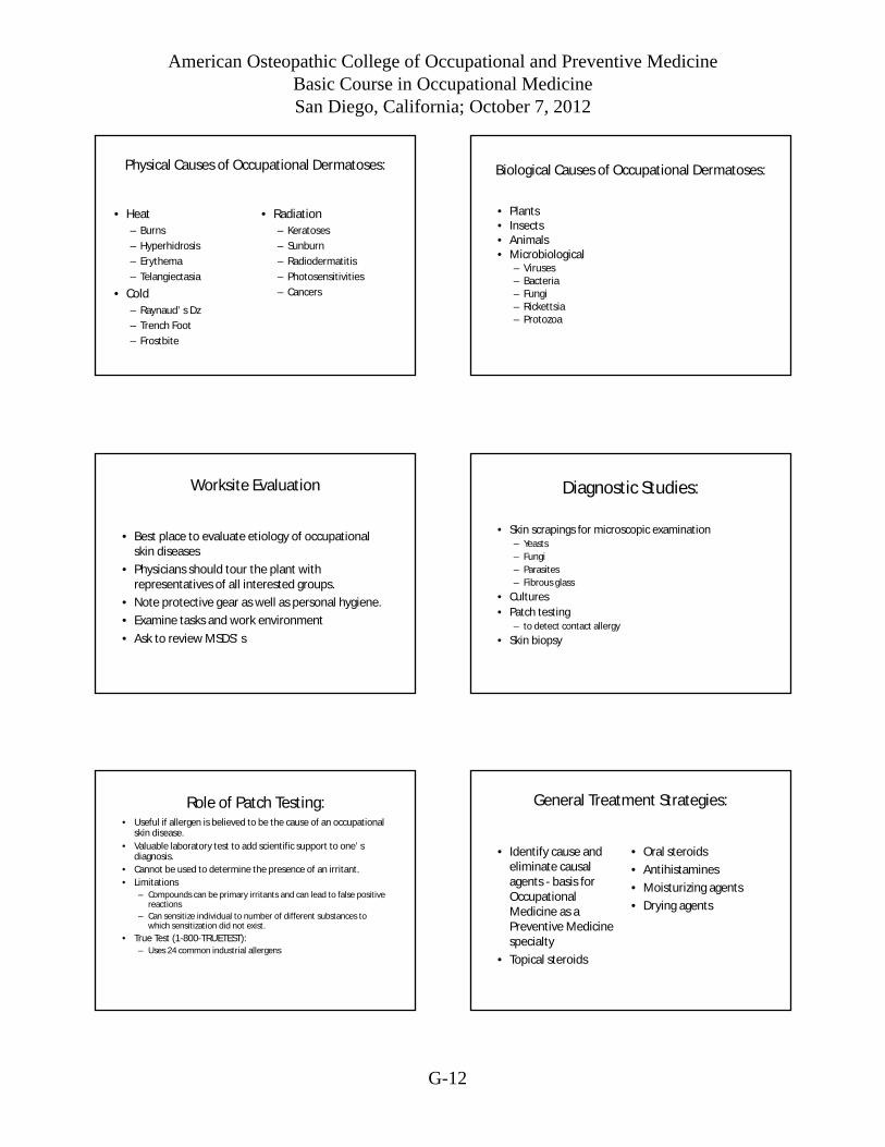
Physical Causes of Occupational Dermatoses:
• Heat
– Burns
– Hyperhidrosis
– Erythema
– Telangiectasia
• Cold
– Raynaud’s Dz
– Trench Foot
– Frostbite
• Radiation
– Keratoses
– Sunburn
– Radiodermatitis
– Photosensitivities
– Cancers
Biological Causes of Occupational Dermatoses:
• Plants• Insects• Animals• Microbiological
– Viruses– Bacteria– Fungi– Rickettsia– Protozoa
Worksite Evaluation
• Best place to evaluate etiology of occupationalskin diseases
• Physicians should tour the plant withrepresentatives of all interested groups.
• Note protective gear as well as personal hygiene.
• Examine tasks and work environment
• Ask to review MSDS’s
Diagnostic Studies:
• Skin scrapings for microscopic examination– Yeasts
– Fungi
– Parasites
– Fibrous glass
• Cultures
• Patch testing– to detect contact allergy
• Skin biopsy
Role of Patch Testing:• Useful if allergen is believed to be the cause of an occupational
skin disease.
• Valuable laboratory test to add scientific support to one’sdiagnosis.
• Cannot be used to determine the presence of an irritant.
• Limitations– Compounds can be primary irritants and can lead to false positive
reactions
– Can sensitize individual to number of different substances towhich sensitization did not exist.
• True Test (1-800-TRUETEST):– Uses 24 common industrial allergens
General Treatment Strategies:
• Identify cause andeliminate causalagents - basis forOccupationalMedicine as aPreventive Medicinespecialty
• Topical steroids
• Oral steroids
• Antihistamines
• Moisturizing agents
• Drying agents
American Osteopathic College of Occupational and Preventive MedicineBasic Course in Occupational MedicineSan Diego, California; October 7, 2012
G-13
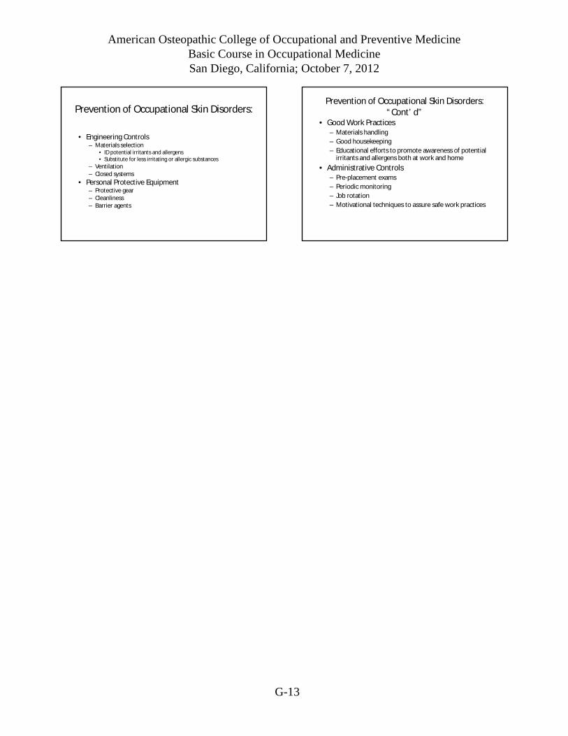
Prevention of Occupational Skin Disorders:
• Engineering Controls– Materials selection
• ID potential irritants and allergens• Substitute for less irritating or allergic substances
– Ventilation– Closed systems
• Personal Protective Equipment– Protective gear– Cleanliness– Barrier agents
Prevention of Occupational Skin Disorders:“Cont’d”
• Good Work Practices– Materials handling
– Good housekeeping
– Educational efforts to promote awareness of potentialirritants and allergens both at work and home
• Administrative Controls– Pre-placement exams
– Periodic monitoring
– Job rotation
– Motivational techniques to assure safe work practices