observations on the gynoecial pathway for pollen tube
TRANSCRIPT

Journal of Applied Botany and Food Quality 80, 6 - 13 (2006)
Institute of Agricultural and Environmental Sciences, Estonian Agricultural University, Tartu, Estonia, andDepartment of Environmental Sciences, Nova Scotia Agricultural College, Truro, Nova Scotia, Canada
Observations on the gynoecial pathway for pollen tube growth in sweet lowbush blueberry (Vaccinium angustifolium Ait.)
Merrit Noormets, A. Randall Olson(Received August 17, 2005)
Summary
Gynoecial structure in sweet lowbush blueberry, Vaccinium angus-tifolium Ait., was investigated in order to characterize the pollentube pathway in order to provide a framework for further studies onpollination and fungal infection. Closed flower buds and pollinatedopen flowers were collected from managed lowbush blueberry fieldsin Colchester County, Nova Scotia, Canada. Following chemicalfixation, the tissue samples were examined histologically using lightand scanning electron microscopy. The continuous pathway wascharacterized by a fluted, exudate-filled stylar canal that connectsthe wet stigmatic surface with the exudate covered surface of theovarian placentae. Following pollen deposition and germination,tubes growing along the pathway eventually arrive at the micropylesof the anatropous ovules; ovule penetration by pollen tubes andfertilization of the female gametophytes ensue. The pollen tubepathway of this taxon conforms to the general pattern reported fromother ericalean taxa.
Introduction
Among the Ericaceae, Vaccinium is a highly polymorphic genuscontaining species that are typically acidophilic, predominantly out-crossing, and distributed throughout many regions of the arctic,temperate and tropical zones (CAMP, 1942, 1945; VANDER KLOET,1988). In eastern North America, the wild blueberry industry dependson the successful management of the sweet lowbush blueberry,Vaccinium angustifolium Ait. This economically important woodyperennial species spreads vegetatively by the formation of rametes.When sexually mature, erect shoots produce numerous invertedpedunculate, racemose inflorescences (CAMP, 1945). Each inflores-cence bears several complete, actinomorphic, pentacyclic flowerscharacterized by an urceolate perianth configuration (PALSER, 1961).During anthesis, the sympetalous corolla will recurve at the tips toexpose the papillate stigma and the porocidal tips of the matureanthers (VANDER KLOET, 1988). The floral morphology, nectarproduction and pendulous orientation of the blossoms of sweetlowbush blueberry are consistent with other entomophilous speciesof this genus (WOOD, 1961; WOOD, 1962; HALL et al., 1971).
The syncarpous, pentacarpellate gynoecium typical of the genusVaccinium consists of a single stigma attached to an elongate, hollowstyle subtended by a pentalocular ovary with axile placentation andnumerous ovules (BELL and BURCHILL, 1955; MUNOZ and LYRENE,1985). The gynoecium is usually surrounded by ten stamens attachedto the base of the corolla near the nectariferous tissue located on theupper ovarian wall; this configuration in conjunction with the po-sition of the poricidal anther tips in relation to the stigma indicatemechanical pollen transfer by insect sonication (MCGREGOR, 1976;JACQUEMART and THOMPSON, 1996). When released from the anthers,Vaccinium pollen is of the dry type, covered with pollenkitt anddispersed in tetrads held together by viscin threads (HESLOP-HARRISON and SHIVANNA, 1977; PACINI, 2000). Each individual pollengrain is bicellular and tricolporate with pollen germination occurring
two to three hours following deposition on a receptive wet stigma(MUNOZ and LYRENE, 1985; VANDER KLOET, 1988). Following suc-cessful pollination, pollen tube growth through the various gynoecialtissues may take three to four days before reaching the ovule-bearingplacentae within the ovary (BELL, 1957).
Typically in angiosperms, determination of pollen compatibility andthe nutritional support of pollen tube growth are mediated by thegynoecial tissues (HERRERO and HORMAZA, 1996). In addition,recent research summarized by LORD and RUSSELL (2002) indicatesthat pollen tubes in some way become predisposed to receive andfollow molecular signals originating from inside of the ovules duringtheir passage through the stigma and style. Thus, an essential pathwayfor pollen tube growth and guidance is established extending fromthe stigmatic surface to the ovular micropyle within the ovary. Certainenvironmental stresses such as frost, however, may interfere withcompatible pollen tube growth in sweet lowbush blueberry due todifferences in tissue sensitivities along this same gynoecial pathway.Even before anthesis, ovule bearing placentae may sustain damagesevere enough to interfere with normal fruit set of this economicallyimportant wild species (OLSON and EATON, 2001).
In addition, gynoecial tissues of sweet lowbush blueberry are sus-ceptible to infection by the fungus Monilinia vaccinii-corymbosiwhich transforms a normally fleshy fruit into a pseudosclerotiumcalled a mummy berry (HILDEBRAND and BRAUN, 1991). Fungalaccess to the inner ovarian tissues is achieved when fungal infectionhyphae from germinating conidia on the stigma grow in conjunctionwith pollen tubes down the style and enter the ovary; in effect thepollen tube pathway functions as the gynoecial infection pathwayfor this economically important disease (SHINNERS and OLSON, 1996).Given the economic importance of this species, there is relativelylittle information available concerning the basic micromorphologyof compatible pollen tube growth so critical for fruit production. Thisstudy, therefore, uses light and scanning electron microscopy todescribe the gynoecial pathway for pollen tube growth in the sweetlowbush blueberry from eastern Canada.
Materials and methods
Flower buds of sweet lowbush blueberry, Vaccinium angustifoliumAit., were collected in May and June in 2000 and in 2001 in fieldsin Kemptown and Debert, Colchester County, N.S., Canada. Forconvenience, the following developmental stages were determinedand followed during the study: (1) young bud – very little of thecorolla is visible; (2) petal elongation – the petals elongate but staytightly closed; (3) petal spread – elongation is completed and theflower bud is just opening at the tips of the petals; (4) open flower –the flower is at anthesis and the petals are open and distinctivelyrecurved with the stigma protruding and exposed; (5) petal drop –the corolla and stamens have senesced and dropped off.
The flower buds were chemically field fixed using either 3% GA(glutaraldehyde) or FAA (Formalin-acetic acid-alcohol) for both light
(LM) and scanning electron microscopy (SEM). Selected gynoecialtissue samples were dissected and placed in a fresh fixative. For SEMthe fixed tissue samples were washed several times in phosphatebuffer (pH 6.8) and then postfixed in 2% osmium tetroxide followedby three washes in phosphate buffer. Then tissue samples weredehydrated in an ethanol series to anhydrous ethanol. Dehydratedspecimens were critical point dried and coated with platinum/goldusing a Samsputter 2a sputter coater. The observations were con-ducted using a Bausch and Lomb Nanolab 2000 SEM at a voltage of15 kv.
For LM histological examination the dehydrated specimens weresubjected to a propylene oxide series, infiltrated with Spurr's resin(SPURR, 1969) and sectioned on a Sorvall MT 6000 ultramicrotomebetween 0.5-1.0 µm. For general observation, certain sections weremounted on gelatin-coated slides and stained with azure II (1%)methylene blue (1%). To determine the presence of insoluble poly-saccharides in the selected plastic sections staining with periodicacid-Schiff's reagent was carried out. Selected sections were sub-jected to a saturated solution of DNPH (2,4 dinitrophenyl-hydrozine)and 1% periodic acid prior to usage of Schiff's reagent (O'BRIEN andMCCULLY, 1981). Then slides were counter stained in 1% anilineblue black to identify the proteins (FISHER, 1968). Additional plasticsections were stained with 1% sudan black B solution to detect lipids(BRONNER, 1975). The stained sections were then mounted with SP15-500 permount (Fisher Scientific) (BERLYN and MIKSCHE, 1976;O'BRIEN and MCCULLY, 1981). Observations were carried out usinga Leitz Diaplan Photomicroscope.
For aniline blue-UV induced fluorescence microscopy to identifycallose associated with pollen tube walls, certain specimens of thefield fixed plant material were selected and hand sectioned. Sectionedtissue samples were stained in a solution of 0.05% aniline blue in0.15 M K2HPO4 at pH 8.6. Observations were made using an OlympusBH2-RFL Photomicroscope with UVFL objectives and barrier filterL-435.
Results
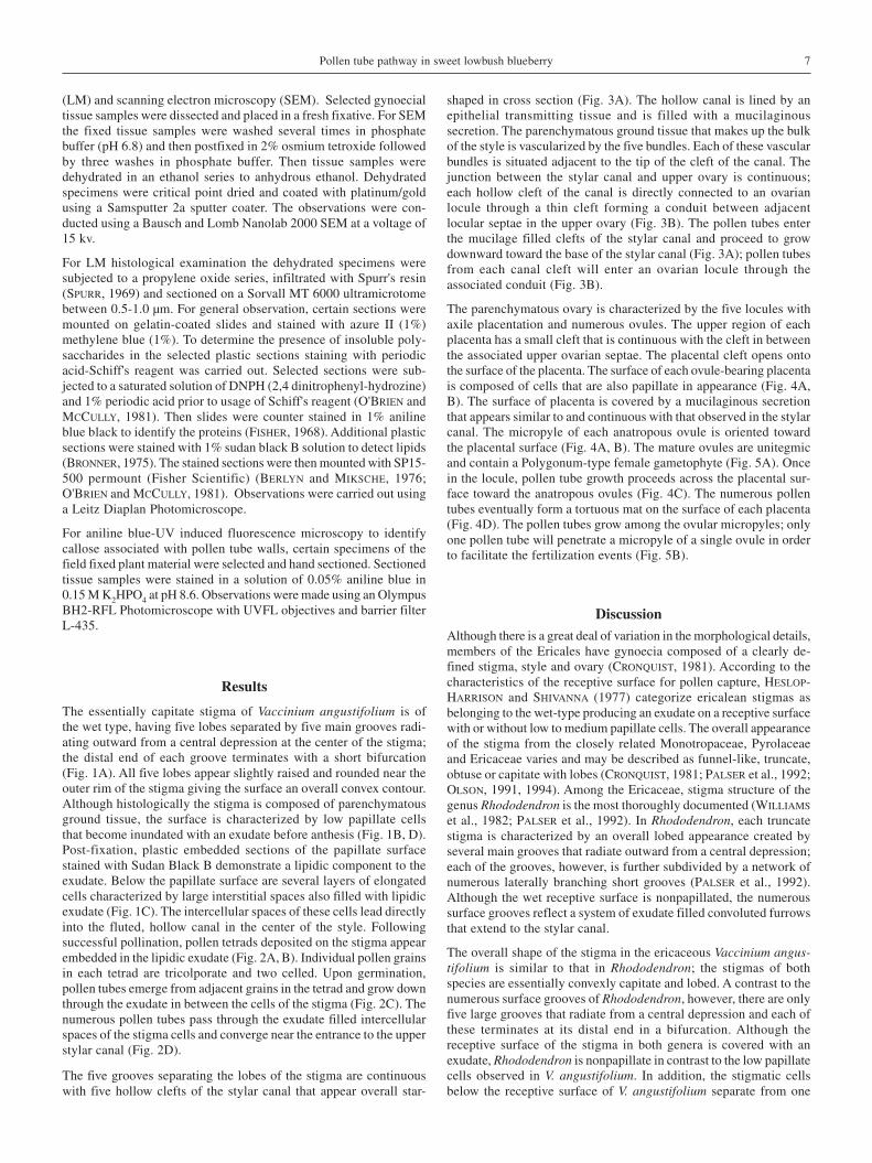
The essentially capitate stigma of Vaccinium angustifolium is ofthe wet type, having five lobes separated by five main grooves radi-ating outward from a central depression at the center of the stigma;the distal end of each groove terminates with a short bifurcation(Fig. 1A). All five lobes appear slightly raised and rounded near theouter rim of the stigma giving the surface an overall convex contour.Although histologically the stigma is composed of parenchymatousground tissue, the surface is characterized by low papillate cellsthat become inundated with an exudate before anthesis (Fig. 1B, D).Post-fixation, plastic embedded sections of the papillate surfacestained with Sudan Black B demonstrate a lipidic component to theexudate. Below the papillate surface are several layers of elongatedcells characterized by large interstitial spaces also filled with lipidicexudate (Fig. 1C). The intercellular spaces of these cells lead directlyinto the fluted, hollow canal in the center of the style. Followingsuccessful pollination, pollen tetrads deposited on the stigma appearembedded in the lipidic exudate (Fig. 2A, B). Individual pollen grainsin each tetrad are tricolporate and two celled. Upon germination,pollen tubes emerge from adjacent grains in the tetrad and grow downthrough the exudate in between the cells of the stigma (Fig. 2C). Thenumerous pollen tubes pass through the exudate filled intercellularspaces of the stigma cells and converge near the entrance to the upperstylar canal (Fig. 2D).
The five grooves separating the lobes of the stigma are continuouswith five hollow clefts of the stylar canal that appear overall star-
shaped in cross section (Fig. 3A). The hollow canal is lined by anepithelial transmitting tissue and is filled with a mucilaginoussecretion. The parenchymatous ground tissue that makes up the bulkof the style is vascularized by the five bundles. Each of these vascularbundles is situated adjacent to the tip of the cleft of the canal. Thejunction between the stylar canal and upper ovary is continuous;each hollow cleft of the canal is directly connected to an ovarianlocule through a thin cleft forming a conduit between adjacentlocular septae in the upper ovary (Fig. 3B). The pollen tubes enterthe mucilage filled clefts of the stylar canal and proceed to growdownward toward the base of the stylar canal (Fig. 3A); pollen tubesfrom each canal cleft will enter an ovarian locule through theassociated conduit (Fig. 3B).
The parenchymatous ovary is characterized by the five locules withaxile placentation and numerous ovules. The upper region of eachplacenta has a small cleft that is continuous with the cleft in betweenthe associated upper ovarian septae. The placental cleft opens ontothe surface of the placenta. The surface of each ovule-bearing placentais composed of cells that are also papillate in appearance (Fig. 4A,B). The surface of placenta is covered by a mucilaginous secretionthat appears similar to and continuous with that observed in the stylarcanal. The micropyle of each anatropous ovule is oriented towardthe placental surface (Fig. 4A, B). The mature ovules are unitegmicand contain a Polygonum-type female gametophyte (Fig. 5A). Oncein the locule, pollen tube growth proceeds across the placental sur-face toward the anatropous ovules (Fig. 4C). The numerous pollentubes eventually form a tortuous mat on the surface of each placenta(Fig. 4D). The pollen tubes grow among the ovular micropyles; onlyone pollen tube will penetrate a micropyle of a single ovule in orderto facilitate the fertilization events (Fig. 5B).
DiscussionAlthough there is a great deal of variation in the morphological details,members of the Ericales have gynoecia composed of a clearly de-fined stigma, style and ovary (CRONQUIST, 1981). According to thecharacteristics of the receptive surface for pollen capture, HESLOP-HARRISON and SHIVANNA (1977) categorize ericalean stigmas asbelonging to the wet-type producing an exudate on a receptive surfacewith or without low to medium papillate cells. The overall appearanceof the stigma from the closely related Monotropaceae, Pyrolaceaeand Ericaceae varies and may be described as funnel-like, truncate,obtuse or capitate with lobes (CRONQUIST, 1981; PALSER et al., 1992;OLSON, 1991, 1994). Among the Ericaceae, stigma structure of thegenus Rhododendron is the most thoroughly documented (WILLIAMS
et al., 1982; PALSER et al., 1992). In Rhododendron, each truncatestigma is characterized by an overall lobed appearance created byseveral main grooves that radiate outward from a central depression;each of the grooves, however, is further subdivided by a network ofnumerous laterally branching short grooves (PALSER et al., 1992).Although the wet receptive surface is nonpapillated, the numeroussurface grooves reflect a system of exudate filled convoluted furrowsthat extend to the stylar canal.
The overall shape of the stigma in the ericaceous Vaccinium angus-tifolium is similar to that in Rhododendron; the stigmas of bothspecies are essentially convexly capitate and lobed. A contrast to thenumerous surface grooves of Rhododendron, however, there are onlyfive large grooves that radiate from a central depression and each ofthese terminates at its distal end in a bifurcation. Although thereceptive surface of the stigma in both genera is covered with anexudate, Rhododendron is nonpapillate in contrast to the low papillatecells observed in V. angustifolium. In addition, the stigmatic cellsbelow the receptive surface of V. angustifolium separate from one
Pollen tube pathway in sweet lowbush blueberry 7
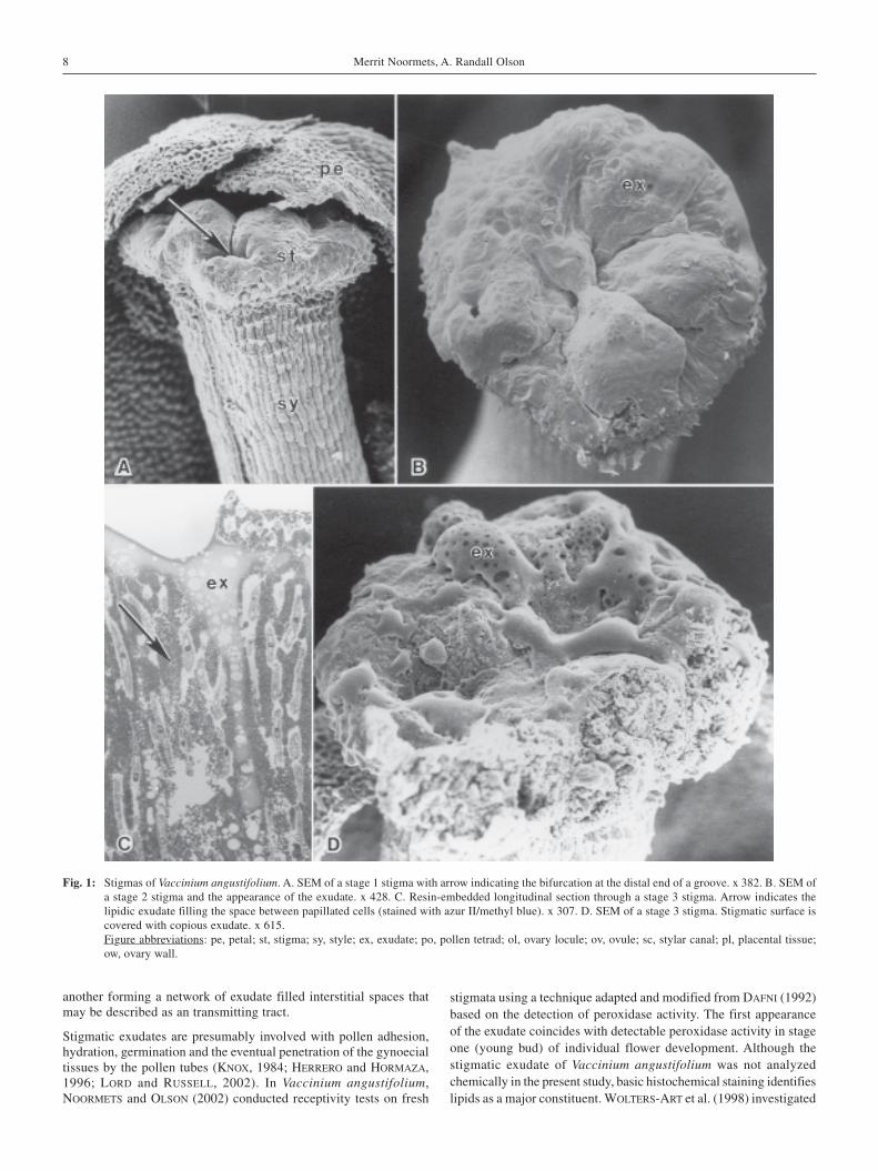
Fig. 1: Stigmas of Vaccinium angustifolium. A. SEM of a stage 1 stigma with arrow indicating the bifurcation at the distal end of a groove. x 382. B. SEM ofa stage 2 stigma and the appearance of the exudate. x 428. C. Resin-embedded longitudinal section through a stage 3 stigma. Arrow indicates thelipidic exudate filling the space between papillated cells (stained with azur II/methyl blue). x 307. D. SEM of a stage 3 stigma. Stigmatic surface iscovered with copious exudate. x 615.Figure abbreviations: pe, petal; st, stigma; sy, style; ex, exudate; po, pollen tetrad; ol, ovary locule; ov, ovule; sc, stylar canal; pl, placental tissue;ow, ovary wall.
another forming a network of exudate filled interstitial spaces thatmay be described as an transmitting tract.
Stigmatic exudates are presumably involved with pollen adhesion,hydration, germination and the eventual penetration of the gynoecialtissues by the pollen tubes (KNOX, 1984; HERRERO and HORMAZA,1996; LORD and RUSSELL, 2002). In Vaccinium angustifolium,NOORMETS and OLSON (2002) conducted receptivity tests on fresh
stigmata using a technique adapted and modified from DAFNI (1992)based on the detection of peroxidase activity. The first appearanceof the exudate coincides with detectable peroxidase activity in stageone (young bud) of individual flower development. Although thestigmatic exudate of Vaccinium angustifolium was not analyzedchemically in the present study, basic histochemical staining identifieslipids as a major constituent. WOLTERS-ART et al. (1998) investigated
8 Merrit Noormets, A. Randall Olson

Fig. 2: V. angustifolium stage 4 stigmas with pollen tetrads. A. SEM of a stigma with pollen tetrads embedded in the surface exudate. Arrows indicatepollen tetrads. x 1800. B. SEM of a stigma showing the emergent pollen tubes penetrating the exudate. Arrows indicate the pollen tubes. x 2995. C.Resin-embedded longitudinal section trough a pollinated stigma. Pollen tetrads embedded in the exudate. Arrows indicate the pollen tubes growingin between the stigmatic surface cells. x 410. D. Aniline blue fluorescent micrograph of a longitudinal free-hand section through the stigma and stylarcanal. Arrows indicate the numerous pollen tubes. x 165.
Pollen tube pathway in sweet lowbush blueberry 9

the possible role lipids play in the complex pollen-stigma inter-actions. Their experiments and careful analyses indicate that lipidsare a crucial factor in establishing an external water gradient requiredfor water uptake by both the pollen grains and the pollen tubes. Itseems reasonable to suggest that lipids on the stigmatic surface of V.angustifolium may also help direct pollen tube penetration and growthin the gynoecial tissues for similar reasons.
Previous observations on the stylar structure in certain members ofthe Ericales reveal a common pattern; the stylar canal is hollow, flutedand filled with a mucilaginous substance presumably derived froman epithelial transmitting tissue that lines the canal. Among thosemembers of the Pyrolaceae and Monotropaceae that were studied,the number of mucilage filled clefts in the fluted canal is five andreflects the number of ovarian locules (PYYKKÖ, 1968; OLSON, 1991,1994). In the Ericaceae, the number of mucilage filled stylar cleftsvaries from five to ten among the various species of Rhododendronbut also reflects the number of ovarian locules (WILLIAMS et al., 1982;PALSER et al., 1992). In Vaccinium elliotii, V. corymbosum and theirhybrids, there are five stylar clefts that become filled with a muci-laginous secretion (MUNOZ and LYRENE, 1985). As expected, theobservations of the style in V. angustifolium conforms to the pre-viously established pattern characterized by five mucilage filledstylar clefts that are aligned with a corresponding ovarian locule.
At the style-ovary junction in members of the Ericales, each cleft ofthe stylar canal is restricted to a narrow cleft between the upperovarian septae that forms a conduit into each ovarian locule. These
septal clefts are continuous with an associated cleft in the placentaltissues bearing the ovules. In various genera studied from the Erica-ceae, Monotropaceae and Pyrolaceae, the clefted placental tissue isrelatively massive and covered with a mucilaginous secretion; eachlocular region has the appearance of containing two protruding, ovulebearing placental lobes (PYYKKÖ, 1968, 1969; PALSER, 1992; OLSON,1991, 1994). In Vaccinium angustifolium, the ovary is clearly dividedinto five locules separated by complete septae with axile placentationthroughout the ovary. There is also a short cleft in the upper placentathat is aligned with the corresponding conduit connecting the stylarcanal with the upper locule. The opening into each locule of V.angustifolium, therefore, leads directly to the secretion-coated surfaceof the ovule bearing placenta. Herein lies the significance of thestructural alignment between the stylar canal and the ovarian loculesin all ericalean taxa studied including V. angustifolium.
Observations on the gynoecial pathway for pollen tube growth inVaccinium angustifolium are in accordance with previous descrip-tions from other ericalean species (WILLIAMS et al., 1982; MUNOZ
and LYRENE, 1985; PALSER et al., 1992; OLSON, 1991, 1994). In theaforementioned studies, the pollen tubes follow an unobstructedpathway characterized by continuous secretions along the entirejourney from the stigma to the ovules. The numerous pollen tubesgrowing from the stigmatic surface, through the stylar canals, intothe locules and along the placental surfaces in V. angustifolium re-main in direct contact with gynoecial secretions until they penetratethe ovular micropyles.
Fig. 3: V. angustifolium. A. Aniline blue fluorescent micrograph of a free-hand cross section through a stage 4 stylar canal. Arrow indicates the pollen tubes.x 206. B. Aniline blue fluorescent micrograph of a free-hand cross section through the junction between the stylar canal and upper ovary at stage 5.Arrow indicates the connecting conduit. x 206.
10 Merrit Noormets, A. Randall Olson

Fig. 4: V. angustifolium. A. SEM of anatropous ovules at stage 4. x 1270. B. SEM of ovule and papillated placental surface at stage 4. Arrow indicates themicropylar opening near the placental surface. Exudate is removed during the chemical preparation. x 2408. C. SEM of the ovules and pollen tubeson a placental surface at stage 5. Arrow indicates a tortous mat of pollen tubes. x 505. D. SEM of the pollen tubes at stage 5 with arrow indicatingcollapsed pollen tubes. x 2535.
Pollen tube pathway in sweet lowbush blueberry 11

Acknowledgements
The authors thank Ms. Sandra Fisk for help with manuscript pre-paration and Dr. Ping Li from the Faculty of Science electron micro-scopy suite of Dalhousie University. This research was supported inpart by the Canadian International Development Agency (CIDA) aspart of the requirements for a doctoral degree at the Estonian Agri-cultural University.
References
BELL, H.P., 1957: The development of the blueberry seed. Can. J. Bot. 35,139-153.
BELL, H.P., BURCHILL, J., 1955: Flower development in the lowbush blueberry.Can. J. Bot. 33, 251-258.
BERLYN, G.P., MIKSCHE, J.P., 1976: Botanical microtechnique and cyto-chemistry. Iowa State University Press, Ames, IA.
BRONNER, R., 1975: Simultaneous demonstration of lipids and starch in planttissues. Stain Technol. 50, 1-4.
CAMP, W.H., 1942: On the structure of populations in the genus Vaccinium.Brittonia 4, 189-204.
CAMP, W.H., 1945: The North American blueberries with notes on othergroups of Vaccinaceae. Brittonia 5, 203-220.
CRONQUIST, A., 1981: An integrated system of classification of floweringplants. Columbia University Press, New York.
DAFNI, A., 1992: Pollination Ecology – A practical approach. OxfordUniversity Press, New York.
FISHER, D.B., 1968: Protein staining of ribboned Epon sections for lightmicroscopy. Histochemie 16, 92-96.
HALL, I.V., FORSYTH, F.R., LIGHTFOOT, H.J., BOCH, R., 1971: Volatiles oflowbush blueberry nectar. HortScience 6, 493-494.
HERRERO, J., HORMANZA, J.I., 1996: Pistil strategies controlling pollen tubegrowth. Sex Plant Reprod. 9,343-347.
HESLOP-HARRISON, Y., SHIVANNA, K.R., 1977: The receptive surface of theangiosperm stigma. Annals Bot. 41,1233-1258.
HILDEBRAND, P.D., BRAUN, P.G., 1991: Factors affecting infection of lowbushblueberry by ascospores of Monilinia vaccinii-corymbosi. Can. J. PlantPathol. 13, 232-240.
JACQUEMART, A.L., THOMPSON, J.D., 1996: Floral and pollination biology ofthree sympatric Vaccinium (Ericaceae) species in the Upper Adrennes,Belgium. Can. J. Bot. 74, 210-221.
KNOX, R.B., 1984: Pollen-pistil interactions. In: Linskens, H.F., Heslop-Harrison, J. (eds.), Cellular interactions. (Encyclopedia of plant physio-logy, new series 17), 508-608. Springer, New York, Berlin, Heidelberg.
LORD, E.M., RUSSELL, S.D., 2002: The mechanisms of pollination andfertilization in plants. Annu. Rev. Cell Dev. Biol. 18, 81-105.
MCGREGOR, S.E., 1976: Insect pollination of cultivated crop plants. USDAAgriculture Handbook No. 494, 110-116.
MUNOZ, C.E., LYRENE, P.M., 1985: Reproductive incompatibility barriers incrosses between Vaccinium corymbosum and V. elliottii. Can. J. Bot. 63,1987-1996.
NOORMETS, M., OLSON, A.R., 2002: Stigma receptivity in Sweet LowbushBlueberry (Vaccinium angustifolium Ait) and in Velvet-Leaf Blueberry(Vaccinium myrtilloides Michx.). Bot. Lith. 8, 117-123.
O'BRIEN, T.P., MCCULLY, M.E., 1981: The study of plant structure. Principles
Fig. 5: V. angustifolium. A. Resin-embedded longitudinal section through a stage 4 ovule. Note the female gametophyte. x 410. B. Aniline blue fluorescentmicrograph of a longitudinal free-hand section of a stage 5 ovary. Arrow indicates a single pollen tube entering the micropyle of an ovule. x 206.
12 Merrit Noormets, A. Randall Olson

and selected methods. Thermarcarpi Pty Ltd., Melbourne.OLSON, A.R., 1991: Gynoecial pathway for pollen tube growth in the genus
Monotropa. Bot. Gaz. 152, 154-163.OLSON, A.R., 1994: Pollen tube pathway through the gynoecium of Mono-
tropsis odorata (Monotropaceae). Am. J. Bot. 81,718-725.OLSON, A.R., EATON, L.J., 2001: Spring frost damage to placental tissues in
lowbush blueberry flower buds. Can. J. Plant Sci. 81, 779-781.PACINI, E., 2000: Anther opening and pollen penetration. In: Dafni, A.,Hesse,
M., Pacini, E. (eds.), Pollen and pollination, 19-43. Springer-Verlag, Wien.PALSER, B.F., 1961: Studies of floral morphology in the Ericales. V. Organo-
graphy and vascular anatomy in several United States species of theVacciniaceae. Bot. Gaz. 123, 79-111.
PALSER, B.F., ROUSE, J.L., WILLIAMS, E.G., 1992: A scanning electronmicroscope study of the pollen tube pathway in pistils of Rhododendron.Can. J. Bot. 70, 1039-1060.
PYYKKÖ, M., 1968: Embryological and anatomical studies on Finnish speciesof the Pyrolaceae. Ann. Bot. Fenn. 5, 153-165.
PYYKKÖ, M., 1969: Placentation in the Ericales. I. Pyrolaceae and Mono-tropaceae. Ann. Bot. Fenn. 6, 255-268.
SHINNERS, T.C., OLSON, A.R., 1996: The gynoecial infection pathway ofMonilinia vaccinii-corymbosi in lowbush blueberry (Vaccinium
angustifolium). Can. J. Plant Sci. 76,493-497.SPURR, A.R., 1969: A low-viscosity epoxy resin embedding medium for
electron microscopy. J. Ultrastruct. Res. 26, 31-43.VANDER KLOET, S.P., 1988: The genus Vaccinium in North America. Ottawa
Research Branch, Agriculture Canada.WILLIAMS, E.G., KNOX, R.B., ROUSE, J.L., 1982: Pollination sub-systems
distinguished by pollen tube arrest after incompatible interspecificcrosses in Rhododendron (Ericaceae). J. Cell. Sci. 53, 255-277.
WOLTERS-ARTS, M., LUSH, W.M., MARIANI, C., 1998: Lipids are required fordirectional pollen tube growth. Nature 392, 818-821.
WOOD, G.W., 1961: The association between age of inflorescence and nectarproduction in the low-bush blueberry Vaccinium angustifolium. Can. J.Bot. 39, 1037-1040.
WOOD, G.W., 1962: The period of receptivity in flowers of the lowbushblueberry. Can. J. Bot. 40, 685-686.
Address of the authors:Merrit Noormets, Institute of Agricultural and Environmental Sciences,Estonian Agricultural University, Tartu 50412, EstoniaProf. Dr. A. R. Olson, Department of Environmental Sciences, Nova ScotiaAgricultural College, Truro, Nova Scotia, Canada, B2N 5E3
Pollen tube pathway in sweet lowbush blueberry 13