nursing care of clients with common skin disorders chapter 45
TRANSCRIPT

Nursing Care of Clients with Common Skin Disorders
Chapter 45

The Client with Psoriasis
Definition– chronic– non-infective– raised reddened round plaques covered by silvery
white scales– most common on scalp, arms, legs
Diagnosed– skin biopsy

Psoriasis

The Client with Psoriasis
Treatment– topical corticosteriods to decrease inflammation– phototherapy
• exposure to ultraviolet light
• decreases the growth rate of epidermal cells
Nursing Care– Impaired skin integrity– Body Image Disturbance

Infections and Infestations Bacterial Skin Infection
– causative agent gram+ staph aureus– and beta-hemolytic streptococci
Furuncle– boils, inflammation of hair follicle
Carbuncles– group of infected hair follicles
Cellulitis - localized infection of dermis

Infections and Infestations
Diagnosis– assessment– culture and sensitivity
Treatment– antibiotics

Fungal Foot Infection

Fungal Infections of the Skin
Tinea pedis - athlete’s foot Tinea capitis - scalp - ringworm Tinea corporis - body Candidiasis Infections
– yeast like fungus, pustules, red rash– skin folds, mouth, peri areas– treatment - antifungal - nystatin, diflucan

Ring Worm

Inflammatory Disorders Dermatitis
– inflammation of the skin characterized by erythema, pain and pruritus
Contact Dermatitis– caused by hypersensitivity response or chemical
irritation
Treatment– topical oints and therapeutic baths

Toxic Epidermal Necrolysis (TEN) Rare, life threatening disease in which the
skin peels off leaves large areas of denuded skin can also occur internally to mucose
membranes Treatment
– ICU, Burn Unit


Toxic Epidermal Necrolysis (TEN Surgery
– skin graphing
Fluid replacement– IV therapy, TPN
Medications– Antibiotics -treat sepsis– Anelgesics - pain management


Neoplastic Skin Disorders
Benign lesions - moles, cysts, keloids, skin tags keratoses
Malignant lesions - skin cancers– over time damage from ultraviolet radiation and
chemicals– basal cell carcinoma, squamous cell and
melanoma

Risk Factors
Environmental– ultraviolet radiation– pollution, chemicals viruses, trauma
Host Factors– skin pigmentation– life style

Skin Changes
Normal Skin
Aged Skin
Sun Damaged Skin

Basal Cell Carcinoma
Tumor that originates from basal layer Most common but least aggressive Tend to recur but rarely metastasize

Basal Cell Carcinoma

Squamous Cell Carinoma
Arises from squamous epithelium Occurs on exposed areas of skin More aggressive, faster growth rate Harden nodule may ulcerate and bleed

Skin Cancer Model

Interdisciplinary Care
Labs and Diagnostics– biopsy
Treatment– surgical excision– curettage and electrodesiccation– cryosurgery– radiation therapy

Malignant Melanoma
Arises from melanocytes is life threatening precursor lesions
– atypical moles (dysplastic nevi)– congenital nevi - present at birth– lentigo freckle - tan or black mole, usually on
the side of the face, slow growing


Interdisciplinary Care
Assessment– A = asymmetry– B = border irregularity– C = color variation– D = diameter >6mm– E = elevation
Labs and Diagnostics– biopsy

Interdisciplinary Care
CT Scan, MRI, CXR, Bone Scan Blood work - CBC, Liver function Surgery
– wide excision of lesion– regional lymph node dissection
Chemotherapy and Radiation Therapy

Interdisciplinary Care
Nursing Care– Impaired skin Integrity– Hopelessness– Anxiety– Teaching for Home Care
• wound care
• avoid exposure
• follow up appointments

Skin Trauma
Pressure Ulcer– ischemic lesions of the skin and underlying
tissue caused by external pressure that impairs blood and lymph flow
– ischemia causes tissue necrosis then ulcerations
Causes– External pressure– Shearing

Pressure areas on bed bound client

Pressure Ulcers
Staging– Stage 1
• intact skin
• non-blanchable erythema
– Stage 2• open, partial layer skin loss
• ulcer, abrasion, blister, shallow crater

Pressure Ulcers
Stage 3– full-thickness, damage or necrosis to
subcutaneous tissue– deep crater
Stage 4– full-thickness skin loss with extensive
destruction, tissue necrosis or damage to muscle, bone, can have sinus tracts

Pressure Ulcers
Eschar– black, leathery necrotic skin– when is present, unable to accurately stage
ulcer– needs to be debrided - surgical, wet-to-dry
normal saline drsg changes.

Pressure Ulcer Staging

Stage ?

Stage 2 Pressure Ulcer

Stage ? On coccyx

Stage ? On coccyx

Heel Ulcer

Stasis Ulcer --Venous Insufficiency
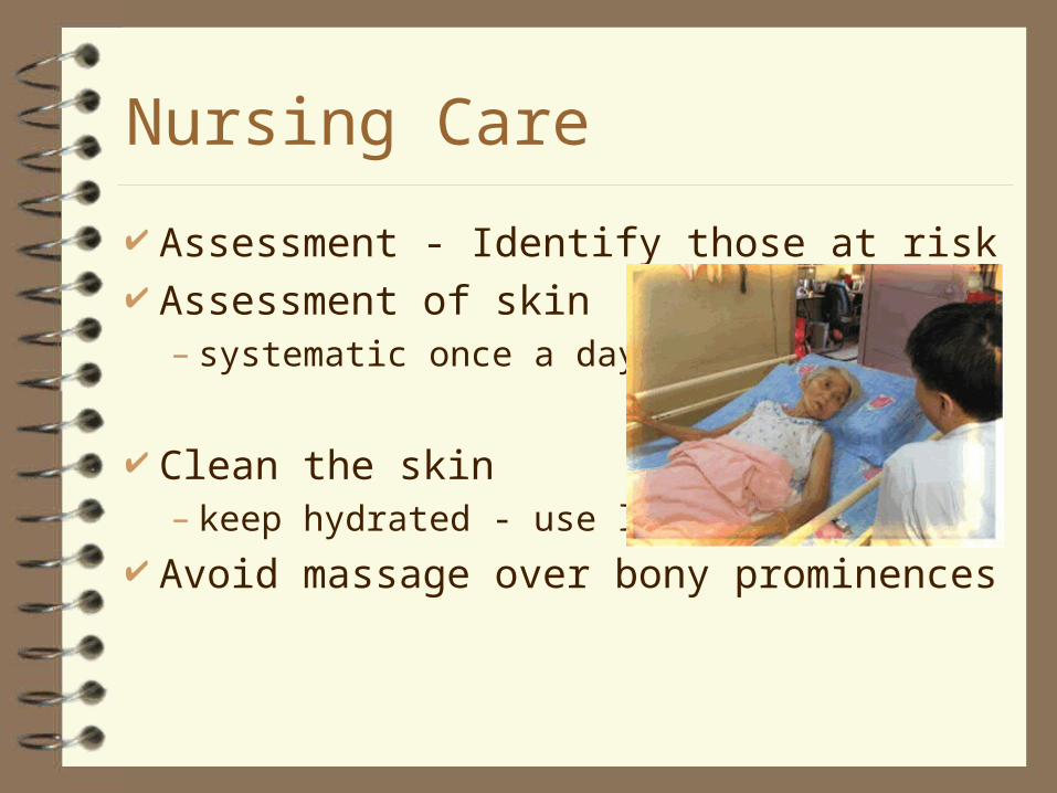
Nursing Care
Assessment - Identify those at risk Assessment of skin
– systematic once a day
Clean the skin– keep hydrated - use lotion
Avoid massage over bony prominences

Nursing Care
Minimize exposure to moisture Avoid friction and shearing Ensure adequate nutritional intake Maintain activity level
– What can you do for a client on bedrest?

Measuring Pressure Ulcers

Documentation
Site Size Stage Appearance
– color– drainage– odor

Turn me


Help me keep my skin intact
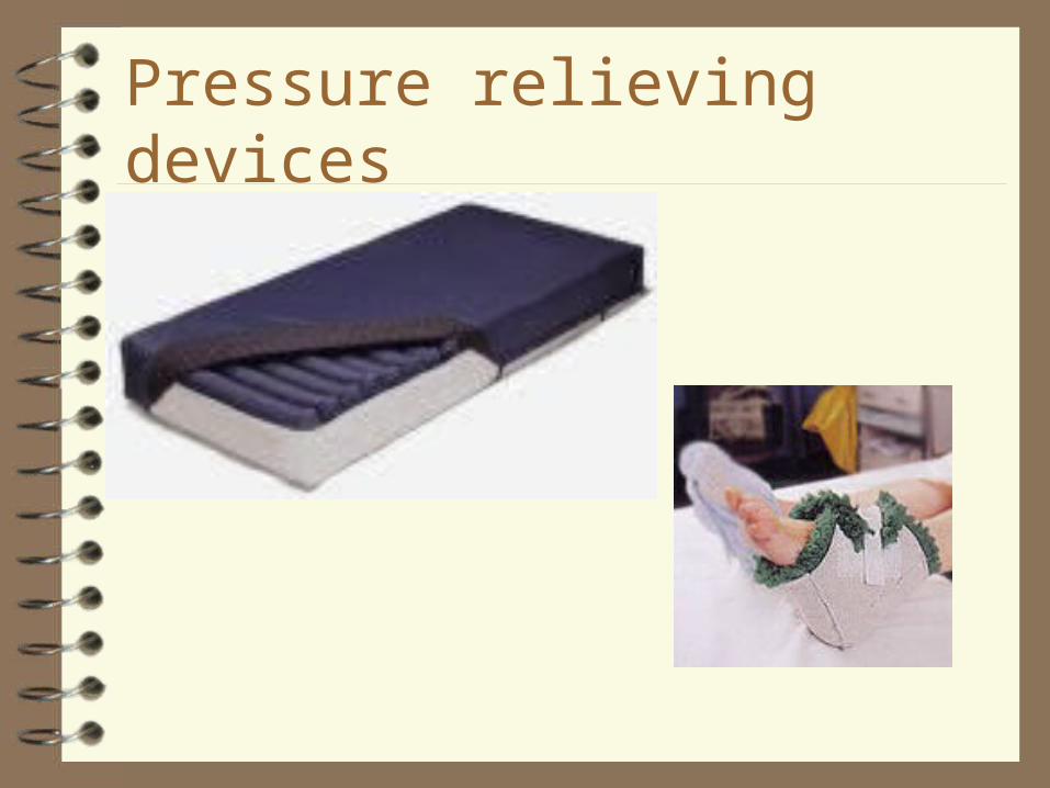
Pressure relieving devices


Hair and Nail Disorders
Hirsutism– excessive body hair
Alopecia– loss of hair or baldness
Nail Disorders– discolored, malformed, infected or separated
from underlying tissue

Fungal Infection of the Toe Nail

Burns – get ready for 3rd semester