nucleotides, nucleic acids and the genetic material this lecture will cover the discovery of the...
TRANSCRIPT
Nucleotides, nucleic acids and the genetic material
This lecture will cover the discovery of the genetic material, DNA, its
structure and the structure of nucleotides and other stuff.
It all started with Mendel
• The beginning of our thinking of the possibility of genetic material begins with Mendel. He described the genetics of pea crosses, although he did not know what genetic material was he referred to “factors” as the things which gave his pea plants their physical characteristics. While Mendel’s work gave a logical description of heredity it was largely ignored for many decades. His work was finally reproduced in the early 1900s and was thereafter regarded as fact
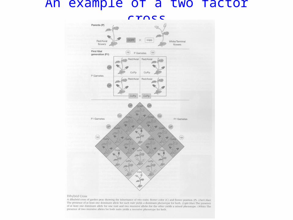
An example of a two factor cross

Nettie Sevens, Walter Sutton and Edmund Wilson, working with the giant chromosomes of Brachystola (grasshoppers) formulated the chromosomal theory
of heredity.
• A variety of chromosome types, as defined by relative size and shape, were found to be present in the nucleus of each cell. Furthermore, there usually were two copies of each type of chromosome. This cell is
called a diploid cell. • All of the cells of an organism, excluding sperm cells, egg cells, and
red blood cells, and all organisms of the same species, were observed to have the same number of chromosomes.
• The number of chromosomes in any cell appeared to double immediately prior to the cell division processes of mitosis and cytokinesis, in which a single cell splits to form two identical offspring
cells.
Chromosome theory of heredity cont.
• The sex or germ cells (i.e., sperm and egg) appeared to have exactly half of the number of chromosomes as were found in the non‑germ or somatic cells of any organism. Furthermore, the germ cells were shown to have just one copy of each chromosome type. Such cells are called haploid cells.
• The fertilization of an egg with a sperm cell produces a diploid cell called a zygote, which has the same number of chromosomes as the somatic cells of that organism.
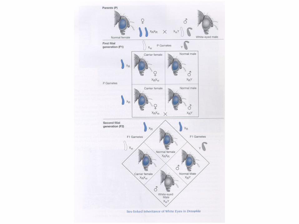
• Further they went on to show that in drosophila male gamates carried a Y chromosome while females carried an X chromosome. Females were found to be XX while males are XY. This assigned a specific trait to inheritance of a specific chromosome. Chromosomes now became the target for carrying the genetic material.
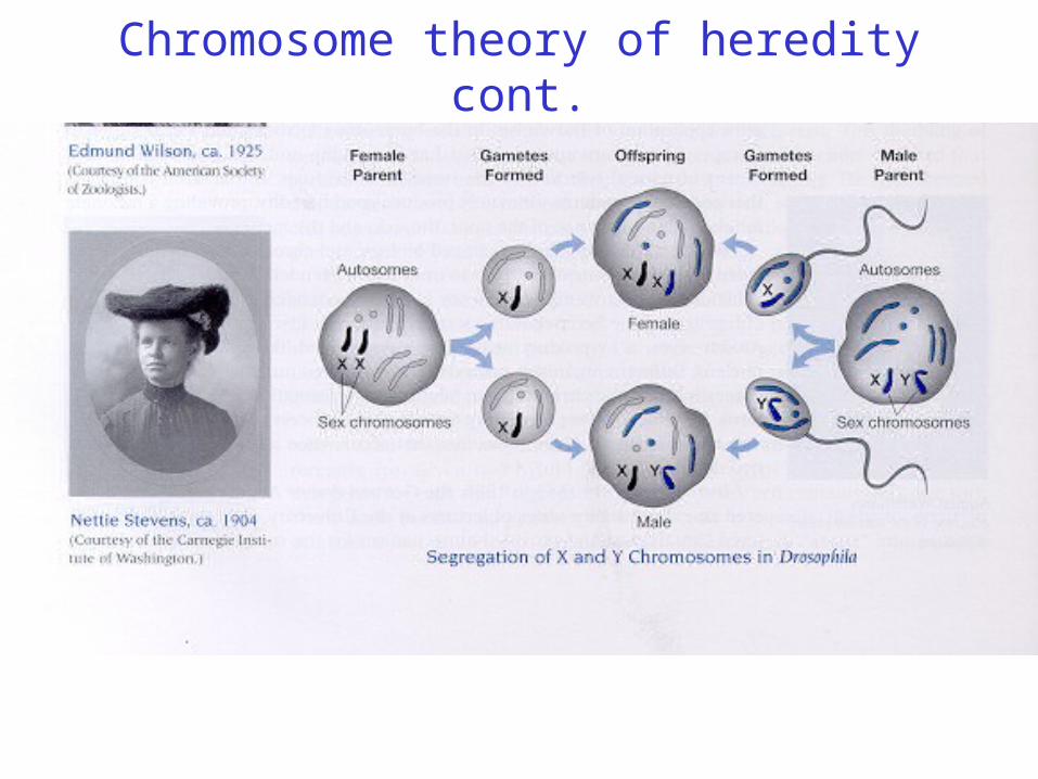
Chromosome theory of heredity cont.
.
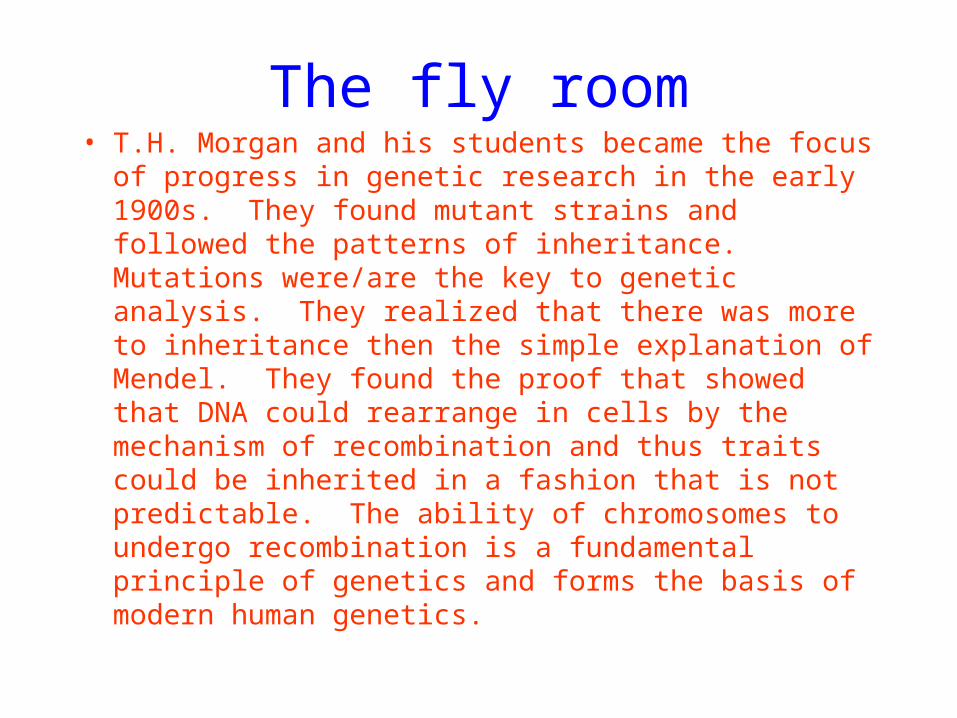
The fly room• T.H. Morgan and his students became the focus of
progress in genetic research in the early 1900s. They found mutant strains and followed the patterns of inheritance. Mutations were/are the key to genetic analysis. They realized that there was more to inheritance then the simple explanation of Mendel. They found the proof that showed that DNA could rearrange in cells by the mechanism of recombination and thus traits could be inherited in a fashion that is not predictable. The ability of chromosomes to undergo recombination is a fundamental principle of genetics and forms the basis of modern human genetics.
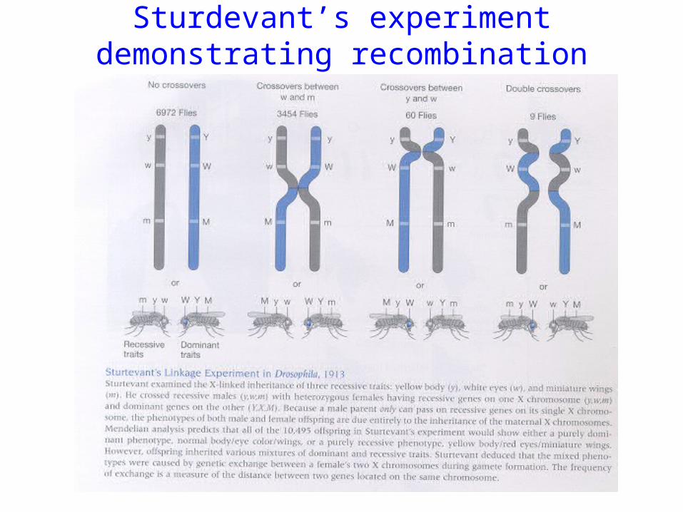
Sturdevant’s experiment demonstrating recombination
What is the actual genetic material, ie what is the composition of chromosomes?
• Quantitative analysis of chromosomes shows a composition of about forty percent DNA and sixty percent protein. At first, it seemed that protein must be responsible for carrying hereditary information, since not only is protein present in larger quantities than DNA, but protein molecules are composed of twenty different subunits while DNA molecules are composed of only four. It seemed clear that a protein molecule could encode not only more information, but a greater variety of information, because it possessed a substantially larger collection of ingredients
with which to work.
Archibald Garrod
• a British physician, hypothesized that various metabolic deficiencies seen in his patients were due to the lack of a specific enzyme missing because of a defect in the genetic material inherited from birth.
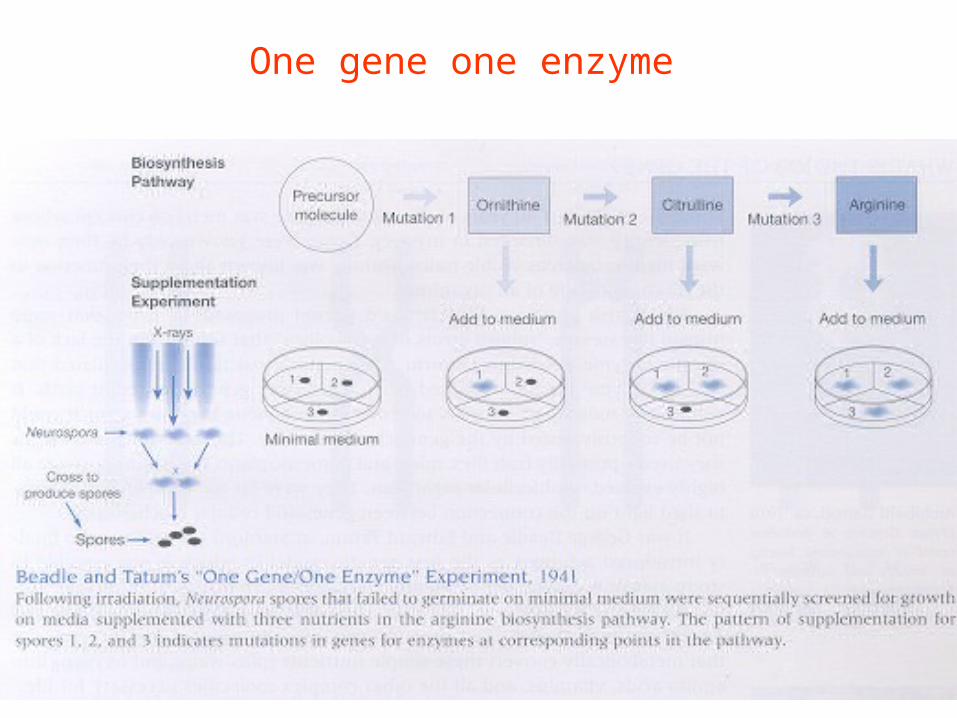
One gene one enzyme
• Three decades Beadle and Tatum would refine this idea with their “one gene one enzyme hypothesis”. These investigators worked on Neurospora and found that if they irradiated spores they “induced” mutations. These mutations were detected as the spores inability to germinate on various defined media in which essential nutrients were omitted. This suggested that a mutation in a specific gene involved in the synthesis of say for instance an amino acid rendered the gene inactive and so no functional protein was made. The experiment shown on
the next slide exemplifies their work.
One gene one enzyme
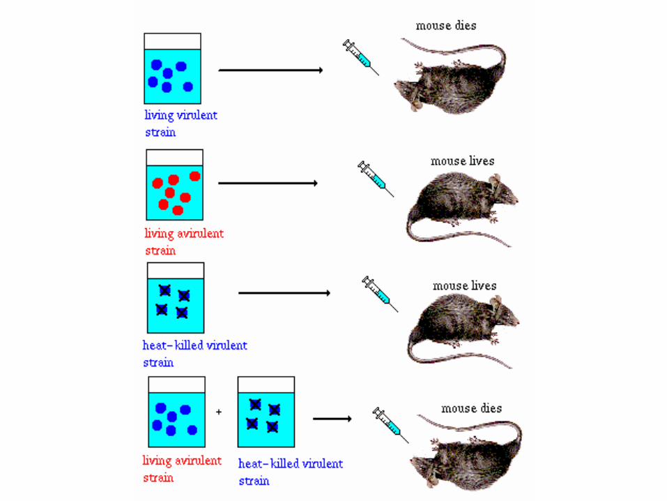
Fred Griffith• In 1928 Fred Griffith, working with the Dipplococcus pneumonia
bacteria found that there was a virulent and nonvirulent form of the bacterium. When injected into mice the virulent bacteria caused death while the mice injected with a non virulent bacteria remained healthy. He next went on to heat kill the virulent bacteria and showed that they could no longer kill the mice. However, if mixed with nonvirulent bacteria the mice again died. Furthermore, bacteria isolated from these mice were virulent having now become virulent. Griffith, postulated that there was a transforming factor which survived heating in the virulent bacteria which could then be transferred to the
nonvirulent a bacteria.
What should have been the definitive experiment
• Griffith’s experiments further defined the gene but brought us no closer to understanding the composition of genes. Then in the 1940s a group of scientists at Rockefeller University carried out a study to finally identify the genetic material. Again working with Dipplococcus pneumonia, these investigators, Avery, McCarthy, and MacLeod first showed that they could convert non infectious rough (R) pneumococcus into smooth (S) virulent pneumococcus by mixing heat killed (S) with live (R) and plating them onto plates got smooth bacteria. This became their assay. Next they isolated the material in (S) that transformed (R). They began with (S) bacteria and isolated DNA by alcohol precipitating and then spooling it out. This material was able to transform (R). This material was exhaustively extracted to remove any protein. And again it transformed. Next they treated this material with RNAse, no effect, protease, no effect and finally DNAse. The DNAse killed the transforming activity and so they concluded that DNA was the genetic material. This was not widely accepted.
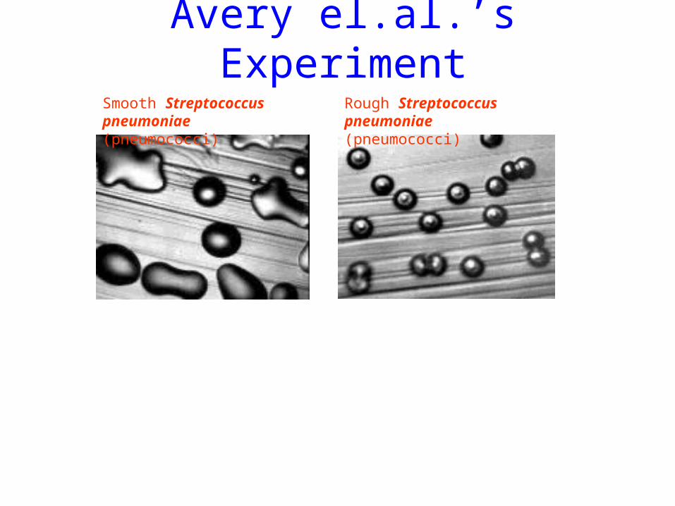
Avery el.al.’s ExperimentSmooth Streptococcus pneumoniae (pneumococci)
Rough Streptococcus pneumoniae (pneumococci)
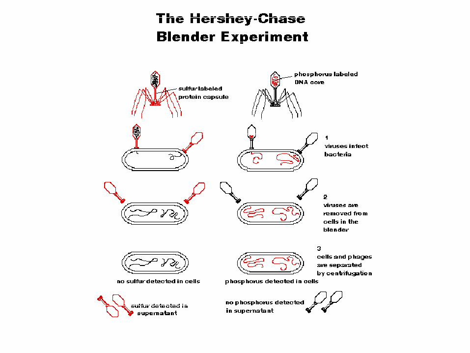
Hershey Chase blender experiment. Okay, okay its DNA• Next Hershey and Chase performed their famous
blender experiment. Here they used radioactivity to label phage DNA with 32P and protein with 35S. These phage were used to infect bacteria, then placed in a blender to remove the phage and the bacteria collected by centrifugation. The 32P was inside the bacteria while the 35S remained on the phage in suspension.
DNA structure
• Now we knew that DNA was the genetic material but how did it work? One approach to figuring this out was to understand its structure and from there Watson and Crick reasoned its function would become apparent. This team of a former physicist and a biologist and an original “Wiz Kid” worked primarily by building models base on the work of others.
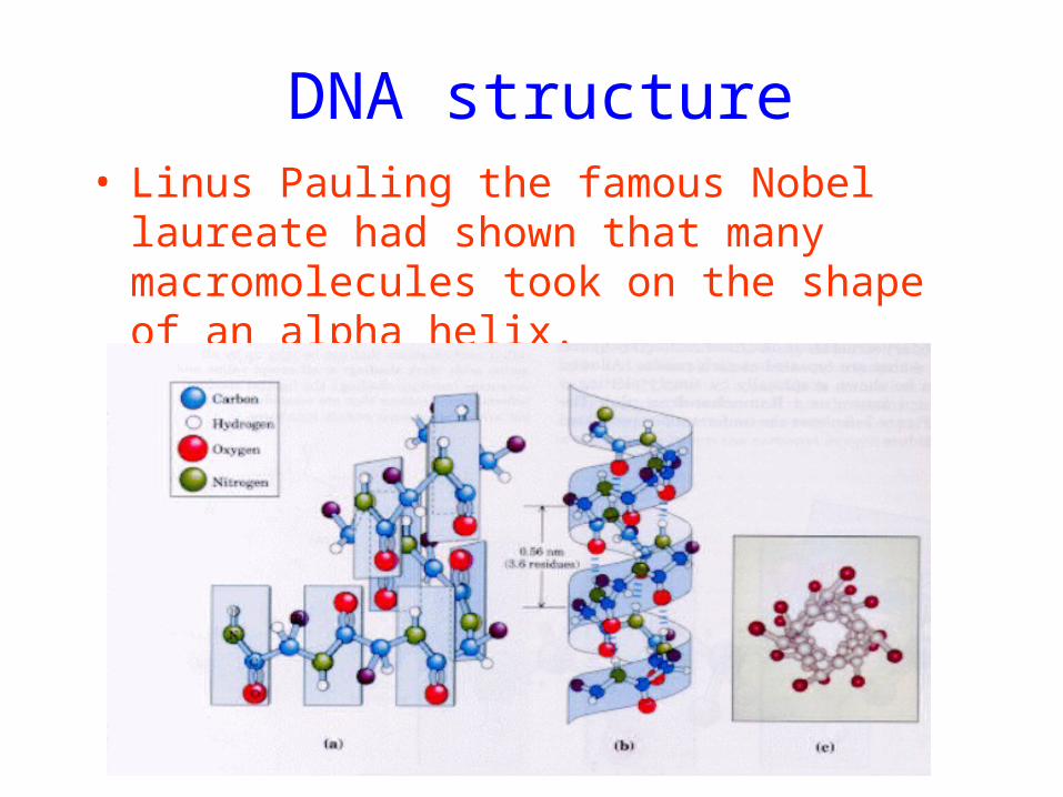
DNA structure• Linus Pauling the famous Nobel laureate had
shown that many macromolecules took on the shape of an alpha helix.
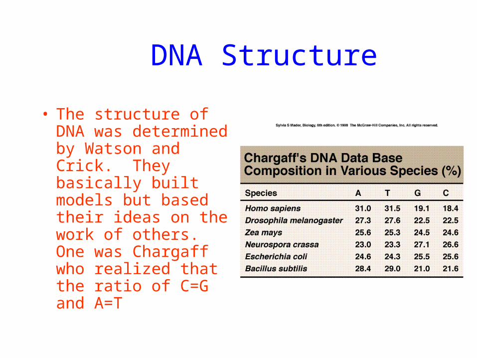
DNA Structure
• The structure of DNA was determined by Watson and Crick. They basically built models but based their ideas on the work of others. One was Chargaff who realized that the ratio of C=G and A=T
DNA structure
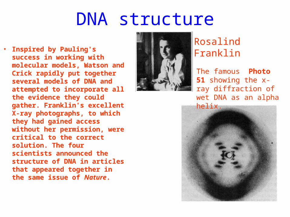
• Inspired by Pauling's success in working with molecular models, Watson and Crick rapidly put together several models of DNA and attempted to incorporate all the evidence they could gather. Franklin's excellent X-ray photographs, to which they had gained access without her permission, were critical to the correct solution. The four scientists announced the structure of DNA in articles that appeared together in the same issue of Nature.
Rosalind Franklin
The famous Photo 51 showing the x-ray diffraction of wet DNA as an alpha helix.
DNA structure
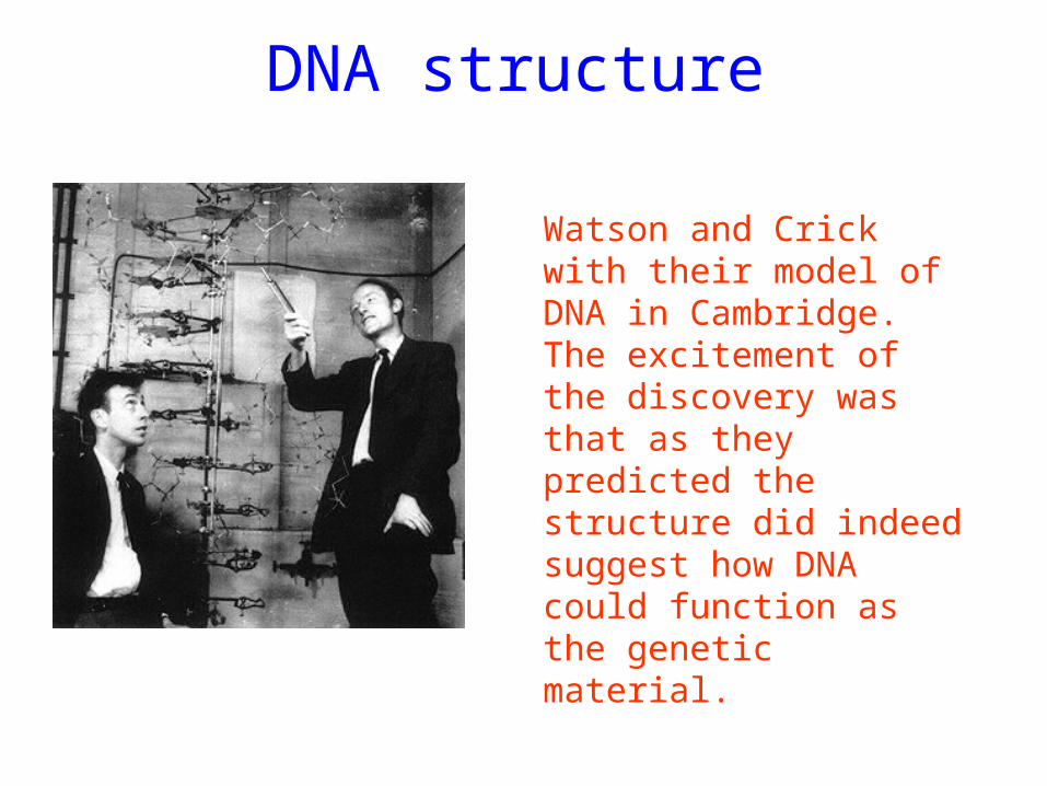
Watson and Crick with their model of DNA in Cambridge. The excitement of the discovery was that as they predicted the structure did indeed suggest how DNA could function as the genetic material.
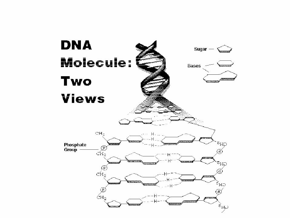
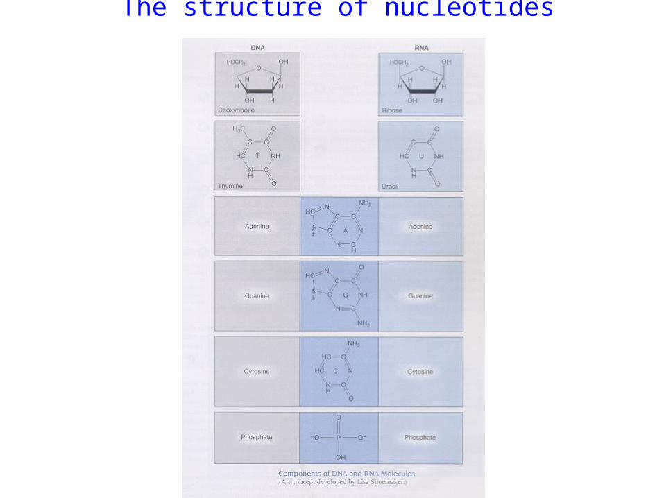
The structure of nucleotides
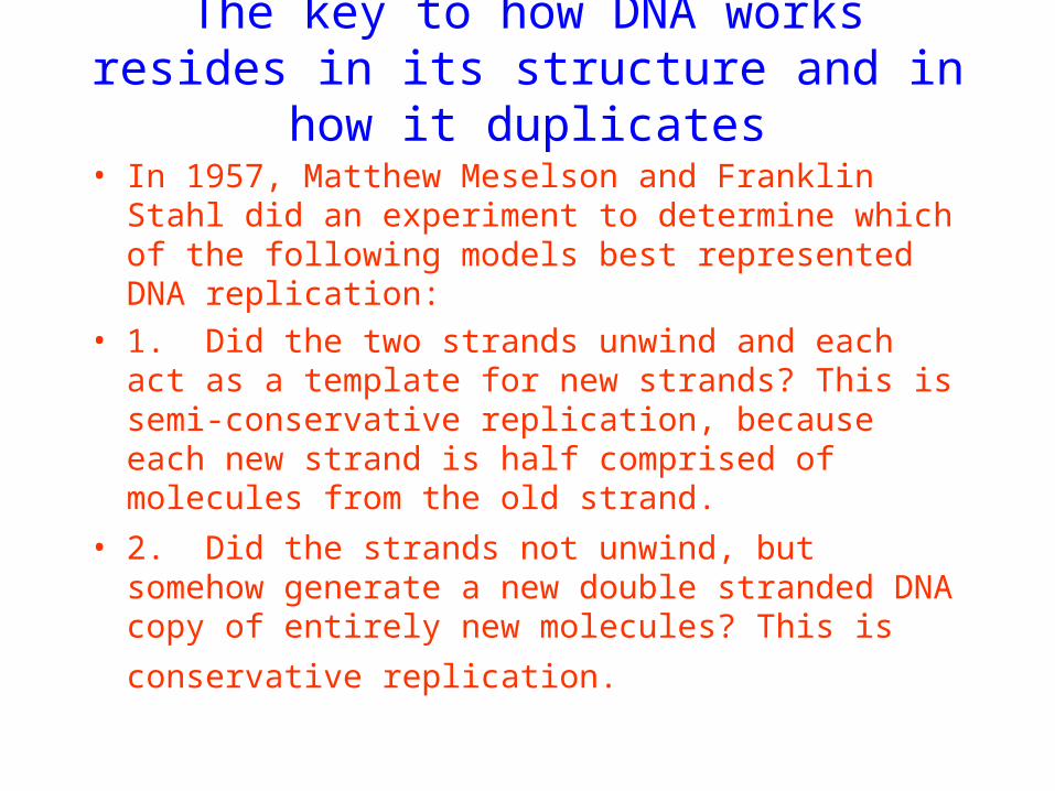
The key to how DNA works resides in its structure and in how it duplicates
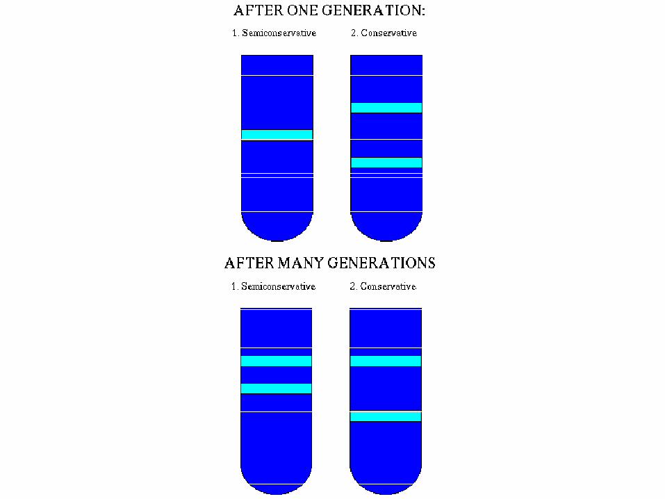
• In 1957, Matthew Meselson and Franklin Stahl did an experiment to determine which of the following models best represented DNA replication:
• 1. Did the two strands unwind and each act as a template for new strands? This is semi-conservative replication, because each new strand is half comprised of molecules from the old strand.
• 2. Did the strands not unwind, but somehow generate a new double stranded DNA copy of entirely new
molecules? This is conservative replication.
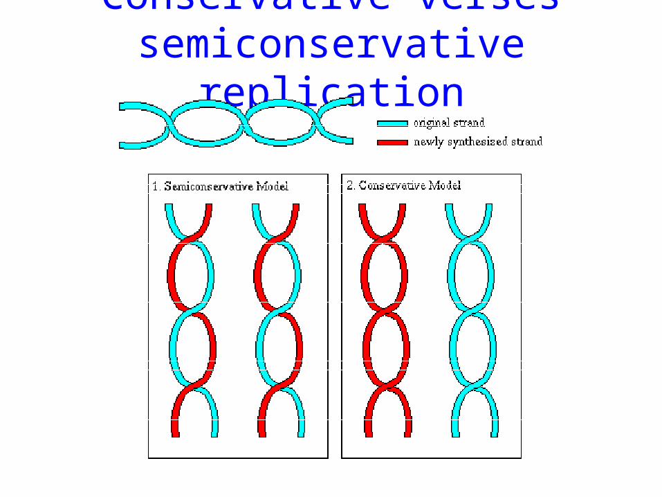
Conservative verses semiconservative replication
The experiment
• In order to determine which of these models was true, the following experiment was performed: The original DNA strand was labelled with the heavy isotope of nitrogen, N‑15. This DNA was allowed to go through one round of replication with N‑14, and then the mixture was centrifuged so that the heavier DNA would form a band lower in the tube, and the intermediate (one N‑15 strand and one N‑14 strand) and light DNA (all N‑14) would appear as a band higher in the tube. The expected results for each model were:
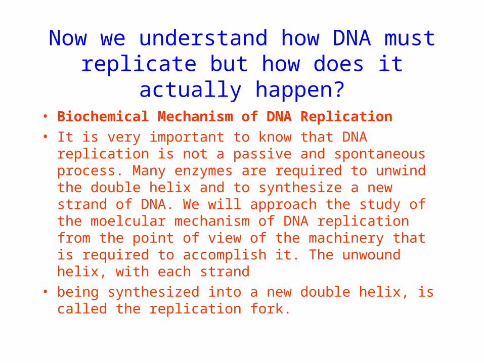
Now we understand how DNA must replicate but how does it actually happen?
• Biochemical Mechanism of DNA Replication
• It is very important to know that DNA replication is not a passive and spontaneous process. Many enzymes are required to unwind the double helix and to synthesize a new strand of DNA. We will approach the study of the moelcular mechanism of DNA replication from the point of view of the machinery that is required to accomplish it. The unwound helix, with each strand
• being synthesized into a new double helix, is called the replication fork.
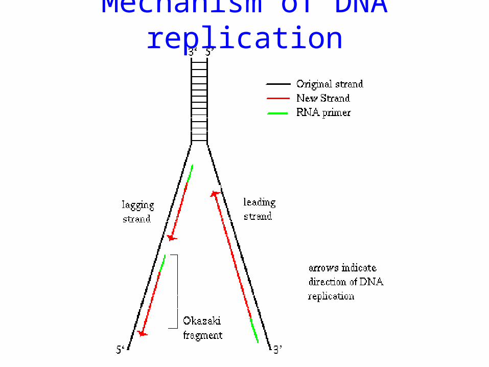
Mechanism of DNA replication
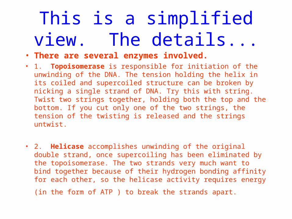
This is a simplified view. The details...
• There are several enzymes involved.• 1. Topoisomerase is responsible for initiation of the unwinding of the
DNA. The tension holding the helix in its coiled and supercoiled structure can be broken by nicking a single strand of DNA. Try this with string. Twist two strings together, holding both the top and the bottom. If you cut only one of the two strings, the tension of the twisting is released and the strings untwist.
• 2. Helicase accomplishes unwinding of the original double strand, once supercoiling has been eliminated by the topoisomerase. The two strands very much want to bind together because of their hydrogen bonding affinity for each other, so the helicase activity requires energy
(in the form of ATP ) to break the strands apart.
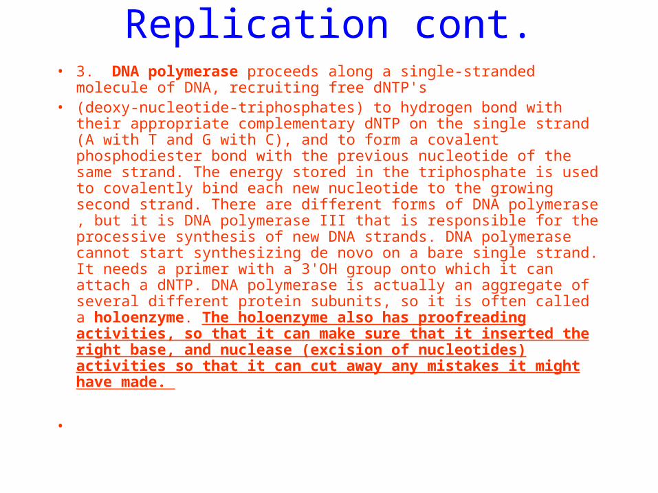
Replication cont.• 3. DNA polymerase proceeds along a single‑stranded molecule of
DNA, recruiting free dNTP's• (deoxy‑nucleotide‑triphosphates) to hydrogen bond with their
appropriate complementary dNTP on the single strand (A with T and G with C), and to form a covalent phosphodiester bond with the previous nucleotide of the same strand. The energy stored in the triphosphate is used to covalently bind each new nucleotide to the growing second strand. There are different forms of DNA polymerase , but it is DNA polymerase III that is responsible for the processive synthesis of new DNA strands. DNA polymerase cannot start synthesizing de novo on a bare single strand. It needs a primer with a 3'OH group onto which it can attach a dNTP. DNA polymerase is actually an aggregate of several different protein subunits, so it is often called a holoenzyme. The holoenzyme also has proofreading activities, so that it can make sure that it inserted the right base, and nuclease (excision of nucleotides) activities so that it can cut away any mistakes it might have made.
•
More replication.
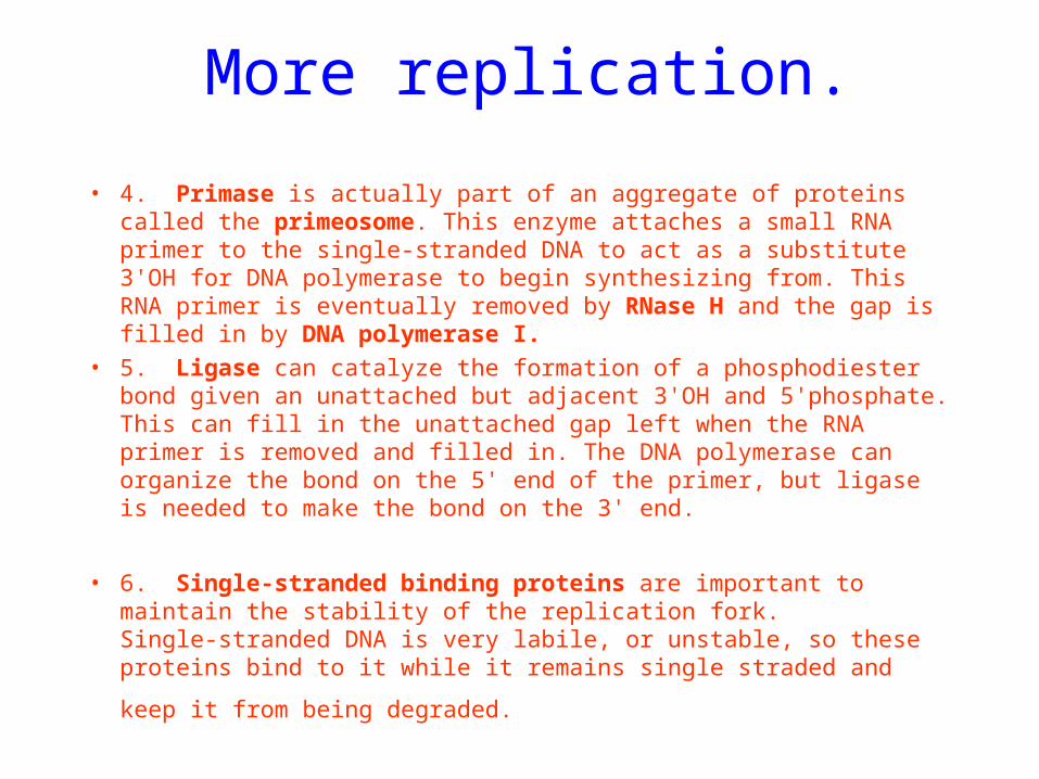
• 4. Primase is actually part of an aggregate of proteins called the primeosome. This enzyme attaches a small RNA primer to the single‑stranded DNA to act as a substitute 3'OH for DNA polymerase to begin synthesizing from. This RNA primer is eventually removed by RNase H and the gap is filled in by DNA polymerase I.
• 5. Ligase can catalyze the formation of a phosphodiester bond given an unattached but adjacent 3'OH and 5'phosphate. This can fill in the unattached gap left when the RNA primer is removed and filled in. The DNA polymerase can organize the bond on the 5' end of the primer, but ligase is needed to make the bond on the 3' end.
• 6. Single‑stranded binding proteins are important to maintain the stability of the replication fork. Single‑stranded DNA is very labile, or unstable, so these proteins bind to it while it remains single straded
and keep it from being degraded.
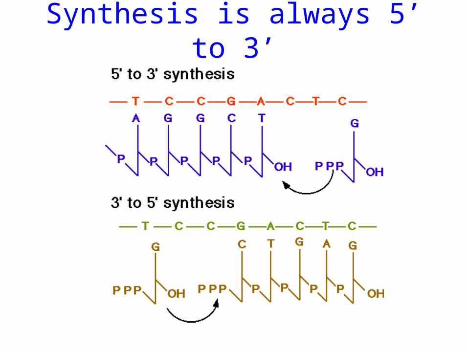
Synthesis is always 5’ to 3’
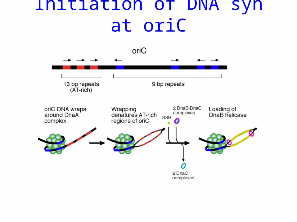
Initiation of DNA syn at oriC
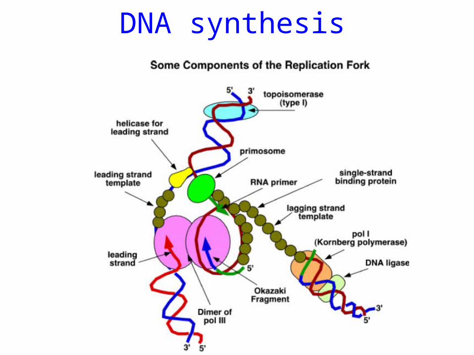
DNA synthesis
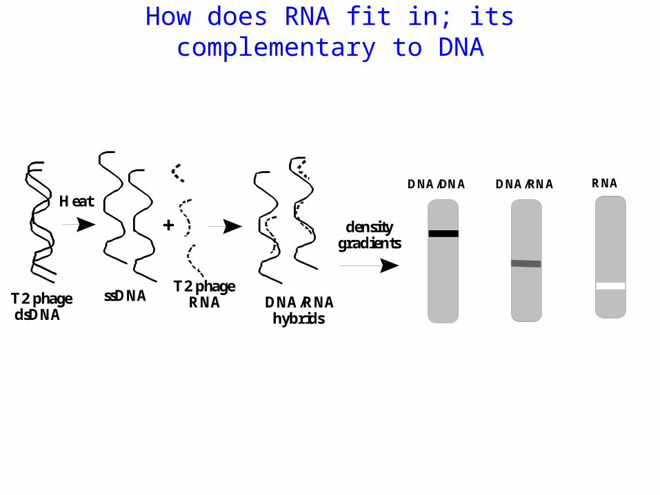
How does RNA fit in; its complementary to DNA
Heat
T2 phage dsDNA
ssDNAT2 phage RNA
+
DNA/RNA hybrids
DNA/DNA DNA/RNA RNA
densitygradients
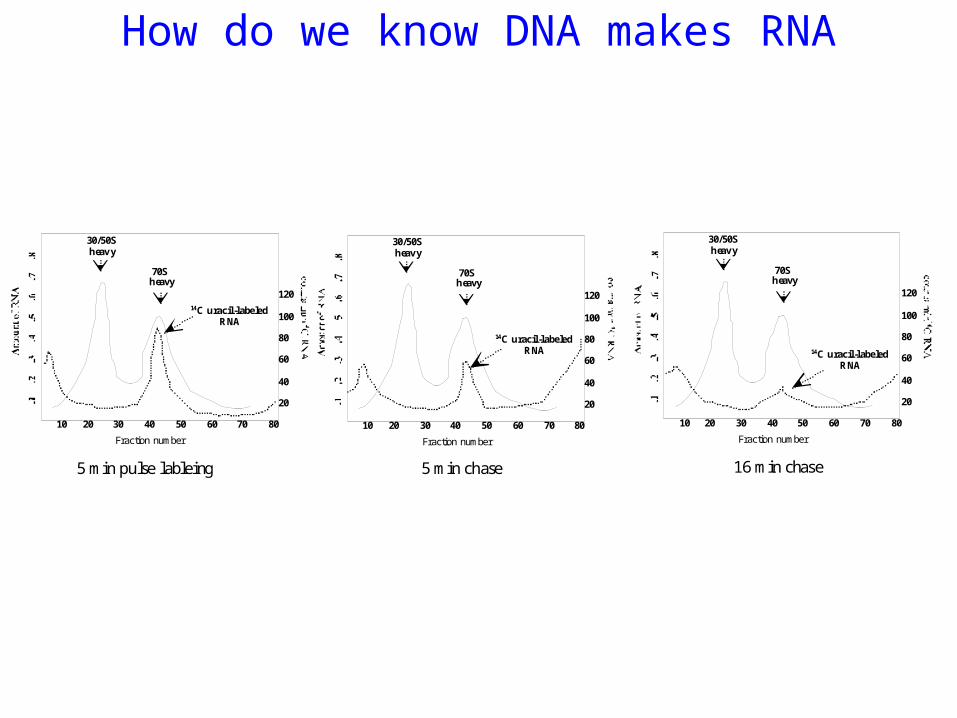
How do we know DNA makes RNA
70S heavy
30/50S heav y
10 20 30 40 50 60 70 80
14C uracil-labeled RNA
120
100
80
60
40
20
Fraction number
70S heavy
30/50S heav y
10 20 30 40 50 60 70 80
14C uracil-labeled RNA
120
100
80
60
40
20
Fraction number
70S heavy
30/50S heav y
10 20 30 40 50 60 70 80
14C uracil-labeled RNA
120
100
80
60
40
20
Fraction number
5 min pulse lableing 5 min chase 16 min chase
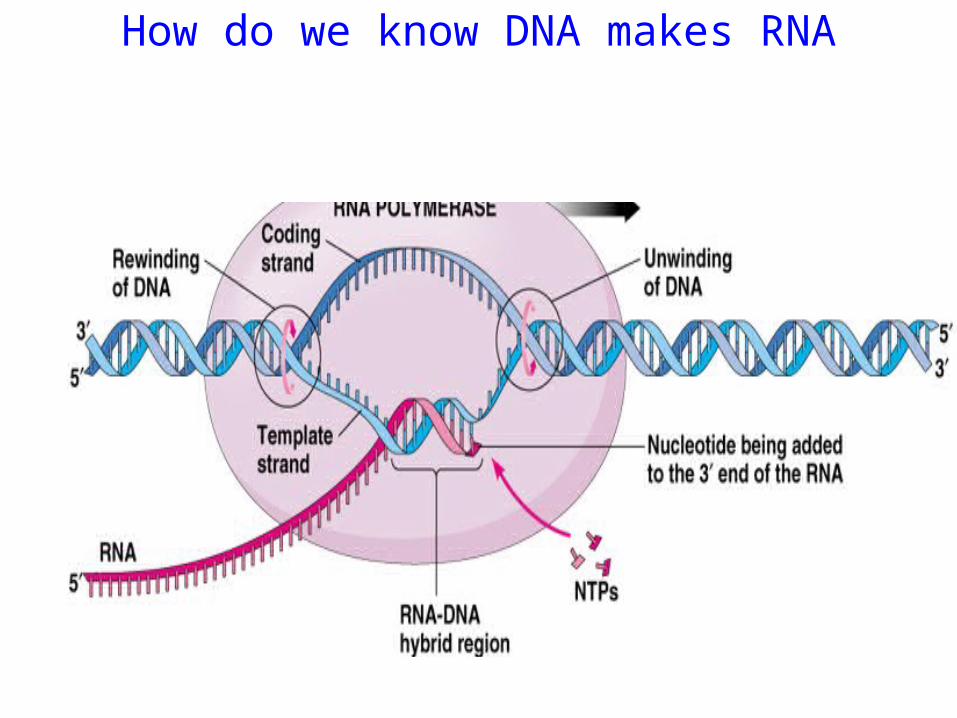
How do we know DNA makes RNA