nuclear matrix protein matrin3 regulates alternative
TRANSCRIPT

Article
Nuclear matrix protein Matrin3 regulatesalternative splicing and forms overlappingregulatory networks with PTBMiguel B Coelho1, Jan Attig2,3, Nicolás Bellora4,5,6, Julian König3,†, Martina Hallegger1,2, Melis Kayikci3,
Eduardo Eyras4,5, Jernej Ule2 & Christopher WJ Smith1,*
Abstract
Matrin3 is an RNA- and DNA-binding nuclear matrix protein foundto be associated with neural and muscular degenerative diseases.A number of possible functions of Matrin3 have been suggested,but no widespread role in RNA metabolism has yet been clearlydemonstrated. We identified Matrin3 by its interaction with thesecond RRM domain of the splicing regulator PTB. Using a combi-nation of RNAi knockdown, transcriptome profiling and iCLIP, wefind that Matrin3 is a regulator of hundreds of alternative splicingevents, principally acting as a splicing repressor with only a smallproportion of targeted events being co-regulated by PTB. Incontrast to other splicing regulators, Matrin3 binds to an extendedregion within repressed exons and flanking introns with no sharplydefined peaks. The identification of this clear molecular function ofMatrin3 should help to clarify the molecular pathology of ALS andother diseases caused by mutations of Matrin3.
Keywords alternative splicing; Matrin3; PTB
Subject Categories RNA Biology
DOI 10.15252/embj.201489852 | Received 19 August 2014 | Revised 12 December
2014 | Accepted 15 December 2014 | Published online 19 January 2015
The EMBO Journal (2015) 34: 653–668
Introduction
Alternative splicing (AS) provides multi-cellular eukaryotes with a
proteomic capacity that far exceeds the number of genes (Nilsen &
Graveley, 2010). AS is an integral part of regulated programs of gene
expression, often acting in concert with transcriptional control, but
affecting different functionally related sets of genes (Blencowe,
2006). Regulation of AS is dictated primarily by RNA-binding
proteins (RBPs) that can bind to specific RNA sequence elements
and which can act as either activators of repressors (Coelho &
Smith, 2014). Splicing predominantly occurs co-transcriptionally
(Carrillo Oesterreich et al, 2011) in a chromatin context, and this
temporal and spatial context provides additional layers of regulatory
input into splicing decisions (Braunschweig et al, 2013). Neverthe-
less, RNA-binding proteins remain the key ‘readers’ of splicing
codes (Barash et al, 2010). RBPs typically have one or more RNA-
binding domains, and exhibit varying degrees of specificity, usually
recognizing sequence motifs of ~3–5 nt (Ray et al, 2013). While
much has been learned about the action of individual RBPs binding
to their cognate binding sites, the combinatorial nature of splicing
regulation has led to an increased focus on the ways in which
groups of regulatory proteins can act together (Barash et al, 2010;
Campbell et al, 2012; Zhang et al, 2013; Cereda et al, 2014).
Polypyrimidine tract binding (PTB/PTBP1/hnRNPI) protein is an
intensively investigated RNA-binding protein, which regulates splic-
ing and other post-transcriptional steps of gene expression
(reviewed in Kafasla et al, 2012; Keppetipola et al, 2012; Sawicka
et al, 2008). PTB binds to pyrimidine-rich motifs with core CU dinu-
cleotides (Singh et al, 1995; Perez et al, 1997; Ray et al, 2013), and
each of its four RRM (RNA recognition motif) family domains
(Fig 1A) can recognize such motifs (Oberstrass et al, 2005).
Although primarily characterized as a repressive splicing regulator,
it can also activate some splice sites and this has been related to
differential positions of binding relative to regulated exons (Xue
et al, 2009; Llorian et al, 2010). Although PTB can act alone as a
regulator (Amir-Ahmady et al, 2005), genome-wide analyses suggest
that it cooperates with a number of other proteins as a component of
‘tissue spicing codes’ (Castle et al, 2008; Wang et al, 2008; Barash
et al, 2010; Bland et al, 2010; Llorian et al, 2010). Structure-function
analysis has indicated that despite their similar RNA-binding prefer-
ences, the four RRMs of PTB show functional diversification (Liu
et al, 2002; Robinson & Smith, 2006; Mickleburgh et al, 2014). Of
particular importance for synergistic action with other regulators,
1 Department of Biochemistry, University of Cambridge, Cambridge, UK2 Department of Molecular Neuroscience, UCL Institute of Neurology, London, UK3 MRC-Laboratory of Molecular Biology, Cambridge, UK4 Computational Genomics, Universitat Pompeu Fabra, Barcelona, Spain5 Catalan Institute for Research and Advanced Studies (ICREA), Barcelona, Spain6 INIBIOMA, CONICET-UNComahue, Bariloche, Argentina
*Corresponding author. Tel: +44 1223 333655; E-mail: [email protected]†Present address: Institute of Molecular Biology gGmbH (IMB), Mainz, Germany
ª 2015 The Authors. Published under the terms of the CC BY 4.0 license The EMBO Journal Vol 34 | No 5 | 2015 653
Published online: January 19, 2015

RRM2 can interact with both RNA via its canonical b-sheet surface,and with short linear PRI (PTB-RRM Interaction) motifs found in the
co-regulator Raver1 (Rideau et al, 2006; Joshi et al, 2011). The PRI
motif is defined by the consensus sequence [S/G][IL]LGxΦP and
binds to the dorsal surface of PTB RRM2, with Tyr247 of PTB partic-
ularly critical for this interaction (Rideau et al, 2006; Joshi et al,
2011). PTB RRM2, along with the following linker sequence, is suffi-
cient for splicing repressor activity when artificially tethered as an
MS2 fusion protein (Robinson & Smith, 2006) (Fig 1A). Despite the
fact that Raver1 can act with PTB as a co-regulator of Tpm1 splicing
(Gromak et al, 2003; Rideau et al, 2006), Raver1 null mice showed
no alteration in Tpm1 splicing (Lahmann et al, 2008) and knock-
down of Raver1 in HeLa cells showed only a few changes in alterna-
tive splicing (Hallegger et al, manuscript in preparation). Therefore,
it remains possible that other co-regulatory proteins with PRI motifs
might interact with PTB RRM2.
Matrin3 is one of the most abundant inner nuclear matrix
proteins (Nakayasu & Berezney, 1991). The main isoforms of
Matrin3 are over 800 amino acids in size, but most of the protein is
not comprised of structurally characterized domains, with the
exception of two DNA-binding C2H2 zinc finger (ZF) and two RRM
domains (Hibino et al, 2006), and a bi-partite nuclear localization
signal (NLS) (Hisada-Ishii et al, 2007) (Fig 2A). Matrin3 is essential
for viability of some cells (Hisada-Ishii et al, 2007; Przygodzka et al,
2010), and alterations in Matrin3 levels are associated with some
diseases (Bernert et al, 2002; Bimpaki et al, 2009). Moreover,
missense mutations in Matrin3 have been associated with asymmet-
ric myopathy with vocal cord paralysis (Senderek et al, 2009) and
amyotrophic lateral sclerosis (ALS) (Johnson et al, 2014). Matrin3 is
located diffusely throughout the nucleoplasm and is concentrated in
the nuclear scaffold (Zeitz et al, 2009), and its DNA- and RNA-
binding domains suggest that it may play roles in processes
associated with the nuclear matrix or nucleoplasm. It can anchor
chromosomes to the nucleus matrix by binding to the MAR/SAR
elements (Hibino et al, 1992). Introduction of MAR/SAR sites
upstream of a promoter stimulates transcription, suggesting Matrin3
binding to these elements might promote transcription (Hibino et al,
2000), a suggestion supported by the proximity of Matrin3 with
RNA Pol II promoters (Malyavantham et al, 2008) and enhancers
(Skowronska-Krawczyk et al, 2014). Matrin has also been shown to
be involved in the early stages of the DNA double-strand break
response (Salton et al, 2010). A number of functional roles
A B
D
C
Figure 1. PTB RRM2 interacts with multiple RNA-binding proteins.
A Schematic representation of PTB (top) and the GST-PTB RRM2 (below), with the limits of the PTB minimal repressor domain indicated. PTB is composed of four RNArecognition motifs (RRM) with three linker regions in between them. It also contains a N-terminus bipartite nuclear localization signal (NLS) as well as a nuclearexport signal (NES). The GST-PTB RRM2 is composed of the second RRM fused to a GST tag in the N-terminus.
B Silverstain of the GST-PTB RRM2 pull-down of the wild-type RRM2 and the Y247Q mutant. Five microlitre of the pull-down was run on a 15% SDS–PAGE and silver-stained. Three strong bands can be seen which are due to the recombinant protein and the beads used for the pull-down. The region encompassing 50 kDa to thetop of the gel was sliced and subjected to in-gel digestion and mass spectrometry. The two strongest bands visible in this region are labelled as Matrin3 and Raver1.
C Proteins identified in the GST-PTB RRM2 pull-down ranked by their unique peptide number. The table shows the different proteins we found binding to RRM2, aswell as when present, the sequence of the PRI motif. The indicated function is only a guideline as many have more functions than shown.
D Western blot of the GST pull-down using antibodies against Matrin3 and Raver1. Lanes 1 and 2 show 5 and 10% of input, respectively, lanes 3 and 4 show GST-PTBfull-length pull-down of wild-type and Y247Q mutant, respectively, and lanes 5 and 6 show GST-PTB RRM2 pull-down of wild-type and Y247Q mutant, respectively,and lane 7 with pull-down using GST alone.
Source data are available online for this figure
The EMBO Journal Vol 34 | No 5 | 2015 ª 2015 The Authors
The EMBO Journal Matrin3 regulates splicing Miguel B Coelho et al
654
Published online: January 19, 2015

associated with cellular and viral RNA have been suggested for
Matrin3 including mRNA stabilization (Salton et al, 2011), nuclear
retention of hyperedited RNA (Zhang & Carmichael, 2001) and Rev-
dependent export of unspliced HIV1 RNA in conjunction with PTB-
associated factor (PSF) (Kula et al; Kula et al, 2011; Yedavalli &
Jeang, 2011). Matrin3 interacts with a number of splicing regulators
including hnRNPK (Salton et al, 2011), hnRNPL, SFRS7, p68 (Zeitz
et al, 2009), NOVA-1/-2 (Polydorides et al, 2000), CTCF (Fujita &
Fujii, 2011; Shukla et al, 2011), as well as the transcription machin-
ery itself (Das et al, 2007). Despite the interactions with splicing
factors, there is no direct evidence for Matrin3 functioning as a
splicing regulator.
Here, we set out to identify nuclear proteins that interact with
PTB RRM2. Matrin3 was the major interacting protein in HeLa
nuclear extracts, interacting via a single PRI motif that is necessary
and sufficient for interaction. Using RNAi knockdown and splice-
sensitive microarray analysis in conjunction with iCLIP of Matrin3
and PTB, we find that Matrin3 acts widely as a splicing regulator.
While a number of its target splicing events are shared with PTB,
the majority are PTB independent and involve Matrin3 action as a
repressor. Matrin3 binding was observed in the introns flanking
repressed exons, but in contrast with other splicing regulators, the
binding occurred to an extended region with no clear peaks. Struc-
ture-function analysis indicates that Matrin3 splicing activity
requires both the RRM domains and the PRI, even for ASEs that are
not co-regulated by PTB.
Results
Identification of PTB RRM2 binding partners
With the aim of understanding better the function of the minimal
PTB repressor domain, we carried out a proteomic screen to identify
interacting protein partners of PTB RRM2, the main component of
the repressor domain (Robinson & Smith, 2006). PTB RRM2 was
fused to GST in wild-type (WT) and Y247Q mutant form, which
impairs interaction with Raver1 PRI peptides (Joshi et al, 2011)
(Fig 1A), and used as bait to pull down interacting proteins from
HeLa nuclear extracts. Numerous proteins bound to WT RRM2 but
not the Y247Q mutant (Fig 1B). Proteins pulled down by WT
GST-RRM2 were identified by mass spectrometry (Fig 1C and
Supplementary Table S1). They include RNA-binding proteins
(MATR3, RAVER1, HNRNPM, RBMX, DDX5, DDX3X, SFRS15,
DDX17, HNRNPH1 and PTB itself), proteins with role in transcrip-
tion regulation (CCAR1, KIAA1967/CCAR2 and RUVBL1/2) and a
protein found in 30 end processing complexes (WDR33). Four of the
five unique PTB peptides are located within RRM2 and so could
derive from the bait protein. Proteins with roles in transcription and
30 end processing may present links to unknown activities of PTB in
the case of transcription regulation, and in the case of WDR33 (Shi
et al, 2009), a molecular link to an already reported function of PTB
in 30 end processing (Moreira et al, 1998; Castelo-Branco et al,
2004).
The strongest protein interaction detected, as indicated by
number of unique peptides and MASCOT score, was the nuclear
matrix protein Matrin3, followed by Raver1 (Fig 1C and D). These
two proteins correspond to the major protein bands interacting
specifically with WT but not mutant RRM2 (Fig 1B, arrows).
Matrin3, CCAR1, KIAA1967 and WDR33 all have potential PRI
motifs similar to those in Raver1 (Figs 1C and 2A). We validated the
Matrin3-PTB interaction by Western blot of GST-RRM2 and full-
length GST-PTB pull-downs, comparing wild-type (WT) and Y247Q
mutant proteins (Fig 1D). Both Matrin3 and Raver1 interacted
strongly with GST-RRM2 and GST-PTB proteins, and in both cases,
the Y247Q mutation abolished the interaction. This indicates that
the RRM2 interaction is sufficient and also necessary in the context
of full-length GST-PTB for interaction with Matrin3 and Raver1
(Fig 1D). However, while Matrin3 interacted equally well with RRM2
or full-length PTB, Raver1 interacted more strongly with full-length
PTB, suggesting that other regions of PTB may also contact Raver1.
Matrin3 PRI motif is necessary and sufficient for PTB interaction
Matrin3 is a large nuclear protein with 847 amino acids that can
bind both to DNA via two C2H2 zinc finger domains (ZF1 and ZF2)
and to RNA by its tandem RNA recognition motifs (RRM1 and
RRM2) (Hibino et al, 2006). A potential PRI motif, GILGPPP, is
located between ZF1 and RRM1. This matches the PRI consensus
(Fig 2A) and is located in a disordered region, which is important
for the function of short linear motifs (Dinkel et al, 2014). More-
over, the motif is absolutely conserved across 84 mammalian, avian,
reptilian and amphibian species (UCSC browser, Vertebrate Multiz
Alignment & Conservation, 100 Species). In order to test whether
the GILGPPP motif is functional, we mutated it to GAAAPPA
(mutated residues underlined) in a FLAG-tagged Matrin3 expression
vector and tested the effect on PTB binding by anti-FLAG co-immu-
noprecipitation. As control, we used wild-type Raver1 and a mutant
with all four PRI motifs mutated (Rideau et al, 2006). FLAG-tagged
Matrin3 and Raver1 both co-immunoprecipitated PTB (Fig 2B).
Mutation of the single PRI in Matrin3 nearly eliminated PTB co-
immunoprecipitation (lane 3), a more emphatic effect than muta-
tion of the Raver1 PRI motifs (Fig 2B, lane 5). We next tested
whether the Matrin3 PRI is sufficient for binding to PTB. We in
vitro transcribed and translated the Matrin3 and the Raver1_1 PRIs
(Fig 2A) fused to the bacteriophage MS2 coat protein. Both the
Raver1 and Matrin3 peptides were pulled down by GST-PTB
(Fig 2C, lanes 1, 3). As negative controls, no binding was observed
to an unrelated RNA-binding protein, GST-SXL, and MS2 alone
was not pulled down by GST-PTB or GST-SXL (lane 2). The speci-
ficity of the interaction was demonstrated by mutation to alanine
of the conserved leucine-3 of the PRI, which strongly impaired
binding to PTB (Fig 2C, lane 4). These data therefore demonstrate
that the PRI motif of Matrin3 is both necessary and sufficient for
interaction with PTB.
Matrin3 is a widespread regulator of alternative splicing
The interaction of Matrin3 with PTB led us to hypothesize that it
may play a role in the co-regulation of some PTB-regulated alterna-
tive splicing events (ASEs). To test this hypothesis, we transfected
HeLa cells with siRNAs targeting the Matrin3 mRNA and observed a
> 90% decrease in the Matrin3 protein levels (Fig 3A). Total RNA
from knockdown and control samples was purified and analysed
using Human Junction microarrays (HJAY), containing probe sets
for all annotated human exons and exon–exon junctions (Llorian
ª 2015 The Authors The EMBO Journal Vol 34 | No 5 | 2015
Miguel B Coelho et al Matrin3 regulates splicing The EMBO Journal
655
Published online: January 19, 2015

et al, 2010). The array data were analysed using the ASPIRE3
pipeline. Only 61 genes showed changes in RNA levels of greater
than twofold, including the expected reduction of Matrin3 levels
(3.7-fold; Supplementary Table S2). This suggests, in contrast to a
previous report (Salton et al, 2011), that Matrin3 does not play a
widespread role in stabilizing mRNAs. We did observe down-
regulation of some of the previously reported mRNAs (Salton et al,
2011), but also observed alteration of alternative splicing events
towards isoforms of these mRNAs with premature termination codons,
which normally leads to nonsense-mediated decay (see Discussion).
Next, we examined the potential role of Matrin3 in regulating
alternative splicing. Significant changes in splicing were predicted
by ASPIRE using a threshold of |dIrank| > 1 (Supplementary Table
S3), which has previously been shown to produce a validation rate
of > 80% (Konig et al, 2010; Wang et al, 2010). This identified 667
ASEs, half of which were cassette exons (n = 331; 50%; Fig 3B). Of
the Matrin3-regulated cassette exons, 75% showed increased inclu-
sion upon Matrin3 knockdown, indicating that Matrin3 represses
inclusion of these exons (Fig 3D). Notably, the degree of confidence
in the changes observed in splicing of the 25% Matrin3-activated
cassette exons was lower when compared to the Matrin3-repressed
ones (Supplementary Fig S1).
We next examined the cassette exons that may be jointly regu-
lated by Matrin3 and PTB, using the HJAY data set produced upon
knockdown of PTB and PTBP2 (Llorian et al, 2010). The double
knockdown is essential as upon PTB knockdown, its neuronal
paralogue PTBP2 is upregulated and can partially compensate for
loss of PTB (Boutz et al, 2007; Makeyev et al, 2007; Spellman et al,
2007). Only 61 (18.4%) of the 331 Matrin3-regulated cassette exons
were also regulated by PTB (Fig 3E). While the number of co-
regulated cassette exons is 2.2-fold greater than expected by chance
(expected 27.4, P = 5.5e�10, hypergeometric test), the majority of
the Matrin3-regulated ASEs are PTB independent.
We validated a number of the cassette exon events predicted to
be regulated by Matrin3 and PTB by knockdown of Matrin3 or
PTBP1/PTBP2. We also tested the effects of combined knockdown
of Matrin3/PTBP1/PTBP2 (Fig 3A). RT–PCR was carried out using
primers in flanking constitutive exons and the percentage exon
inclusion determined (Fig 3F). Four different classes of events were
observed, depending on their response to Matrin3 and PTB knock-
down: Matrin3 repressed, PTB independent (ST7 exon 11, ACSL3
exon 3 and PLEKHA3 exon 4); Matrin3 activated, PTB repressed
(TCF12 exon 18, VWA5A exon 2, PTBP2 exon 10 and PTBP3 exon
2); Matrin3 repressed, PTB activated (C3orf17 exon 2, ZMYND8
exon 33 and VEZT exon 11); and repressed by both Matrin3 and
PTB (PIGX exon 3 and DMD exon 78). In the cases where Matrin3
and PTB activities were opposed, knockdown of the repressor
protein had a larger effect and tended to be dominant over the acti-
vator. In some cases, knockdown of the activator had no effect in
the absence of the repressor (e.g. VEZT exon 11 and PTBP2 exon
A
B C
Figure 2. Matrin3 interacts with PTB via a PRI motif.
A Schematic representation of Matrin3. Matrin3 is composed by two zinc finger (ZF) domains, two tandem RNA recognition motifs (RRM), as well as a N-terminalnuclear export signal (NES) and a C-terminal nuclear localization signal (NLS). A PRI motif is localized between the first ZF and the first RRM, and the sequence isaligned with the sequence from the two functional PRIs from Raver1. Conserved PRI residues are in bold.
B FLAG immunoprecipitation of Matrin3 and Raver1, both with wild-type and with PRI mutated, and FLAG-MS2 as a negative control. The immunoprecipitated complexwas separated in a SDS–PAGE and subjected to Western blot using antibody against PTB which showed interaction to wild-type Matrin3 (lane 2) and Raver1 (lane 4).The input was also analysed by Western blot with antibodies against PTB as a loading control and against the FLAG tag to ensure equal expression of proteins.
C The 20-residue Raver1 491–511 (lane 1) and the Matrin3 346–365 (lane 3) peptide fused to MS2 were transcribed and translated in vitro (Input) and then pulled downwith GST-PTB or with GST-SXL as a control. Effects of single mutation of the Matrin3 PRI GILPPP to GIAPPP were also tested (lane 4).
Source data are available online for this figure
The EMBO Journal Vol 34 | No 5 | 2015 ª 2015 The Authors
The EMBO Journal Matrin3 regulates splicing Miguel B Coelho et al
656
Published online: January 19, 2015

A B
C
F
D E
Figure 3
ª 2015 The Authors The EMBO Journal Vol 34 | No 5 | 2015
Miguel B Coelho et al Matrin3 regulates splicing The EMBO Journal
657
Published online: January 19, 2015

10), suggesting that that the sole function of the activator is to
antagonize the repressor.
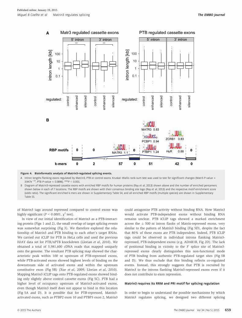
Properties of Matrin3-regulated exons
In order to assess whether the exons regulated by Matrin3 possess any
specific splicing features, we examined 50 and 30 splice sites, branch
points, pyrimidine tracts, and nucleotide composition (Supplementary
Fig S3) and flanking intron lengths (Fig 4A). Few significant differ-
ences were observed compared to a control set of annotated cassette
exons unaffected by knockdown of Matrin3 (or PTB) knockdown. One
striking difference was that the introns flanking Matrin3 repressed
exons are on average 1 kb longer than introns flanking Matrin3-
activated, PTB-repressed, PTB-activated or control exons (Fig 4A).
We next looked for enrichment of pentamer sequence motifs
associated with Matrin3-regulated exons, compared to control unreg-
ulated cassette exons, across seven transcript locations (cassette
exons, flanking constitutive exons, 50 and 30 end of each flanking
intron). Numerous motifs were enriched (FDR < 0.05) in the introns
flanking Matrin3-repressed exons, but none within exons or in any
location associated with Matrin3-activated exons (Fig 4B, Supple-
mentary Fig S4, Supplementary Table S4). Motifs associated with
Matrin3-repressed exons were heterogeneous and included a
number of pyrimidine motifs associated with PTB (e.g. TTCTT,
TCTTT). The enrichment was also observed using a control set
consisting of exons including PTB-regulated, Matrin3-independent
exons. Most of the remaining motifs had high pyrimidine content,
with one or two interrupting purines; more than half of the motifs
immediately flanking Matrin3 repressed exons had a single purine.
Individual analyses of RNA binding by Matrin3 have not revealed a
clear consensus sequence (Hibino et al, 2006; Salton et al, 2011;
Yamazaki et al, 2014). However, Matrin3 was one of 207 RBPs
whose optimal sequence was determined by the RNA-compete
array-based selection (Ray et al, 2013). We therefore used heptamer
position frequency matrices to look for enrichment of RNA-compete
motifs. Once again, enriched motifs were found only in the introns
flanking Matrin3-repressed exons, and in no locations associated
with Matrin3 activation (Fig 4B, Supplementary Table S5). Strik-
ingly, the Matrin3 motif was the only enriched RNA-compete motif
upstream of repressed exons and was one of only four motifs on the
immediate downstream side. Other enriched motifs included PTBP1,
PCBP1, HuR and ZCRB1; this may be either due to a consequence of
these proteins functioning as a co-regulator of Matrin3 or simply
due to the similarity between the sequences of the binding sites.
Taken together, the k-mer and RNA-compete motif enrichments
suggest that Matrin3 might bind directly to the longer introns flank-
ing repressed exons, but that activation by Matrin3 might be
indirect.
Matrin3 binds widely around repressed exons
To directly address the relationship between Matrin3 splicing activ-
ity and RNA binding, we carried out crosslinking and immunopre-
cipitation (iCLIP) in HeLa cells (Konig et al, 2010). We obtained a
total of 3,496,801 cDNA reads after collapsing PCR duplicates that
mapped uniquely onto the genome. A Matrin3 splicing map was
generated using the Matrin3 iCLIP binding microarray data sets
(Fig 5). Matrin3 binding was elevated in long intronic regions
immediately flanking repressed exons (Fig 5A, blue). In contrast,
the flanking constitutive exons, their immediate intron flanks and
all regions associated with Matrin-activated exons (Fig 5A, red)
showed binding levels only slightly elevated above control cassette
exons (Fig 5A, grey). This differential observed binding agrees well
with the motif enrichments (Fig 4B, Supplementary Tables S4 and
S5). In contrast to many other splicing regulators (Licatalosi &
Darnell, 2010; Witten & Ule, 2011), Matrin3 binding was uniformly
elevated within 500 nt of repressed exons, with no discrete peaks
(Fig 5A). This uniform binding was not simply a result of higher
steady-state levels of these RNA regions, because TIA1 (Wang et al,
2010) and U2AF65 (Zarnack et al, 2013) iCLIP tags showed a similar
density on control and Matrin3-regulated exons (Supplementary Fig
S2A). The uniform binding was also observed around individual
exons (e.g. ZMYND8 and ADAR1B, Supplementary Fig S2D) and so
did not result from averaging across large numbers of introns with
discreet peaks at different locations. We also noted that elevated
Matrin3 binding extended into the repressed exons (Fig 5A), even
though no enriched motifs had been observed in this location
(Fig 4B). A possible explanation for the extended region of elevated
binding including the exon is that binding initiates at high affinity
specific motifs in the flanking introns and then extends by Matrin3
oligomerization with less specific RNA binding (see Discussion). We
did not observe a clear correlation between tag density and degree
of splicing change upon Matrin3 knockdown, which may be related
to lack of saturation of the iCLIP library. Nevertheless, the density
Figure 3. Global splicing effects upon Matrin3 knockdown.
A Western blot probed for Matrin3 (top panel), PTB (middle panel) and actin (lower panel). Lanes 1–4 contain a twofold dilution of the control C2 sample (lane 1—12.5%, lane 2—25%, lane 3—50%, lane 4—100%). Lanes 4–7 contain equal amount of proteins, as can be confirmed by the anti-actin (lower panel), of control sample(lane 4), double-PTB and nPTB siRNA-treated sample (lane 5), Matrin3 siRNA-treated sample (lane 6) and triple knockdown of Matrin3, PTB and nPTB (lane 7). Lane 7is from the same gel and exposure, but some lanes present in the original gel were cropped for clarity, and a black line to indicate cropping was placed.
B Pie chart of the different categories of Matrin3-regulated alternative splicing events (ASEs), 330 cassette exons (50%), 116 alternative promoter usage (17%), 78terminal exons (11%), 32 alternative 50 (5%) and 34 30 (5%) splice site and 18 intron retention (IR; 3%) events.
C Gene ontology (GO) analysis of the Matrin3-regulated cassette exons. The x-axis represents the P-value in a logarithmic scale as shown.D Pie chart of the activated and repressed cassette exons by Matrin3.E Venn diagram of the overlap between the PTB- (blue) and Matrin3- (yellow) regulated cassette exons, showing the 813 events regulated only by PTB, 270 by Matrin3
only and the 61 that overlap.F RT–PCR validation of Matrin3-regulated alternative spicing events in the ST7, ACSL3, PLEKHA3, TCF12, VWA5A, PTBP2, PTBP3, C3orf17, ZMYND8, VEZT, PIGX and DMD
genes. In each case, triplicates for each condition (C—control, M—Matrin3, PTB/nPTB and Matrin3/PTB/nPTB siRNA transfection samples) were analysed and exoninclusion (EI) percentage is shown beneath the corresponding lane, along with the standard deviation (s.d.).
Source data are available online for this figure
◀
The EMBO Journal Vol 34 | No 5 | 2015 ª 2015 The Authors
The EMBO Journal Matrin3 regulates splicing Miguel B Coelho et al
658
Published online: January 19, 2015
of Matrin3 tags around repressed compared to control exons was
highly significant (P < 0.0001, v2 test).In view of our initial identification of Matrin3 as a PTB-interact-
ing protein (Figs 1 and 2), the small overlap of target splicing events
was somewhat surprising (Fig 3). We therefore explored the rela-
tionship of Matrin3 and PTB binding to each other’s target RNAs.
We carried out iCLIP for PTB in HeLa cells and used the previous
HJAY data set for PTB/nPTB knockdown (Llorian et al, 2010). We
obtained a total of 5,981,600 cDNA reads that mapped uniquely
onto the genome. The resultant PTB splicing map showed the char-
acteristic peak within 100 nt upstream of PTB-repressed exons,
while PTB-activated exons showed highest levels of binding on the
downstream side of activated exons and within the upstream
constitutive exon (Fig 5B) (Xue et al, 2009; Llorian et al, 2010).
Mapping Matrin3 iCLIP tags onto PTB-regulated exons showed bind-
ing only slightly above control cassette exons (Fig 5C). PTB had a
higher level of occupancy upstream of Matrin3-activated exons,
even though Matrin3 itself does not appear to bind in this location
(Fig 5A and D). It is possible that for PTB-repressed, Matrin3-
activated exons, such as PTBP2 exon 10 and PTBP3 exon 2, Matrin3
could antagonize PTB activity without binding RNA. How Matrin3
would activate PTB-independent exons without binding RNA
remains unclear. PTB iCLIP tags showed a marked enrichment
across the � 500 nt intron flanks of Matrin-repressed exons, very
similar to the pattern of Matrin3 binding (Fig 5D), despite the fact
that 80% of these exons are PTB independent. Indeed, PTB iCLIP
tags could be observed in individual introns flanking Matrin3-
repressed, PTB-independent exons (e.g. ADAR1B, Fig 2D). The lack
of positional binding in vicinity to the 30 splice site of Matrin3-
repressed exons clearly distinguishes this non-functional mode
of PTB binding from authentic PTB-regulated target sites (Fig 5B
and D). We thus exclude that this binding reflects co-regulated
events. Instead, this strongly suggests that PTB is recruited by
Matrin3 to the introns flanking Matrin3-repressed exons even if it
does not contribute to exon repression.
Matrin3 requires its RRM and PRI motif for splicing regulation
In order to begin to understand the possible mechanisms by which
Matrin3 regulates splicing, we designed two different splicing
A
B
Figure 4. Bioinformatic analysis of Matrin3-regulated splicing events.
A Intron lengths flanking exons regulated by Matrin3, PTB or control exons. Kruskal–Wallis rank-sum test was used to test for significant changes (Matr3 P-value =3.947e�15, PTB P-value = 0.3896). ***P < 0.001.
B Diagram of Matrin3-repressed cassette exons with enriched RBP motifs for human proteins (Ray et al, 2013) shown above and the number of enriched pentamersshown below in each of 7 locations. The RBP motifs are shown with their consensus binding site logo (Ray et al, 2013) and the respective motif enrichment score(odds ratio). The significant enriched k-mers are shown in Supplementary Table S4, and all enriched RBP motifs (multiple species) are shown in SupplementaryTable S5.
ª 2015 The Authors The EMBO Journal Vol 34 | No 5 | 2015
Miguel B Coelho et al Matrin3 regulates splicing The EMBO Journal
659
Published online: January 19, 2015

A
B
C
D
Figure 5. Matrin3 and PTB splicing maps.
A Matrin3 crosslinking in Matrin3-regulated pre-mRNAs where position of crosslinked nucleotides was mapped onto the regulated exon and the 500 nucleotidesupstream and downstream of its 30ss and 50ss, respectively, the upstream flanking exon and 500 nucleotides downstream of its 50ss and the downstream flankingexon with 500 nucleotides upstream of its 50ss. The iCLIP tags were mapped onto silenced ASE (blue, n = 421), enhanced ASE (red, n = 187) and to control ASE (grey,n = 16,242), and percentage of occupancy is plotted.
B Same as in (A) but using iCLIP tags obtained from PTB iCLIP and mapped onto PTB/nPTB-regulated ASE, silenced ASE (blue, n = 729), enhanced (red, n = 820) andcontrol ASE (grey, n = 14,599).
C Matrin3 crosslinked nucleotides mapped onto PTB-regulated ASE.D PTB crosslinked nucleotides mapped onto Matrin3-regulated ASE.
The EMBO Journal Vol 34 | No 5 | 2015 ª 2015 The Authors
The EMBO Journal Matrin3 regulates splicing Miguel B Coelho et al
660
Published online: January 19, 2015

reporters based on ABI2 exon 8, which is activated by Matrin3
and repressed by PTB, and ST7 exon 11, which is Matrin3
repressed and PTB independent. The exons with flanking intronic
regions were cloned into an exon-trapping vector with flanking
constitutive exons and splice sites to generate a 3 exon construct.
The splicing reporters were co-transfected into HeLa cells along
with Matrin3 expression vectors with a range of domain deletions
and mutations (Fig 6A). All FLAG-tagged Matrin3 proteins were
expressed at comparable levels (Fig 6B), allowing direct compari-
son of their activities. The ABI2 exon 8 showed increased inclu-
sion in response to Matrin3 co-transfection (Fig 6D, lanes 1, 2).
This activity was unaffected by deletion of zinc finger 2 and
apparently increased by deletion of zinc finger 1 (Fig 6D, lanes 4
and 6). In contrast, deletion of the RRMs impaired activity
(Fig 6D, lane 5). This shows that although Matrin3-activated
exons showed no association with enriched motifs (Fig 4B) or
observed binding (Fig 5), Matrin3 still requires its RRM domains
for activity. Mutation of the PRI led to a complete loss of activity
(Fig 6D, lane 3), suggesting that the ability of Matrin3 to interact
with PTB is important for its antagonistic activity upon this exon.
The ST7 reporter alone showed ~50% exon inclusion and, as
expected, this level decreased upon overexpression of Matrin3
(Fig 6C, lanes 1 and 2). The response to the various mutations
was very similar to ABI2 exon 8. Deletion of the zinc finger
domains (Fig 6C, lanes 4 and 6) did not impair Matrin3 repressor
activity, while deletion of the RRMs (Fig 6C, lane 5) or mutation
of the PRI (Fig 6C, lane 3) abolished activity. The response to
RRM mutation is consistent with the observed direct binding
around Matrin3-repressed exons (Fig 5). However, the effect of
the PRI mutation is striking in view of the fact that ST7 exon 11
is independent of PTB (Fig 3F). This suggests that other proteins
might bind to this motif and be required to co-regulate events
independent of PTB.
To address whether the effect of the Matrin3 PRI mutation is
related to its interaction with PTB or other PRI-interacting proteins,
we tested the effects of Matrin3 overexpression in combination with
PTB knockdown upon the minigenes (Fig 6E). The ST7 minigene,
like the endogenous gene (Fig 3F), was unresponsive to PTB/nPTB
knockdown and responded to Matrin3 overexpression indepen-
dently of PTB levels (Fig 6F, lanes 1, 2, 4, 5). In contrast, the ABI2
minigene responded to both overexpression and knockdown of PTB
(Fig 6G, lanes 1, 3, 4, 6). Although Matrin3 overexpression
promoted ABI2 exon 8 inclusion in the presence of PTB, upon PTB
knockdown Matrin3 had no effect (Fig 6G, compare lanes 1, 2 with
4, 5). This suggests that Matrin3 acts mainly to antagonize PTB
when regulating ABI2 exon 8. In summary, the structure-function
analysis indicates that Matrin3 requires its PRI motif and one or
both RRM domains to act as a splicing regulator.
Discussion
Matrin3 is a splicing regulator
Matrin3 has long been suspected to play a role in RNA metabolism
due to the presence of RNA-binding domains and its interactions
with multiple other RNA-binding proteins, some with known roles
in splicing regulation and other RNA processing roles (Polydorides
et al, 2000; Zeitz et al, 2009; Salton et al, 2011). Nevertheless,
evidence for a direct functional role of Matrin3 in cellular mRNA
metabolism has been missing. Using a combination of RNAi knock-
down, transcriptome profiling and iCLIP, we show unequivocally
that Matrin3 is a strong regulator of multiple splicing events. Knock-
down of HeLa cell Matrin3 leads to dysregulation of 667 ASEs, while
iCLIP shows that Matrin3 binds directly to the introns flanking
repressed exons. Analysis of functional terms associated with the
regulated cassette exons revealed enrichment of genes encoding
chromatin/chromatin-binding proteins and cytoskeletal proteins
(Fig 3C). Matrin3 has a number of previously reported roles includ-
ing involvement in DNA damage response (Salton et al, 2010), and
as a co-factor of Rev-mediated export of HIV1 RNA (Kula et al,
2011, 2012; Yedavalli & Jeang, 2011). The only other suggested
widespread RNA-related function for Matrin3 is mRNA stabilization
(Salton et al, 2011). Among the relatively few transcript level
changes that we observed upon Matrin3 knockdown, we were able
to identify nine of the 77 previously reported down-regulated
mRNAs (Salton et al, 2011). Notably in five of the nine mRNAs, we
observed ASEs that shifted upon Matrin3 knockdown towards
isoforms with premature termination codons (PTCs) that would be
expected to lead to nonsense-mediated decay (NMD). For example,
in THUMPD2 pre-mRNA, Matrin3 represses splicing of the
PTC-containing exon 7, while in ACAD9 upon Matrin3 knockdown,
50 splice site selection on exon 1 shifts to an alternative down-
stream site which leads to inclusion of a PTC. Indeed, of the 18
Figure 6. Matrin3 splicing function requires PRI and RRMs.
A Schematic representation of Matrin3 with its domains in the wild-type construct (WT) and the mutants used in overexpression, mPRI which has correspondingdomain deleted in each one of the mutants, mPRI where the PRI motif is mutated from GILGPPP to GAAAPPA, DZ1 where the first zinc finger is deleted, DRRMwhere both the RRM domains are deleted and DZ2 where the second zinc finger is deleted.
B Western blot of the overexpressed Matrin3 and its mutants probed with anti-FLAG and anti-actin antibodies.C, D RT–PCR analysis using primers specific to the ST7 exon 11 (C) and ABI2 exon 8 (D) splicing reporters, respectively, of samples where FLAG-tagged Matrin3 and its
mutants have been overexpressed. Quantification of at least three replicates for each condition is shown as a histogram of the percentage of exon inclusion.*P < 0.01 compared with control sample (�).
E Western blot of overexpressed FLAG-tagged Matrin3 and PTB, in control siRNA and PTB/nPTB siRNA-transfected cells, probed with anti-FLAG to detectoverexpressed proteins, anti-PTB to detect the knockdown levels and anti-actin to confirm protein loading. A titration of control sample (lane 1—12.5%, lane 2—25%, lane 3—50% and lane 4—100%) is also included to assess the levels of knockdown. The band present above the PTB doublet in lane 6 is FLAG-tagged PTB.
F, G RT–PCR analysis using primers specific to the ST7 exon 11 (F) and ABI2 exon 8 (G) splicing reporters, respectively, of samples where Matrin3 or PTB expressionvectors were co-transfected, in control siRNA and PTB/nPTB siRNA-transfected cells. Quantification of at least three replicates for each condition is shown as ahistogram of the percentage of exon inclusion. *P < 0.01 when compared with control sample in each condition (-).
Source data are available online for this figure
▸
ª 2015 The Authors The EMBO Journal Vol 34 | No 5 | 2015
Miguel B Coelho et al Matrin3 regulates splicing The EMBO Journal
661
Published online: January 19, 2015

A
B E
C F
D G
Figure 6.
The EMBO Journal Vol 34 | No 5 | 2015 ª 2015 The Authors
The EMBO Journal Matrin3 regulates splicing Miguel B Coelho et al
662
Published online: January 19, 2015

Matrin3-repressed cassette exons in genes whose expression is
down-regulated more than 1.5-fold, 13 are predicted to lead to NMD
upon exon inclusion and hence to be destabilized upon Matrin3
depletion. Likewise, two of the four Matrin3-activated exons in
genes that are more than 1.5-fold down-regulated upon Matrin3
knockdown cause NMD upon skipping, including the exon 10 of
nPTB (Fig 3F). While we cannot rule out Matrin3 acting directly to
stabilize some mRNAs, our data suggest that at least in some cases
alterations of mRNA levels are attributable to Matrin3 action as a
splicing regulator of ASEs which lead to NMD. Another possible
indirect influence of Matrin3 upon mRNA levels is via its regulation
ASEs in chromatin-related proteins (Fig 3C).
The characterization of Matrin3 as a splicing regulator is relevant
for interpreting the basis of Matrin3-associated pathologies, such as
ALS (Johnson et al, 2014) and distal myopathies (Senderek et al,
2009). Like many other RNA-binding proteins associated with
neurodegenerative diseases, Matrin3 has extensive low-complexity
disordered segments. Mutations in these regions of other proteins,
such as TDP43, can lead to intracellular insoluble protein inclusions
which can be directly proteotoxic or can lead to dysregulated RNA
metabolism (Buratti & Baralle, 2012). Likewise, Matrin3 mutations
might directly affect its splicing regulatory activity or reduce its
effective concentration, either of which could have consequences
for target ASEs. Notably, nuclear and cytoplasmic localization of
isoforms of dystrophin (Gonzalez et al, 2000), the protein primarily
affected in Duchenne’s and Becker’s muscular dystrophies, is regu-
lated by AS of exon 78. Both Matrin3 and PTB repress inclusion of
this exon (Fig 3F).
Mechanism of Matrin regulation of splicing
Matrin3 function as a direct splicing repressor is supported by its
observed binding around repressed exons (Fig 5) along with the
enrichment in flanking intron segments of optimal binding motifs
for its RRM motifs (Fig 4B). Consistent with this, it requires intact
RNA-binding domains, but not DNA-binding domains, for its splic-
ing repressor activity (Fig 6). Nevertheless, further evidence of its
direct mode of action could be provided by in vitro analyses of its
binding to regulated RNAs, demonstrating that specific binding sites
are required for its splicing repressor activity, and more detailed
analysis of the roles of the individual RRM domains. Unexpectedly,
deletion of the ZF1 DNA-binding domain actually enhanced the
activity of transfected Matrin3 (Fig 6). One possible explanation for
this observation is that deletion of ZF1 affects the distribution of
Matrin3 between pools that are active for splicing regulation or that
are localized elsewhere.
The majority of Matrin3 target exons are unaffected by PTB
(Fig 3), even though we initially identified Matrin3 via its interac-
tion with PTB RRM2, part of the minimal repressor domain of PTB
(Figs 1 and 2) (Robinson & Smith, 2006). Indeed, we observed
enhanced recruitment of PTB around Matrin3-repressed exons and,
importantly, the distribution of PTB resembled the broad distribu-
tion of Matrin3 around its own targets (Fig 5A and D) and lacked
the distinctive peak of PTB binding observed upstream of PTB-
repressed exons (Fig 5B). This suggests that PTB can be recruited to
Matrin3-regulated exons, even when it is not functionally required,
as in the ST7 exon 11 (Figs 3F and 6). However, we observed that
repression of ST7 exon 11 depended absolutely upon the Matrin3
PRI motif (Fig 6), which suggests that Matrin3 repressor activity at
this exon might require interaction with proteins other than PTB via
its PRI. The binding of Matrin3 across extensive regions flanking
and within repressed exons (Fig 5A) is suggestive of a mechanism
of initial binding to specific sites followed by spreading. This is
consistent with the observed enrichment of k-mers and the RNA-
compete motif exclusively in the immediate 250 nucleotides of the
flanking introns (Fig 4), and with its reported propensity for self-
association (Zeitz et al, 2009). Similar models were originally
suggested for repression by PTB (Wagner & Garcia-Blanco, 2001)
and hnRNPA1 (Zhu et al, 2001), but analysis of numerous model
systems has shown that this is not a common mechanism for these
proteins (e.g. Cherny et al, 2010), and their splicing maps show
distinct peaks of enriched binding (Xue et al, 2009; Llorian et al,
2010; Huelga et al, 2012). In contrast, the Matrin3 splicing map
(Fig 5A) is consistent with a general mechanism of action in which
initial binding of Matrin3 at specific sites is followed by propagative
binding across a wide region of RNA, leading to repression of the
targeted exon. This model is amenable to testing by various meth-
ods including single molecule analysis (Cherny et al, 2010).
How Matrin3 might promote exon inclusion is less clear. Splicing
maps indicate that binding around activated exons is not much
above background (Fig 5A) and no motifs were enriched (Fig 4).
Nevertheless, in the ABI2 minigene assay, the RRMs were required,
along with the PRI motif (Fig 6). One explanation could be that acti-
vated exons are mainly indirect targets, and changes in their splicing
result from the primary actions of Matrin3, which require RNA bind-
ing. Another possibility, in cases where Matrin3 activation directly
opposes PTB repression, is that interaction with Matrin3 might
antagonize PTB activity. Although Matrin3 binding is low around
Matrin3-activated exons (Fig 5A), PTB binding shows an upstream
peak (Fig 5B) consistent with PTB repression. A notable example of
this is the Matrin3-activated nPTB exon 10 (Fig 3F), a well-known
target of PTB repression (Boutz et al, 2007; Makeyev et al, 2007;
Spellman et al, 2007).
PRI-protein interactions
In addition to Matrin3 and Raver1, we found a number of other
interesting factors that bound to PTB RRM2 and were sensitive to
the Y247Q mutation (Joshi et al, 2011), suggesting that their interac-
tions might also be mediated by PRI motifs (Fig 1). These included
splicing regulators, proteins involved in the co-transcriptional-
dependent regulation of splicing and 30 end processing factors. We
did not identify a number of previously identified PTB interactors,
including hnRNP-L, PSF, hnRNPA1, hnRNPA2B1, hnRNPC and
MRG15 (Patton et al, 1993; Hahm et al, 1998; Luco et al, 2010; King
et al, 2014), although we did identify the helicase DDX3X (King
et al, 2014). The lack of overlap could be because we focused on
proteins that interact primarily via RRM2. Interesting novel PTB
interactors with possible relevance for PTB’s splicing regulatory
activities include KIAA1967/DBC1/CCAR2 and its paralogue
CCAR1, both of which bound strongly to PTB RRM2 in a manner
that was sensitive to the PTB Y247Q mutation (data not shown).
CCAR1 is a spliceosomal A-complex component that interacts with
U2AF65 (Hegele et al, 2012). DBC1 together with ZNF326/ZIRD
forms the DBIRD complex, which integrates alternative splicing with
RNA polymerase II transcription (Close et al, 2012). This complex is
ª 2015 The Authors The EMBO Journal Vol 34 | No 5 | 2015
Miguel B Coelho et al Matrin3 regulates splicing The EMBO Journal
663
Published online: January 19, 2015

required for RNA Pol II to efficiently transcribe AT-rich regions
upstream of some exons. Depletion of the DBIRD complex, by DBC1
or ZIRD knockdown, leads to accumulation of stalled RNA Pol II in
these AT-rich regions, which enhances the inclusion of the proximal
exon (Close et al, 2012). It will be interesting to determine whether
interaction of PTB with DBC1 occurs as part of the DBIRD complex,
as a distinct DBC1-PTB regulatory axis, or possibly in coordination
with chromatin association PTB-MRG15 interaction (Luco et al,
2010). DBC1 and CCAR1 both have potential N-terminal PRI motif,
and DBC1 has an additional one. These motifs are conserved across
multiple vertebrate species, but we have yet to determine whether
they are functional. While we have identified a number of poten-
tially interesting PTB-interacting proteins, it is also likely that we
have failed to identify other interacting proteins for which the RRM2
interface is either not sufficient or not necessary. Notably, Matrin3
interacted more strongly with full-length PTB than RRM2 alone,
while Raver1 had the opposite response (Fig 1D). This suggests that
identification of the full range of proteins that interact with PTB will
require additional analyses using full-length PTB and possibly its
PTB1 and 4 spliced isoforms (Wollerton et al, 2001).
While the Matrin3 PRI matches the consensus [S/G][I/L]LGx/Pbased on mutagenesis of Raver1 motifs, it is notable that it shows
absolute conservation (GILGPPP) across all available vertebrate
species, whereas the two main Raver1 motifs show variation at posi-
tions 5 and 6. This suggests that while Raver1 interacts only with
PTB through its PRI, Matrin3 might bind also to other RRM contain-
ing proteins and is therefore subject to greater sequence constraints.
Consistent with this, we observe that activity of Matrin3 on ST7
splicing reporter requires the PRI motif (Fig 6C), even though this
event is PTB independent (Fig 3F). Comparison of proteins
co-immunoprecipitated with wild-type and PRI mutant Matrin3
showed only two obvious differences, which mass spectrometry
revealed to be both isoforms of PTB (Supplementary Fig S5).
Further investigation will be carried in the future to determine the
additional PRI-interacting factors for Matrin3 activity. Several
proteins from previous Matrin3 interactome studies contain RRM
domains and might be candidate PRI-interacting proteins, including
RBMX, SAFB, HNRNPL, U1SNRNBP, SFRS7, SLTM, PABPC1, PSF
and TDP43 (Zhang & Carmichael, 2001; Zeitz et al, 2009; Salton
et al, 2011; Johnson et al, 2014). Additional candidates suggested
by association of their RNA-compete motifs (Ray et al, 2013) with
Matrin3-repressed exons are HuR and ZCRB1 (Fig 4). Strikingly, the
strongest interaction seen in yeast two-hybrid screens was Matrin3
binding to itself (Zeitz et al, 2009), raising the possibility that its PRI
could interact with an RRM domain of another Matrin3 monomer.
We tested this hypothesis and saw no binding between the PRI and
RRM domains of Matrin3 (data not shown). A further possibility is
that the Matrin3 PRI interacts with domains other than RRMs, a
possibility which could be addressed by unbiased interaction
screening of wild-type and PRI mutant Matrin3 by quantitative
proteomics.
In summary, we have established a clear role for the nuclear
matrix protein Matrin3 in directly regulating a network of alterna-
tive splicing events, a small proportion of which are also co-
regulated by PTB. This insight should assist future investigations
into the cellular roles of Matrin3, as well as the basis of molecular
pathologies associated with Matrin3 mutations (Senderek et al,
2009; Johnson et al, 2014).
Materials and Methods
Tissue culture, DNA and siRNA transfection and analysis
HeLa S3 and HEK-293T cell lines were cultured using standard
procedures. siRNA-mediated knockdown was carried out by doing a
2-hit transfection of control, Matrin3 or PTB targeting siRNAs. Cells
were plated in a 35-mm well in day 1, followed by transfection of
40 pmol of siRNA (40 pmol of control siRNA, 40 pmol of Matrin3
siRNA, and for PTB/nPTB double-knockdown 20 pmol of PTB
siRNA and 20 pmol of nPTB siRNA) in day 2 and day 3, using Oligo-
fectamine (Invitrogen) and Lipofectamine (Invitrogen) in each day,
respectively, with cells being harvested on day 5. siRNAs were
purchased from Dharmacon, and mRNA targets for gene-specific
knockdown were (50-30) Matrin3 M3 AAAGACUUCCAUGGACU-
CUUA (Salton et al, 2010), PTB P1 AACUUCCAUCAUUCCAGAGAA,
nPTB N1 AAGAGAGGAUCUGACGAACUA and control C2
AAGGUCCGGCUCCCCCAAAUG (Spellman et al, 2007). PTB knock-
down was carried out by transfecting siRNAs targeting both PTB
(P1) and its neuronal paralogue (N1), nPTB, which otherwise
partially compensates for PTB (Spellman et al, 2007). Knockdown
efficiency was monitored by lysing a 35-mm well for each condition
with protein loading buffer directly, followed by SDS–PAGE and
Western blot using anti-Matrin3 (sc-55723; Santa Cruz), anti-PTB
(Spellman et al, 2007) and anti-actin antibodies (A2066; Sigma).
Other antibodies used were FLAG M2 monoclonal (Sigma) and
Raver1 rabbit polyclonal against the RRM domains. For immunopre-
cipitation, cells were transfected using Lipofectamine 2000 (Invitro-
gen) and harvested 48 h post-transfection in 200 ll of lysis buffer
(Huttelmaier et al, 2001), 5% of cell extract was analysed by
Western blot to check for expression of transfected proteins, and the
remainder was used for immunoprecipitation using 5 ll of anti-
FLAG antibody (Sigma M2) with 10 ll of protein G sepharose, previ-
ously blocked with 2% BSA. Antibody–protein complex was
allowed to bind for 2 h at 4°C, followed by extensive washes, and
eluted in 30 ll of protein loading buffer. The entire sample was
subjected to Western blot and probed against PTB. For splicing
reporter analysis, we transfected cells in triplicates with 2 lg of
effector DNA encoding Matrin3 and its mutants together with
200 ng of the indicated splicing reporter. Forty-eight hours post-
transfection cells were harvested using TRI reagent (Sigma) and
2 lg of total RNA was used for reverse transcription using either
splicing reporter-specific oligo, or in the case of validation of
microarray ASE, oligo dT, using superscript II (Invitrogen). For
the splicing reporters, the splicing pattern was analysed by PCR
using the primers GFPN and CG3 as described previously (Llorian
et al, 2010). For endogenous ASE analysis, gene-specific primers
were used (Supplementary Materials and Methods). All PCRs were
carried out using the Jumpstart Taq polymerase (Sigma), and the
products were separated and quantified on a QIAXcel capillary
electrophoresis system (Qiagen).
DNA constructs
GST-PTB1 and GST-SXL used for pull-down of PRI motifs have
been described previously (Rideau et al, 2006). GST-RRM2 and
GST-RRM2 Y247Q were PCR-amplified and cloned into the EcoRI
site in pGEX3 as described (Joshi et al, 2011). To generate the
The EMBO Journal Vol 34 | No 5 | 2015 ª 2015 The Authors
The EMBO Journal Matrin3 regulates splicing Miguel B Coelho et al
664
Published online: January 19, 2015

FLAG-Matrin3 PRI-MS2, we cloned into the AvrII and MluI sites
pre-annealed oligos encoding the indicated protein sequence. FLAG-
MS2, FLAG-Raver1 full-length wild-type and mutant, FLAG-Raver1
PRI have all been described previously (Rideau et al, 2006). Full-
length Matrin3 was PCR-amplified from human cDNA and cloned
into the MluI sites of a pCI-FLAG vector (Promega). Deletion
mutants were generated by divergent PCR and mPRI by site-directed
mutagenesis (Promega). The splicing reporters were constructed by
PCR amplifying the regulated exon and the flanking region and clon-
ing them into the Asp718I and EcoRV sites of a GFP expression
cassette where an intron has been inserted into the second codon.
The cloning sites are localized in the middle of this intron so will
generate three exon splicing reporters upon cloning (Wollerton et al,
2004). All DNA constructs were confirmed by sequencing.
GST expression, purification and pull-down
Pull-down of nuclear proteins using GST-RRM2 and GST-RRM2
Y247Q was carried out as follows: 2 lg of purified recombinant
protein was pre-bound to glutathione sepharose 4B beads for 1 h.
To each GST protein, 300 ll of HeLa nuclear extract was added,
together with RNase A to a final concentration of 5 lg/ml. The
mixture was incubated for 2 h at 4°C rotating, followed by extensive
washes and elution using 30 ll of protein loading buffer. Five
microlitre was used for silverstain analysis, and the remainder of
the pull-down was run on a SDS–PAGE followed by mass spectro-
metry of the entire section of the lane above the recombinant
protein (see Fig 1A). Expression and purification of GST-RRM
proteins has been described elsewhere (Joshi et al, 2011). Pull-
downs of in vitro translated protein by GST-PTB were carried out as
described (Rideau et al, 2006) using GST-SXL as negative controls.
GST-SXL protein was produced from the plasmid pGEX CS NR SXL
XW II, which was a kind gift from J. Valcarcel, Centre de Regulacio
Genomica. RNase treatment of pull-downs was carried out in two
steps: the in vitro translations were terminated with a 15-min incu-
bation at 30°C with 25 lg/ml RNase A, and RNase A was added at
0.5 lg/ml to the pull-down pre-incubation for 3 h at 4°C.
Human Junction microarray experiments and validation
RNA from four biological replicates of control or Matrin3
siRNA-transfected HeLa cells was isolated using TRI reagent (Life
Technologies). Total RNA was hybridized to Human Affymetrix
Exon-Junction Array (HJAY) by Genecore (EMBL). The microarray
data were analysed using ASPIRE (Analysis of SPlicing Isoform
REciprocity) 3.0 (Konig et al, 2010; Wang et al, 2010). It analyses
signal in reciprocal probe sets to monitor changes in alternative
splicing events, from which we applied a threshold of |dIrank| ≥ 1
and obtained 667 alternative splicing events (Supplementary Table
S3). Matrin3, PTB/nPTB and triple Matrin3/PTB/nPTB were carried
out and cDNA was produced from extracted RNA using oligo dT.
The splicing pattern of each ASE was analysed by RT–PCR (for
primer sequence, see Supplementary Materials and Methods).
Bioinformatic analysis of Matrin3 regulated ASE
Analysis of enriched functional gene categories was carried out using
PANTHER (Mi et al, 2013). Analysis of Matr3 intron length was
done using ASPIRE [CS1] annotation in R version 3.0.1 and the
ggplot2 and pgirmess packages. Since some alternative splice events
have 0-length introns, intron length was analysed for alternative
cassette exons only. For statistic analysis, data were tested with
Kruskal–Wallis rank-sum test before doing multiple comparison test-
ing. Motif enrichments were calculated using 100 bp of the flanking
exons and the complete sequence of the cassette exons. For introns,
we used maximum intronic flanks of 250 nt, removing SS context to
avoid BP, SS and PPT signals, 9 nt at donor side and 30 nt at the
acceptor side (Bland et al, 2010), retrieving introns with a minimum
length of 60 nt. We assess the enrichment of 5-mers and RNA-
compete motif matches (Ray et al, 2013) with the following proce-
dure implemented in a custom PHP script: Given a sample set S of N
sequences and a control set S(0) of N(0) sequences, the number of
times na,i that each motif a appeared in each sequence i was calcu-
lated. Likewise, for the control set, the number of occurrences nð0Þa;i of
each motif a per sequence i was also calculated. The expected
density dð0Þa of each motif was also calculated as the ratio between
the total number of occurrences in the control set over the total
sequence length of the control set:
dð0Þa ¼
P
iISð0Þnð0Þa;i
P
iISð0Þli
;
where li is the length of each sequence in the control set. For each
sequence i in the sample set each motif a, it was recorded whether
the observed motif count (na,i) is greater than the calculated
expected count (dð0Þa li):
di;a ¼ 1 if ni;a [dð0Þa li
di;a ¼ 0 otherwise
Similarly, for the counts in the control set:
dð0Þi;a ¼ 1 if nð0Þa;i [dð0Þa li
dð0Þi;a ¼ 0 otherwise
The sum of the di,a values over the sequences i represents the
number of sequences for which the motif a has an observed count
greater than expected. Thus, for each motif, the odds ratio (motif
score) and corresponding P-value were the motif a has an obtained
by performing a Fisher’s test (one-tailed) with these sums counts for
the sample set and the control set:
5-mera More than expected Less than expected
SP
iIS
di;a N�P
iIS
di;a
S(0)P
iIS
dð0Þa;i Nð0Þ �P
iIS
dð0Þa;i
Statistically over-represented motifs were selected based on the
Benjamini’s and Hochberg’s false discovery rate multiple test-
corrected P-value (BH-FDR < 0.05). Statistical tests were performed
and graphics were generated with R Development Core Team
(2011). Additional scripts were written in PHP and Awk, and
sequence logos were generated with seqlogo (Crooks et al, 2004).
ª 2015 The Authors The EMBO Journal Vol 34 | No 5 | 2015
Miguel B Coelho et al Matrin3 regulates splicing The EMBO Journal
665
Published online: January 19, 2015

CLIP and splicing maps
iCLIP experiments were performed for Matr3 and PTB using anti-
bodies targeting the endogenous protein, GTX47279 (GeneTex) and
polyclonal anti-PTB serum (Spellman et al, 2007). Exponentially
growing HeLa cells were washed once with PBS and cross-linked at
0.15 mJ/cm2 with a Stratalinker 2400 equipped with 254 nm light
bulbs. Retrieval of protein-bound RNAs and preparation of Illumina-
compatible DNA libraries were done as described in Huppertz et al
(2014). To compute RNAmaps of Matrin3 and PTB binding on
exon–intron boundaries, we assessed the positioning of cross-link
sites. Cross-linked nucleotides are defined as the nucleotide
upstream of mapped iCLIP cDNA tags as described before (Konig
et al, 2010). For each position within the RNAmap, the number of
cross-link nucleotides was counted as 1 if one or more cDNA tags
matched the position, and then summed across all splice events.
The summed cross-link count was divided by the number of splice
events and plotted in 10 nucleotide bins. Thus, the resulting occur-
rence value reflects the number of exons with cross-linked nucleo-
tides within the 10-nt window.
Accession numbersE-MTAB-3092, E-MTAB-3107, E-MTAB-3108.
Supplementary information for this article is available online:
http://emboj.embopress.org
AcknowledgementsWe thank Nejc Haberman (UCL) for assisting in the generation of the splicing
maps and preparation of the iCLIP data. This work was supported by Wellcome
Trust programme grants to CWJS (077877 and 092900) and by grants to EE
and NB BIO2011-23920 and RNAREG (CSD2009-00080) from the Spanish
Government and by the Sandra Ibarra Foundation for Cancer (FSI2013). JA was
supported by a Boehringer Ingelheim Fonds studentship.
Author contributionsMBC and CWJS conceived and designed the investigation. MBC carried out
proteomic and molecular biology experiments. MK and JU analysed microarray
data. JA, JK and MH carried out iCLIP experiments. JA analysed iCLIP data and
created splicing maps with input from JU. NB carried out bioinformatic
analyses of regulated exons with input from EE. MBC and CWJS wrote the
manuscript with input from all other authors.
Conflict of interestThe authors declare that they have no conflict of interest.
References
Amir-Ahmady B, Boutz PL, Markovtsov V, Phillips ML, Black DL (2005) Exon
repression by polypyrimidine tract binding protein. RNA 11: 699 – 716
Barash Y, Calarco JA, Gao W, Pan Q, Wang X, Shai O, Blencowe BJ, Frey BJ
(2010) Deciphering the splicing code. Nature 465: 53 – 59
Bernert G, Fountoulakis M, Lubec G (2002) Manifold decreased protein levels
of matrin 3, reduced motor protein HMP and hlark in fetal Down’s
syndrome brain. Proteomics 2: 1752 – 1757
Bimpaki EI, Iliopoulos D, Moraitis A, Stratakis CA (2009) MicroRNA
signature in massive macronodular adrenocortical disease and
implications for adrenocortical tumourigenesis. Clin Endocrinol (Oxf) 72:
744 – 751
Bland CS, Wang ET, Vu A, David MP, Castle JC, Johnson JM, Burge CB, Cooper
TA (2010) Global regulation of alternative splicing during myogenic
differentiation. Nucleic Acids Res 38: 7651 – 7664
Blencowe BJ (2006) Alternative splicing: new insights from global analyses.
Cell 126: 37 – 47
Boutz PL, Stoilov P, Li Q, Lin CH, Chawla G, Ostrow K, Shiue L, Ares M Jr,
Black DL (2007) A post-transcriptional regulatory switch in polypyrimidine
tract-binding proteins reprograms alternative splicing in developing
neurons. Genes Dev 21: 1636 – 1652
Braunschweig U, Gueroussov S, Plocik AM, Graveley BR, Blencowe BJ (2013)
Dynamic integration of splicing within gene regulatory pathways. Cell 152:
1252 – 1269
Buratti E, Baralle FE (2012) TDP-43: gumming up neurons through
protein-protein and protein-RNA interactions. Trends Biochem Sci 37:
237 – 247
Campbell ZT, Bhimsaria D, Valley CT, Rodriguez-Martinez JA, Menichelli E,
Williamson JR, Ansari AZ, Wickens M (2012) Cooperativity in RNA-protein
interactions: global analysis of RNA binding specificity. Cell Rep 1:
570 – 581
Carrillo Oesterreich F, Bieberstein N, Neugebauer KM (2011) Pause locally,
splice globally. Trends Cell Biol 21: 328 – 335
Castelo-Branco P, Furger A, Wollerton M, Smith C, Moreira A, Proudfoot N
(2004) Polypyrimidine tract binding protein modulates efficiency of
polyadenylation. Mol Cell Biol 24: 4174 – 4183
Castle JC, Zhang C, Shah JK, Kulkarni AV, Kalsotra A, Cooper TA, Johnson JM
(2008) Expression of 24,426 human alternative splicing events and
predicted cis regulation in 48 tissues and cell lines. Nat Genet 40:
1416 – 1425
Cereda M, Pozzoli U, Rot G, Juvan P, Schweitzer A, Clark T, Ule J (2014)
RNAmotifs: prediction of multivalent RNA motifs that control alternative
splicing. Genome Biol 15: R20
Cherny D, Gooding C, Eperon GE, Coelho MB, Bagshaw CR, Smith CW, Eperon
IC (2010) Stoichiometry of a regulatory splicing complex revealed by
single-molecule analyses. EMBO J 29: 2161 – 2172
Close P, East P, Dirac-Svejstrup AB, Hartmann H, Heron M, Maslen S, Chariot
A, Soding J, Skehel M, Svejstrup JQ (2012) DBIRD complex integrates
alternative mRNA splicing with RNA polymerase II transcript elongation.
Nature 484: 386 – 389
Coelho MB, Smith CW (2014) Regulation of alternative pre-mRNA splicing.
Methods Mol Biol 1126: 55 – 82
Crooks GE, Hon G, Chandonia JM, Brenner SE (2004) WebLogo: a sequence
logo generator. Genome Res 14: 1188 – 1190
Das R, Yu J, Zhang Z, Gygi MP, Krainer AR, Gygi SP, Reed R (2007) SR proteins
function in coupling RNAP II transcription to pre-mRNA splicing. Mol Cell
26: 867 – 881
Dinkel H, Van Roey K, Michael S, Davey NE, Weatheritt RJ, Born D, Speck T,
Krüger D, Grebnev G, Kuban M, Strumillo M, Uyar B, Budd A, Altenberg B,
Seiler M, Chemes LB, Glavina J, Sánchez IE, Diella F, Gibson TJ (2014) The
eukaryotic linear motif resource ELM: 10 years and counting. Nucleic Acids
Res 42: D259 –D266
Fujita T, Fujii H (2011) Direct identification of insulator components
by insertional chromatin immunoprecipitation. PLoS One 6:
e26109
Gonzalez E, Montanez C, Ray PN, Howard PL, Garcia-Sierra F, Mornet D,
Cisneros B (2000) Alternative splicing regulates the nuclear or cytoplasmic
localization of dystrophin Dp71. FEBS Lett 482: 209 – 214
The EMBO Journal Vol 34 | No 5 | 2015 ª 2015 The Authors
The EMBO Journal Matrin3 regulates splicing Miguel B Coelho et al
666
Published online: January 19, 2015

Gromak N, Rideau A, Southby J, Scadden AD, Gooding C, Huttelmaier S, Singer
RH, Smith CW (2003) The PTB interacting protein raver1 regulates alpha-
tropomyosin alternative splicing. EMBO J 22: 6356 – 6364
Hahm B, Cho OH, Kim JE, Kim YK, Kim JH, Oh YL, Jang SK (1998)
Polypyrimidine tract-binding protein interacts with HnRNP L. FEBS Lett
425: 401 – 406
Hegele A, Kamburov A, Grossmann A, Sourlis C, Wowro S, Weimann M, Will
CL, Pena V, Luhrmann R, Stelzl U (2012) Dynamic protein-protein
interaction wiring of the human spliceosome. Mol Cell 45: 567 – 580
Hibino Y, Nakamura K, Asano S, Sugano N (1992) Affinity of a highly
repetitive bent DNA for nuclear scaffold proteins from rat liver. Biochem
Biophys Res Commun 184: 853 – 858
Hibino Y, Ohzeki H, Sugano N, Hiraga K (2000) Transcription modulation
by a rat nuclear scaffold protein, P130, and a rat highly repetitive
DNA component or various types of animal and plant matrix or
scaffold attachment regions. Biochem Biophys Res Commun 279:
282 – 287
Hibino Y, Usui T, Morita Y, Hirose N, Okazaki M, Sugano N, Hiraga K (2006)
Molecular properties and intracellular localization of rat liver nuclear
scaffold protein P130. Biochim Biophys Acta 1759: 195 – 207
Hisada-Ishii S, Ebihara M, Kobayashi N, Kitagawa Y (2007) Bipartite nuclear
localization signal of matrin 3 is essential for vertebrate cells. Biochem
Biophys Res Commun 354: 72 – 76
Huelga SC, Vu AQ, Arnold JD, Liang TY, Liu PP, Yan BY, Donohue JP, Shiue L,
Hoon S, Brenner S, Ares M Jr, Yeo GW (2012) Integrative genome-wide
analysis reveals cooperative regulation of alternative splicing by hnRNP
proteins. Cell Rep 1: 167 – 178
Huppertz I, Attig J, D’Ambrogio A, Easton LE, Sibley CR, Sugimoto Y, Tajnik M,
Konig J, Ule J (2014) iCLIP: protein-RNA interactions at nucleotide
resolution. Methods 65: 274 – 287
Huttelmaier S, Illenberger S, Grosheva I, Rudiger M, Singer RH, Jockusch
BM (2001) Raver1, a dual compartment protein, is a ligand for PTB/
hnRNPI and microfilament attachment proteins. J Cell Biol 155:
775 – 786
Johnson JO, Pioro EP, Boehringer A, Chia R, Feit H, Renton AE, Pliner HA,
Abramzon Y, Marangi G, Winborn BJ, Gibbs JR, Nalls MA, Morgan S, Shoai
M, Hardy J, Pittman A, Orrell RW, Malaspina A, Sidle KC, Fratta P et al
(2014) Mutations in the Matrin 3 gene cause familial amyotrophic lateral
sclerosis. Nat Neurosci 17: 664 – 666
Joshi A, Coelho MB, Kotik-Kogan O, Simpson PJ, Matthews SJ, Smith CW,
Curry S (2011) Crystallographic analysis of polypyrimidine tract-binding
protein-Raver1 interactions involved in regulation of alternative splicing.
Structure 19: 1816 – 1825
Kafasla P, Mickleburgh I, Llorian M, Coelho M, Gooding C, Cherny D, Joshi A,
Kotik-Kogan O, Curry S, Eperon IC, Jackson RJ, Smith CW (2012)
Defining the roles and interactions of PTB. Biochem Soc Trans 40:
815 – 820
Keppetipola N, Sharma S, Li Q, Black DL (2012) Neuronal regulation of pre-
mRNA splicing by polypyrimidine tract binding proteins, PTBP1 and
PTBP2. Crit Rev Biochem Mol Biol 47: 360 – 378
King HA, Cobbold LC, Pichon X, Poyry T, Wilson LA, Booden H, Jukes-Jones R,
Cain K, Lilley KS, Bushell M, Willis AE (2014) Remodelling of a
polypyrimidine tract-binding protein complex during apoptosis activates
cellular IRESs. Cell Death Differ 21: 161 – 171
König J, Zarnack K, Rot G, Curk T, Kayikci M, Zupan B, Turner DJ,
Luscombe NM, Ule J (2010) iCLIP reveals the function of hnRNP
particles in splicing at individual nucleotide resolution. Nat Struct Mol
Biol 17: 909 – 915
Kula A, Guerra J, Knezevich A, Kleva D, Myers MP, Marcello A (2011)
Characterization of the HIV-1 RNA associated proteome identifies Matrin 3
as a nuclear cofactor of Rev function. Retrovirology 8: 60
Kula A, Gharu L, Marcello A (2012) HIV-1 pre-mRNA commitment to Rev
mediated export through PSF and Matrin 3. Virology 435: 329 – 340
Lahmann I, Fabienke M, Henneberg B, Pabst O, Vauti F, Minge D, Illenberger
S, Jockusch BM, Korte M, Arnold HH (2008) The hnRNP and cytoskeletal
protein raver1 contributes to synaptic plasticity. Exp Cell Res 314:
1048 – 1060
Licatalosi DD, Darnell RB (2010) RNA processing and its regulation: global
insights into biological networks. Nat Rev Genet 11: 75 – 87
Liu H, Zhang W, Reed RB, Liu W, Grabowski PJ (2002) Mutations in RRM4
uncouple the splicing repression and RNA-binding activities of
polypyrimidine tract binding protein. RNA 8: 137 – 149
Llorian M, Schwartz S, Clark TA, Hollander D, Tan LY, Spellman R, Gordon A,
Schweitzer AC, de la Grange P, Ast G, Smith CW (2010) Position-
dependent alternative splicing activity revealed by global profiling of
alternative splicing events regulated by PTB. Nat Struct Mol Biol 17:
1114 – 1123
Luco RF, Pan Q, Tominaga K, Blencowe BJ, Pereira-Smith OM, Misteli T (2010)
Regulation of alternative splicing by histone modifications. Science 327:
996 – 1000
Makeyev EV, Zhang J, Carrasco MA, Maniatis T (2007) The MicroRNA miR-124
promotes neuronal differentiation by triggering brain-specific alternative
pre-mRNA splicing. Mol Cell 27: 435 – 448
Malyavantham KS, Bhattacharya S, Barbeitos M, Mukherjee L, Xu J,
Fackelmayer FO, Berezney R (2008) Identifying functional neighborhoods
within the cell nucleus: proximity analysis of early S-phase replicating
chromatin domains to sites of transcription, RNA polymerase II,
HP1gamma, matrin 3 and SAF-A. J Cell Biochem 105: 391 – 403
Mi H, Muruganujan A, Casagrande JT, Thomas PD (2013) Large-scale gene
function analysis with the PANTHER classification system. Nat Protoc 8:
1551 – 1566
Mickleburgh I, Kafasla P, Cherny D, Llorian M, Curry S, Jackson RJ, Smith CW
(2014) The organization of RNA contacts by PTB for regulation of FAS
splicing. Nucleic Acids Res 42: 8605 – 8620
Moreira A, Takagaki Y, Brackenridge S, Wollerton M, Manley JL, Proudfoot NJ
(1998) The upstream sequence element of the C2 complement poly(A)
signal activates mRNA 30 end formation by two distinct mechanisms.
Genes Dev 12: 2522 – 2534
Nakayasu H, Berezney R (1991) Nuclear matrins: identification of the major
nuclear matrix proteins. Proc Natl Acad Sci USA 88: 10312 – 10316
Nilsen TW, Graveley BR (2010) Expansion of the eukaryotic proteome by
alternative splicing. Nature 463: 457 – 463
Oberstrass FC, Auweter SD, Erat M, Hargous Y, Henning A, Wenter P,
Reymond L, Amir-Ahmady B, Pitsch S, Black DL, Allain FH (2005) Structure
of PTB bound to RNA: specific binding and implications for splicing
regulation. Science 309: 2054 – 2057
Patton JG, Porro EB, Galceran J, Tempst P, Nadal-Ginard B (1993) Cloning and
characterization of PSF, a novel pre-mRNA splicing factor. Genes Dev 7:
393 – 406
Perez I, McAfee JG, Patton JG (1997) Multiple RRMs contribute to RNA
binding specificity and affinity for polypyrimidine tract binding protein.
Biochemistry 36: 11881 – 11890
Polydorides AD, Okano HJ, Yang YY, Stefani G, Darnell RB (2000) A brain-
enriched polypyrimidine tract-binding protein antagonizes the ability of
Nova to regulate neuron-specific alternative splicing. Proc Natl Acad Sci
USA 97: 6350 – 6355
ª 2015 The Authors The EMBO Journal Vol 34 | No 5 | 2015
Miguel B Coelho et al Matrin3 regulates splicing The EMBO Journal
667
Published online: January 19, 2015

Przygodzka P, Boncela J, Cierniewski CS (2010) Matrin 3 as a key regulator of
endothelial cell survival. Exp Cell Res 317: 802 – 811
R Development Core Team (2011) R: A language and Environment for
Statistical Computing. R Foundation for Statistical Computing Vienna,
Austria: ISBN 3-900051-07-0, URL http://www.R-project.org/.
Ray D, Kazan H, Cook KB, Weirauch MT, Najafabadi HS, Li X, Gueroussov S,
Albu M, Zheng H, Yang A, Na H, Irimia M, Matzat LH, Dale RK, Smith SA,
Yarosh CA, Kelly SM, Nabet B, Mecenas D, Li W et al (2013) A
compendium of RNA-binding motifs for decoding gene regulation. Nature
499: 172 – 177
Rideau AP, Gooding C, Simpson PJ, Monie TP, Lorenz M, Huttelmaier S, Singer
RH, Matthews S, Curry S, Smith CW (2006) A peptide motif in Raver1
mediates splicing repression by interaction with the PTB RRM2 domain.
Nat Struct Mol Biol 13: 839 – 848
Robinson F, Smith CW (2006) A splicing repressor domain in polypyrimidine
tract-binding protein. J Biol Chem 281: 800 – 806
Salton M, Lerenthal Y, Wang SY, Chen DJ, Shiloh Y (2010) Involvement of
Matrin 3 and SFPQ/NONO in the DNA damage response. Cell Cycle 9:
1568 – 1576
Salton M, Elkon R, Borodina T, Davydov A, Yaspo M-L, Halperin E, Shiloh Y
(2011) Matrin 3 binds and stabilizes mRNA. PLoS One 6: e23882
Sawicka K, Bushell M, Spriggs KA, Willis AE (2008) Polypyrimidine-tract-
binding protein: a multifunctional RNA-binding protein. Biochem Soc Trans
36: 641 – 647
Senderek J, Garvey SM, Krieger M, Guergueltcheva V, Urtizberea A, Roos A,
Elbracht M, Stendel C, Tournev I, Mihailova V, Feit H, Tramonte J, Hedera
P, Crooks K, Bergmann C, Rudnik-Schoneborn S, Zerres K, Lochmuller H,
Seboun E, Weis J et al (2009) Autosomal-dominant distal myopathy
associated with a recurrent missense mutation in the gene encoding the
nuclear matrix protein, matrin 3. Am J Hum Genet 84: 511 – 518
Shi Y, Di Giammartino DC, Taylor D, Sarkeshik A, Rice WJ, Yates JR 3rd, Frank
J, Manley JL (2009) Molecular architecture of the human pre-mRNA 30
processing complex. Mol Cell 33: 365 – 376
Shukla S, Kavak E, Gregory M, Imashimizu M, Shutinoski B, Kashlev M,
Oberdoerffer P, Sandberg R, Oberdoerffer S (2011) CTCF-promoted RNA
polymerase II pausing links DNA methylation to splicing. Nature 479:
74 – 79
Singh R, Valcarcel J, Green MR (1995) Distinct binding specificities and
functions of higher eukaryotic polypyrimidine tract-binding proteins.
Science 268: 1173 – 1176
Skowronska-Krawczyk D, Ma Q, Schwartz M, Scully K, Li W, Liu Z,
Taylor H, Tollkuhn J, Ohgi KA, Notani D, Kohwi Y, Kohwi-Shigematsu
T, Rosenfeld MG (2014) Required enhancer-matrin-3 network
interactions for a homeodomain transcription program. Nature 514:
257 – 261
Spellman R, Llorian M, Smith CW (2007) Crossregulation and functional
redundancy between the splicing regulator PTB and its paralogs nPTB and
ROD1. Mol Cell 27: 420 – 434
Wagner EJ, Garcia-Blanco MA (2001) Polypyrimidine tract binding protein
antagonizes exon definition. Mol Cell Biol 21: 3281 – 3288
Wang ET, Sandberg R, Luo S, Khrebtukova I, Zhang L, Mayr C, Kingsmore SF,
Schroth GP, Burge CB (2008) Alternative isoform regulation in human
tissue transcriptomes. Nature 456: 470 – 476
Wang Z, Kayikci M, Briese M, Zarnack K, Luscombe NM, Rot G, Zupan B, Curk
T, Ule J (2010) iCLIP predicts the dual splicing effects of TIA-RNA
interactions. PLoS Biol 8: e1000530
Witten JT, Ule J (2011) Understanding splicing regulation through RNA
splicing maps. Trends Genet 27: 89 – 97
Wollerton MC, Gooding C, Robinson F, Brown EC, Jackson RJ, Smith CW
(2001) Differential alternative splicing activity of isoforms of
polypyrimidine tract binding protein (PTB). RNA 7: 819 – 832
Wollerton MC, Gooding C, Wagner EJ, Garcia-Blanco MA, Smith CW (2004)
Autoregulation of polypyrimidine tract binding protein by alternative
splicing leading to nonsense-mediated decay. Mol Cell 13: 91 – 100
Xue Y, Zhou Y, Wu T, Zhu T, Ji X, Kwon YS, Zhang C, Yeo G, Black DL, Sun H,
Fu XD, Zhang Y (2009) Genome-wide analysis of PTB-RNA interactions
reveals a strategy used by the general splicing repressor to modulate exon
inclusion or skipping. Mol Cell 36: 996 – 1006
Yamazaki F, Kim HH, Lau P, Hwang CK, Iuvone PM, Klein D, Clokie SJH (2014)
pY RNA1-s2: a highly retina-enriched small RNA that selectively binds to
Matrin 3 (Matr3). PLoS One 9: e88217
Yedavalli VS, Jeang KT (2011) Matrin 3 is a co-factor for HIV-1 Rev in
regulating post-transcriptional viral gene expression. Retrovirology 8:
61
Zarnack K, Konig J, Tajnik M, Martincorena I, Eustermann S, Stevant I, Reyes
A, Anders S, Luscombe NM, Ule J (2013) Direct competition between
hnRNP C and U2AF65 protects the transcriptome from the exonization of
Alu elements. Cell 152: 453 – 466
Zeitz MJ, Malyavantham KS, Seifert B, Berezney R (2009) Matrin 3:
chromosomal distribution and protein interactions. J Cell Biochem 108:
125 – 133
Zhang C, Lee KY, Swanson MS, Darnell RB (2013) Prediction of clustered RNA-
binding protein motif sites in the mammalian genome. Nucleic Acids Res
41: 6793 – 6807
Zhang Z, Carmichael GG (2001) The fate of dsRNA in the nucleus: a p54(nrb)-
containing complex mediates the nuclear retention of promiscuously A-to-
I edited RNAs. Cell 106: 465 – 475
Zhu J, Mayeda A, Krainer AR (2001) Exon identity established through
differential antagonism between exonic splicing silencer-bound hnRNP A1
and enhancer-bound SR proteins. Mol Cell 8: 1351 – 1361
License: This is an open access article under the
terms of the Creative Commons Attribution 4.0
License, which permits use, distribution and reproduc-
tion in any medium, provided the original work is
properly cited.
The EMBO Journal Vol 34 | No 5 | 2015 ª 2015 The Authors
The EMBO Journal Matrin3 regulates splicing Miguel B Coelho et al
668
Published online: January 19, 2015