noviosense in eye glucose monitor
TRANSCRIPT
NovioSense in Eye Glucose Monitor
MohamadBoveiri
The material in this tutorial is based in part on
mentioned IEEE papers and my own research.
For more information, please write to
© 2019 K. N. Toosi University of Technology
Abstract
This article reviews the development of a
noninvasive diagnostic for diabetes by detecting
ocular glucose. Early diagnosis and daily
management are very important to diabetes
patients to ensure a healthy life. Commercial
blood glucose sensors have been used since the
1970s. Millions of diabetes patients have to
prick their finger for a drop of blood 4–5 times a
day to check blood glucose levels—almost 1800
times annually. There is a strong need to have a
noninvasive device to help patients to manage
the disease easily and painlessly. Instead of
detecting the glucose in blood, monitoring the
glucose level in other body fluids may provide a
feasible approach for noninvasive diagnosis and
diabetes control. Tear glucose has been studied
for several decades. This article reviews studies
on ocular glucose and its monitoring methods.
Attempts to continuously monitor the
concentration of tear glucose by using contact
lens-based sensors are discussed as well as our
current development of a nanostructured lens-
based sensor for diabetes. This disposable
biosensor for the detection of tear glucose may
provide an alternative method to help patients
manage the disease conveniently.
1-1) Introduction and Background
A statistical analysis by the World Health
Organization (WHO) indicates that more than
422 million people have to live with
diabetes.1Diabetes caused over 1.6 million
deaths in 2016, and diabetes death is estimated
to double by 2030.Early diagnosis and
continuous management are vital to patients to
ensure a healthy life and to avoid circulatory
problems and other diseases caused by
diabetes, such as kidney failure, heart disease,
and blindness. Current practice for diabetes
management relies on monitoring blood
glucose. Patients have to prick their finger for a
drop of blood multiple times a day, about 1800

times per year, to check glucose levels.
Furthermore, the reports of blood-borne
infection have been noted with these invasive
glucose sensors. For these reasons, new
techniques have been employed to develop a
minorly invasive and/or noninvasive device for
blood glucose monitoring, including infrared (IR)
spectroscopy, fluorescence
spectroscopy, Raman spectroscopy, optical
polarization rotation measurement, photo-
acoustic probes, and surface plasmon
resonance. However, results from these
techniques need to be checked frequently
against direct blood glucose measurements,
that is, they cannot replace the direct measure
of a blood glucose sensor. As an alternative,
approaches to measure the concentration of
glucose in an accessible body fluid, including
urine, saliva, and tear fluid, have great potential
for noninvasive diagnosis and management of
diabetes. Compared to other body fluids, tears
are more accessible than blood or interstitial
fluid, more continuously obtainable, and less
susceptible to dilution than urine.
Tear fluid is the aqueous layer on the ocular
surface, and has many functions as part of the
optical system, such as lubricating the eye and
nourishing the cornea. The average rate of tear
product is in the range of 0.5–2.2 µl/min; about
0.72–3.2 ml of tears are secreted per day. Tear
fluid normally consists of over 20 components,
including salt water, proteins, glucose, some
small metallic ions, etc. Tear glucose has been
studied since 1930. Many reports have
demonstrated that tear glucose is higher in
diabetic subjects than in healthy ones.
Furthermore, the correlation of tear glucose
and blood glucose has been studied by different
methods in both human and animals, e.g.,
rabbits.
Glucometer
Google lens
1-2) Tear Glucose and Its Measures in Diabetes Patients
Since the 1930s, human models and animals
have been studied to estimate the glucose level
in tears and the correlation between tear
glucose and blood glucose. The results indicate
that the level of glucose in tears is often
elevated in diabetes patients. Lewis and
Stephens studied tear fluids of diabetic and
nondiabetic subjects by using a commercialized
blood test tool quantitatively and qualitatively,
and concluded that tear fluid is a sample that
can be used in the diagnosis of
diabetes. Gasset et al. and Motoji et al. found a
definite relationship between tear glucose and
blood glucose and concluded that

hyperglycemia could be detected by measuring
tear glucose levels. Furthermore, Sen and Sarin
compared the glucose levels in the tears and
blood of both diabetic and nondiabetic subjects.
Their study indicated that blood glucose was
about two times higher in diabetic subjects than
in nondiabetic subjects, whereas tear glucose
was about five times higher in diabetic versus
nondiabetic subjects. Dawn and Hill reported
the correlation coefficient (r) between blood
glucose and tear glucose was +0.53. Other
studies indicate that diabetic and nondiabetic
tear glucose mean values were 6.34 ± 0.7 mg/dl
and 2.9 ± 0.5 mg/dl, respectively. It should be
noted that the discrepancy of the correlation
coefficient (r) between blood glucose and tear
glucose is caused by the different tear collection
methods, e.g., filter paper or microcapillary.
Obviously, the development of an in situ ocular
glucose sensor for diabetes control is heavily
limited by the following factors:
1- it takes time to collect enough tear sample,
i.e., at least 10 minutes is required to collect 10
µl of tear sample by using glass capillaries.
2- the concentration of the glucose in tears is
lower than that in blood.
3- and very few suitable devices are able to
compete against commercial blood glucose
testers to quantitatively measure tear glucose
in a short period of time.
A couple of methods have been developed to
overcome the difficulties of in situ testing of
tear glucose. Coté and colleagues developed a
polarimetric glucose sensor for monitoring
ocular glucose. It indicated that the time lag
between blood glucose and anterior aqueous
humor glucose concentrations was an average
of five minutes. The other approach is related to
the contact lens-based sensor, which has been
receiving a lot of attention because the device is
portable and disposable. It is likely that contact
lens-based glucose sensors have great potential
to realize continuous and noninvasive diabetes
control. Furthermore, hydrogel-based soft
contact lenses are approved as safe daily wear
lenses in diabetes patients.
2 ) Biosensor
What is a biosensor? Various definitions and
terminologies are used depending on the field
of application. Biosensors are known as:
immunosensors, optrodes, chemical canaries,
resonant mirrors, glucometers, biochips,
biocomputers, and so on. A commonly cited
definition is: “a biosensor is a chemical sensing
device in which a biologically derived
recognition entity is coupled to a transducer, to
allow the quantitative development of some
complex biochemical parameter”, and also: “a
biosensor is an analytical device incorporating a
deliberate and intimate combination of a
specific biological element (that creates a
recognition event) and a physical element (that
transduces the recognition event)”. The name
“biosensor” signifies that the device is a
combination of two parts:

(i) a bio-element, and (ii) a sensor-element. The
basic concepts of a biosensor’s operation can be
illustrated with the help of Fig. 1. A specific “bio”
element (say, enzyme) recognizes a specific
analyte and the “sensor” element transduces
the change in the biomolecule into an electrical
signal.
Fig.1 :A Shematic Representation of Biosensor
The bio element is very specific to the analyte to
which it is sensitive. It does not recognize other
analytes. Biosensors can have a variety of
biomedical, industry, and military applications
as shown in Fig. 2.
Fig.2:Potential Aplications of Biosensors
The major application so far is in blood glucose
sensing because of its abundant market
potential. However, biosensors have
tremendous potential for commercialization in
other fields of application as well. In spite of this
potential, however, commercial adoption has
been slow because of several technological
difficulties. For example, due to the presence of
biomolecules along with semiconductor
materials, biosensor contamination is a major
issue.
2 -2) Glucose Biosensor
The most commercially successful biosensors
are amperometric glucose biosensors. These
biosensors have been made available in the
market in various shapes and forms such as
glucose pens, glucose displays, etc.
The first historic experiment that served as the
origin of glucose biosensors was carried out by
Leland C. Clark. He used platinum (Pt)
electrodes to detect oxygen. The enzyme
glucose oxidase (GOD) was placed very close to
the surface of platinum by physically trapping it
against the electrodes with a piece of dialysis
membrane.
The enzyme activity changes depending on the
surrounding oxygen concentration. Fig. 3 shows
the reaction catalyzed by GOD.
Fig.3
Glucose reacts withglucose oxidase (GOD) to
form gluconic acid while producing two
electrons and two protons, thus reducing GOD.
The reduced GOD, surrounding
oxygen,electrons and protons (produced above)
react to form hydrogen peroxide and oxidized
GOD (the original form). This GOD can again
react with more glucose. The higher the glucose
content, more oxygen is consumed. On the
other hand, lower glucose content results in
more hydrogen peroxide. Hence, either the
consumption of oxygen or the production of
hydrogen peroxide can be detected by the help
of platinum electrodes and this can serve as a
measure for glucose concentration. Disposable
amperometric biosensors for the detection of
glucose are also available. The typical
configuration is a button-shaped biosensor
consisting of the following layers: metallic
substrate, graphite layer, isolating layer,
mediator modified membrane, immobilized
enzyme membrane (GOD), and a cellulose
acetate membrane. This biosensor uses
graphite electrodes instead of platinum
electrodes (as originally used by Clark). The
isolating layer is placed on the graphite
electrodes which can filter out certain
interfering substances (ascorbic acid, uric acid)
while allowing the passage of hydrogen
peroxide and oxygen. The mediator modified
membrane helps in keeping the GOD membrane
attached to the graphite electrode when the
electrochemical reaction takes place at a
specific applied potential. The cellulose acetate
outer layer placed over the GOD membrane also
provides a barrier for interfering substances.
The amperometric reading of the biosensor
(current versus glucose concentration) shows
that the relationship is linear up to a specific
glucose concentration. In other words current
increases linearly with glucose concentration,
hence it can be used for detection.
The current and future applications of glucose
biosensors are very broad due to their
immediate use in diabetic self-monitoring of
capillary blood glucose. These types of
monitoring devices comprise one of the largest
markets for biosensors today and their
existence has dramatically improved the quality
of life of diabetics.
2 -2) Noviosense BIOSENSOR
NovioSense was founded in 2012 and has
continually developed and refined their sensor
and coating technology in response to pre-
clinical trials as well as competitor testing and
benchmarking.
NovioSense combines robust enzymatic
detection technology with innovative
immobilisation and wireless sensor
technologies to provide a flexible sensor
platform for the non-invasive detection of
biomarkers. NovioSense is a spring like device
made of multiple wires that form a micro-
electrochemical cell. The coiled form factor
means that the sensor is flexible and can
conform to the contour of the environment into
which it is placed such as the curvature of the
eye. The hollow core of the coil provides
housing for nano-potentiostat and nano-
transponder chips that allow us to carry out
wireless measurements. Coated with a
protective layer of soft hydrogel material the
device forms a smooth interface between the
sensor coil and the surrounding biological
material. The flexibility and soft gel coating

means that the sensor may be used as a non-
invasive monitor device in a number of key
diagnostic areas without discomfort
Fig.4
Fig.5
NovioSense is a small and flexible spring like
device measuring less than 2 cm in length and
1.5mm in diameter. The whole device is coated
with a soft hydrogel layer. The flexible form
factor allows the device to bend to conform to
the surface of the lower eye lid where the
sensor is placed. Utilising state of the art low-
power and highly sensitive ASIC technology
developed by Fraunhofer IMS together with
NovioSense the device can measure minute
changes in glucose levels in the tears that are
representative of blood glucose levels.
The micro electrochemical cell is depicted by
three wires. The first wire shown in green (the
working electrode) is coated with an
immobilized enzyme, the enzyme converts
glucose into gluconic acid leaving the co-
enzyme FAD reduced to FADH. A molecule of
oxygen oxidises the co-factor and produces a
short lived molecule of hydrogen peroxide that
is converted on the electrode surface to water.
The conversion of peroxide to water results in
an electric current that can be measured using
the two remaining electrodes (red and blue in
the video). The device converts the electrical
signal into a radio frequency signal that can
transmit to the outside world via the antenna
(Black). The glucose sensor may then be
connected into a closed loop system with an
insulin pump to provide a truly closed loop
system.
fig.6
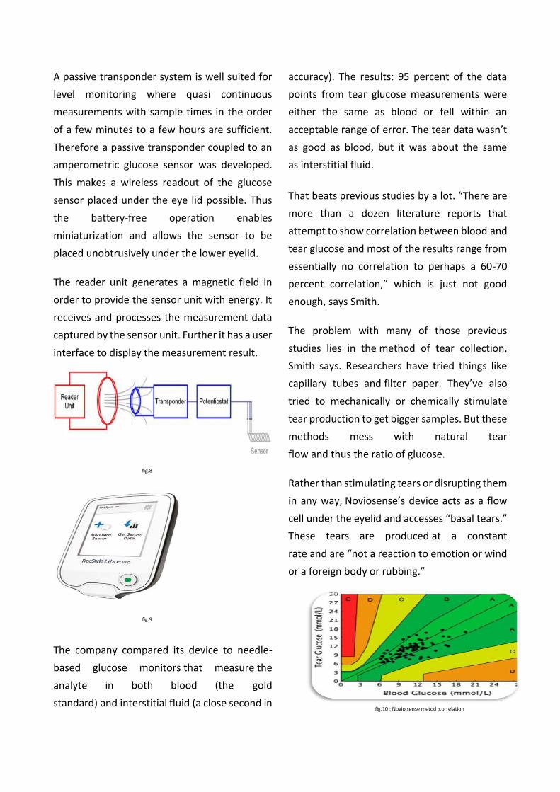
A passive transponder system is well suited for
level monitoring where quasi continuous
measurements with sample times in the order
of a few minutes to a few hours are sufficient.
Therefore a passive transponder coupled to an
amperometric glucose sensor was developed.
This makes a wireless readout of the glucose
sensor placed under the eye lid possible. Thus
the battery-free operation enables
miniaturization and allows the sensor to be
placed unobtrusively under the lower eyelid.
The reader unit generates a magnetic field in
order to provide the sensor unit with energy. It
receives and processes the measurement data
captured by the sensor unit. Further it has a user
interface to display the measurement result.
fig.8
fig.9
The company compared its device to needle-
based glucose monitors that measure the
analyte in both blood (the gold
standard) and interstitial fluid (a close second in
accuracy). The results: 95 percent of the data
points from tear glucose measurements were
either the same as blood or fell within an
acceptable range of error. The tear data wasn’t
as good as blood, but it was about the same
as interstitial fluid.
That beats previous studies by a lot. “There are
more than a dozen literature reports that
attempt to show correlation between blood and
tear glucose and most of the results range from
essentially no correlation to perhaps a 60-70
percent correlation,” which is just not good
enough, says Smith.
The problem with many of those previous
studies lies in the method of tear collection,
Smith says. Researchers have tried things like
capillary tubes and filter paper. They’ve also
tried to mechanically or chemically stimulate
tear production to get bigger samples. But these
methods mess with natural tear
flow and thus the ratio of glucose.
Rather than stimulating tears or disrupting them
in any way, Noviosense’s device acts as a flow
cell under the eyelid and accesses “basal tears.”
These tears are produced at a constant
rate and are “not a reaction to emotion or wind
or a foreign body or rubbing.”
fig.10 : Novio sense metod :correlation
Refrences
1.World Health Organization. Diabetes: Fact sheet N°312. Geneva (Switzerland): Available
from: http://www.who.int/mediacentre/factsheets/fs312/en/. Accessed on 2019/5/25.
2. Shen YC, Davies AG, Linfield EH, Elsey TS, Taday PF, Arnone DD. The use of Fourier-transform
infrared spectroscopy for the quantitative determination of glucose concentration in whole
blood. Phys Med Biol. 2003;48(13):2023–2032.[Google Scholar]
3. Burmeister JJ, Arnold MA, Small GW. Noninvasive blood glucose measurements by near-infrared
transmission spectroscopy across human tongues. [Google Scholar]
4. Nelson LA, McCann JC, Loepke AW, Wu J, Ben Dor B, Kurth CD. Development and validation of a
multiwavelength spatial domain near-infrared oximeter to detect cerebral hypoxia-
ischemia. [Google Scholar]
5. Evans ND, Gnudi L, Rolinski OJ, Birch DJ, Pickup JC. Non-invasive glucose monitoring by NAD(P)H
autofluorescence spectroscopy in fibroblasts and adipocytes: a model for skin glucose
sensing. [Google Scholar]
6. Katika KM, Pilon L. Feasibility analysis of an epidermal glucose sensor based on time-resolved
fluorescence.[Google Scholar]
7. Lambert JL, Morookian JM, Sirk SJ, Borchert MS. Measurement of aqueous glucose in a model
anterior chamber using Raman spectroscopy. J Raman Spectrosc. [Google Scholar]
8. Enejder AM, Scecina TG, Oh J, Hunter M, Shih WC, Sasic S, Horowitz GL, Feld MS. Raman
spectroscopy for noninvasive glucose measurements.[Google Scholar]
9. Rabinovitch B, March WF, Adams RL. Noninvasive glucose monitoring of the aqueous humor of
the eye: Part I. Measurement of very small optical rotations. Diabetes Care. [Google Scholar]
10. March WF, Rabinovitch B, Adams RL. Noninvasive glucose monitoring of the aqueous humor of
the eye: Part II. Animal studies and the scleral lens. Diabetes Care. [Google Scholar]
11. Coté GL, Fox MD, Northrop RB. Noninvasive optical polarimetric glucose sensing using a true
phase measurement technique. IEEE Trans Biomed Eng. [Google Scholar]
12. MacKenzie HA, Ashton HS, Spiers S, Shen Y, Freeborn SS, Hannigan J, Lindberg J, Rae P. Advances
in photoacoustic noninvasive glucose testing.[Google Scholar]
13. Gasset AR, Braverman LE, Fleming MC, Arky RA, Alter BR. Tear glucose detection of
hyperglycemia. Am J Ophthalmol. [Google Scholar]
14. Motoji K. The glucose content of the tear fluid in normal and diabetic subjects. Jpn J Clin
Ophthalmol. [Google Scholar]
15. Sen DK, Sarin GS. Tear glucose levels in normal people and in diabetic patients. Br J
Ophthalmol. [Google Scholar]
16. Malik BH, Coté GL. Modeling the corneal birefringence of the eye toward the development of a
polarimetric glucose sensor. [Google Scholar]
17. O’Donnell C, Efron N, Boulton AJ. A prospective study of contact lens wear in diabetes
mellitus. [Google Scholar]
18. March W, Long B, Hofmann W, Keys D, McKenney C. Safety of contact lenses in patients with
diabetes. Diabetes Technol Ther. [Google Scholar]
19.NovioSense Company. Available from: http://www.noviosense.com/publications/ Accessed on
2019/5/28.