novel strategies for soil-borne diseases: exploiting the
TRANSCRIPT

fmicb-10-01296 June 7, 2019 Time: 17:10 # 1
ORIGINAL RESEARCHpublished: 07 June 2019
doi: 10.3389/fmicb.2019.01296
Edited by:Jesús Mercado-Blanco,
Instituto de Agricultura Sostenible(IAS), Spain
Reviewed by:Miguel Talavera,
Andalusian Institute for Researchand Training in Agriculture, Fisheries,
Food and Ecological Production(IFAPA), Spain
Antonio José Fernández-González,Estación Experimental del Zaidín
(EEZ), SpainIsabel Abrantes,
University of Coimbra, Portugal
*Correspondence:Gabriele Berg
Specialty section:This article was submitted to
Plant Microbe Interactions,a section of the journal
Frontiers in Microbiology
Received: 06 March 2019Accepted: 23 May 2019
Published: 07 June 2019
Citation:Wolfgang A, Taffner J,
Guimarães RA, Coyne D and Berg G(2019) Novel Strategies for Soil-Borne
Diseases: Exploiting the Microbiomeand Volatile-Based Mechanisms
Toward ControllingMeloidogyne-Based Disease
Complexes.Front. Microbiol. 10:1296.
doi: 10.3389/fmicb.2019.01296
Novel Strategies for Soil-BorneDiseases: Exploiting the Microbiomeand Volatile-Based MechanismsToward ControllingMeloidogyne-Based DiseaseComplexesAdrian Wolfgang1, Julian Taffner1, Rafaela Araújo Guimarães2, Danny Coyne3 andGabriele Berg1*
1 Institute of Environmental Biotechnology, Graz University of Technology, Graz, Austria, 2 Department of Phytopathology,Universidade Federal de Lavras, Lavras, Brazil, 3 International Institute of Tropical Agriculture, Nairobi, Kenya
Under more intensified cropping conditions agriculture will face increasing incidencesof soil-borne plant pests and pathogens, leading to increasingly higher yield lossesworld-wide. Soil-borne disease complexes, in particular, are especially difficult to control.In order to better understand soil-borne Meloidogyne-based disease complexes, westudied the volatile-based control mechanism of associated bacteria as well as therhizospheric microbiome on Ugandan tomato plants presenting different levels of root-galling damage, using a multiphasic approach. The experimental design was based onrepresentative samplings of healthy and infected tomato plants from two field locationsin Uganda, to establish species collections and DNA libraries. Root galling symptomson tomato resulted from a multispecies infection of root-knot nematodes (Meloidogynespp.). Results revealed that 16.5% of the bacterial strain collection produced nematicidalvolatile organic compounds (nVOC) active against Meloidogyne. Using SPME GC-MS, diverse VOC were identified, including sulfuric compounds, alkenes and onepyrazine. Around 28% of the bacterial strains were also antagonistic toward at leastone fungal pathogen of the disease complex. However, antagonistic interactions appearhighly specific. Nematicidal antagonists included Pseudomonas, Comamonas, andVariovorax and fungicidal antagonists belonged to Bacillus, which interestingly, wereprimarily recovered from healthy roots, while nematode antagonists were prominent inthe rhizosphere and roots of diseased roots. In summary, all antagonists comprisedup to 6.4% of the tomato root microbiota. In general, the microbiota of healthyand diseased root endospheres differed significantly in alpha and quantitative betadiversity indices. Bacteria-derived volatiles appear to provide a remarkable, yet whollyunexploited, potential to control Meloidogyne-based soil-borne disease complexes.The highly specific observed antagonism indicates that a combination of volatiles orVOC-producing bacteria are necessary to counter the range of pathogens involvedin such complexes.
Keywords: root-knot nematodes, tomato microbiome, biocontrol, antagonists, Pseudomonas, Comamonas,Variovorax, Bacillus
Frontiers in Microbiology | www.frontiersin.org 1 June 2019 | Volume 10 | Article 1296

fmicb-10-01296 June 7, 2019 Time: 17:10 # 2
Wolfgang et al. Meloidogyne-Based Disease Complexes
INTRODUCTION
Agriculture causes long-lasting anthropogenic environmentalimpacts as it replaces natural vegetation, alters biogeochemicalcycles, and decreases biodiversity; this defined a new human-dominated geological epoch, the Anthropocene. The soildisturbance by the conversion of land to agriculture has resultedin species extinctions 100–1,000 times higher than naturalrates, and likely constitutes the beginning of the sixth massextinction in Earth’s history (Lewis and Maslin, 2015). However,much less is known about the loss of microbial diversity(Berg et al., 2017) due to human activity than the loss ofmacroscopic diversity. In particular, bacteria occupy importantniches and roles, linking plant microbial diversity and ecosystemfunctioning, such as productivity, host plant fitness and resilience(Bais et al., 2004; Tilman et al., 2012; Vandenkoornhuyse et al.,2015; Laforest-Lapointe et al., 2017). Among the most strikingdirect consequences of agricultural intensification is the elevatedpresence and impact of soil-borne pests and pathogens (Mendeset al., 2012). Soil-borne diseases are often “microbiome diseases”;they signify the result of a loss of microbial diversity and dysbiosisin soil and consequently in the rhizosphere and endosphereof plants (van Elsas et al., 2012). Once established, bacteria,fungi, and nematode pathogens accumulate, often as a synergisticcombination, leading to high yield losses, which prove difficult tocontrol (Oerke, 2006; Bennett et al., 2012). The soil environmentis a complex arena, the biological permutations of which arelittle understood (Bais et al., 2004), especially in areas such asAfrica, where assessments and understanding of the microfloraand microbiota remain negligible.
Root-knot nematodes Göldi 1892 (RKN, genus Meloidogyne)infect over 5500 host plants, including plant species from nearlyevery extant known plant family (Trudgill and Blok, 2001).Species of Meloidogyne can be extremely polyphagous, are mainlyparthenogenetic, and are highly adapted obligate sedentaryplant parasites. They are regarded as the most economicallyimportant plant-pathogenic nematode group worldwide (Joneset al., 2013) and in the tropics, are viewed as the mostsignificant biotic threat to crop production (Karssen et al., 2013;Coyne et al., 2018). Host roots are infected by freshly hatched,motile second stage juveniles (J2), which, upon establishinga feeding site behind the root tip, become sedentary, feedingfrom cells which it modifies to provide a constant supply ofnutrients. Infection by RKN often leads to typical symptoms ofroot damage and gall formation, with above-ground symptomsof stunting, wilting, leaf chlorosis, reduced yield, which aresymptomatic of water or nutrient scarcity (Karssen et al., 2013).Nematode-infected plants tend to be more susceptible to otherdiseases (Coyne et al., 2018). These unspecific above-groundsymptoms, however, lead to an excess overuse of fertilizersand ineffective treatment with pesticides (Karungi et al., 2011).Excessive and frequent pesticide applications, in combinationwith inappropriate handling, increase the risks to human healthas well as to water resources and the ecological system (Coyneet al., 2018). The impact of RKN on the host is significantlyexaggerated through secondary pathogen infections, such asroot rot pathogens, bacterial and fungal wilts, e.g., Fusarium
oxysporum and Verticillium dahliae (Back et al., 2002; Karssenet al., 2013). These plant pathogens do not necessarily need thepresence of RKN to successfully infect their hosts, but since RKNmay act as casual agents, the disease can be seen as Meloidogyne-based disease complex. The interactions between RKN, secondarypathogens, host plant and plant-associated microorganisms leadto the resulting effects on plant health (Karssen et al., 2013).The management of RKN would therefore benefit from a moreholistic approach, taking into consideration the management ofsoil-borne microbial complexes. A deep understanding of theplant-associated microbiomes would be beneficial, given that aproportion of microorganisms are antagonistic toward soil-bornepests and pathogens (Berg et al., 2002). One recently discoveredindirect mode of plant disease prevention in bacteria is theproduction of volatile organic compounds (VOC) (Effmert et al.,2012; Cernava et al., 2015). VOC are semiochemicals that actas “long-range” allelochemicals in soil, which can have growth-promoting or -inhibiting effects on other microorganisms(Effmert et al., 2012) and plants (Ryu et al., 2003). The factthat VOC can have communicational, controlling or inhibitoryeffects that act inter- and/or intra-specifically, make them a highlyinteresting field of study for biological control (Effmert et al.,2012; Torto et al., 2018). However, despite their potential, VOChave received only limited attention and are yet to be fullyexploited for biocontrol strategies. Our hypothesis was that RKNcan be negatively affected and controlled by bacteria-derivedVOC in the microbiome.
We selected tomato as a model plant, given its susceptibility toRKN and soil-borne disease complexes, using two RKN-infectedfield sites in Uganda. Tomatoes in Uganda are a key sourceof income and food security for smallholders (Ssekyewa, 2006)who often own less than 2 ha of land (Karungi et al., 2011).A major challenge in controlling the RKN-disease complex is theneed for simultaneous control of all involved pathogens. Novelmechanisms are urgently needed to address these soil-bornechallenges, especially in Africa where the need to sustainablyintensify cropping production systems is critical (Bennett et al.,2012; Vanlauwe et al., 2014). Our current study focused on threemain objectives: (i) identify the Meloidogyne species present,(ii) screen bacterial strains capable of producing nematicidalVOC or fungicidal metabolites, (iii) analyze the microbiome shiftin the root endosphere due to RKN activity. Results of thisstudy will create a better understanding of the soil-pathogen-hostinteractions in the RKN-disease complex, which will be translatedto developing novel control strategies.
MATERIALS AND METHODS
Sampling DesignThree bulk samples, each comprising ten roots of fruit-bearingtomato plants (Solanum lycopersicum L.) with adhering soil,were collected from two field sites in Uganda in April 2017.Gall formation was categorized according to a root galling index(RGI) from 1 (no visible galling damage) to 5 (severe/lethaldamage) (Coyne et al., 2007); two plants were selected foreach RGI score from each site during uprooting. Sampling site
Frontiers in Microbiology | www.frontiersin.org 2 June 2019 | Volume 10 | Article 1296
fmicb-10-01296 June 7, 2019 Time: 17:10 # 3
Wolfgang et al. Meloidogyne-Based Disease Complexes
“Luwero” (0◦39′20′′ N, 32◦24′38′′ E, 1187 m) was a rural farmer’sopen field with 1-year-old virgin soil. Tomato cv. “Rio Grande,”received unknown application levels of pesticides but includedgeneric fungicides, mainly mancozeb-based and the insecticidescypermethrin and chlorpyriphos. Sampling site “Namulonge”was at the IITA research station (0◦31′46′′ N, 32◦36′45′′ E,1170 m) and consisted of a RKN-infected sandy soil withina concrete tomato outdoor bed with no direct connectionto surrounding soils. Tomato cv. “Moneymaker” received nopesticide applications. A 300 g soil sample from each samplesite was assessed for pH, nutrient (K, P, Mg, organic matter)content and soil type by “AGROLAB Agrar und Umwelt GmbH”(Sarstedt, Germany) to compare soil composition between thetwo sampling sites.
Bacterial Strains and Isolation of TotalCommunity DNASamples were recovered from different microhabitats associatedwith RKN infection: bulk soil constituting the native bacterialcommunity in the field, rhizosphere representing root-associatedbacteria, galled and non-galled rhizoendosphere. Bacterialsuspensions were recovered using 0.9% NaCl from a 5 g sub-sample of the bulk soil (soil between tomato plants, threesamples/site, n = 6), from rhizosphere (root adhering soil,one/plant, n = 20) and surface sterilized sections of rootsfrom both galled (RE-D, n = 16) and non-galled (RE-H,n = 17) roots. Suspensions were used for DNA extractions forboth amplicon analysis and isolation of bacterial strains (fordetails, see Supplementary Material – Additional Methods).Suspensions were plated onto NA plates (nutrient terestinglyagar; Sifin GmbH, Berlin, Germany); in total 260 strains wereisolated and screened for nematicidal (see section “Screening fornVOC-Producing Strains”) and fungicidal properties (see section“Bacterial Antagonistic Activity Against Fungal Pathogens”).Extraction of the DNA pellet was conducted using “FastDNASpin Kit for soil” (MP Biomedical, Eschwege, GER). PCR-products were cleaned with GENECLEAN TurboTM Kit (MPBiomedicals, Eschwege, GER) following the manufacturer’sinstructions for genomic DNA. 16S rRNA gene amplificationswere carried out in 3 × 30 µl reactions with the Illuminabarcode universal bacterial primer set 515f-806r (Caporasoet al., 2011) and PNA Mix (Lundberg et al., 2013) to removeplastid DNA. PCR products of barcoded samples were pooledto equimolarity; sequencing was carried out by Eurofins MWGOperon (Ebersberg, GER1) with an Illumina HiSeq 2500 system(for details, see Supplementary Material – Additional Methods).
Identification of Nematodes and InoculaProductionRandomly selected females with eggs were dissected fromdiseased roots. Perineal patterns of females were used formorphological diagnosis; body content of the same crushedfemales were used for molecular identification using themolecular key method of Adam et al. (2007). Furthermore,
1http://www.eurofinsgenomics.eu/
the region for NAD dehydrogenase subunit 5 was amplifiedfor genetic determination using the body content of individualfemales (Janssen et al., 2016). Data were combined to determinespecies identification. Eggs were used to re-infect tomatoseedlings in the fourth-true-leaf stadium to establish purecultures. J2 of identified pure cultures were partially used infurther experiments. For extracting J2, roots were rinsed freeof adhering soil. Diseased root sections were chopped coarsely,placed in 1.2% NaOCl solution and blended with a hand blenderfor 3 min. The suspension was rinsed with tap water on nested100–25 µm sieves. Eggs were caught on 25 µm sieve and collectedinto a beaker, which was aerated for 10 days to allow hatching.The J2 suspensions were placed on a Baermann funnel filter for24 h at room temperature (Supplementary Figure S1A). Theresulting ∼30 ml J2 suspension was stored horizontally in 50 mlSarstedt tubes at 6◦C until use.
Screening for nVOC-Producing StrainsWe used a variation of the two clamp VOC assay (TCVA,Cernava et al., 2015): Bacterial strains were streaked on 12-wellplates containing NA and incubated at 30◦C for 24 h. Eachplate had a blank well, containing NA only. Plates were inversedonto another 12-well plate containing ∼100 J2 of M. incognita(provided from Julius Kühn-Institut, Münster, Germany) on 2%-tap water agar. A silicon foil with a 5 mm hole between the twoopposing chambers separated the two 12-well plates. The twoplates were clamped together to provide airtight test conditions(Supplementary Figure S1B) and then maintained for 24 h atroom temperature. Dead J2 were assigned dead if the body wasstraight and did not react when prodded with a dissection needle.Percentage J2 mortality was calculated after correcting for theblank value of the corresponding plate blank. Bacterial strainswere categorized according to their activity: non-active (<10%mortality), slightly active (>10–80%), active (>80–95%), andhighly active (>95%). Distributions of the number of bacterialstrains within the categories were compared between samplingsites, microhabitat (healthy/diseased root, rhizosphere) and RGI.The experiment was repeated two more times for those samplesdemonstrating >80% nematicidal activity. The strains showingconsistent nematicidal activity were identified by sequencingof 16S rRNA gene.
Bacterial Antagonistic Activity AgainstFungal PathogensAll 260 isolated bacterial strains were tested for theirantagonistic activity against the fungal pathogens Botrytiscinerea, F. oxysporum, Fusarium verticillioides, Sclerotiumrolfsii, and V. dahliae (provided by Institute of EnvironmentalBiotechnology Graz) in dual cultures on Waxman agar in threereplicates. Antifungal activity was categorized according to: 0(fungi overgrow bacterial colony), +1 (hyphae reach bacteria,but do not overgrow), +2 (lateral inhibition zone <0.5 cm), and+3 (lateral inhibition zone >0.5 cm). The mean category acrossthe three repeat assessments was calculated. Bacterial strainsshowing a strong antifungal effect (category +3) were compared
Frontiers in Microbiology | www.frontiersin.org 3 June 2019 | Volume 10 | Article 1296
fmicb-10-01296 June 7, 2019 Time: 17:10 # 4
Wolfgang et al. Meloidogyne-Based Disease Complexes
using VENN2. DNA of strains with a mean antifungal activity of+3 against at least four pathogens (n = 23, see also Figure 2A)was extracted. A BOX-PCR was carried out to identify clones,resulting in five genotypes. 16S rRNA gene of the resulting fivegenotypes was amplified and sequenced for identification.
SPME GC-MS of nVOC and NematicidalEffect of Single CompoundsNematicidal volatile organic compounds were identified usingan adapted version of the method from Verginer et al. (2010)(for details, see Supplementary Material – Additional Methods).A total of nine compounds (purity >98%) partially foundin the GC-MS samples were tested against M. javanica J2in a chambered Petri dish, namely decene (10en), undecene(11en), undecane-2-on (11on), dodecene (12en), 2-methoxy-3-methyl pyrazine (2M3MP), 2,5-dimethyl pyrazine (25DP),5-isobutyl-2,3-dimethyl pyrazine (5I23DP), 2-ethyl-3-methylpyrazine (2E3MP), and 2-isobutyl-3-methoxy pyrazine (2I3MP)(all Sigma-Aldrich, Darmstadt, Germany). 2M3MP was usedas a substitute for 3-methoxy-2,5-dimethyl pyrazine, whichwas consistently detected in nVOC-volatilome of Pseudomonaskoreensis T3GI1 (see also Table 2). The compound 2-undecanonewas used as a positive control, due to its known nematicidaleffects on M. incognita (Huang et al., 2010) and other nematodes(Gu et al., 2007). On one side of the Petri dish, a 500 µlsuspension of M. javanica (∼250 J2) was placed on 8 ml of 2%-tapwater agar. On the opposite side, three concentrations (1, 5, and20 µl) of a single compound were placed on a microscopic slide,which prevented interactions of the compound with the Petridish plastic. A Petri dish with 20 µl distilled water representedthe control. Plates were maintained at room temperature for24 h and the experiment repeated a further two times and theblank-corrected mortality rate calculated for each compound.
Amplicon Analysis and StatisticsPre-processing of the reads obtained by the sequencing companyof Eurofins MWG Operon was carried out using QIIME 2(2017.12 release) and QIIME 1 (Caporaso et al., 2010) followingthe protocol of Schwendner et al. (2017). Demultiplexing,denoising (400 bp length, including phiX and chimera filtering)and generation of ribosomal sequence variants (RSVs) wascarried out with the DADA2 algorithm (Callahan et al., 2016).RSVs were summarized in a feature table. The taxonomic analysiswas based on a customized naïve-bayes classifier trained on16S rRNA gene features clustered at 97% similarities within theSilva128 database release and trimmed to a length of 400 bp.Reads for mitochondria and chloroplasts were filtered usingQIIME 2 before analysis. The feature table was rarefied to6,890 reads for core metrics analysis. Alpha diversity indiceswere analyzed using Pairwise Kruskal–Wallis test and betadiversity indices with PERMANOVA. Bray–Curtis dissimilarityand unweighted UniFrac dissimilarity between habitats werevisualized using EMPEROR3. One rhizosphere sample (T4R)was removed due to poor quality. Differential abundances
2http://bioinformatics.psb.ugent.be/webtools/Venn/3https://view.qiime2.org
were subjected to ANCOM and Gneiss test, implementedin QIIME 2. Bacterial network of the core microbiome ofeach habitat (>1% relative abundance within habitat) wasvisualized using Cytoscape 3.3.0 (Shannon et al., 2003) onorder level. The core microbiomes were defined with anoccurence of >75% throughout the replicates for each habitat.Mean relative abundance of fungal and nematode antagonistsfound in this study was calculated for each data set. Bacterialabundances on family level were compared between healthy anddiseased root samples to visualize bacterial community shift dueto RKN infection.
RESULTS
Meloidogyne Were the Causal Agents ofthe Disease Complex in TomatoThe combination of identification approaches (SCAR-primermolecular key, morphological examination, NAD5 sequences)identified 10 adult females as M. incognita and two as M. incognitasensu lato. in Luwero (site 1). Just M. incognita was reliablyidentified from Luwero. In samples from Namulonge (site 2),three females were identified as M. incognita, six as M. javanicaand five as M. incognita s. lat. Thus, a multispecies infectionwas confirmed in Namulonge (see Supplementary Material –Additional Results, Supplementary Figures S2, S3).
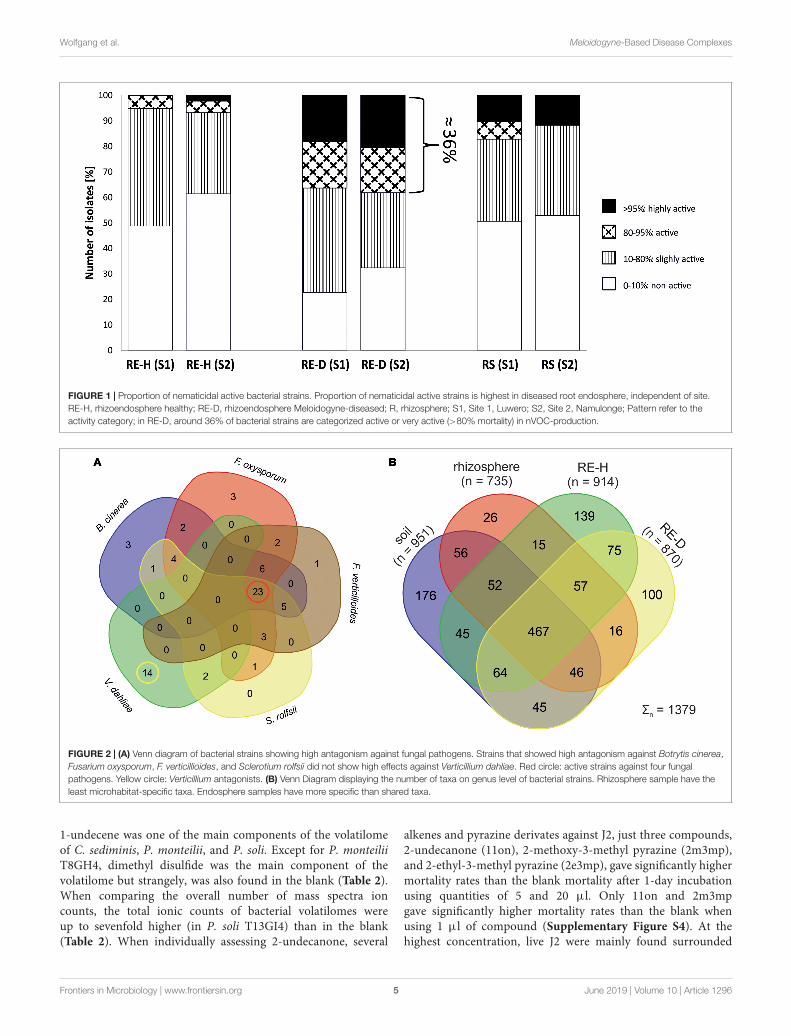
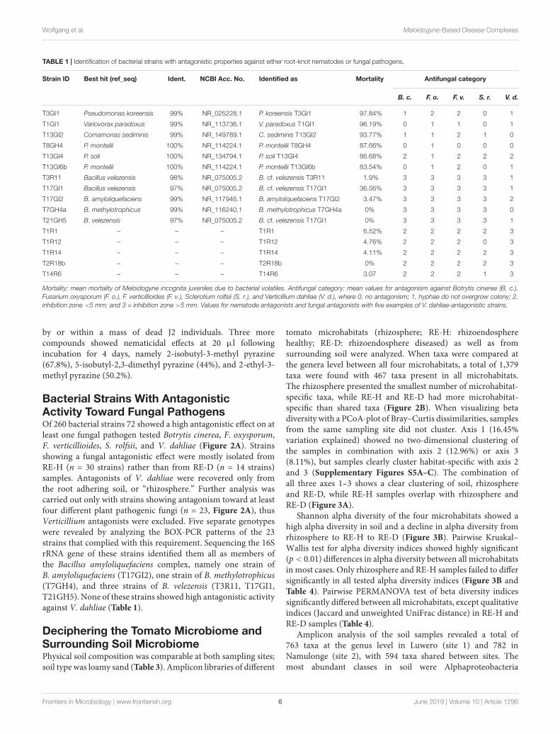
Bacterial Strains With AntagonisticActivity Toward MeloidogyneRepresentative bacterial strains from each of the two samplingsites caused similar mean nematode mortality rates throughnVOC (32.1 ± 25.4% in Luwero, 30.9 ± 28.1% in Namulonge).Most strains showed no effect (mortality <10%) or only slightnematicidal effects. In total, 43 strains were categorized as active(80–95% mortality; n = 20) or highly active (>95% mortality;n = 23). When comparing the overall nematicidal activity ofbacteria from plants of different RGI, a trend was apparent forhigher mortality rates of strains from highly diseased plants.Most active and highly active strains (n = 43) were recoveredfrom diseased rhizoendosphere (RE-D) samples, independent ofsite (Figure 1). Of these 43 strains, six demonstrated repeatednematicidal activity (>80% mean mortality). Five out of these sixhighly active strains were bacteria isolated from RE-D. Moreover,three of these strains were collected from the same tomato plant(T13), a heavily galled (RGI = 4.5) plant from Namulonge. Justone strain was isolated from healthy, non-galled rhizoendosphere(RE-H) from Luwero. Sequences of the 16S rRNA genes identifiedthe nematicidal bacteria as Pseudomonas koreensis, Comamonassediminis, Variovorax paradoxus, P. soli and two strains ofP. monteilii. Strains with high nematicidal activity showed nohigh antifungal activity and vice versa (Table 1).
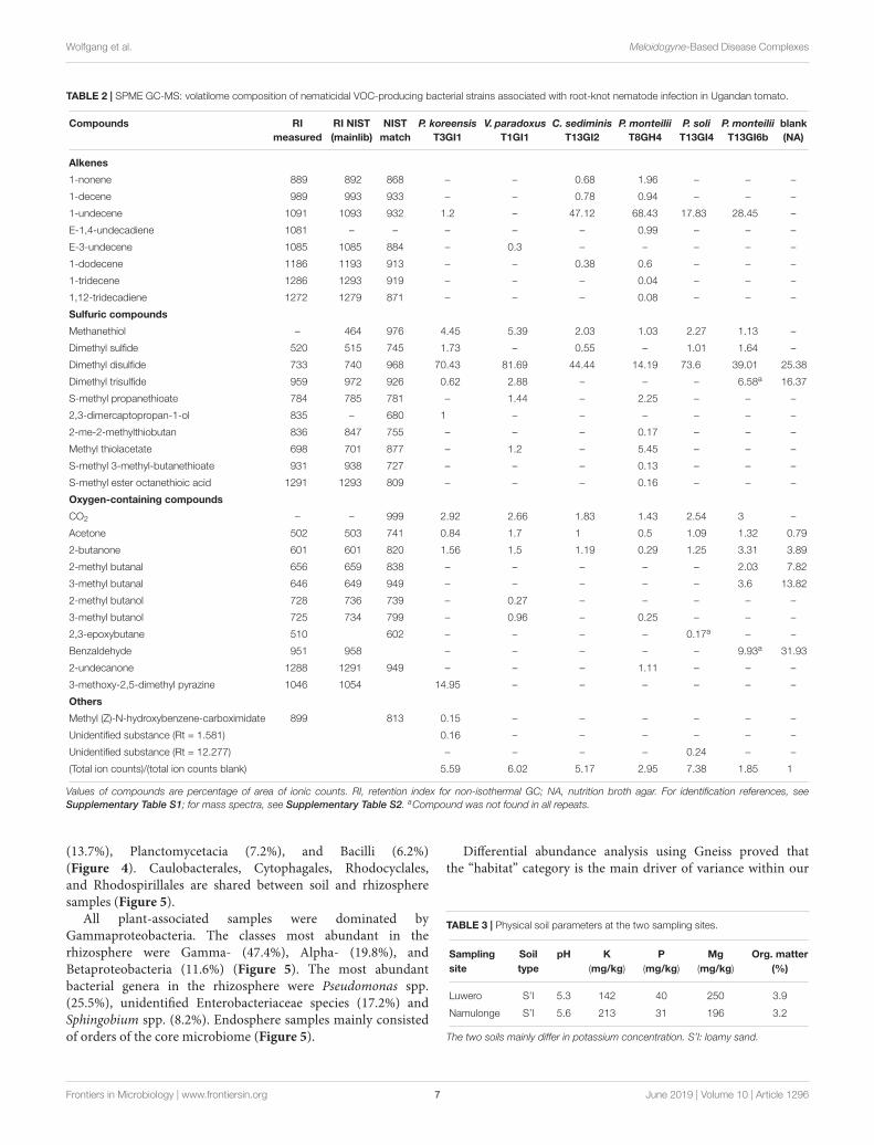
The volatilomes of the nVOC-producing strains mainlyconsisted of alkenes, sulfuric compounds, alcohols, ketones,and aldehydes. Additionally, one pyrazine (3-methoxy-2,5-dimethyl pyrazine) was consistently detected in P. koreensisT3GI1, the strain with the highest nematicidal effect (Table 2).
Frontiers in Microbiology | www.frontiersin.org 4 June 2019 | Volume 10 | Article 1296
fmicb-10-01296 June 7, 2019 Time: 17:10 # 5
Wolfgang et al. Meloidogyne-Based Disease Complexes
FIGURE 1 | Proportion of nematicidal active bacterial strains. Proportion of nematicidal active strains is highest in diseased root endosphere, independent of site.RE-H, rhizoendosphere healthy; RE-D, rhizoendosphere Meloidogyne-diseased; R, rhizosphere; S1, Site 1, Luwero; S2, Site 2, Namulonge; Pattern refer to theactivity category; in RE-D, around 36% of bacterial strains are categorized active or very active (>80% mortality) in nVOC-production.
FIGURE 2 | (A) Venn diagram of bacterial strains showing high antagonism against fungal pathogens. Strains that showed high antagonism against Botrytis cinerea,Fusarium oxysporum, F. verticillioides, and Sclerotium rolfsii did not show high effects against Verticillium dahliae. Red circle: active strains against four fungalpathogens. Yellow circle: Verticillium antagonists. (B) Venn Diagram displaying the number of taxa on genus level of bacterial strains. Rhizosphere sample have theleast microhabitat-specific taxa. Endosphere samples have more specific than shared taxa.
1-undecene was one of the main components of the volatilomeof C. sediminis, P. monteilii, and P. soli. Except for P. monteiliiT8GH4, dimethyl disulfide was the main component of thevolatilome but strangely, was also found in the blank (Table 2).When comparing the overall number of mass spectra ioncounts, the total ionic counts of bacterial volatilomes wereup to sevenfold higher (in P. soli T13GI4) than in the blank(Table 2). When individually assessing 2-undecanone, several
alkenes and pyrazine derivates against J2, just three compounds,2-undecanone (11on), 2-methoxy-3-methyl pyrazine (2m3mp),and 2-ethyl-3-methyl pyrazine (2e3mp), gave significantly highermortality rates than the blank mortality after 1-day incubationusing quantities of 5 and 20 µl. Only 11on and 2m3mpgave significantly higher mortality rates than the blank whenusing 1 µl of compound (Supplementary Figure S4). At thehighest concentration, live J2 were mainly found surrounded
Frontiers in Microbiology | www.frontiersin.org 5 June 2019 | Volume 10 | Article 1296
fmicb-10-01296 June 7, 2019 Time: 17:10 # 6
Wolfgang et al. Meloidogyne-Based Disease Complexes
TABLE 1 | Identification of bacterial strains with antagonistic properties against either root-knot nematodes or fungal pathogens.
Strain ID Best hit (ref_seq) Ident. NCBI Acc. No. Identified as Mortality Antifungal category
B. c. F. o. F. v. S. r. V. d.
T3GI1 Pseudomonas koreensis 99% NR_025228.1 P. koreensis T3GI1 97.84% 1 2 2 0 1
T1GI1 Variovorax paradoxus 99% NR_113736.1 V. paradoxus T1GI1 96.19% 0 1 1 0 1
T13GI2 Comamonas sediminis 99% NR_149789.1 C. sediminis T13GI2 93.77% 1 1 2 1 0
T8GH4 P. monteilii 100% NR_114224.1 P. monteilii T8GH4 87.66% 0 1 0 0 0
T13GI4 P. soli 100% NR_134794.1 P. soli T13GI4 86.68% 2 1 2 2 2
T13GI6b P. monteilii 100% NR_114224.1 P. monteilii T13GI6b 83.54% 0 1 2 0 1
T3R11 Bacillus velezensis 98% NR_075005.2 B. cf. velezensis T3R11 1.9% 3 3 3 3 1
T17GI1 Bacillus velezensis 97% NR_075005.2 B. cf. velezensis T17GI1 36.56% 3 3 3 3 1
T17GI2 B. amyloliquefaciens 99% NR_117946.1 B. amyloliquefaciens T17GI2 3.47% 3 3 3 3 2
T7GH4a B. methylotrophicus 99% NR_116240.1 B. methylotrophicus T7GH4a 0% 3 3 3 3 0
T21GH5 B. velezensis 97% NR_075005.2 B. cf. velezensis T17GI1 0% 3 3 3 3 1
T1R1 – – – T1R1 6.52% 2 2 2 2 3
T1R12 – – – T1R12 4.76% 2 2 2 0 3
T1R14 – – – T1R14 4.11% 2 2 2 2 3
T2R18b – – – T2R18b 0% 2 2 2 2 3
T14R6 – – – T14R6 3.07 2 2 2 1 3
Mortality: mean mortality of Meloidogyne incognita juveniles due to bacterial volatiles. Antifungal category: mean values for antagonism against Botrytis cinerea (B. c.),Fusarium oxysporum (F. o.), F. verticillioides (F. v.), Sclerotium rolfsii (S. r.), and Verticillium dahliae (V. d.), where 0, no antagonism; 1, hyphae do not overgrow colony; 2,inhibition zone <5 mm; and 3 = inhibition zone >5 mm. Values for nematode antagonists and fungal antagonists with five examples of V. dahliae-antagonistic strains.
by or within a mass of dead J2 individuals. Three morecompounds showed nematicidal effects at 20 µl followingincubation for 4 days, namely 2-isobutyl-3-methyl pyrazine(67.8%), 5-isobutyl-2,3-dimethyl pyrazine (44%), and 2-ethyl-3-methyl pyrazine (50.2%).
Bacterial Strains With AntagonisticActivity Toward Fungal PathogensOf 260 bacterial strains 72 showed a high antagonistic effect on atleast one fungal pathogen tested Botrytis cinerea, F. oxysporum,F. verticillioides, S. rolfsii, and V. dahliae (Figure 2A). Strainsshowing a fungal antagonistic effect were mostly isolated fromRE-H (n = 30 strains) rather than from RE-D (n = 14 strains)samples. Antagonists of V. dahliae were recovered only fromthe root adhering soil, or “rhizosphere.” Further analysis wascarried out only with strains showing antagonism toward at leastfour different plant pathogenic fungi (n = 23, Figure 2A), thusVerticillium antagonists were excluded. Five separate genotypeswere revealed by analyzing the BOX-PCR patterns of the 23strains that complied with this requirement. Sequencing the 16SrRNA gene of these strains identified them all as members ofthe Bacillus amyloliquefaciens complex, namely one strain ofB. amyloliquefaciens (T17GI2), one strain of B. methylotrophicus(T7GH4), and three strains of B. velezensis (T3R11, T17GI1,T21GH5). None of these strains showed high antagonistic activityagainst V. dahliae (Table 1).
Deciphering the Tomato Microbiome andSurrounding Soil MicrobiomePhysical soil composition was comparable at both sampling sites;soil type was loamy sand (Table 3). Amplicon libraries of different
tomato microhabitats (rhizosphere; RE-H: rhizoendospherehealthy; RE-D: rhizoendosphere diseased) as well as fromsurrounding soil were analyzed. When taxa were compared atthe genera level between all four microhabitats, a total of 1,379taxa were found with 467 taxa present in all microhabitats.The rhizosphere presented the smallest number of microhabitat-specific taxa, while RE-H and RE-D had more microhabitat-specific than shared taxa (Figure 2B). When visualizing betadiversity with a PCoA-plot of Bray–Curtis dissimilarities, samplesfrom the same sampling site did not cluster. Axis 1 (16.45%variation explained) showed no two-dimensional clustering ofthe samples in combination with axis 2 (12.96%) or axis 3(8.11%), but samples clearly cluster habitat-specific with axis 2and 3 (Supplementary Figures S5A–C). The combination ofall three axes 1–3 shows a clear clustering of soil, rhizosphereand RE-D, while RE-H samples overlap with rhizosphere andRE-D (Figure 3A).
Shannon alpha diversity of the four microhabitats showed ahigh alpha diversity in soil and a decline in alpha diversity fromrhizosphere to RE-H to RE-D (Figure 3B). Pairwise Kruskal–Wallis test for alpha diversity indices showed highly significant(p < 0.01) differences in alpha diversity between all microhabitatsin most cases. Only rhizosphere and RE-H samples failed to differsignificantly in all tested alpha diversity indices (Figure 3B andTable 4). Pairwise PERMANOVA test of beta diversity indicessignificantly differed between all microhabitats, except qualitativeindices (Jaccard and unweighted UniFrac distance) in RE-H andRE-D samples (Table 4).
Amplicon analysis of the soil samples revealed a total of763 taxa at the genus level in Luwero (site 1) and 782 inNamulonge (site 2), with 594 taxa shared between sites. Themost abundant classes in soil were Alphaproteobacteria
Frontiers in Microbiology | www.frontiersin.org 6 June 2019 | Volume 10 | Article 1296
fmicb-10-01296 June 7, 2019 Time: 17:10 # 7
Wolfgang et al. Meloidogyne-Based Disease Complexes
TABLE 2 | SPME GC-MS: volatilome composition of nematicidal VOC-producing bacterial strains associated with root-knot nematode infection in Ugandan tomato.
Compounds RImeasured
RI NIST(mainlib)
NISTmatch
P. koreensisT3GI1
V. paradoxusT1GI1
C. sediminisT13GI2
P. monteiliiT8GH4
P. soliT13GI4
P. monteiliiT13GI6b
blank(NA)
Alkenes
1-nonene 889 892 868 – – 0.68 1.96 – – –
1-decene 989 993 933 – – 0.78 0.94 – – –
1-undecene 1091 1093 932 1.2 – 47.12 68.43 17.83 28.45 –
E-1,4-undecadiene 1081 – – – – – 0.99 – – –
E-3-undecene 1085 1085 884 – 0.3 – – – – –
1-dodecene 1186 1193 913 – – 0.38 0.6 – – –
1-tridecene 1286 1293 919 – – – 0.04 – – –
1,12-tridecadiene 1272 1279 871 – – – 0.08 – – –
Sulfuric compounds
Methanethiol – 464 976 4.45 5.39 2.03 1.03 2.27 1.13 –
Dimethyl sulfide 520 515 745 1.73 – 0.55 – 1.01 1.64 –
Dimethyl disulfide 733 740 968 70.43 81.69 44.44 14.19 73.6 39.01 25.38
Dimethyl trisulfide 959 972 926 0.62 2.88 – – – 6.58a 16.37
S-methyl propanethioate 784 785 781 – 1.44 – 2.25 – – –
2,3-dimercaptopropan-1-ol 835 – 680 1 – – – – – –
2-me-2-methylthiobutan 836 847 755 – – – 0.17 – – –
Methyl thiolacetate 698 701 877 – 1.2 – 5.45 – – –
S-methyl 3-methyl-butanethioate 931 938 727 – – – 0.13 – – –
S-methyl ester octanethioic acid 1291 1293 809 – – – 0.16 – – –
Oxygen-containing compounds
CO2 – – 999 2.92 2.66 1.83 1.43 2.54 3 –
Acetone 502 503 741 0.84 1.7 1 0.5 1.09 1.32 0.79
2-butanone 601 601 820 1.56 1.5 1.19 0.29 1.25 3.31 3.89
2-methyl butanal 656 659 838 – – – – – 2.03 7.82
3-methyl butanal 646 649 949 – – – – – 3.6 13.82
2-methyl butanol 728 736 739 – 0.27 – – – – –
3-methyl butanol 725 734 799 – 0.96 – 0.25 – – –
2,3-epoxybutane 510 602 – – – – 0.17a – –
Benzaldehyde 951 958 – – – – – 9.93a 31.93
2-undecanone 1288 1291 949 – – – 1.11 – – –
3-methoxy-2,5-dimethyl pyrazine 1046 1054 14.95 – – – – – –
Others
Methyl (Z)-N-hydroxybenzene-carboximidate 899 813 0.15 – – – – – –
Unidentified substance (Rt = 1.581) 0.16 – – – – – –
Unidentified substance (Rt = 12.277) – – – – 0.24 – –
(Total ion counts)/(total ion counts blank) 5.59 6.02 5.17 2.95 7.38 1.85 1
Values of compounds are percentage of area of ionic counts. RI, retention index for non-isothermal GC; NA, nutrition broth agar. For identification references, seeSupplementary Table S1; for mass spectra, see Supplementary Table S2. aCompound was not found in all repeats.
(13.7%), Planctomycetacia (7.2%), and Bacilli (6.2%)(Figure 4). Caulobacterales, Cytophagales, Rhodocyclales,and Rhodospirillales are shared between soil and rhizospheresamples (Figure 5).
All plant-associated samples were dominated byGammaproteobacteria. The classes most abundant in therhizosphere were Gamma- (47.4%), Alpha- (19.8%), andBetaproteobacteria (11.6%) (Figure 5). The most abundantbacterial genera in the rhizosphere were Pseudomonas spp.(25.5%), unidentified Enterobacteriaceae species (17.2%) andSphingobium spp. (8.2%). Endosphere samples mainly consistedof orders of the core microbiome (Figure 5).
Differential abundance analysis using Gneiss proved thatthe “habitat” category is the main driver of variance within our
TABLE 3 | Physical soil parameters at the two sampling sites.
Sampling Soil pH K P Mg Org. mattersite type (mg/kg) (mg/kg) (mg/kg) (%)
Luwero S’l 5.3 142 40 250 3.9
Namulonge S’l 5.6 213 31 196 3.2
The two soils mainly differ in potassium concentration. S’l: loamy sand.
Frontiers in Microbiology | www.frontiersin.org 7 June 2019 | Volume 10 | Article 1296

fmicb-10-01296 June 7, 2019 Time: 17:10 # 8
Wolfgang et al. Meloidogyne-Based Disease Complexes
FIGURE 3 | (A) PCoA plot of Bray–Curtis dissimilarity between microhabitats. PCoA-plot shows clear clustering in soil (brown) in rhizosphere (blue) while healthyrhizoendosphere (green) sampling points (a-c) overlap with Meloidogyne-diseased rhizoendosphere (red) and rhizosphere; sample point d (rhizosphere) does notcluster with other rhizosphere samples and was removed in statistical analysis. (B) Barplots of Shannon diversity of microhabitats. Alpha diversity is highest in soiland lowest in diseased rhizoendosphere (galls). Alpha diversity index values are given.
TABLE 4 | P-values of diversity indices reveal significant microhabitat-specific bacterial community differences in root-knot nematode-diseased tomato roots.
Alpha diversity indices Beta diversity indices
Shannon Observed OTUs Faith’s PD Evenness Jaccard Bray–Curtis Unweighted Unifrac Weighted UniFrac
RE-H/RE-D 0.028 0.505 0.746 0.002 0.061 0.022 0.396 0.013
RE-H/R 0.350 0.084 0.788 0.579 0.001 0.001 0.006 0.003
RE-H/S <0.001 <0.001 <0.001 <0.001 0.001 0.001 0.001 0.001
RE-D/R 0.002 0.004 0.289 0.001 0.001 0.001 0.001 0.001
RE-D/S <0.001 0.001 <0.001 <0.001 0.001 0.001 0.001 0.001
R/S <0.001 <0.001 <0.001 <0.001 0.001 0.001 0.001 0.001
P-values for Kruskal–Wallis and PERMANOVA results of alpha and beta diversity indices comparing microhabitats, significant (<0.05) p-values formatted italic, highlysignificant (p < 0.01) values formatted bold. RE-H, healthy rhizoendosphere; RE-D, Meloidogyne-diseased rhizoendosphere; R, rhizosphere; S, bulk soil.
dataset, changes of the microbiome composition through higherRGI were not significant. Enterobacteriaceae, Burkholderiaceae,Pasteuriaceae, and Rhizobiaceae were identified as themain drivers of microbiome changes in rhizoendosphere(Supplementary Figures S6A–D). When comparing core taxa(present in >75% of the samples) on family level, these familiesshowed a strong abundance shift (Figure 6).
Linking the Microbiome WithAntagonistic StrainsAntagonists differ in their overall abundance betweenmicrohabitats. Bacillus spp. had the highest mean abundance inRE-H (4.1%), Variovorax spp. in RE-D (1%), Comamonas spp.(0.3%), and Pseudomonas spp. (22%) across the 19 rhizospheresamples. Taxa showing >98% identity with 16S rRNA sequencesof isolated antagonists were represented by 6% of the rootendosphere microbiome (Table 5). Focusing on the three mostabundant antagonistic genera (Pseudomonas, Pasteuria, Bacillus),no direct correlation between their abundance and RGI wasdetected (Figure 7). Pseudomonas was especially abundant indiseased plants with a RGI of 5, whereas Pasteuria showed
highest abundance in moderately diseased (RGI = 3 ± 1)plants (Figure 7).
DISCUSSION
Bacteria and nVOCIn our comprehensive study of soil-borne diseases of tomatofrom Uganda, we discovered novel principles, which help toexplain the disease complex and offer new potential strategiesas to how to suppress them. Diseased tomatoes suffered from amultispecies infection of Meloidogyne. We clearly identified twospecies (M. incognita, M. javanica) but results indicate that morespecies are involved (Adam et al., 2007; Janssen et al., 2016).
Our novel VOC-based screening method toward RKNresulted in the identification of several nematicidal antagonists,namely Comamonas sediminis, P. koreensis, P. monteilii, P. soli,and Variovorax paradoxus. P. koreensis and P. monteilii haveknown potential for biocontrol against oomycetes (Hultberget al., 2010a,b) and fungi in general through VOC, respectively(Dharni et al., 2014). In contrast, P. soli and C. sediminishave not previously been identified as potential biocontrol
Frontiers in Microbiology | www.frontiersin.org 8 June 2019 | Volume 10 | Article 1296

fmicb-10-01296 June 7, 2019 Time: 17:10 # 9
Wolfgang et al. Meloidogyne-Based Disease Complexes
FIGURE 4 | Microbial composition on class level within the four microhabitats. In diseased rhizoendosphere (RE-D), mainly Bacilli and Gammaproteobacteria changein their abundances compared to healthy rhizoendosphere (RE-D). RE-D, Meloidogyne-diseased rhizoendosphere; RE-H, healthy rhizoendosphere; RS, rhizosphere.
candidates. However, the closely related C. acidovorans hasshown antagonistic effects toward plant pathogens (El-Banna,2007; Liu et al., 2007). Variovorax paradoxus has great potentialfor bioremediation, biotechnology (Satola et al., 2013), and plant-protection (Chen et al., 2013). Variovorax spp. and Comamonasspp. occurred in relatively low abundance in endosphere samples(Table 5). The question remains as to whether they can becomesufficiently abundant to affect RKN through their nematicidalnVOC and support the plant indirectly, or if they inherit directplant-promoting traits.
A considerable aspect of the cultivable bacteria isolated in ourstudy demonstrated their negative impact on RKN by producingnVOC. VOC produced by the bacterial community – amongother factors – may contribute to the overall suppressivenesstoward RKN of different soils. This may also explain the efficacyof RKN management methods that promote bacterial growth anddiversity, e.g., the use of soil amendments (Viaene et al., 2013).We identified six nVOC-producing strains and 72 potentialfungal antagonists. However, our tests did not result in any singlestrain that controlled both RKN and phytopathogenic fungi toany great extent. Furthermore, individual antagonists tendedto attain high abundances in different, separate microhabitats.Fungal antagonists were mainly isolated from, and abundantin, RE-H, as supported by the amplicon data, indicating animportant role of these strains for host-plant protection. Thestrong antagonistic effects on fungal pathogens by members ofthe B. amyloliquefaciens complex is well-known (Chowdhuryet al., 2015). The strains found in our study did not effectivelyinhibit growth of V. dahliae in dual cultures, although in vitro
antagonism toward V. dahliae was been reported for otherBacillus strains (Danielsson et al., 2007). The antimicrobialactivity of their metabolites is well-studied, showing thatinduced systemic resistance (ISR) and antimicrobial metaboliteproduction are their main mechanism of plant protection(Chowdhury et al., 2015). Our findings exclude nVOC of Bacillusstrains as a controlling component for RKN, though they mayhave a repellent effect in vitro. We question that Bacillus spp. inthe root endosphere are enriched from surrounding soil, becauseBacillus spp. are a part of the core microbiome of the tomato seedsitself (Bergna et al., 2018). In this case, susceptibility of tomatoestoward diseases may also be a consequence of declining bacterialdiversity through breeding practices or the inability to establishnative antagonists.
One component of our most effective nVOC-producing strainwas a pyrazine, which have known antimicrobial effects (Haidaret al., 2016; Kusstatscher et al., 2017), we therefore focused ondifferent pyrazines in the single compound test. We found thatthe effect of pyrazines on RKN appears dependent on theirfunctional groups. Although all pyrazines were not as effectiveas 2-undecanone, they may enhance the nematicidal effect of thetotal volatilome of P. koreensis T3GI1. Alkenes were consistentlyfound in high concentrations in the nVOC spectra but theyshowed no nematicidal effect. This may be a result of theirhydrophobicity, as J2 were protected by a thicker water layer inthe single compound test, than in the modified TCVA-screening.Therefore, hydrophobic compounds would be less able to affectthe nematode cuticle. We also found several sulfuric compoundswith strong odors (e.g., dimethyl sulfide, octanethioic acid
Frontiers in Microbiology | www.frontiersin.org 9 June 2019 | Volume 10 | Article 1296

fmicb-10-01296 June 7, 2019 Time: 17:10 # 10
Wolfgang et al. Meloidogyne-Based Disease Complexes
FIGURE 5 | Feature network of core taxa (≥80% of samples) at class level (>1% relative abundance) found within the four microhabitats. Bacterial communityin soil is very diverse, all endosphere taxa belong to the core microbiome, Methylophilales are <1% abundant in diseased endosphere. Core microbiome coversaround 90% of all abundant orders except for soil (soil: 68.9%; rhizosphere: 92.7%; healthy rhizoendosphere/RE-H: 91.27%; Meloidogyne-diseasedrhizoendosphere/RE-D: 91.24%).
S-methylester). Nematicidal properties may mostly arise fromdimethyl disulfide, which is an effective nematicide (Gómez-Tenorio et al., 2015) and was the main volatilome component inall strains except P. monteilii T8GH4 in our assessment. Sulfuris generally regarded to be in low concentrations in tropicalsoils (Blair et al., 1980). As elemental sulfur is known to reduceRKN densities in non-sterile soil (Rumiani et al., 2016), VOCof the microbial sulfur metabolism may enhance RKN control.Although our study revealed the potential of VOC in suppressingmultispecies plant diseases, in order to be able to translate thisconcept into plant protection a greater understanding and moreresearch is needed.
The Microbiome of Meloidogyne DiseaseComplexesOur study provides the first in-depth analysis of the influenceof RKN on the bacterial community under field conditions.
Results of Tian et al. (2015), which studied microbiomes ofRKN-diseased tomatoes under controlled, greenhouse conditionsdiffer heavily from our study in terms of the dominating taxa,microbiome composition, and microbiome shift due to RKNinfection. These differences are not entirely surprising, however,given that our study was performed on plants removed fromfields following natural infection by RKN, compared with thecontrolled pot environment and artificial inoculation of RKNby Tian et al. (2015). Some of this variance may also be dueto cultivar differences (Rybakova et al., 2017) but which is notexpected to be apparent at this taxonomic level. Infection withnematodes was correlated with a strong bacterial communityshift in tomato roots, with a microbiome from healthy plantsdiffering from infected roots, even though this was not necessarilydependent upon the RGI. Regarding the beta diversity, onlyquantitative indices revealed significant differences betweenRE-D and RE-H. Thus, nematode feeding site (NFS) induction
Frontiers in Microbiology | www.frontiersin.org 10 June 2019 | Volume 10 | Article 1296

fmicb-10-01296 June 7, 2019 Time: 17:10 # 11
Wolfgang et al. Meloidogyne-Based Disease Complexes
FIGURE 6 | Changes of relative abundance of bacteria families due to root-knot nematode (Meloidogyne spp.) infection. Only families with >1% relative abundanceshown. +, obligate nematode parasites; f, fungi antagonists; ∗, nematode antagonists found in this study.
TABLE 5 | Relative abundance of taxa most similar to 16S rRNA sequences of isolated bacterial antagonists in different microhabitats.
Relative abundance of antagonists Ident. (%) n(taxa) Soil Rhizosphere RE-H RE-D
Best hit Pseudomonas koreensis T3GI1 98.97 7 0 0.176 0.189 0.000
Pseudomonas putida-group >98 18 0.331 23.295 5.515 5.338
Best hit P. monteilii T8GH4 98.97 0 0.335 0.131 0.084
Best hit P. soli T13GI4 98.63 0.031 1.691 0.069 0
Best hit P. monteilii TT13GI6b 98.97 0.044 2.828 0.185 0
Variovorax >98 5 0.052 0.677 0.538 1.060
Best hit V. paradoxus T1GI1 98.97 0.008 0.084 0.046 0
Comamonas >98 4 0 0.138 0.033 0.017
Best hit C. sediminis T13GI2 98.97 0.025 0 0.004 0
nVOC antagonists total >98 0.382 23.972 6.053 6.398
Pasteuria >98 7 0.002 0.118 3.115 17.948
Best hit P. penetrans (AF077672.1) 98.97 0 0 0.136 1.104
Bacillus amyloliquefaciens-group >98 6 0.008 0.141 0.286 0.128
Best hit B. amyloliquefaciens s. lat. 99.32 0 0 0.007 0
n(taxa) refers to the number of taxa with >98% identic 16S rRNA sequences. RE-H, rhizoendosphere healthy; RE-D, Meloidogyne-diseased rhizoendosphere.
would appear to have a greater impact on the abundance ofbacterial taxa that are present and highly abundant in bothRE-D and RE-H (Figure 6), rather than the microhabitat-specific colonization pattern of low-abundant taxa (Figure 2B).A higher diversity of endophytes due to RKN infection (Tianet al., 2015) could not be confirmed; cultivable bacteria andhabitat-specific taxa were more abundant in RE-H than inRE-D. Further, RE-H and rhizosphere did not significantly differin alpha diversity indices. This appears to be extraordinary:the rhizosphere is regarded as a biodiversity hotspot and rootendosphere diversity in tomato was found to be lower than inrhizosphere. Nevertheless, both rhizosphere and to a lesser extent
root endosphere diversity are influenced by the surrounding soil(Bergna et al., 2018). Since tomato are not indigenous to Uganda,the rhizoendophytic alpha diversity may be raised because ofthe combination of tomato core microbiota and an uptake ofnative soil bacteria. Interestingly, alpha diversity in RE-D issignificantly lowered compared to RE-H despite comparablenumbers of species. Therefore, RKN may favor roots with lowermicrobiome diversity for NFS selection. Diversity of endophyticmicrobiota is regarded as a key factor for plant health (Berget al., 2017) but RGI and Shannon diversity do not clearlycorrelate with healthy and diseased roots (data not shown).Furthermore, RE-D microbiomes of moderately damaged plants
Frontiers in Microbiology | www.frontiersin.org 11 June 2019 | Volume 10 | Article 1296

fmicb-10-01296 June 7, 2019 Time: 17:10 # 12
Wolfgang et al. Meloidogyne-Based Disease Complexes
FIGURE 7 | Abundance of selected nematode-antagonistic genera in relation to severity of Meloidogyne-infection. 1rA, difference of relative abundances,= abundance (diseased root) – abundance (healthy root); RGI, root galling index; 1, symptomless; 5, severely damaged roots; antagonistic genera are generallyhigher in healthy roots, except for obligate nematode parasites Pasteuria spp. The high abundance of Pseudomonas in the RG5 plant may be due to saprophyticspecies, which already degrade the plant material.
(RGI 2–3.5) show a more asymmetric composition with fewerbut more dominant taxa than the severely damaged plants(RGI = 4.5–5). This may be due to delayed establishment ofslower growing saprophytic or commensal species, although,Tian et al. (2015) also found a specific enrichment of somebacterial groups in the NFS, which indicated specific associationof these groups with the NFS and nematode pathogenesis. Ourresults indicate that the changed physiological conditions of plantcells at the NFS is responsible for microbiome changes. Themicrobial community within the NFS is influenced by microbesthat are able to adhere to the nematode cuticle (Elhady et al.,2017). This is most obvious when looking at the abundanceof obligate parasites of plant pathogenic nematodes, such asPasteuria spp. within RE-D samples (six samples with >20%relative abundance). Their abundance did not correlate withRGI (Figure 7), maybe because it is dependent on a successfultransportation adhered to the cuticle of RKN to the NFS. Theincrease of Rhizobiaceae in RKN galls seems to be a constanteffect (Hallmann et al., 2001; Tian et al., 2015). There arethree possible explanations: (i) Rhizobiaceae have a preferenceto attach to the nematode surface during soil migration, asreported for Neorhizobium (Elhady et al., 2017); (ii) it is aside effect of NFS induction, since RKN manipulate the geneexpression of plant hormones and nodulation factors (Gheysenand Fenoll, 2002; Jones et al., 2013); (iii) it contributes to adefense reaction of the host plant, since Rhizobiaceae are knownto closely interact with plants. The latter two hypotheses areconnected to the regulation of plant flavonoids, which have
several important functions in the plant-nematode interaction(Hutangura et al., 1999; Weston and Mathesius, 2013) andthere is evidence that RKN counteract the effects of Rhizobium,since RKN reduce nodulation in legumes (Kimenju et al., 1999).Enterobacteriaceae and Burkholderiaceae were the families withthe highest negative shift in abundance in RE-D. Whether therewas a lower abundance at the NFS beforehand, or this waslowered following RKN infection remains unclear. However,Enterobacteriaceae and Burkholderiaceae appear to be lesscompetitive following the physiological and physical changesthat occur as galls develop in the roots. Due to the highabundance of Enterobacteriaceae (31.8%) and Burkholderiaceae(5.3%) in RE-H their abundance shift is most obvious. Still, whenexamining the abundance changes separately, the percentagechange in abundances is higher in low-abundant taxa, such asCaulobacterales or Methylophilales (Supplementary Table S3).
Managing the RKN disease complexes through biocontrolrequires a detailed knowledge on the antagonists and theireffects. Since antagonistic strains of bacteria are here shownto prefer different microhabitats, they would likely affectRKN at different stages of their life cycle, which wouldindicate the need for a more holistic consortia of biologicalcontrol agents. Further, some bacterial strains are known toimpact on multiple targets, such as both fungi and RKN(Adam et al., 2014). As we did not observe this in thecurrent study, we hypothesize that the fungal antagonistsin our study would affect RKN with alternative mode ofaction than with nVOC.
Frontiers in Microbiology | www.frontiersin.org 12 June 2019 | Volume 10 | Article 1296
fmicb-10-01296 June 7, 2019 Time: 17:10 # 13
Wolfgang et al. Meloidogyne-Based Disease Complexes
CONCLUSION
Our results indicate that VOC and plant-associated microbialdiversity offers promise for RKN-defense management. Basedon our data, we suggest three methods for RKN-control:(i) application of a consortia containing bacterial, fungi andnematode antagonists; (ii) application of sulfur-containingfertilizers to enhance sulfur-containing VOC in the rhizospherefor J2-reduction; (iii) screening and application of Rhizobiaceae-strains that produce nematicidal metabolites to take advantageof their increased abundance in galls. The combination ofdifferent management methods can lead to synergistic beneficialeffects in tropical climates (Viaene et al., 2013). Implementingenvironmentally sensitive biocontrol strategies in agriculturalprograms, especially on smallholder farms, is an alternative to theharmful and often unspecific toxic biocides, toward preservingthe stability and diversity of macro- and microhabitats. It wouldalso help alleviate agriculture-related health issues, hunger andsocial conflicts while simultaneously providing economic andnutritional needs of the local people.
DATA AVAILABILITY
The datasets generated and/or analyzed during the currentstudy are available in the European Nucleotide Archive (ENA)under project no. PRJEB28436 under the accession numbersERS2856266–ERS2856324. Reference sequences of Meloidogyneincognita (KJ476151.1), M. javanica (KP202352.1), M. arenaria(KP202350.1), M. ethiopica (KU372360), and M. chitwoodi(KJ476150.1) are publicly available at the NCBI database(https://www.ncbi.nlm.nih.gov/) under the correspondingaccession numbers.
AUTHOR CONTRIBUTIONS
DC, GB, and AW designed the study. AW, JT, DC, RG, and GBperformed the sample process. AW and JT analyzed the data.AW and GB wrote the manuscript. All authors improved andapproved the final manuscript.
FUNDING
This work was undertaken as part of the research project “IITA –Healthy seedling systems for a safer, more productive vegetablesin East Africa” (F37139), funded by the Austrian DevelopmentAgency (ADA) to DC and GB.
ACKNOWLEDGMENTS
We thank Günther Raspotnig (Graz), Johannes Hallmann(Münster), and Wim Bert (Ghent) for their expertise in nematodeidentification. We also thank Tobija Glawogger, NikolinaTodorovic, Jelena Gagic, Ingrid Matzer, Barbara Fetz, IsabellaWrolli, Monica Schneider-Trampitsch, and Doreen Nampamya(Graz, Kampala) for their help in the laboratory. AlexanderMahnert, Henry Müller, and Tomislav Cernava (Graz) supportedthe bioinformatic analysis.
SUPPLEMENTARY MATERIAL
The Supplementary Material for this article can be foundonline at: https://www.frontiersin.org/articles/10.3389/fmicb.2019.01296/full#supplementary-material
REFERENCESAdam, M., Heuer, H., and Hallmann, J. (2014). Bacterial antagonists of fungal
pathogens also control root-knot nematodes by induced systemic resistance oftomato plants. PLoS One 9:e90402. doi: 10.1371/journal.pone.0090402
Adam, M. A. M., Phillips, M. S., and Blok, V. C. (2007). Molecular diagnostickey for identification of single juveniles of seven common and economicallyimportant species of root-knot nematode (Meloidogyne spp.). Plant Pathol. 56,190–197. doi: 10.1111/j.1365-3059.2006.01455.x
Back, M. A., Haydock, P. P. J., and Jenkinson, P. (2002). Disease complexesinvolving plant parasitic nematodes and soilborne pathogens. Plant Pathol. 51,683–697. doi: 10.1046/j.1365-3059.2002.00785.x
Bais, H. P., Park, S. W., Weir, T. L., Callaway, R. M., and Vivanco, J. M. (2004). Howplants communicate using the underground information superhighway. TrendsPlant Sci. 9, 26–32. doi: 10.1016/j.tplants.2003.11.008
Bennett, A. J., Bending, G. D., Chandler, D., Hilton, S., and Mills, P. (2012).Meeting the demand for crop production: the challenge of yield decline in cropsgrown in short rotations. Biol. Rev. 87, 52–71. doi: 10.1111/j.1469-185X.2011.00184.x
Berg, G., Köberl, M., Rybakova, D., Müller, H., Grosch, R., and Smalla, K. (2017).Plant microbial diversity is suggested as the key to future biocontrol and healthtrends. FEMS Microbiol. Ecol. 93, 1–9. doi: 10.1093/femsec/fix050
Berg, G., Roskot, N., Steidle, A., Eberl, L., Zock, A., and Smalla, K. (2002). Plant-dependent genotypic and phenotypic diversity of antagonistic rhizobacteriaisolated from different Verticillium host plants. Appl. Environ. Microbiol. 68,3328–3338. doi: 10.1128/AEM.68.7.3328
Bergna, A., Cernava, T., Rändler, M., Grosch, R., Zachow, C., and Berg, G. (2018).Tomato seeds preferably transmit plant beneficial endophytes. Phytobiomes. J.2, 183–193. doi: 10.1094/PBIOMES-06-18-0029-R
Blair, G. J., Mamaril, C. P., and Ismunadji, M. (1980). “Sulfur deficiency in soilsin the tropics as a constraint to food production,” in Priorities for AlleviatingSoil-Related Constraints to Food Production in the Tropics, ed. M. Drosdoff (LosBaños: International Rice Research Institute), 233–251.
Callahan, B. J., McMurdie, P. J., Rosen, M. J., Han, A. W., Johnson, A. J. A., andHolmes, S. P. (2016). DADA2: high-resolution sample inference from Illuminaamplicon data. Nat. Methods 13:581. doi: 10.1038/nmeth.3869
Caporaso, J. G., Kuczynski, J., Stombaugh, J., Bittinger, K., Bushman, F. D.,Costello, E. K., et al. (2010). QIIME allows analysis of high-throughputcommunity sequencing data. Nat. Methods 7:335.
Caporaso, J. G., Lauber, C. L., Walters, W. A., Berg-Lyons, D., Lozupone, C. A.,Turnbaugh, P. J., et al. (2011). Global patterns of 16S rRNA diversity at adepth of millions of sequences per sample. Proc. Natl. Acad. Sci. U.S.A. 108,4516–4522. doi: 10.1073/pnas.1000080107
Cernava, T., Aschenbrenner, I. A., Grube, M., Liebminger, S., and Berg, G. (2015).A novel assay for the detection of bioactive volatiles evaluated by screeningof lichen-associated bacteria. Front. Microbiol. 6:398. doi: 10.3389/fmicb.2015.00398
Chen, L., Dodd, I. C., Theobald, J. C., Belimov, A. A., and Davies, W. J.(2013). The rhizobacterium Variovorax paradoxus 5C-2, containing ACCdeaminase, promotes growth and development of Arabidopsis thaliana viaan ethylene-dependent pathway. J. Exp. Bot. 64, 1565–1573. doi: 10.1093/jxb/ert031
Frontiers in Microbiology | www.frontiersin.org 13 June 2019 | Volume 10 | Article 1296

fmicb-10-01296 June 7, 2019 Time: 17:10 # 14
Wolfgang et al. Meloidogyne-Based Disease Complexes
Chowdhury, S. P., Hartmann, A., Gao, X. W., and Borriss, R. (2015). Biocontrolmechanism by root-associated Bacillus amyloliquefaciens FZB42 - A review.Front. Microbiol. 6:780. doi: 10.3389/fmicb.2015.00780
Coyne, D. L., Cortada, L., Dalzell, J. J., Claudius-cole, A. O., Haukeland, S.,Luambano, N., et al. (2018). Plant-parasitic nematodes and food security in Sub-Saharan Africa. Annu. Rev. Phytopathol. 56, 1–23. doi: 10.1146/annurev-phyto-080417-45833
Coyne, D. L., Nicol, J. M., and Claudius-Cole, B. (2007). Practical plant nematology:A field and laboratory guide. Cotonou: SP-IPM Secretariat, InternationalInstitute of Tropical Agriculture (IITA).
Danielsson, J., Reva, O., and Meijer, J. (2007). Protection of oilseed rape(Brassica napus) toward fungal pathogens by strains of plant-associated Bacillusamyloliquefaciens. Microb. Ecol. 54, 134–140. doi: 10.1007/s00248-006-9181-9182
Dharni, S., Sanchita, S., Maurya, A., Samad, A., Srivastava, S. K., Sharma, A.,et al. (2014). Purification, characterization, and in vitro activity of 2,4-Di- tert-butylphenol from Pseudomonas monteilii psf84: conformational and moleculardocking studies. J. Agric. Food Chem. 62, 6138–6146. doi: 10.1021/jf5001138
Effmert, U., Kalderás, J., Warnke, R., and Piechulla, B. (2012). Volatile mediatedinteractions between bacteria and fungi in the soil. J. Chem. Ecol. 38, 665–703.doi: 10.1007/s10886-012-0135-135
El-Banna, N. M. (2007). Antifungal activity of Comamonas acidovorans isolatedfrom water pond in south Jordan. Afr. J. Biotechnol. 6, 2216–2219. doi: 10.5897/ajb2007.000-2347
Elhady, A., Giné, A., Topalovic, O., Jacquiod, S., Sørensen, S. J., Sorribas, F. J., et al.(2017). Microbiomes associated with infective stages of root-knot and lesionnematodes in soil. PLoS One 12:e0177145. doi: 10.1371/journal.pone.0177145
Gheysen, G., and Fenoll, C. (2002). Gene expression in nematode feeding sites.Annu. Rev. Phytopathol. 40, 191–219. doi: 10.1146/annurev.phyto.40.121201.093719
Gómez-Tenorio, M. A., Zanón, M. J., de Cara, M., Lupión, B., and Tello, J. C.(2015). Efficacy of dimethyl disulfide (DMDS) against Meloidogyne sp. and threeformae speciales of Fusarium oxysporum under controlled conditions. CropProt. 78, 263–269. doi: 10.1016/j.cropro.2015.09.013
Gu, Y. Q., Mo, M. H., Zhou, J. P., Zou, C. S., and Zhang, K. Q. (2007). Evaluationand identification of potential organic nematicidal volatiles from soil bacteria.Soil Biol. Biochem. 39, 2567–2575. doi: 10.1016/j.soilbio.2007.05.011
Haidar, R., Roudet, J., Bonnard, O., Dufour, M. C., Corio-Costet, M. F., Fert, M.,et al. (2016). Screening and modes of action of antagonistic bacteria to controlthe fungal pathogen Phaeomoniella chlamydospora involved in grapevinetrunk diseases. Microbiol. Res. 192, 172–184. doi: 10.1016/j.micres.2016.07.003
Hallmann, J., Quadt-Hallmann, A., Miller, W. G., Sikora, R. A., and Lindow, S. E.(2001). Endophytic colonization of plants by the biocontrol agent Rhizobiumetli G12 in relation to Meloidogyne incognita infection. Phytopathology 91,415–422. doi: 10.1094/PHYTO.2001.91.4.415
Huang, Y., Xu, C. K., Ma, L., Zhang, K. Q., Duan, C. Q., and Mo, M. H. (2010).Characterisation of volatiles produced from Bacillus megaterium YFM3.25 andtheir nematicidal activity against Meloidogyne incognita. Eur. J. Plant Pathol.126, 417–422. doi: 10.1007/s10658-009-9550-z
Hultberg, M., Alsberg, T., Khalil, S., and Alsanius, B. (2010a). Suppression ofdisease in tomato infected by Pythium ultimum with a biosurfactant producedby Pseudomonas koreensis. BioControl 55, 435–444. doi: 10.1007/s10526-009-9261-9266
Hultberg, M., Bengtsson, T., and Liljeroth, E. (2010b). Late blight on potato issuppressed by the biosurfactant-producing strain Pseudomonas koreensis 2.74and its biosurfactant. BioControl 55, 543–550. doi: 10.1007/s10526-010-9289-9287
Hutangura, P., Mathesius, U., Jones, M. G. K., and Rolfe, B. G. (1999). Auxininduction is a trigger for root gall formation caused by root-knot nematodesin white clover and is associated with the activation of the flavonoid pathway.Funct. Plant Biol. 26, 221–231.
Janssen, T., Karssen, G., Verhaeven, M., Coyne, D., and Bert, W. (2016).Mitochondrial coding genome analysis of tropical root-knot nematodes(Meloidogyne) supports haplotype based diagnostics and reveals evidence ofrecent reticulate evolution. Sci. Rep. 6, 1–13. doi: 10.1038/srep22591
Jones, J. T., Haegeman, A., Danchin, E. G. J., Gaur, H. S., Helder, J., Jones, M. G. K.,et al. (2013). Top 10 plant-parasitic nematodes in molecular plant pathology.Mol. Plant Pathol. 14, 946–961. doi: 10.1111/mpp.12057
Karssen, G., Wesemael, W., and Moens, M. (2013). “Root-knot nematodes,” inPlant Nematology, eds R. N. Perry and M. Moens (Wallingford, UK: CABInternational), 73–108. doi: 10.1079/9781780641515.0073
Karungi, J., Kyamanywa, S., Adipala, E., and Erbaugh, M. (2011). “Pesticideutilisation, regulation and future prospects in small scale horticultural cropproduction systems in a developing country,” in Pesticides in the Modern World,ed. M. Stoytcheva (London: InTechopen), 19–34.
Kimenju, J. W., Karanja, N. K., and Macharia, I. (1999). Plant parasitic nematodesassociated with common bean in Kenya and the effect of Meloidogyne infectionon bean nodulation. Afr. Crop Sci. J. 7, 503–510. doi: 10.4314/acsj.v7i4.27744
Kusstatscher, P., Cernava, T., Liebminger, S., and Berg, G. (2017). Replacingconventional decontamination of hatching eggs with a natural defense strategybased on antimicrobial, volatile pyrazines. Sci. Rep. 7, 1–8. doi: 10.1038/s41598-017-13579-13577
Laforest-Lapointe, I., Paquette, A., Messier, C., and Kembel, S. W. (2017).Leaf bacterial diversity mediates plant diversity and ecosystem functionrelationships. Nature 546, 145–147. doi: 10.1038/nature22399
Lewis, S. L., and Maslin, M. A. (2015). Defining the anthropocene. Nature 519,171–180. doi: 10.1038/nature14258
Liu, W., Sutton, J. C., Grodzinski, B., Kloepper, J. W., and Reddy, M. S. (2007).Biological control of pythium root rot of chrysanthemum in small-scalehydroponic units. Phytoparasitica 35:159. doi: 10.1007/BF02981111
Lundberg, D. S., Yourstone, S., Mieczkowski, P., Jones, C. D., and Dangl, J. L.(2013). Practical innovations for high-throughput amplicon sequencing. Nat.Methods 10:999. doi: 10.1038/nmeth.2634
Mendes, R., Kruijt, M., de Bruijn, I., Dekkers, E., van der Voort, M., Schneider,J. H. M., et al. (2012). Deciphering the rhizosphere microbiome for disease-suppressive bacteria. Science 332, 1097–1100. doi: 10.1126/science.1203980
Oerke, E.-C. (2006). Crop losses to pests. J. Agric. Sci. 144, 31–43. doi: 10.1017/S0021859605005708
Rumiani, M., Karegar, A., Hamzehzarghani, H., and Banihashemi, Z. (2016). Effectof elemental sulfur on the root-knot nematode. Meloidogyne incognita, activitiesin cucumber plants. Iran. J. Plant Pathol. 52, 85–98.
Rybakova, D., Mancinelli, R., Wikström, M., Birch-Jensen, A. S., Postma, J., Ehlers,R. U., et al. (2017). The structure of the Brassica napus seed microbiome iscultivar-dependent and affects the interactions of symbionts and pathogens.Microbiome 5:104. doi: 10.1186/s40168-017-0310-316
Ryu, C.-M., Farag, M. A., Hu, C.-H., Reddy, M. S., Wei, H.-X., Paré, P. W., et al.(2003). Bacterial volatiles promote growth in Arabidopsis. Proc. Natl. Acad. Sci.U.S.A. 100, 4927–4932. doi: 10.1073/pnas.0730845100
Satola, B., Wübbeler, J. H., and Steinbüchel, A. (2013). Metabolic characteristicsof the species Variovorax paradoxus. Appl. Microbiol. Biotechnol. 97, 541–560.doi: 10.1007/s00253-012-4585-z
Schwendner, P., Mahnert, A., Koskinen, K., Moissl-Eichinger, C., Barczyk, S.,Wirth, R., et al. (2017). Preparing for the crewed Mars journey: microbiotadynamics in the confined Mars500 habitat during simulated Mars flight andlanding. Microbiome 5:129. doi: 10.1186/s40168-017-0345-348
Shannon, P., Markiel, A., Ozier, O., Baliga, N. S., Wang, J. T., Ramage, D.,et al. (2003). Cytoscape: a software environment for integrated models ofbiomolecular interaction networks. Genome Res. 13, 2498–2504. doi: 10.1101/gr.1239303.metabolite
Ssekyewa, C. (2006). Incidence, Distribution and Characteristics of Major TomatoLeaf Curl and Mosaic Virus Diseases in Uganda. PhD-thesis. Faculty ofBioscience Engineering, Ghent University, Ghent.
Tian, B. Y., Cao, Y., and Zhang, K. Q. (2015). Metagenomic insights intocommunities, functions of endophytes, and their associates with infection byroot-knot nematode. Meloidogyne incognita, in tomato roots. Sci. Rep. 5, 1–15.doi: 10.1038/srep17087
Tilman, D., Reich, P. B., and Isbell, F. (2012). Biodiversity impacts ecosystemproductivity as much as resources, disturbance, or herbivory. Proc. Natl. Acad.Sci. U.S.A. 109, 10394–10397. doi: 10.1073/pnas.1208240109
Torto, B., Cortada-Gonzalez, L., Murungi, L. K., Haukeland, S., and Coyne, D.(2018). Management of cyst and root knot nematodes: a chemical ecologyperspective. J. Agric. Food Chem. 66, 8672–8678. doi: 10.1021/acs.jafc.8b01940
Trudgill, D. L., and Blok, V. C. (2001). Apomicitc, polyphagous root-knotnematodes: exceptionally successful and damaging biotrophic root pathogens.Annu. Rev. Phytopathol. 39, 53–77. doi: 10.1146/annurev.phyto.39.1.53
van Elsas, J. D., Chiurazzi, M., Mallon, C. A., Elhottova, D., Kristufek, V., andSalles, J. F. (2012). Microbial diversity determines the invasion of soil by a
Frontiers in Microbiology | www.frontiersin.org 14 June 2019 | Volume 10 | Article 1296

fmicb-10-01296 June 7, 2019 Time: 17:10 # 15
Wolfgang et al. Meloidogyne-Based Disease Complexes
bacterial pathogen. Proc. Natl. Acad. Sci. U.S.A. 109, 1159–1164. doi: 10.1073/pnas.1109326109
Vandenkoornhuyse, P., Quaiser, A., Duhamel, M., Le Van, A., and Dufresne, A.(2015). The importance of the microbiome of the plant holobiont. New Phytol.206, 1196–1206. doi: 10.1111/nph.13312
Vanlauwe, B., Coyne, D., Gockowski, J., Hauser, S., Huising, J., Masso, C., et al.(2014). Sustainable intensification and the African smallholder farmer. Curr.Opin. Environ. Sustain. 8, 15–22. doi: 10.1016/j.cosust.2014.06.001
Verginer, M., Leitner, E., and Berg, G. (2010). Production of volatile metabolitesby grape-associated microorganisms. J. Agric. Food Chem. 58, 8344–8350. doi:10.1021/jf100393w
Viaene, N., Coyne, D. L., and Davies, K. G. (2013). “Biological and CulturalManagement,” in Plant Nematology, eds M. Moens and R. N. Perry(Wallingford, UK: CAB International), 383–410. doi: 10.1079/9781780641515.0383
Weston, L. A., and Mathesius, U. (2013). Flavonoids: their structure, biosynthesisand role in the rhizosphere, including allelopathy. J. Chem. Ecol. 39, 283–297.doi: 10.1007/s10886-013-0248-245
Conflict of Interest Statement: The authors declare that the research wasconducted in the absence of any commercial or financial relationships that couldbe construed as a potential conflict of interest.
Copyright © 2019 Wolfgang, Taffner, Guimarães, Coyne and Berg. This is an open-access article distributed under the terms of the Creative Commons AttributionLicense (CC BY). The use, distribution or reproduction in other forums is permitted,provided the original author(s) and the copyright owner(s) are credited and that theoriginal publication in this journal is cited, in accordance with accepted academicpractice. No use, distribution or reproduction is permitted which does not complywith these terms.
Frontiers in Microbiology | www.frontiersin.org 15 June 2019 | Volume 10 | Article 1296