novel regulation of aquaporins during osmotic - plant physiology
TRANSCRIPT

Novel Regulation of Aquaporins during Osmotic Stress1
Rosario Vera-Estrella*, Bronwyn J. Barkla, Hans J. Bohnert, and Omar Pantoja
Departamento de Biologıa Molecular de Plantas, Instituto de Biotecnologıa, Universidad NacionalAutonoma de Mexico, Cuernavaca, Morelos, 62250, Mexico (R.V.-E., B.J.B., O.P.); and Departmentsof Plant Biology and Crop Sciences, University of Illinois, Urbana, Illinois 61801–3838 (H.J.B.)
Aquaporin protein regulation and redistribution in response to osmotic stress was investigated. Ice plant (Mesembryanthemumcrystallinum) McTIP1;2 (McMIPF) mediated water flux when expressed in Xenopus leavis oocytes. Mannitol-induced waterimbalance resulted in increased protein amounts in tonoplast fractions and a shift in protein distribution to other membranefractions, suggesting aquaporin relocalization. Indirect immunofluorescence labeling also supports a change in membranedistribution for McTIP1;2 and the appearance of a unique compartment where McTIP1;2 is expressed. Mannitol-inducedredistribution of McTIP1;2 was arrested by pretreatment with brefeldin A, wortmannin, and cytochalasin D, inhibitors ofvesicle trafficking-related processes. Evidence suggests a role for glycosylation and involvement of a cAMP-dependent signal-ing pathway in McTIP1;2 redistribution. McTIP1;2 redistribution to endosomal compartments may be part of a homeostaticprocess to restore and maintain cellular osmolarity under osmotic-stress conditions.
It seems intuitively obvious that plant waterchannels, aquaporins (AQP), ought to play a dynamicrole in maintaining cellular water homeostasis underconditions that necessitate modifications in water flux.Changes in water uptake and allocation would berequired to balance alterations in the cellular osmoticpotential and, therefore, AQP activity and/or expres-sion should be tightly regulated.
Environmental stimuli, including drought, dehy-dration, desiccation, and salinity, as well as a rise inabscisic acid (ABA), which accompanies the percep-tion of osmotic stress, have been shown to regulate theexpression of both tonoplast (vacuolar membrane; TP)and plasma membrane (PM) AQP at the transcriptlevel (for review, see Maurel et al., 2002).
Recent evidence supports a direct role for aquapor-ins in plant water relations and provides informationon their involvement in drought stress tolerance.Manipulation of PM intrinsic protein (PIP) transcriptlevels through overexpression (Aharon et al., 2003),or through gene silencing by antisense suppression(Kaldenhoff et al., 1998; Martre et al., 2002; Siefritzet al., 2002) or T-DNA insertion (Javot et al., 2003),resulted in changes in root hydraulic conductivity,transpiration rates, and cellular osmotic water poten-tial and, in some cases, directly affected the plant’sability to recover from water deficit conditions (Martreet al., 2002; Siefritz et al., 2002; Aharon et al., 2003).
Few studies have addressed the direct regulation ofAQP expression by osmotic stress at the protein level,and fewer still have focused on AQP dynamic behav-ior. However, when factors such as mRNA stabilityand, conceivably, altered turnover under stress con-ditions are considered, evidence for transcriptionalregulation may be less compelling than observationsindicating that protein amounts are directly affected.
In the ice plant (Mesembryanthemum crystallinum),regulation of AQP protein amount by salinity stresswas observed using peptide-specific antibodies.McTIP1;2 amounts in leaf TP decreased during saltstress and levels of McPIP2;1 (McMIPC) in root PMfractions were induced, while McPIP1;4 (McMIPA)showed no change (Kirch et al., 2000). For McTIP1;2,the decrease in protein amount in the presence of NaClwas contrasted by an increase in the presence ofmannitol (Vera-Estrella et al., 2000), suggesting precisedeciphering and discrimination of osmotic and ionicsignaling pathways.
Little is known about the synthesis of AQP and thepathway delivering them to their destination mem-branes. Endosomal trafficking of plant AQP may playa role in the regulation and turnover of these proteinsat their target membrane under conditions of osmoticstress. In animals, regulatory cycling between mem-branes has been demonstrated for several AQP.Vasopressin-induced increases in water permeabilityof kidney-collecting duct cells have been shown tooccur via shuttling of AQP2 from intracellular vesiclesto the apical PM through exocytosis (Nielsen et al.,1995; for review, see Knepper and Inoue, 1997). Also,acetylcholine-induced increases in cytoplasmic Ca21
appeared to trigger the translocation of AQP5 fromintracellular membranes to the apical PM of ratparotid tissue (Ishikawa et al., 1999). In the ice plant,changes in membrane distribution of the TP AQPMcTIP1;2, correlatedwith changes in osmotic potential,
1 This work was supported by the Consejo Nacional de Ciencia yTecnologıa (grant nos. 31794N to R.V.-E. and 33054N to O.P.) andby the National Science Foundation International Program (USA-Mexico; to O.P. and H.J.B.).
* Corresponding author; e-mail [email protected]; fax 777–3114691.
Article, publication date, and citation information can be found atwww.plantphysiol.org/cgi/doi/10.1104/pp.104.044891.
2318 Plant Physiology, August 2004, Vol. 135, pp. 2318–2329, www.plantphysiol.org � 2004 American Society of Plant Biologists www.plantphysiol.orgon April 3, 2019 - Published by Downloaded from
Copyright © 2004 American Society of Plant Biologists. All rights reserved.
have been suggested (Barkla et al., 1999) and observed(Vera-Estrella et al., 2000). Here, we further character-ize regulation of the ice plant AQP McTIP1;2 underosmotic stress, and begin to dissect the mechanismsresponsible for the stress-induced changes inMcTIP1;2membrane distribution.
RESULTS
Ice Plant McTIP1;2 Is a Functional Water Channel
in Xenopus Oocytes
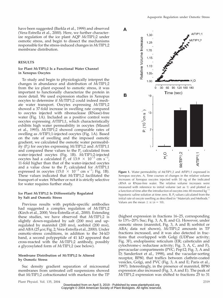
To study and begin to physiologically interpret thechanges in abundance and distribution of McTIP1;2from the ice plant exposed to osmotic stress, it wasimportant to functionally characterize the protein inmore detail. We used expression studies in Xenopusoocytes to determine if McTIP1;2 could indeed medi-ate water transport. Oocytes expressing McTIP1;2showed a 37-fold increase in swelling rate comparedto oocytes injected with ribonuclease (RNase)-freewater (Fig. 1A). Included as a positive control wereoocytes expressing AtTIP1;1, which characteristicallyexhibits high water permeability in oocytes (Maurelet al., 1993). McTIP1;2 showed comparable rates ofswelling as AtTIP1;1-injected oocytes (Fig. 1A). Basedon the rate of swelling and the imposed osmoticgradient, we calculated the osmotic water permeabil-ity (Pf) for oocytes expressing McTIP1;2 and AtTIP1;1and compared these values to the Pf calculated fromwater-injected oocytes (Fig. 1B). McTIP1;2-injectedoocytes had a calculated Pf of 13.9 3 1023 cm s21,11-fold higher than that of the water-injected oocytesand a value close to the Pf calculated for AtTIP1;1expressed in oocytes (15.0 3 1023 cm s21; Fig. 1B).These values indicated that McTIP1;2 facilitated thetransport of water. Whether or not it is strictly selectivefor water requires further study.
Ice Plant McTIP1;2 Is Differentially Regulated
by Salt and Osmotic Stress
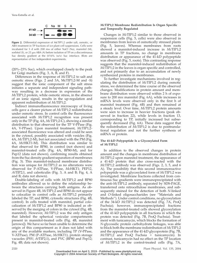
Previous results with peptide-specific antibodieshad suggested a complex regulation of McTIP1;2(Kirch et al., 2000; Vera-Estrella et al., 2000). Extendingthese studies, we have observed that McTIP1;2 isslightly down-regulated by salt (200 mM) and up-regulated by mannitol (200 mM), sorbitol (200 mM),andABA (25mM; Fig. 2; Vera-Estrella et al., 2000).Underosmotic-stress conditions, in addition to the 34-kDband, a second polypeptide of 41 kD appeared thatcross-reacted with the McTIP1;2 antibody, possiblya glycosylated form of McTIP1;2 (see below).
Membrane Distribution of McTIP1;2 Is Alteredby Osmotic Stress
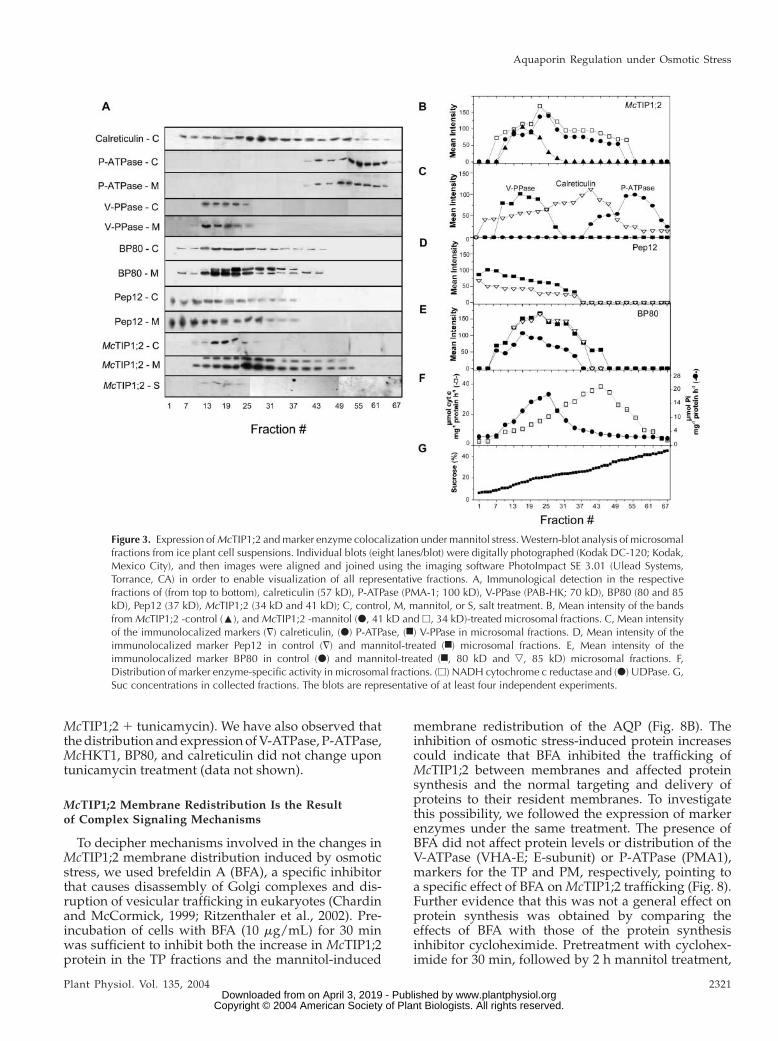
Suc density gradient separation of microsomalmembranes from untreated cell suspensions showedthat McTIP1;2 cofractionated with markers for the TP
(highest expression in fractions 16–25, correspondingto 13%–20% Suc; Fig. 3, A, B, and G). However, underosmotic stress (mannitol, Fig. 3, A and B; sorbitol orABA; data not shown), McTIP1;2 amounts in TPfractions increased, and it was also detected in frac-tions that overlapped with Golgi (UDPase activity;Fig. 3F), endoplasmic reticulum (ER; calreticulin andcytochrome-c reductase activity; Fig. 3, A, C, and F),prevacuolar compartments (PVC; Pep12; Fig. 3, A andD; Sanderfoot et al., 1998), and the vacuolar-sortingreceptor, BP80, that traffics between clathrin-coatedvesicles, Golgi, and PVC (Fig. 3, A and E; Paris et al.,1997). Interestingly, in the presence of mannitol, BP80expression also increased (Fig. 3, A and E). The peak ofMcTIP1;2 expression was shifted to fractions 25 to 31
Figure 1. Water permeability of McTIP1;2 and AtTIP1;1 expressed inXenopus oocytes. A, Time courses of changes in the relative volumeincreases of Xenopus oocytes injected with 50 ng of the indicatedcRNA or RNase-free water. The relative volume increases weremeasured with reference to initial volume (set as 1) and plotted asa function of time after the introduction of oocytes into 40 mosmol kg21
hypotonic saline solution at time zero. B, Pf values calculated from theinitial rate of oocyte swelling as described in ‘‘Materials and Methods.’’Values are the mean 6 SE (n 5 10).
Aquaporin Regulation under Osmotic Stress
Plant Physiol. Vol. 135, 2004 2319 www.plantphysiol.orgon April 3, 2019 - Published by Downloaded from
Copyright © 2004 American Society of Plant Biologists. All rights reserved.
(20%–23% Suc), which overlapped closely to the peakfor Golgi markers (Fig. 3, A, B, and F).
Differences in the response of McTIP1;2 to salt andosmotic stress (Figs. 2 and 3A, McTIP1;2-M and -S)suggest that the ionic component of the salt stressinitiates a separate and independent signaling path-way resulting in a decrease in expression of theMcTIP1;2 protein, while osmotic stress, in the absenceof an ionic signal, results in the up-regulation andapparent redistribution of McTIP1;2.
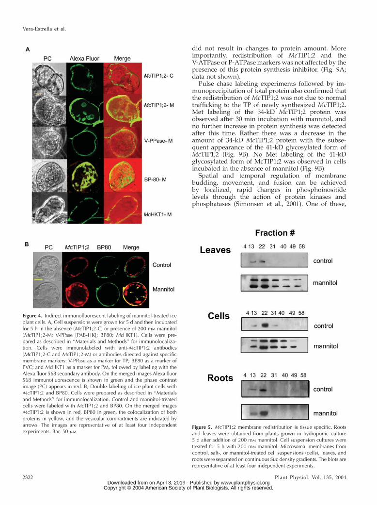
Indirect immunofluorescence microscopy of livingcells gave a clearer picture of McTIP1;2 redistributionunder osmotic stress. In control cells the fluorescenceassociated with McTIP1;2 recognition was presentonly in the TP (Fig. 4A,McTIP1;2-C), showing a similardistribution to that observed for the TP V-PPase (Fig.4A, V-PPase). Upon mannitol treatment, McTIP1;2-associated fluorescence was altered and could be seenin the cytosol, possibly associated with vesicles (Fig.4A, McTIP1;2-M), but not associated with the PM (Fig.4A, McHKT1-M). This distribution was similar tothat observed for BP80, in control (not shown) andmannitol-treated cells (Fig. 4A, BP80-M), and forPep12 (data not shown), confirming results obtainedfrom the Sucdensity gradient separation ofmembranes(Fig. 3). This mannitol-induced membrane distribu-tion was unique for McTIP1;2 as no alterations wereobserved for P-ATPase, V-PPase, McHKT1; PEP12,AtTIP2;1, and calreticulin (Fig. 3, A and B; Fig. 4, Aand B; data not shown).
Double-labeling of cells with McTIP1;2 and BP80antibodies allowed us to define the relationship be-tween the structures carrying both antigens. As ob-served in Figure 4B,McTIP1;2 and BP80 do not appearto colocalize in control cells as McTIP1;2 primarilylabeled the TP, and BP80, cytosolic structures (Fig. 4B,control). In cells treated with mannitol, partial colo-calization of McTIP1;2 and BP80 is indicated as ob-served by the merging of colors in the cytosol (Fig. 4B,mannitol). However, McTIP1;2 was the only antigenthat labeled the spherical vesicular compartmentspresent in mannitol-treated cells (Fig. 4B, mannitol[arrows]). We have so far been unable to determine theorigin of this compartment as it does not label withany of the available markers, including TP (V-PPase,V-ATPase), PM (P-ATPase, McHKT1), protein storagevacuoles (PSV; AtTIP2;1), and PVC (BP80 and Pep12;Fig. 4B; data not shown).
McTIP1;2 Membrane Redistribution Is Organ Specific
and Temporally Regulated
Changes in McTIP1;2 similar to those observed insuspension cells (Fig. 5, cells) were also observed inmembranes from leaves of osmotically stressed plants(Fig. 5, leaves). Whereas membranes from rootsshowed a mannitol-induced increase in McTIP1;2amounts in TP fractions, no change in membranedistribution or appearance of the 41-kD polypeptidewas observed (Fig. 5, roots). This contrasting responsesuggests that the mannitol-induced redistribution ofMcTIP1;2 in the leaves is organ specific and controlled,and not primarily due to an accumulation of newlysynthesized proteins in membranes.
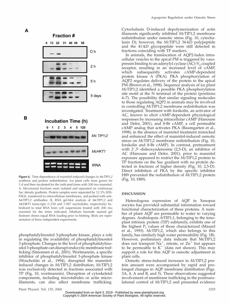
To further investigate mechanisms involved in reg-ulating the distribution of McTIP1;2 during osmoticstress, we determined the time course of the observedchanges. Modifications in protein amount and mem-brane distribution were observed within 2 h of expo-sure to 200 mM mannitol (Fig. 6A), while increases inmRNA levels were observed only in the first h ofmannitol treatment (Fig. 6B) and then remained ata steady level. Over time, McTIP1;2 protein amountswere seen to increase in heavier fractions (peak ob-served in fraction 22), while levels in fraction 13,corresponding to TP, initially increased but subse-quently decreased (Fig. 6A). These data suggest thatthe redistribution of McTIP1;2 is due to posttransla-tional regulation and not the further synthesis ofmRNA or protein.
The 41-kD Polypeptide Is a Glycosylated Formof McTIP1;2
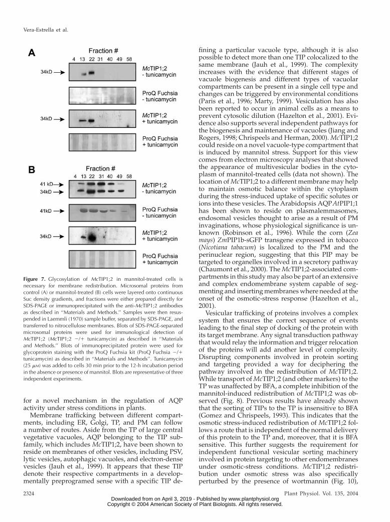
In addition to the observed changes in proteinamount and the changes in membrane distribution ofMcTIP1;2 upon mannitol treatment, the appearance ofa 41-kD protein that also cross-reacted with theMcTIP1;2 antibody was observed (Figs. 2, 3, 5, and 6A). The possibility that this second immunoreactivepolypeptide was a glycosylated form of McTIP1;2 wasinvestigated. Membrane fractions collected from con-tinuous Suc gradients were immunoprecipitated withthe anti-McTIP1;2 antibody, separated by SDS-PAGE,transferred onto nitrocellulose membranes, and sub-sequently stained for the detection of both N-linkedand O-linked oligosaccharides (see ‘‘Materials andMethods’’). Under control conditions, no glycosylationof the 34-kD McTIP1;2 was detected (Fig. 7A, ProQFuchsia); however, immunoprecipitated fractionsfrom the mannitol-treated cells showed glycosylationof the 41-kD polypeptide in all fractions in which theprotein was detected (Fig. 7B, ProQ Fuchsia). Treat-ment with tunicamycin, which blocks the formation ofN-glycosidic protein carbohydrate linkages, was ableto block both the membrane redistribution ofMcTIP1;2and the appearance of the 41-kD glycoprotein (Fig. 7B,McTIP1;2 and ProQ Fuchsia 1 tunicamycin). Incontrast, tunicamycin had no effect on the TP locationof McTIP1;2 in the control-treated cells (Fig. 7A,
Figure 2. Differential expression of McTIP1;2 under salt, osmotic, orABA treatment in TP fractions of ice plant cell suspensions. Cells wereincubated for 5 d with 200 mM of either NaCl (Na), mannitol (M),sorbitol (S), or 25 mM ABA (A) before isolation of TP on a discontinuousSuc density gradient at the 0/16% (w/v) Suc interface. Blots arerepresentative of five independent experiments.
Vera-Estrella et al.
2320 Plant Physiol. Vol. 135, 2004 www.plantphysiol.orgon April 3, 2019 - Published by Downloaded from
Copyright © 2004 American Society of Plant Biologists. All rights reserved.
McTIP1;2 1 tunicamycin). We have also observed thatthedistribution andexpression ofV-ATPase, P-ATPase,McHKT1, BP80, and calreticulin did not change upontunicamycin treatment (data not shown).
McTIP1;2 Membrane Redistribution Is the Resultof Complex Signaling Mechanisms
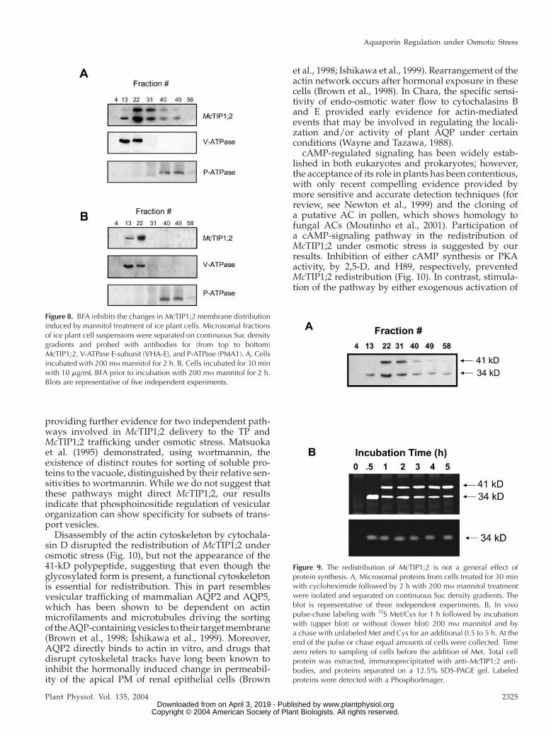
To decipher mechanisms involved in the changes inMcTIP1;2 membrane distribution induced by osmoticstress, we used brefeldin A (BFA), a specific inhibitorthat causes disassembly of Golgi complexes and dis-ruption of vesicular trafficking in eukaryotes (Chardinand McCormick, 1999; Ritzenthaler et al., 2002). Pre-incubation of cells with BFA (10 mg/mL) for 30 minwas sufficient to inhibit both the increase in McTIP1;2protein in the TP fractions and the mannitol-induced
membrane redistribution of the AQP (Fig. 8B). Theinhibition of osmotic stress-induced protein increasescould indicate that BFA inhibited the trafficking ofMcTIP1;2 between membranes and affected proteinsynthesis and the normal targeting and delivery ofproteins to their resident membranes. To investigatethis possibility, we followed the expression of markerenzymes under the same treatment. The presence ofBFA did not affect protein levels or distribution of theV-ATPase (VHA-E; E-subunit) or P-ATPase (PMA1),markers for the TP and PM, respectively, pointing toa specific effect of BFA onMcTIP1;2 trafficking (Fig. 8).Further evidence that this was not a general effect onprotein synthesis was obtained by comparing theeffects of BFA with those of the protein synthesisinhibitor cycloheximide. Pretreatment with cyclohex-imide for 30 min, followed by 2 h mannitol treatment,
Figure 3. Expression of McTIP1;2 and marker enzyme colocalization under mannitol stress. Western-blot analysis of microsomalfractions from ice plant cell suspensions. Individual blots (eight lanes/blot) were digitally photographed (Kodak DC-120; Kodak,Mexico City), and then images were aligned and joined using the imaging software PhotoImpact SE 3.01 (Ulead Systems,Torrance, CA) in order to enable visualization of all representative fractions. A, Immunological detection in the respectivefractions of (from top to bottom), calreticulin (57 kD), P-ATPase (PMA-1; 100 kD), V-PPase (PAB-HK; 70 kD), BP80 (80 and 85kD), Pep12 (37 kD), McTIP1;2 (34 kD and 41 kD); C, control, M, mannitol, or S, salt treatment. B, Mean intensity of the bandsfrom McTIP1;2 -control (:), and McTIP1;2 -mannitol (d, 41 kD and h, 34 kD)-treated microsomal fractions. C, Mean intensityof the immunolocalized markers (=) calreticulin, (d) P-ATPase, (n) V-PPase in microsomal fractions. D, Mean intensity of theimmunolocalized marker Pep12 in control (=) and mannitol-treated (n) microsomal fractions. E, Mean intensity of theimmunolocalized marker BP80 in control (d) and mannitol-treated (n, 80 kD and ,, 85 kD) microsomal fractions. F,Distribution of marker enzyme-specific activity in microsomal fractions. (h) NADH cytochrome c reductase and (d) UDPase. G,Suc concentrations in collected fractions. The blots are representative of at least four independent experiments.
Aquaporin Regulation under Osmotic Stress
Plant Physiol. Vol. 135, 2004 2321 www.plantphysiol.orgon April 3, 2019 - Published by Downloaded from
Copyright © 2004 American Society of Plant Biologists. All rights reserved.
did not result in changes to protein amount. Moreimportantly, redistribution of McTIP1;2 and theV-ATPase or P-ATPasemarkers was not affected by thepresence of this protein synthesis inhibitor. (Fig. 9A;data not shown).
Pulse chase labeling experiments followed by im-munoprecipitation of total protein also confirmed thatthe redistribution of McTIP1;2 was not due to normaltrafficking to the TP of newly synthesized McTIP1;2.Met labeling of the 34-kD McTIP1;2 protein wasobserved after 30 min incubation with mannitol, andno further increase in protein synthesis was detectedafter this time. Rather there was a decrease in theamount of 34-kD McTIP1;2 protein with the subse-quent appearance of the 41-kD glycosylated form ofMcTIP1;2 (Fig. 9B). No Met labeling of the 41-kDglycosylated form of McTIP1;2 was observed in cellsincubated in the absence of mannitol (Fig. 9B).
Spatial and temporal regulation of membranebudding, movement, and fusion can be achievedby localized, rapid changes in phosphoinositidelevels through the action of protein kinases andphosphatases (Simonsen et al., 2001). One of these,
Figure 5. McTIP1;2 membrane redistribution is tissue specific. Rootsand leaves were obtained from plants grown in hydroponic culture5 d after addition of 200 mM mannitol. Cell suspension cultures weretreated for 5 h with 200 mM mannitol. Microsomal membranes fromcontrol, salt-, or mannitol-treated cell suspensions (cells), leaves, androots were separated on continuous Suc density gradients. The blots arerepresentative of at least four independent experiments.
Figure 4. Indirect immunofluorescent labeling of mannitol-treated iceplant cells. A, Cell suspensions were grown for 5 d and then incubatedfor 5 h in the absence (McTIP1;2-C) or presence of 200 mM mannitol(McTIP1;2-M; V-PPase [PAB-HK]; BP80; McHKT1). Cells were pre-pared as described in ‘‘Materials and Methods’’ for immunolocaliza-tion. Cells were immunolabeled with anti-McTIP1;2 antibodies(McTIP1;2-C and McTIP1;2-M) or antibodies directed against specificmembrane markers: V-PPase as a marker for TP; BP80 as a marker ofPVC; and McHKT1 as a marker for PM, followed by labeling with theAlexa fluor 568 secondary antibody. On the merged images Alexa fluor568 immunofluorescence is shown in green and the phase contrastimage (PC) appears in red. B, Double labeling of ice plant cells withMcTIP1;2 and BP80. Cells were prepared as described in ‘‘Materialsand Methods’’ for immunolocalization. Control and mannitol-treatedcells were labeled with McTIP1;2 and BP80. On the merged imagesMcTIP1;2 is shown in red, BP80 in green, the colocalization of bothproteins in yellow, and the vesicular compartments are indicated byarrows. The images are representative of at least four independentexperiments. Bar, 50 mM.
Vera-Estrella et al.
2322 Plant Physiol. Vol. 135, 2004 www.plantphysiol.orgon April 3, 2019 - Published by Downloaded from
Copyright © 2004 American Society of Plant Biologists. All rights reserved.
phosphatidylinositol 3-phosphate kinase, plays a rolein regulating the availability of phosphatidylinositol3-phosphate. Changes in the level of phosphatidylino-sitol 3-phosphate candisrupt endocyticmembrane traf-ficking (Simonsen et al., 2001). Wortmannin, a specificinhibitor of phosphatidylinositol 3-phosphate kinase(Woscholski et al., 1994), disrupted the mannitol-induced changes in McTIP1;2 distribution; McTIP1;2was exclusively detected in fractions associated withTP (Fig. 10, wortmannin). Disruption of cytoskeletalcomponents, including microtubules or actin micro-filaments, can also affect membrane trafficking.
Cytochalasin D-induced depolymerization of actinfilaments significantly inhibited McTIP1;2 membraneredistribution under osmotic stress (Fig. 10, cytocha-lasin D); however, the McTIP1;2 34-kD polypeptideand the 41-kD glycopeptide were still detected infractions coinciding with TP markers.
In animals, the translocation of AQP2-laden intra-cellular vesicles to the apical PM is triggered by vaso-pressin binding to an adenylyl cyclase (AC)-V2 coupledreceptor, resulting in an increased level of cAMP,which subsequently activates cAMP-dependentprotein kinase A (PKA). PKA phosphorylation ofAQP2 regulates delivery of the protein to the apicalPM (Brown et al., 1998). Sequence analysis of ice plantMcTIP1;2 identified a possible PKA phosphorylationsite motif at the N terminal of the protein (positions4–7). The possibility that similar signaling moleculesto those regulating AQP2 in animals may be involvedin controlling McTIP1;2 membrane redistribution wasinvestigated. Treatment with forskolin, an activator ofAC, known to elicit cAMP-dependent physiologicalresponses by increasing intracellular cAMP (Hanouneand Defer, 2001), and 8-Br cAMP, a cell permeablecAMP analog that activates PKA (Baumgarten et al.,1998), in the absence of mannitol treatment mimickedand enhanced the effect of mannitol-induced osmoticstress on McTIP1;2 membrane redistribution (Fig. 10,forskolin and 8-Br cAMP). In contrast, pretreatmentwith 2#,5#-dideoxyadenosine (2,5-D), an inhibitor ofAC (Hanoune and Defer, 2001), prior to mannitolexposure appeared to restrict the McTIP1;2 protein toTP fractions on the Suc gradient with no protein de-tected in fractions of higher density (Fig. 10, 2,5-D).Direct inhibition of PKA by the specific inhibitorH89 prevented the redistribution of McTIP1;2 protein(Fig. 10, H89).
DISCUSSION
Heterologous expression of AQP in Xenopusoocytes has provided substantial information towardfunctional characterization of these proteins. A num-ber of plant AQP are permeable to water to varyingdegrees. Arabidopsis AtTIP1;1, belonging to the tono-plast intrinsic protein (TIP) subfamily, exhibits one ofthe highest Pf values of those characterized (Maurelet al., 1993). McTIP1;2, which also belongs to thisfamily, has similarly high water permeability (Fig. 1B).However, preliminary data indicate that McTIP1;2does not transport Na1, nitrate, or Zn1 but appearsto be permeable to K1 (data not shown). This maysupport a role for this AQP in osmotic adjustment inplant cells.
Osmotic stress-induced increases in McTIP1;2 pro-tein amount were accompanied by rapid and pro-longed changes in AQP membrane distribution (Figs.3A, 4, A and B, and 5). These observations suggestedinvolvement of membrane trafficking in the posttrans-lational control of McTIP1;2 and presented evidence
Figure 6. Time dependence of mannitol-induced changes in McTIP1;2synthesis and protein redistribution. Ice plant cells were grown for5 d and then incubated for the indicated times with 200 mM mannitol.A, Microsomal fractions were isolated and separated on continuousSuc density gradients. Protein samples were separated by 12.5% SDS-PAGE, transferred to nitrocellulose membranes, and probed with anti-McTIP1;2 antibodies. B, RNA gel-blot analysis of McTIP1;2 andMcHKT1 transcripts (1,250 and 1,907 nucleotides, respectively) hy-bridized to total RNA from cell suspensions treated with 200 mM
mannitol for the times indicated. Ethidium bromide stained gel(bottom) shows equal RNA loading prior to blotting. Blots are repre-sentative of three independent experiments.
Aquaporin Regulation under Osmotic Stress
Plant Physiol. Vol. 135, 2004 2323 www.plantphysiol.orgon April 3, 2019 - Published by Downloaded from
Copyright © 2004 American Society of Plant Biologists. All rights reserved.
for a novel mechanism in the regulation of AQPactivity under stress conditions in plants.
Membrane trafficking between different compart-ments, including ER, Golgi, TP, and PM can followa number of routes. Aside from the TP of large centralvegetative vacuoles, AQP belonging to the TIP sub-family, which includes McTIP1;2, have been shown toreside on membranes of other vesicles, including PSV,lytic vesicles, autophagic vacuoles, and electron-densevesicles (Jauh et al., 1999). It appears that these TIPdenote their respective compartments in a develop-mentally preprogramed sense with a specific TIP de-
fining a particular vacuole type, although it is alsopossible to detect more than one TIP colocalized to thesame membrane (Jauh et al., 1999). The complexityincreases with the evidence that different stages ofvacuole biogenesis and different types of vacuolarcompartments can be present in a single cell type andchanges can be triggered by environmental conditions(Paris et al., 1996; Marty, 1999). Vesiculation has alsobeen reported to occur in animal cells as a means toprevent cytosolic dilution (Hazelton et al., 2001). Evi-dence also supports several independent pathways forthe biogenesis and maintenance of vacuoles (Jiang andRogers, 1998; Chrispeels and Herman, 2000).McTIP1;2could reside on a novel vacuole-type compartment thatis induced by mannitol stress. Support for this viewcomes from electron microscopy analyses that showedthe appearance of multivesicular bodies in the cyto-plasm of mannitol-treated cells (data not shown). Thelocation ofMcTIP1;2 to a different membranemay helpto maintain osmotic balance within the cytoplasmduring the stress-induced uptake of specific solutes orions into these vesicles. The Arabidopsis AQPAtPIP1;1has been shown to reside on plasmalemmasomes,endosomal vesicles thought to arise as a result of PMinvaginations, whose physiological significance is un-known (Robinson et al., 1996). While the corn (Zeamays) ZmPIP1b-sGFP transgene expressed in tobacco(Nicotiana tabacum) is localized to the PM and theperinuclear region, suggesting that this PIP may betargeted to organelles involved in a secretory pathway(Chaumont et al., 2000). TheMcTIP1;2-associated com-partments in this studymay also be part of an extensiveand complex endomembrane system capable of seg-menting and insertingmembraneswhere needed at theonset of the osmotic-stress response (Hazelton et al.,2001).
Vesicular trafficking of proteins involves a complexsystem that ensures the correct sequence of eventsleading to the final step of docking of the protein withits target membrane. Any signal transduction pathwaythat would relay the information and trigger relocationof the proteins will add another level of complexity.Disrupting components involved in protein sortingand targeting provided a way for deciphering thepathway involved in the redistribution of McTIP1;2.While transport ofMcTIP1;2 (and other markers) to theTP was unaffected by BFA, a complete inhibition of themannitol-induced redistribution of McTIP1;2 was ob-served (Fig. 8). Previous results have already shownthat the sorting of TIPs to the TP is insensitive to BFA(Gomez and Chrispeels, 1993). This indicates that theosmotic stress-induced redistribution of McTIP1;2 fol-lows a route that is independent of the normal deliveryof this protein to the TP and, moreover, that it is BFAsensitive. This further suggests the requirement forindependent functional vesicular sorting machineryinvolved in protein targeting to other endomembranesunder osmotic-stress conditions. McTIP1;2 redistri-bution under osmotic stress was also specificallyperturbed by the presence of wortmannin (Fig. 10),
Figure 7. Glycosylation of McTIP1;2 in mannitol-treated cells isnecessary for membrane redistribution. Microsomal proteins fromcontrol (A) or mannitol-treated (B) cells were layered onto continuousSuc density gradients, and fractions were either prepared directly forSDS-PAGE or immunoprecipitated with the anti-McTIP1;2 antibodiesas described in ‘‘Materials and Methods.’’ Samples were then resus-pended in Laemmli (1970) sample buffer, separated by SDS-PAGE, andtransferred to nitrocellulose membranes. Blots of SDS-PAGE-separatedmicrosomal proteins were used for immunological detection ofMcTIP1;2 (McTIP1;2 2/1 tunicamycin) as described in ‘‘Materialsand Methods.’’ Blots of immunoprecipitated protein were used forglycoprotein staining with the ProQ Fuchsia kit (ProQ Fuchsia 2/1tunicamycin) as described in ‘‘Materials and Methods’’. Tunicamycin(25 mM) was added to cells 30 min prior to the 12-h incubation periodin the absence or presence of mannitol. Blots are representative of threeindependent experiments.
Vera-Estrella et al.
2324 Plant Physiol. Vol. 135, 2004 www.plantphysiol.orgon April 3, 2019 - Published by Downloaded from
Copyright © 2004 American Society of Plant Biologists. All rights reserved.
providing further evidence for two independent path-ways involved in McTIP1;2 delivery to the TP andMcTIP1;2 trafficking under osmotic stress. Matsuokaet al. (1995) demonstrated, using wortmannin, theexistence of distinct routes for sorting of soluble pro-teins to the vacuole, distinguished by their relative sen-sitivities to wortmannin. While we do not suggest thatthese pathways might direct McTIP1;2, our resultsindicate that phosphoinositide regulation of vesicularorganization can show specificity for subsets of trans-port vesicles.Disassembly of the actin cytoskeleton by cytochala-
sin D disrupted the redistribution of McTIP1;2 underosmotic stress (Fig. 10), but not the appearance of the41-kD polypeptide, suggesting that even though theglycosylated form is present, a functional cytoskeletonis essential for redistribution. This in part resemblesvesicular trafficking of mammalian AQP2 and AQP5,which has been shown to be dependent on actinmicrofilaments and microtubules driving the sortingof theAQP-containingvesicles to their targetmembrane(Brown et al., 1998; Ishikawa et al., 1999). Moreover,AQP2 directly binds to actin in vitro, and drugs thatdisrupt cytoskeletal tracks have long been known toinhibit the hormonally induced change in permeabil-ity of the apical PM of renal epithelial cells (Brown
et al., 1998; Ishikawa et al., 1999). Rearrangement of theactin network occurs after hormonal exposure in thesecells (Brown et al., 1998). In Chara, the specific sensi-tivity of endo-osmotic water flow to cytochalasins Band E provided early evidence for actin-mediatedevents that may be involved in regulating the locali-zation and/or activity of plant AQP under certainconditions (Wayne and Tazawa, 1988).
cAMP-regulated signaling has been widely estab-lished in both eukaryotes and prokaryotes; however,the acceptance of its role in plants has been contentious,with only recent compelling evidence provided bymore sensitive and accurate detection techniques (forreview, see Newton et al., 1999) and the cloning ofa putative AC in pollen, which shows homology tofungal ACs (Moutinho et al., 2001). Participation ofa cAMP-signaling pathway in the redistribution ofMcTIP1;2 under osmotic stress is suggested by ourresults. Inhibition of either cAMP synthesis or PKAactivity, by 2,5-D, and H89, respectively, preventedMcTIP1;2 redistribution (Fig. 10). In contrast, stimula-tion of the pathway by either exogenous activation of
Figure 9. The redistribution of McTIP1;2 is not a general effect ofprotein synthesis. A, Microsomal proteins from cells treated for 30 minwith cycloheximide followed by 2 h with 200 mM mannitol treatmentwere isolated and separated on continuous Suc density gradients. Theblot is representative of three independent experiments. B, In vivopulse-chase labeling with 35S Met/Cys for 1 h followed by incubationwith (upper blot) or without (lower blot) 200 mM mannitol and bya chase with unlabeled Met and Cys for an additional 0.5 to 5 h. At theend of the pulse or chase equal amounts of cells were collected. Timezero refers to sampling of cells before the addition of Met. Total cellprotein was extracted, immunoprecipitated with anti-McTIP1;2 anti-bodies, and proteins separated on a 12.5% SDS-PAGE gel. Labeledproteins were detected with a PhosphorImager.
Figure 8. BFA inhibits the changes in McTIP1;2 membrane distributioninduced by mannitol treatment of ice plant cells. Microsomal fractionsof ice plant cell suspensions were separated on continuous Suc densitygradients and probed with antibodies for (from top to bottom)McTIP1;2, V-ATPase E-subunit (VHA-E), and P-ATPase (PMA1). A, Cellsincubated with 200 mM mannitol for 2 h. B, Cells incubated for 30 minwith 10 mg/mL BFA prior to incubation with 200 mM mannitol for 2 h.Blots are representative of five independent experiments.
Aquaporin Regulation under Osmotic Stress
Plant Physiol. Vol. 135, 2004 2325 www.plantphysiol.orgon April 3, 2019 - Published by Downloaded from
Copyright © 2004 American Society of Plant Biologists. All rights reserved.
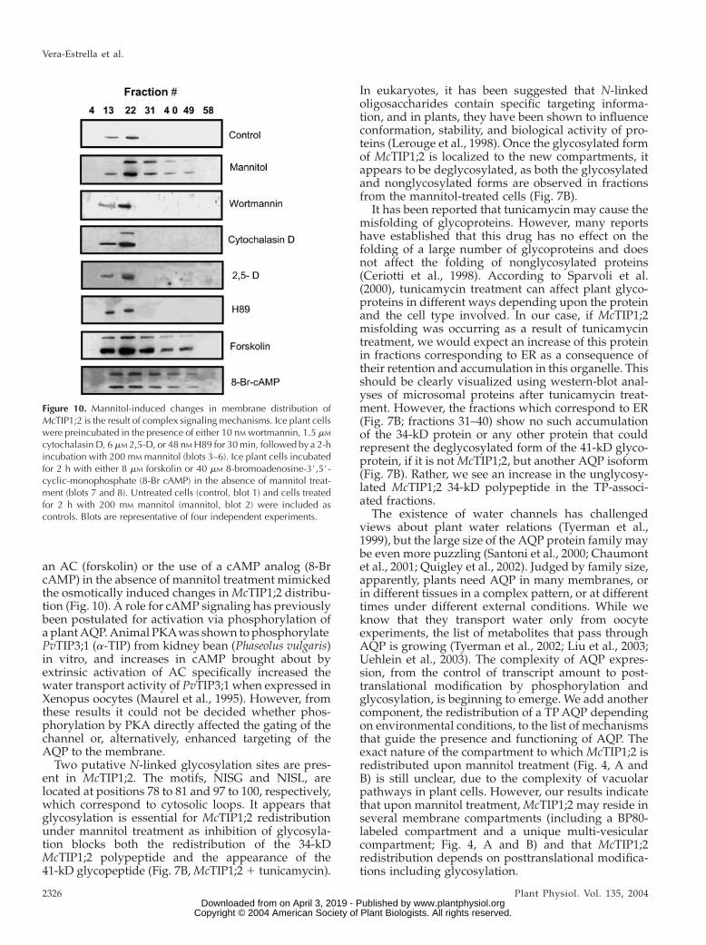
an AC (forskolin) or the use of a cAMP analog (8-BrcAMP) in the absence of mannitol treatment mimickedthe osmotically induced changes inMcTIP1;2 distribu-tion (Fig. 10). A role for cAMP signaling has previouslybeen postulated for activation via phosphorylation ofa plantAQP.Animal PKAwas shown tophosphorylatePvTIP3;1 (a-TIP) from kidney bean (Phaseolus vulgaris)in vitro, and increases in cAMP brought about byextrinsic activation of AC specifically increased thewater transport activity of PvTIP3;1 when expressed inXenopus oocytes (Maurel et al., 1995). However, fromthese results it could not be decided whether phos-phorylation by PKA directly affected the gating of thechannel or, alternatively, enhanced targeting of theAQP to the membrane.
Two putative N-linked glycosylation sites are pres-ent in McTIP1;2. The motifs, NISG and NISL, arelocated at positions 78 to 81 and 97 to 100, respectively,which correspond to cytosolic loops. It appears thatglycosylation is essential for McTIP1;2 redistributionunder mannitol treatment as inhibition of glycosyla-tion blocks both the redistribution of the 34-kDMcTIP1;2 polypeptide and the appearance of the41-kD glycopeptide (Fig. 7B,McTIP1;21 tunicamycin).
In eukaryotes, it has been suggested that N-linkedoligosaccharides contain specific targeting informa-tion, and in plants, they have been shown to influenceconformation, stability, and biological activity of pro-teins (Lerouge et al., 1998). Once the glycosylated formof McTIP1;2 is localized to the new compartments, itappears to be deglycosylated, as both the glycosylatedand nonglycosylated forms are observed in fractionsfrom the mannitol-treated cells (Fig. 7B).
It has been reported that tunicamycin may cause themisfolding of glycoproteins. However, many reportshave established that this drug has no effect on thefolding of a large number of glycoproteins and doesnot affect the folding of nonglycosylated proteins(Ceriotti et al., 1998). According to Sparvoli et al.(2000), tunicamycin treatment can affect plant glyco-proteins in different ways depending upon the proteinand the cell type involved. In our case, if McTIP1;2misfolding was occurring as a result of tunicamycintreatment, we would expect an increase of this proteinin fractions corresponding to ER as a consequence oftheir retention and accumulation in this organelle. Thisshould be clearly visualized using western-blot anal-yses of microsomal proteins after tunicamycin treat-ment. However, the fractions which correspond to ER(Fig. 7B; fractions 31–40) show no such accumulationof the 34-kD protein or any other protein that couldrepresent the deglycosylated form of the 41-kD glyco-protein, if it is notMcTIP1;2, but another AQP isoform(Fig. 7B). Rather, we see an increase in the unglycosy-lated McTIP1;2 34-kD polypeptide in the TP-associ-ated fractions.
The existence of water channels has challengedviews about plant water relations (Tyerman et al.,1999), but the large size of the AQP protein family maybe even more puzzling (Santoni et al., 2000; Chaumontet al., 2001; Quigley et al., 2002). Judged by family size,apparently, plants need AQP in many membranes, orin different tissues in a complex pattern, or at differenttimes under different external conditions. While weknow that they transport water only from oocyteexperiments, the list of metabolites that pass throughAQP is growing (Tyerman et al., 2002; Liu et al., 2003;Uehlein et al., 2003). The complexity of AQP expres-sion, from the control of transcript amount to post-translational modification by phosphorylation andglycosylation, is beginning to emerge. We add anothercomponent, the redistribution of a TPAQP dependingon environmental conditions, to the list of mechanismsthat guide the presence and functioning of AQP. Theexact nature of the compartment to which McTIP1;2 isredistributed upon mannitol treatment (Fig. 4, A andB) is still unclear, due to the complexity of vacuolarpathways in plant cells. However, our results indicatethat upon mannitol treatment,McTIP1;2 may reside inseveral membrane compartments (including a BP80-labeled compartment and a unique multi-vesicularcompartment; Fig. 4, A and B) and that McTIP1;2redistribution depends on posttranslational modifica-tions including glycosylation.
Figure 10. Mannitol-induced changes in membrane distribution ofMcTIP1;2 is the result of complex signaling mechanisms. Ice plant cellswere preincubated in the presence of either 10 nM wortmannin, 1.5 mM
cytochalasin D, 6 mM 2,5-D, or 48 nM H89 for 30 min, followed by a 2-hincubation with 200 mM mannitol (blots 3–6). Ice plant cells incubatedfor 2 h with either 8 mM forskolin or 40 mM 8-bromoadenosine-3#,5#-cyclic-monophosphate (8-Br cAMP) in the absence of mannitol treat-ment (blots 7 and 8). Untreated cells (control, blot 1) and cells treatedfor 2 h with 200 mM mannitol (mannitol, blot 2) were included ascontrols. Blots are representative of four independent experiments.
Vera-Estrella et al.
2326 Plant Physiol. Vol. 135, 2004 www.plantphysiol.orgon April 3, 2019 - Published by Downloaded from
Copyright © 2004 American Society of Plant Biologists. All rights reserved.
MATERIALS AND METHODS
Plant Materials and Growth Conditions
Ice plant (Mesembryanthemum crystallinum) plants and cell suspensions
were grown as previously described (Barkla et al., 1999; Vera-Estrella et al.,
1999). Cells were grown for 5 d following transfer to fresh Murashige and
Skoog-supplemented medium (Murashige and Skoog, 1962) before treatment
with 200 mM NaCl, or 200 mM mannitol or sorbitol, or 25 mM ABA. Treatments
were added to the existing cell medium as sterile crystals and/or powders to
avoid changes in volume. Cells incubated in the presence of agonists and/or
antagonists were observed microscopically for possible cellular damage, and
cells were used only under conditions where cell viability was not affected.
Membrane Isolation and Purification
Membranes were isolated from ice plant plants and cell suspension
cultures as previously described (Barkla et al., 1999; Vera-Estrella et al.,
1999). Microsomes were layered onto either continuous (5% to 46% [w/v] Suc)
or discontinuous Suc gradients (consisting of a top layer of 9mL of 16% or 22%
[w/v] Suc, over 9 mL of 32% [w/v] Suc, on a cushion of 9 mL of 38% [w/v]
Suc). Gradients were centrifuged at 100,000g (3 h at 4�C) using a Beckman
SW 28 swinging bucket rotor in a Beckman L8-M ultracentrifuge (Beckman,
Mexico City). On a discontinuous Suc gradient TP from cell suspension
cultures separates at the 0/16% Suc interface while PM is collected from the
32%/38% Suc interface (Vera-Estrella et al., 1999). TP isolated from root and
leaf tissue is collected at the 0%/22% Suc interface (Barkla et al., 1999). The
purity of the TP fraction was calculated by assaying for the relative
contribution of PM, TP, and mitochondrial ATPase enzyme activities. From
these studies it is estimated that the 0%/16% Suc interface had 85%
bafilomycin and nitrate-sensitive ATPase activity (attributed to V-ATPase
activity on the TP). No mitochondrial marker activity was detected (data not
shown). Bands from the discontinuous gradient or fractions (0.5 mL) from the
continuous Suc gradient were collected, frozen in liquid N2, and stored at
280�C. The Suc concentration of fractions from continuous gradients was
measured using a Zeiss refractometer (Zeiss, Mexico City). Previously we
have shown that ice plant cell suspensions show similar adaptive responses as
those of leaves to salt and osmotic stress (Vera-Estrella et al., 1999).
Pulse-Chase Labeling and Extraction of Total Protein
Cells of ice plant labeled for 1 h with 35S Met/Cys followed by incubation
from 0.5 to 5 h in the presence of 200 mM mannitol, were filtered onto
Whatman Number 1 filter paper, frozen in liquid N2, homogenized in
extraction buffer (100 mM Tris-MES, pH 8.0, 1 mM EGTA, 5 mM dithiothreitol,
4 mM MgSO4, 5% [w/v] insoluble PVP), and vortexed for 1 min. The samples
were then filtered through one layer of Miracloth (Calbiochem, La Jolla, CA),
and the crude protein extracts were centrifuged at 10,000g for 15 min using
a SS34 rotor in a Sorvall 5C high speed centrifuge (DuPont, Newton, CT) to
remove cellular debris. Samples were used for immunoprecipitation (see
below) and resolved by SDS-PAGE on 12.5% (w/v) linear acrylamide gels.
Signals were detected with a PhosphorImager (ImageQuant, Molecular
Dynamics, Sunnyvale, CA).
Preparation of Template DNA, In Vitro Transcription,
and Capping of mRNA
The coding region ofMcTIP1;2was cloned into the pGEM-HE vector, while
AtTIP1;1, provided by Dr. C. Maurel, was cloned into the XbG-ev2 vector
(Maurel et al., 1993). Vectors contained either a T7 RNA polymerase promoter
(pGEM-HE) or a T3 RNA polymerase promoter (XbG-ev2), and 5#- and 3#-untranslated region of the Xenopus laevis b-globin gene for enhanced expres-
sion. The plasmid DNAwas purified by polyethylene glycol precipitation and
digested with either Pst1 (pGEM-HE) or Xba1 (XbG-ev2) that cleave imme-
diately downstream of the inserts to produce a linear template. The linearized
DNA was extracted with phenol-chloroform, precipitated with ethanol, and
resuspended in RNase free water. The complementary RNAs (cRNA) were
synthesized in vitro using the mCAP(TM) mRNA capping kit (Stratagene,
LaJolla, CA).
Oocyte Expression
Oocytes injected with 50 ng of cRNAwere used 2 to 5 d post injection. The
osmotic permeability of oocytes was measured by assaying the rates of oocyte
swelling upon rapid dilution from iso-osmotic Barth’s solution [10 mM
HEPES-NaOH, pH 7.4, 88 mM NaCl, 1 mM KCl, 2.4 mM NaHCO3, 0.33 mM
Ca(NO3)2, 0.41 mM CaCl2, 0.82 mM MgSO4, 200 mosmol/kg] to hypoosmotic
dilute Barth’s solution (40 mosmol/kg). Swelling was measured by video
imaging on a Nikon Eclipse TE 300 microscope (Nikon, Mexico City),
equipped with a Hitachi KP-D50 color video camera (Hitachi Denshi, Wood-
bury, NY). Images were captured and digitized by the Image-Pro Plus
software (Version 4, Media Cybernetics, Silver Spring, MD).
The osmotic water permeability (Pf, cm/s) was calculated by the relation,
Pf 5V0ðd½V=V0�=dtÞSVwðosmi 2 osmoÞ
where V0 is the initial oocyte volume at time zero measured for each
individual oocyte, V/Vo is the relative volume, Vw is the partial molar volume
of water (18 cm3/mol), S is the initial oocyte surface area calculated for each
individual oocyte, osmi is the osmolarity inside the oocyte, and osmo is the
osmolarity in the external medium. The time interval used for the calculations
of Pf was 0 to 60 s.
Enzyme Assays
NADH cytochrome c reductase was measured spectrophotometrically at
25�C by following the reduction of cytochrome c at 550 nm as described
(Hodges and Leonard, 1974). UDPase activity was measured in a 0.5 mL
reaction volume containing 30 mM Tris/MES pH 7.3, 3 mM UDP-Tris, 3 mM
MnSO4 in the presence of 0.3%Triton X-100. Release of Pi was detected at
820 nm following the precipitation of Triton X-100 with TCA and HClO4
(Manolson et al., 1985). Spectrophotometric measurements were taken using
a diode array spectrophotometer (Hewlett-Packard, Mexico City).
Protein Determination
Protein content in microsomal and purified PM or TP fractions was
measured by a modification of the dye-binding method of Bradford (1976), in
which membrane protein was solubilized by the addition of 0.5% (v/v) Triton
X-100 for 5 min before the addition of the dye reagent concentrate (Bio-Rad,
Cuernavaca, Mexico).
Primary and Secondary Antibodies
A peptide representing the carboxy terminus of the deduced amino acid
sequence of the ice plant McTIP1;2 was synthesized, coupled to keyhole
limpet hemocyanin, and antibodies were generated as previously described
(Kirch et al., 2000). Anti-McTIP1;2 was used at a dilution of 1:500. Antibodies
against the V-ATPase E-subunit (VMA-E) from barley (Hordeum vulgare),
V-PPase (PAB-HK) from sugar beet (Beta vulgaris), and the P-ATPase (PMA1)
from Arabidopsis were kindly supplied by K.-J. Dietz, P.A. Rea, and R.
Serrano, respectively (Pardo and Serrano, 1989; Kim et al., 1994; Dietz and
Arbinger, 1996). Antibodies against BP80 were kindly supplied by J.C. Rogers
(Jauh et al., 1999). Calreticulin antibodies used were against the protein from
Arabidopsis (Nelson et al., 1997). Pep12 monoclonal antibodies were pur-
chased from Molecular Probes (Eugene, OR). Immunocomplexes were de-
tected using a 1/5,000 dilution of either peroxidase labeled goat anti-mouse
IgG secondary antibodies (BP80 and PEP12) or goat anti-rabbit IgG secondary
antibodies (for detection of all other primary antibodies), and developed using
the chemiluminescent ECL detection substrate (Amersham, Cuernavaca,
Mexico). Film exposure time was kept constant for all blots.
Immunoprecipitation
Protein (30 mg) was incubated with 500 mL of NET-gel buffer (50 mM Tris/
HCl, pH 7.5, 150 mMNaCl, 0.1%Nonidet P-40, 1 mM EDTA, and 0.25% gelatin)
containing anti-McTIP1;2 antibodies (1/500 dilution) for 1 h at 4�C on
a rotating table. Protein A Sepharose CL-4B (20 mL; Amersham Pharmacia
Biotech, Uppsala) was added and the slurry rocked for a further 2 h at 4�C.Samples were then centrifuged at 12,000g for 20 s and the supernatant
eliminated using a 1-mL syringe. The protein pellet was washed twice with
NET-gel buffer followed by a final rinse with wash buffer (10 mM Tris/HCl,
pH 7.5, 0.1% Nonidet P-40). The pellet was then resuspended with 20 mL of
2.5%-Laemmli sample buffer (Laemmli, 1970), and heated at 95�C for 3 min.
Aquaporin Regulation under Osmotic Stress
Plant Physiol. Vol. 135, 2004 2327 www.plantphysiol.orgon April 3, 2019 - Published by Downloaded from
Copyright © 2004 American Society of Plant Biologists. All rights reserved.
Samples were centrifuged at 10,000g prior to loading onto a 12.5%-linear
acrylamide gel for SDS-PAGE and subsequent protein blotting.
SDS-PAGE and Protein Immunoblotting
Samples were prepared according to the method of Parry et al. (1989).
Protein was precipitated by dilution of the samples 50-fold in 1:1 (v/v)
ethanol:acetone and incubated overnight at 230�C. Samples were then
centrifuged at 13,000g for 20 min at 4�C using an F2402 rotor in a GS-15R
table-top centrifuge (Beckman). Pellets were air dried, resuspended with
Laemmli (1970) sample buffer (2.5% SDS final concentration), and heated at
60�C for 2 min before loading onto 12.5%-(w/v) linear acrylamide mini-gels.
Unless stated in the figure legends, 12 mg of protein was loaded per lane. After
electrophoresis, the gels were either stained with Coomassie Brilliant Blue
R250 (0.25% [w/v] in 50% [v/v] methanol/7% [v/v] acetic-acid), destained in
10%methanol/10% acetic-acid (v/v), and dried under vacuum at 80�C for 2 h,
or prepared for immunoblotting. SDS-PAGE-separated proteins were electro-
phoretically transferred onto nitrocellulose membranes (ECL, Amersham,
Buckinghamshire, UK) as previously described (Vera-Estrella et al., 1999).
Following transfer, proteins were stained with Ponceau S protein stain (0.1%
w/v in 1% v/v acetic acid for 30 s) to check for equal loading/transfer of
proteins. Membranes were then blocked with TBS (100 mM Tris, 150 mM NaCl)
containing 0.02% (w/v) Na-azide, and 5% (w/v) fat-free milk powder for 2 h
at room temperature. Blocked membranes were incubated for a minimum of
3 h at room temperature with the appropriate primary antibodies, followed by
the addition of a 1:5,000 dilution of secondary antibodies (goat anti-rabbit or -
mouse) conjugated to horse radish (Armoracia lapathifolia) peroxidase. Immu-
nodection was carried out using the chemiluminescent ECL western-blotting
analysis system (Amersham, UK). Mean intensity of the immunodetected
protein bands was calculated using ECL Mr markers as loading control
standards (Amersham,Mexico). Images were captured using Kodak 1D image
analysis software (Eastman Kodak, Rochester, NY).
Staining of Glycoproteins
In-blot staining of glycoproteins was preformed using the Pro-Q fuchsia
glycoprotein staining kit (Molecular Probes, Eugene, OR) according to
manufacturer’s instructions. Tunicamycin (25 mM) was added to cells 30 min
prior to the 12 h incubation period in the presence or absence of mannitol.
Immunofluorescence
Cells were treated for 5 h with 200 mM mannitol and prepared for
immunolabeling according to Brown and Lemmon (1995) with some mod-
ifications. Cells were fixed by incubation for 15 min at room temperature with
1.4% paraformaldehyde in stabilizing buffer containing 10 mM Bis-Tris pro-
pane, 25 mM HEPES, 2 mM MgCl2, 10 mM EGTA, pH 6.8. For control cells all
solutions were adjusted to 200 mosmoles kg21, while solutions for mannitol-
treated cells were adjusted to 500 mosmoles kg21. Fixed cells were then
washed three times and cell walls were partially removed with an enzymatic
cocktail (10 mM KCl, 2 mM MgCl2, 1 mM CaCl2, 10 mM ascorbic acid, 10 mM
MES, 0.05% BSA [w/v], 1% cellulase [w/v], 0.02% [w/v] pectolyase, pH 6.8)
for 20 min at room temperature. Following digestion, cells were washed and
permeabilized with 1% Triton X-100 (v/v) in stabilizing buffer for 10 min with
respective washes. Triton X-100 treatment also served to eliminate autofluo-
rescence. Before addition of the primary antibodies the cells were incubated for
1 h with 0.5% (w/v) fat-free milk powder. Primary antibodies were added at
a 1:100 dilution and all cells were incubated overnight at 4�C with gentle
shaking, followed by addition of goat anti-rabbit or anti-mouse secondary
Alexa fluor 568 antibodies (Molecular Probes) for 2 h at room temperature. For
double-labeling experiments, cells were incubated sequentially with the
primary antibody, followed by a mix of the corresponding secondary anti-
bodies coupled to different fluorochromes (Alexa Fluor 488 or 568). The cells
were imaged with a Bio-Rad Confocal laser microimager (Bio-Rad, Hercules,
CA) mounted on a Carl Zeiss Axioskop fitted with a Neo-Fluar 40X/0.74
objective and using Cosmos 7.0 Software (Bio-Rad). The stored digital images
were pseudo-colored as red or green images, using Photoshop 3.0 (Adobe,
Mountain View, CA). For double-labeled cells, the separate images were
pseudocolored, one as red, the other one as green, and then overlayed and/or
merged using the confocal assistance software (Bio-Rad). This resulted in
regions of colocalization appearing as yellow. To enable comparison, all
images were recorded using the same parameters of laser power and photo-
multiplier sensitivity. Images shown are representative of at least three
independent experiments in each condition and were processed by using
identical values for contrast and brightness.
RNA Isolation and Northern-Blot Analysis
Total RNAwas isolated from cells treated for different times with 200 mM
mannitol using Trizol reagent (Life Technologies, Mexico City) according to
manufacturer’s instructions. Typically, 200 mg of frozen cells were ground in
a chilled mortar and suspended in 2 mL of Trizol reagent. After alcohol
precipitation and washes, the RNA pellet was resuspended in diethyl
pyrocarbonate-treated water, and LiCl was added to a final concentration of
1 M. Following centrifugation, the resulting RNA pellet was resuspended in
diethyl pyrocarbonate-treated water and the amount of RNAwas determined
spectrophotometrically. Ten micrograms of purified total RNA was resolved
on formaldehyde-agarose gels and transferred to Hybond-N1 nylon mem-
brane (Amersham). Blots were screened with 32P-labeled probes derived from
McTIP1;2 and McHKT1 full-length cDNAs. After hybridization, blots were
washed under high-stringency conditions (40 mM Na2HPO4, pH 7.2, 1 mM
EDTA, 1% SDS at 65�C) and subjected to autoradiography.
ACKNOWLEDGMENTS
We thank Drs. Ramon Serrano (Valencia, Spain), Phil Rea (Philadelphia),
Karl-Josef Dietz (Bielefeld, Germany), and John C. Rogers (Pullman, WA) for
antibodies against PMA1 (PM H1-ATPase), PAB-HK (vacuolar H1-pyrophos-
phatase), VMA-E (vacuolar H1-ATPase), and BP80, respectively, and Chris-
tophe Maurel for the AtTIP1;1 construct. We also thank Enrique Balderas for
help with the oocytes, Chris Michalowski for help with the cDNA constructs,
and Xochitl Alvarado for help with the confocal microscope.
Received April 22, 2004; returned for revision May 31, 2004; accepted June 4,
2004.
LITERATURE CITED
Aharon R, Shahak Y, Wininger S, Bendov R, Kapulnik Y, Galili G (2003)
Overexpression of a plasma membrane aquaporin in transgenic tobacco
improves plant vigor under favorable growth conditions but not under
drought or salt stress. Plant Cell 15: 439–447
Barkla BJ, Vera-Estrella R, Maldonado-Gama M, Pantoja O (1999)
Abscisic acid induction of vacuolar H1-ATPase activity in Mesembryan-
themum crystallinum is developmentally regulated. Plant Physiol 120:
811–819
Baumgarten R, van de Pol MH, Wetzels JF, van Os CH, Deen PM (1998)
Glycosylation is not essential for vasopressin-dependent routing of
aquaporin-2 in transfected Madison-Darby canine kidney cells. J Am
Soc Nephrol 9: 1553–1559
Bradford MM (1976) A rapid and sensitive method for the quantitation of
microgram quantities of protein utilizing the principle of protein-dye
binding. Anal Biochem 72: 248–254
Brown BE, Lemmon BE (1995) Methods in plant immunolight microscopy.
In DW Galbraith, HJ Bohnert, DP Bourque, eds, Methods in Plant Cell
Biology. Academic Press, New York, pp 85–107
Brown D, Katsura T, Gustafson CE (1998) Cellular mechanisms of aqua-
porin trafficking. Am J Physiol 275: F328–F331
Ceriotti A, Duranti M, Bollini R (1998) Effects of N-glycosylation on the
folding and structure of plant proteins. J Exp Bot 49: 1091–1103
Chardin P, McCormick F (1999) Brefeldin A: the advantage of being
uncompetitive. Cell 97: 153–155
Chaumont F, Barrieu F, Jung R, Chrispeels MJ (2000) Plasma membrane
intrinsic proteins from maize cluster in two sequence subgroups with
differential aquaporin activity. Plant Physiol 122: 1025–1034
Chaumont F, Barrieu F, Wojcik E, Chrispeels MJ, Jung R (2001) Aqua-
porins constitute a large and highly divergent protein family in maize.
Plant Physiol 125: 1206–1215
Chrispeels MJ, Herman EM (2000) Endoplasmic reticulum-derived com-
partments function in storage and as mediators of vacuolar remodeling
via a new type of organelle, precursor protease vesicles. Plant Physiol
123: 1227–1233
Vera-Estrella et al.
2328 Plant Physiol. Vol. 135, 2004 www.plantphysiol.orgon April 3, 2019 - Published by Downloaded from
Copyright © 2004 American Society of Plant Biologists. All rights reserved.
Dietz KJ, Arbinger B (1996) cDNA sequence and expression of subunit E of
the vacuolar H(1)-ATPase in the inducible Crassulacean acid metabo-
lism plant Mesembryanthemum crystallinum. Biochim Biophys Acta 1281:
134–138
Gomez L, Chrispeels MJ (1993) Tonoplast and soluble vacuolar proteins
are targeted by different mechanisms. Plant Cell 5: 1113–1124
Hanoune J, Defer N (2001) Regulation and role of adenylyl cyclase
isoforms. Annu Rev Pharmacol Toxicol 41: 145–174
Hazelton SR, Felgenhauer BE, Spring JH (2001) Ultrastructural changes in
the Malpighian tubules of the House Cricket, Acheta domesticus, at the
onset of diuresis: a time study. J Morphol 247: 80–92Hodges TK, Leonard RT (1974) Purification of a plasma membrane-bound
triphosphate from plant roots. Methods Enzymol 321: 392–406Ishikawa Y, Skowronski MT, Inoue N, Ishida H (1999) a1-Adrenoceptor-
induced trafficking of aquaporin-5 to the apical plasma membrane of rat
parotid cells. Biochem Biophys Res Commun 265: 94–100
Jauh G-Y, Phillips TE, Rogers JC (1999) Tonoplast intrinsic protein iso-
forms as markers for vacuolar functions. Plant Cell 11: 1867–1882
Javot H, Lauvergeat V, Santoni V, Martin-Laurent F, Gucxlu J, Vinh J,
Heyes J, Franck KI, Schaffner AR, Bouchez D, et al (2003) Role of
a single aquaporin isoform in root water uptake. Plant Cell 15: 509–522
Jiang L, Rogers JC (1998) Integral membrane protein sorting to vacuoles in
plant cells: evidence for two pathways. J Cell Biol 30: 1183–1199
Kaldenhoff R, Grote K, Zhu J-J, Zimmermann U (1998) Significance of
plasmalemma aquaporins for water-transport in Arabidopsis thaliana.
Plant J 14: 121–128Kim EJ, Zhen R-G, Rea PA (1994) Heterologous expression of plant
vacuolar pyrophosphatase in yeast demonstrates sufficiency of the
substrate-binding subunit for proton transport. Proc Natl Acad Sci
USA 91: 6128–6132Kirch H-H, Vera-Estrella R, Golldack D, Quigley F, Michalowski CB,
Barkla BJ, Bohnert HJ (2000) Expression of water channel proteins in
Mesembryanthemum crystallinum. Plant Physiol 123: 111–124Knepper MA, Inoue T (1997) Regulation of aquaporin-2 water channel
trafficking by vasopressin. Curr Opin Cell Biol 9: 560–564
Laemmli UK (1970) Cleavage of structural proteins during assembly of the
head of bacteriophage T4. Nature 227: 680–685Lerouge P, Cabanes-Macheteau M, Rayon C, Fischette-Laire AC, Gomord
V, Faye L (1998) N-glycoprotein biosynthesis in plants: recent develop-
ments and future trends. Plant Mol Biol 38: 31–48
Liu LH, Ludewig U, Gasset B, Frommer WB, Von Wiren N (2003) Urea
transport by nitrogen-regulated tonoplast intrinsic proteins in Arabi-
dopsis. Plant Physiol 133: 1220–1228Manolson MF, Rea PA, Poole RJ (1985) Identification of 3-O-(4-benzoyl)
benzoyladenosine 5#-triphosphate- and N,N#-dicyclohexylcarbodii-mide-binding subunits of a higher plant H1-translocating tonoplast
ATPase. J Biol Chem 260: 12273–12279
Martre P, Morillon R, Barrieu F, North GB, Nobel PS, Chrispeels MJ
(2002) Plasma membrane aquaporins play a significant role during
recovery from water deficit. Plant Physiol 130: 2101–2110Marty F (1999) Plant vacuoles. Plant Cell 11: 587–599
Matsuoka K, Bassham DC, Raikhel NV, Nakamura K (1995) Different
sensitivity to wortmannin of two vacuolar sorting signals indicates the
presence of distinct sorting machineries in tobacco cells. J Cell Biol 130:
1307–1318Maurel C, Javot H, Lauvergeat V, Gerbeau P, Taournaire C, Santoni V,
Heyes J (2002) Molecular physiology of aquaporins in plants. Int Rev
Cytol 215: 105–148
Maurel C, Kado RT, Guern J, Chrispeels MJ (1995) Phosphorylation
regulates the water channel activity of the seed-specific aquaporin
a-TIP. EMBO J 14: 3028–3035
Maurel C, Reizer J, Schroeder JI, Chrispeels MJ (1993) The vacuolar
membrane protein a-TIP creates water specific channels in Xenopus
oocytes. EMBO J 12: 2241–2247Moutinho A, Hussey PJ, Trewavas AJ, Malho R (2001) cAMP acts as
a second messenger in pollen tube growth and reorientation. Proc Natl
Acad Sci USA 98: 10481–10486Murashige T, Skoog F (1962) A revised medium for rapid growth and
bioassay with tobacco tissue cultures. Physiol Plant 15: 473–479Nelson DE, Glaunsinger B, Bohnert HJ (1997) Abundant accumulation of
the calcium-binding molecular chaperone calreticulin in specific floral
tissues of Arabidopsis thaliana. Plant Physiol 114: 29–37
Newton RP, Roef L, Witters E, Van Onckelen H (1999) Cyclic nucleotides
in higher plants: the enduring paradox. New Phytol 143: 427–455
Nielsen S, Chou CL, Marples D, Christensen EI, Kishore BK, Knepper
MA (1995) Vasopressin increases water permeability of kidney collect-
ing duct by inducing translocation of aquaporin-CD water channels to
plasma membrane. Proc Natl Acad Sci USA 92: 1013–1017
Pardo JM, Serrano R (1989) Structure of a plasma membrane H1-ATPase
gene from the plant Arabidopsis thaliana. J Biol Chem 264: 8557–8562
Paris N, Rogers SW, Jiang L, Kirsch T, Beevers L, Phillips TE, Rogers JC
(1997) Molecular cloning and further characterization of a probable
plant vacuolar sorting receptor. Plant Physiol 115: 29–39
Paris N, Stanley CM, Jones RL, Rogers JC (1996) Plant cells contain two
functionally distinct vacuolar compartments. Cell 85: 563–572
Parry RV, Turner JC, Rea PA (1989) High purity preparations of higher
plant vacuolar H1-ATPase reveal additional subunits: revised subunit
composition. J Biol Chem 264: 20025–20032
Quigley F, Rosenberg JM, Shachar-Hill Y, Bohnert HJ (2002) From genome
to function: the Arabidopsis aquaporins. Genome Biol 3: 1–17
Ritzenthaler C, Nebenfuhr A, Movafeghi A, Stussi-Garaud C, Behnia L,
Pimpl P, Staehelin LA, Robinson DG (2002) Reevaluation of the effects
of brefeldin A on plant cells using tobacco bright yellow 2 cells
expressing Golgi-targeted green fluorescent protein and COP1 antisera.
Plant Cell 14: 237–261
Robinson DG, Sieber H, Kammerloher W, Schaffner AR (1996) PIP1
aquaporins are concentrated in plasmalemmasomes of Arabidopsis
mesophyll. Plant Physiol 111: 645–649
Sanderfoot AA, Ahmed SU, Marty-Mazars D, Rapoport I, Kirchhausen T,
Marty F, Raikhel NV (1998) A putative vacuolar cargo receptor partially
colocalizes with AtPEP12p on a prevacuolar compartment in Arabidopsis
roots. Proc Natl Acad Sci USA 95: 9920–9925
Santoni V, Gerbeau P, Javot H, Maurel C (2000) The high diversity of
aquaporins reveals novel facets of plant membrane functions. Curr Opin
Plant Biol 3: 476–481
Siefritz F, Tyree MT, Lovisolo C, Schubert A, Kaldenhoff R (2002) PIP1
plasma membrane aquaporins in tobacco: from cellular effects to
function in plants. Plant Cell 14: 869–876
Simonsen A, Wurmser AE, Emr SD, Stenmark H (2001) The role of
phosphoinositides in membrane transport. Curr Opin Cell Biol 13:
485–492
Sparvoli F, Faoro F, Daminati MG, Ceriotti A, Bollini R (2000) Misfolding
and aggregation of vacuolar glycoproteins in plant cells. Plant J 24:
825–836
Tyerman SD, Bohnert HJ, Maurel C, Steudle E, Smith JAC (1999) Plant
aquaporins: their molecular biology, biophysics and significance for
plant water relations. J Exp Bot 50: 1055–1071
Tyerman SD, Niemietz CM, Bramley H (2002) Plant aquaporins: multi-
functional water and solute channels with expanding roles. Plant Cell
Environ 25: 173–194
Uehlein N, Lovisolo C, Siefritz F, Kaldenhoff R (2003) The tobacco
aquaporin NtAQP1 is a membrane CO2 pore with physiological func-
tions. Nature 16: 734–738
Vera-Estrella R, Barkla BJ, Bohnert HJ, Pantoja O (1999) Salt stress in
Mesembryanthemum crystallinum L. cell suspensions activates adaptive
mechanisms similar to those observed in the whole plant. Planta 207:
426–435
Vera-Estrella R, Barkla BJ, Gallardo-Amarillas C, Bohnert HJ, Pantoja O
(2000) Aquaporin regulation under salt and osmotic stress in
the halophyte Mesembryanthemum crystallinum L. In S Hohmann, S
Nielsen, eds, Molecular Biology and Physiology of Water and Solute
Transport. Kluwer Academic/Plenum Publishers, New York, pp
339–346
Wayne R, Tazawa M (1988) The actin cytoskeleton and polar water
permeability in Characean cells. Protoplasma Suppl 2: 116–130
Woscholski R, Kodaki T, McKinnonM,WaterfieldMD, Parker PJ (1994) A
comparison of demethoxyviridin and wortmannin as inhibitors of
phosphatidylinositol 3-kinase. FEBS Lett 342: 109–114
Aquaporin Regulation under Osmotic Stress
Plant Physiol. Vol. 135, 2004 2329 www.plantphysiol.orgon April 3, 2019 - Published by Downloaded from
Copyright © 2004 American Society of Plant Biologists. All rights reserved.