novel orthoreovirus detected in a child hospitalized with ... · 6 adela fratnik steyer 1, nata a...
TRANSCRIPT

1
Novel orthoreovirus detected in a child hospitalized with acute 1
gastroenteritis; high similarity to mammalian orthoreoviruses 2
found in European bats 3
Andrej Steyer1,*, Ion Gutiérrez-Aguire2,5, Marko Kolenc1, Simon Koren3, Denis Kutnjak2, 4
Marko Pokorn4, Mateja Poljšak-Prijatelj1, Nejc Rački2, Maja Ravnikar2,5, Martin Sagadin1, 5
Adela Fratnik Steyer1, Nataša Toplak3 6
7
1Institute of Microbiology and Immunology, Faculty of Medicine, University of Ljubljana, 8
Ljubljana, Slovenia 9
2National Institute of Biology, Ljubljana, Slovenia 10
3Omega d.o.o., Ljubljana, Slovenia 11
4Department of Infectious Diseases, University Medical Centre Ljubljana, Ljubljana, Slovenia 12
5 The Centre of Excellence for Biosensors, Instrumentation and Process Control-COBIK, 13
Solkan, Slovenia 14
15
Running title: Bat MRV in hospitalized child with diarrhea 16
17
*Corresponding author: Andrej Steyer, PhD, Institute of Microbiology and Immunology, 18
Faculty of Medicine, University of Ljubljana, Zaloška 4, SI-1000 Ljubljana, Slovenia; e-mail: 19
[email protected]; Tel.: +386 1 543 7459; Fax: +386 1 543 7401 20
JCM Accepts, published online ahead of print on 11 September 2013J. Clin. Microbiol. doi:10.1128/JCM.01531-13Copyright © 2013, American Society for Microbiology. All Rights Reserved.
on January 24, 2020 by guesthttp://jcm
.asm.org/
Dow
nloaded from

2
Abstract 21
Mammalian orthoreoviruses (MRV) are known to cause mild enteric and respiratory 22
infections in humans. They are widespread and infect a broad spectrum of mammals. We 23
report here the first case of MRV detected in a child with acute gastroenteritis, which showed 24
the highest similarity to MRV reported recently in European bats. Stool sample examination 25
of the child was negative for most common viral and bacterial pathogens. Reovirus particles 26
were identified by electron microscopic examination of both stool suspension and cell culture 27
supernatant. The whole genome sequence was obtained with the Ion Torrent next generation 28
sequencing platform. Prior to sequencing, stool sample suspension and cell culture 29
supernatant were pre-treated with nucleases and/or the convective interaction media (CIM) 30
monolithic chromatographic method to purify and concentrate the target viral nucleic acid. 31
Whole genome sequence analysis revealed that the Slovenian SI-MRV01 isolate was most 32
similar to MRV found in bat in Germany. High similarity was shared in all genome segments, 33
with nucleotide and amino acid identities between 93.8-99.0% and 98.4-99.7%, respectively. 34
It was shown that CIM monolithic chromatography alone is an efficient method for enriching 35
the sample in viral particles before nucleic acid isolation and next generation sequencing 36
application. 37
38
39
40
41
42
43
on January 24, 2020 by guesthttp://jcm
.asm.org/
Dow
nloaded from
3
Introduction 44
Reoviridae is a highly diverse viral family, including viruses capable of infecting various host 45
species (mammals, reptiles, fish, birds, protozoa, fungi, plants and insects). These viruses 46
consist of an icosahedric capsid and a segmented genome of 10-12 dsRNA segments (1). 47
They are unenveloped and relatively stable in the environment (2). Two subfamilies have 48
been described within the Reoviridae family: Spinareovirinae and Sedoreovirinae, including 9 49
and 6 genera, respectively (3). In human medicine, the most recognized are Rotaviruses 50
within the Sedoreovirinae subfamily and Orthoreoviruses within Spinoreovirinae. 51
Orthoreoviruses, with mammalian orthoreovirus (MRV) as the type species, were already 52
recognized in the 1950s as respiratory and enteric orphan viruses (4). They were found in 53
hosts, with or without clinical manifestations (1). MRV have been reported to date in various 54
mammalian hosts, including human and animal species. In the last few years, they have often 55
been described as a sole pathogen in various hosts presenting severe clinical manifestations, 56
such as hemorrhagic enteritis, acute respiratory infections, central nerve system implications 57
and others (5-10). There is consequently increasing concern about the widespread nature and 58
pathogenesis of these viruses. A German group of researchers recently reported infections of 59
bat species with MRV, which resulted in evident pathologic signs in the organs of infected 60
bats (11). This was the first report of reoviruses in bats that were not clustered into the species 61
Pteropine orthoreovirus but in MRV. Almost at the same time, an Italian group published 62
data on reovirus detection in various bat species, reporting nucleotide sequences of partial L1 63
and complete S1 segments, which showed the highest similarity to the German bat isolate 64
(12). Both research groups speculated on bat-to-human interspecies transmission but there 65
was no evidence to support this hypothesis. A closely related MRV had been previously 66
described by another Italian group, in a dog with hemorrhagic enteritis. The authors proposed 67
that these viruses might be important zoonotic pathogens (13). 68
on January 24, 2020 by guesthttp://jcm
.asm.org/
Dow
nloaded from

4
The zoonotic potential of reoviruses has already been described and discussed elsewhere (6, 7, 69
11). The transmission of reoviruses from one host to another is not limited to close contacts 70
but extends to indirect transmission. Infection through contaminated food, water or other 71
factors in the environment is highly possible, since infective reovirus particles have been 72
found in environmental samples (14-17). Viral persistence outside the host is one of the 73
advantageous features that enables them to spread efficiently. 74
Researchers throughout the world have recently focused very intensively on zoonotic or 75
potentially zoonotic viruses. Bats are of special interest because of their diversity, the wide 76
spectrum of virus populations found in different bat samples and a special virus-host 77
interaction (18, 19). Screening animals for potentially zoonotic viruses has resulted in the 78
discovery of novel viruses. In the last few years, the pathogen discovery field has made a 79
major step forward. Applying new technologies, such as next generation sequencing (NGS), 80
enables researchers to take a different approach to pathogen discovery, gathering a huge 81
amount of genomic information from a sample of interest in a short period of time (20). In the 82
last decade, the price of sequencing technology has decreased drastically, while the capacity 83
of NGS data obtained has increased sharply (20). This research tool has also major potential 84
as part of microbiological diagnostics in combination with classical or standard 85
microbiological methodologies (21). In the future, NGS could help resolve clinical cases of 86
infections with undetermined etiology. However, when using NGS in pathogen discovery or 87
diagnostics, the preparation of high quality and quantity nucleic acid is of major concern (22). 88
Improved and optimized sample preparation, purification and concentration of the target are 89
essential for achieving the best results in NGS. Isolation of as pure as possible viral nucleic 90
acid enables easier sequencing of the target of interest and faster bioinformatics workflow. 91
Convective interaction media (CIM) chromatography is the method of choice for virus 92
concentration/purification from different samples (23, 24). 93
on January 24, 2020 by guesthttp://jcm
.asm.org/
Dow
nloaded from

5
In this work, we discovered a MRV strain with high similarity to orthoreoviruses detected in 94
European bats. The MRV was found in a child with acute gastroenteritis requiring 95
hospitalization. This is an indication of zoonotic transmission. The whole virus genome was 96
sequenced using the Ion Torrent platform. It is highly possible that this group of MRV 97
detected in bats in Europe is widespread and could easily cross species barriers. However, the 98
detailed pathogenesis has yet to be determined. To the best of our knowledge, this is the first 99
report of bat MRV detected in a human with clinical manifestation. In addition, a new 100
approach for sample preparation prior to NGS is presented. A chromatographic method using 101
CIM monoliths was used, which proved to be effective in enriching and purifying the sample 102
of MRV prior to RNA extraction and NGS application. 103
Case Report 104
A 17-month old boy presented at the Department of Infectious Diseases with a 5-day history 105
of non-bloody diarrhea. Stools were frequent; the child refused to drink and after 4 days 106
developed fever. His previous medical history was unremarkable, with only a fever-related 107
thrombocytopenia at the age of 3 months. He had been vaccinated against rotavirus. He 108
attended a day care center at which no gastroenteritis cases were observed. 109
On admission, the child was mildly dehydrated, afebrile and the abdomen was not sensitive to 110
palpation. CRP concentration was 20 mg/L, WBC count was 8.4 × 10⁹/L (with normal 111
differential), RBC 4.94 x 10¹²/L, Hb 124 g/L, Hct 0.384, blood sugar, blood urea nitrogen, 112
creatinine and electrolytes were normal. The child was given 500 mL of intravenous fluids 113
and discharged home. The next day he returned to the department because of ongoing fever, 114
frequent non-bloody stools, accompanied by colicky pains and red and swollen gums with 115
oral ulcers. Aphtous stomatitis was diagnosed, in addition to gastroenteritis, and the child was 116
on January 24, 2020 by guesthttp://jcm
.asm.org/
Dow
nloaded from

6
admitted for parenteral rehydration and discharged the next day. He made an uneventful 117
recovery. The total duration of diarrhea was 8 days. 118
A stool sample was analyzed according to the standard diagnostic protocol, including classical 119
bacterial examination with culturing techniques, checking for the most common bacterial 120
pathogens (Campylobacter spp., Salmonella sp., pathogenic E. coli, Yersinia sp., Shigella 121
sp.). A virological examination was carried out at the same time: negative staining electron 122
microscopy (EM), ELISA tests for group A rotaviruses and adenoviruses 40/41 (Meridian 123
Bioscience Inc., Cincinati, OH), in-house real time RT-PCR for noroviruses genogroups I and 124
II and astroviruses as described previously (25, 26). Reoviruses were observed by electron 125
microscopy in the stool sample taken at the first admission. 126
Materials and Methods 127
Virus identification and cell culture propagation 128
Cell line LLC-MK2 (Rhesus Monkey Kidney Epithelial Cells) grown in MEM Eagle's media 129
supplemented with 10% of fetal calf serum was used for virus propagation. A 10% stool 130
suspension was prepared and clarified by centrifugation at 1,600 × g. Clarified supernatant of 131
a 10% stool suspension was filtered through a 0.22 µm filter (Millipore, Billerica, MA) and 132
inoculated on a 48h cell culture monolayer in 25 cm2 flasks (TPP, Trasadingen, Switzerland). 133
After incubation for 1h at 37 °C, the inoculum was removed and cells were supplemented 134
with original growth media and incubated further at 37 °C in an atmosphere with 5% CO2 135
until the appearance of a cytopathic effect (CPE). A 10-fold serial dilution of reovirus isolate 136
was prepared to determine the TCID50 value (27). A known concentration of reovirus isolate 137
helped us to estimate reovirus concentration in child’s stool sample using real time RT-PCR, 138
described previously (11). 139
on January 24, 2020 by guesthttp://jcm
.asm.org/
Dow
nloaded from

7
In addition, infected cells were prepared for thin sectioning. Briefly, the specimen was fixed 140
with 2% (v/v) glutaraldehyde in 0.1 M phosphate buffer (PBS), pH 7.4 for 2h, rinsed with 3 141
changes of PBS and postfixed with 1% (v/v) OsO4 in PBS for 2h. After washing, the 142
specimen was dehydrated in a graded series of ethanol and embedded in epoxy (Agar Low 143
Viscosity) resin, following the standard protocol. Ultrathin sections were collected on carbon 144
coated 200 mesh copper grids and stained with 1% uranyl acetate and 1% lead citrate. In 145
addition, clarified stool suspension was used for direct EM examination of the sample after 146
negative staining with 2% phosphotungstic acid (pH 4.5). EM grids were screened at 80 kV in 147
a JEM 1200 EXII transmission electron microscope (JEOL, Tokio, Japan). 148
Sample preparation for NGS 149
The RNA for the sequencing procedure was obtained from two sources: clarified stool 150
suspension and cell culture supernatant after virus propagation. Two samples (6 ml of stool 151
and 6 ml of cell culture supernatant) were incubated with 1000 U/ml of benzonase (Novagen, 152
San Diego, CA) for 1 hour at room temperature (sample 1 and 2) to digest non-encapsidated 153
nucleic acid. Samples were then diluted 6x in chromatography running buffer (Hepes 50 mM, 154
pH 7) and left overnight at 4 °C. An additional 6 ml sample of cell culture supernatant was 155
diluted in the same way but without benzonase treatment (sample 3). Ten ml of all three 156
samples was loaded into a 0.34 ml volume CIM-QA disk (BIA Separations, Ajdovščina, 157
Slovenia), using an AKTA purifier chromatographic system (GE Healthcare, Uppsala, 158
Sweden). After loading, non-bound material was washed with running buffer and the elution 159
of bound viruses proceeded in two different ways. The benzonase treated stool suspension and 160
cell culture samples (samples 1 and 2) were eluted in a single step, by including 1M NaCl in 161
the running buffer. The benzonase untreated cell culture sample (sample 3) was eluted by 162
using a gradient of 0-500 mM NaCl in ≈ 60 column volumes in order to purify the viruses as 163
much as possible. In all three cases, the elution was collected in 1 ml fractions. The presence 164
on January 24, 2020 by guesthttp://jcm
.asm.org/
Dow
nloaded from

8
and approximate number of putative reoviruses in the elution fractions were estimated by the 165
virus particle counting method under an electron microscope using a negative staining 166
technique (28). To have an indication of the presence of eukaryotic nucleic acid, RNA was 167
extracted from the fractions using a QIAamp virus RNA kit (QIAgen, Valencia, CA) and 168
applied to an eukaryotic 18S assay (Life Technologies, Applied Biosystems Division Foster 169
City, CA), in a one-step real time RT-PCR format, using an Ag-Path master mix (Life 170
Technologies) and an ABI 7900 HT real time PCR system. To overcome the lower sensitivity 171
of the electron microscopy technique and in order to confirm the presence of reovirus, the 172
RNA from fractions 6 to 10 obtained in the stool sample run, in which no virus particles were 173
observed, was applied to RT-PCR using a set of reovirus generic primers (5). Fractions that 174
were selected for the sequencing procedure (Fractions 6 to 10, 3 to 6 and 15 to 18, for runs 175
involving samples 1, 2 and 3 respectively) were pooled and RNA was extracted from the 176
whole pooled volume using a TRIzol LS plus RNA purification kit (Life Technologies, 177
Invitrogen Division, Carlsbad, CA). The final purified RNA solution obtained from each of 178
the three samples was further evaporated to a final volume of ≈ 100μl in a GeneVac personal 179
evaporator (Genevac Ltd., Ipswitch, UK). 180
Ion Torrent library preparation and sequencing 181
The RNA library was prepared using an Ion Total RNA-Seq Kit v2 (Life Technologies, 182
Invitrogen Division, Darmstadt, Germany) according to the manufacturer’s protocol 183
(4476286, Revision D) for a low input of starting material (between 10 and 100 ng). The 184
amount and size distribution of library DNA fragments were determined with a Labchip GX 185
instrument (Caliper, Life Sciences, MA). Emulsion PCR and enrichment steps were carried 186
out using an Ion OneTouch™200 Template Kit v2 DL as described in the protocol 187
(MAN0006957, Revision 5.0). Assessment of the Ion Sphere Particle quality was undertaken 188
between the emulsion PCR and enrichment steps with an Ion Sphere Quality Control Kit 189
on January 24, 2020 by guesthttp://jcm
.asm.org/
Dow
nloaded from

9
using a Qubit 2.0 fluorometer (Life Technologies). Each library was sequenced on a separate 190
Ion 314 Chip (Life Technologies). Signal processing and base calling was performed with 191
Torrent Suite software version 3.4.2. Adapter sequences were trimmed using the same 192
software. 193
Complete genome sequence generation 194
Trimmed reads were used for de novo assembly using CLC Genomic Workbench 6.0 (CLC 195
Bio, Aarhus, Denmark), with default program parameters (Table S2). The generated contigs 196
were compared for similarity against all virus sequences deposited in the NCBI GenBank 197
database using Basic Local Alignment Search Tool (BLASTn, BLASTx). In all three samples, 198
the majority of the contigs from all ten viral genome segments showed the highest identity 199
values with MRV strain 342/08 genome, deposited in GenBank under accession number 200
JQ412755-JQ412764 (11). This strain was therefore selected as reference genome. Reads 201
obtained from each sample were initially mapped separately to the reference sequence using 202
default parameters (Table S2). The generated consensus sequences were additionally 203
compared and no difference was observed between the consensus sequences of the three 204
different samples. The reads of all three samples were subsequently mapped together to the 205
reference. After removal of duplicate reads, all the SNPs and indels (in comparison to the 206
reference genome sequence) were explored visually. They were considered to be reliable if 207
they were covered with at least 30 reads and were derived from both positive and negative 208
strand of RNA. 209
In order to compare the efficiency of host (LLC-MK2 cell culture) nucleic acid removal 210
between samples 2 and 3, reads were mapped to the Macaca mulatta genome (rheMac3, 211
Beijing Genomics Institute, Shenzhen, GenBank accession ID: GCA_000230795.1). To 212
prevent unspecific mapping, reads smaller than 25 bp were removed and highly stringent 213
parameters were used for these mappings (given in Table S2). 214
on January 24, 2020 by guesthttp://jcm
.asm.org/
Dow
nloaded from

10
Sequence analysis 215
Consensus genome segments were further analyzed for ORFs, which were determined using 216
Geneious® 6.0.5 version (Biomatters Ltd., Auckland, New Zealand) and deduced amino acid 217
sequences were obtained. 218
For phylogenetic analysis, whole genome MRV sequences available in GenBank were 219
downloaded and multiple sequence alignments (ClustalW) were carried out. A Neighbor-220
Joining phylogenetic tree (Kimura-2 parameter system) was made to show the phylogenetic 221
relationships, using the MEGA 4 software system (29). Branch support was assessed by 222
bootstrap analysis of 1000 replicates. For reassortment/recombination events, the 223
concatenated whole genome sequence of Slovenian MRV was compared to other whole 224
genome MRVs from GenBank. A similarity plot was constructed using SimPlot software, 225
version 3.5.1 (30). 226
Nucleotide sequences obtained in this study were deposited in GenBank under accession 227
numbers KF154724-KF154733. 228
Results 229
Bacterial laboratory examination of the patient’s stool sample was negative at first and second 230
admission. Virological examination was performed only on the sample at first admission and 231
was negative for specific antigen detection tests for group A rotaviruses, adenoviruses 40/41 232
and molecular tests for noroviruses genogroup I and II and astroviruses, which are the most 233
prevalent pathogens detected in the age group of the case. However, clear 75 nm reovirus 234
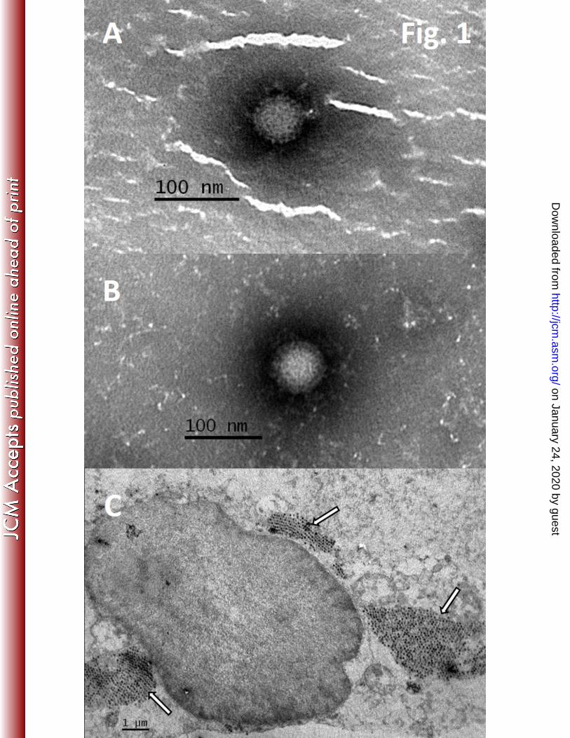
particles were observed upon EM examination of the stool suspension (Fig. 1A). After 235
inoculation of the filtered stool suspension on a LLC-MK2 cell culture, CPE appeared 48 236
hours post-inoculation, comprising cell rounding with markable membrane and final cell lysis. 237
on January 24, 2020 by guesthttp://jcm
.asm.org/
Dow
nloaded from
11
Again, reovirus particles were observed under an electron microscope, on examining the cell 238
culture supernatant without a prior concentration step (Fig. 1B) and in a thin section of 239
infected cell culture (Fig. 1C). In ultrathin sections, several cytoplasmic inclusions with 240
densely packed virus particles were found. Electron-dense center and a clear outer rim were 241
observed in all particles. 242
Cell culture supernatant was cleared and reovirus concentration was determined to be 243
2.43×10⁸ TCID50/ml. Regression equation was obtained from the real time RT-PCR analysis 244
of serial 10-fold dilutions of reovirus isolate ( 40.73 3.49 ; 0.999). The 245
theoretically TCID50/ml was calculated for reoviruses in child’s stool sample, using Cq value 246
of the reovirus real time RT-PCR analysis. The concentration was estimated to 2.26×10⁷ 247
TCID50/ml, considering the 10% stool sample dilution. 248
Sample preparation and sequencing on the Ion Torrent PGM platform 249
The high 18S real time RT-PCR Cq values (≈ 34) observed in the load of the first two 250
samples treated with nucleases (in comparison with non-treated sample 3, Cq ≈ 25) indicates 251
that benzonase degraded the nucleic acids present in the sample (Fig. 2). Comparing the 18S 252
Cq values of the load and elution fractions in these two samples, it can be concluded that one-253
step elution did not further clean the sample from eukaryotic NA, although it allowed a single 254
order of magnitude concentration of the viruses in sample 2 (1 virus per field of 400 mesh EM 255
grid in the load compared to 12-22 in the eluted fractions). In sample 1 (stool suspension), the 256
virus amount was below the LOD of the electron microscopy already in the load and the 257
presence of viruses was only detected by conventional RT-PCR (Fig. 2), in which a certain 258
concentration was again observed when comparing the intensities of the agarose gel bands 259
from load and elution fractions. Sample 3 showed the highest virus amount in the eluted 260
fractions (up to 59 viruses per field of 400 mash EM grid) and it also allowed concentration of 261
on January 24, 2020 by guesthttp://jcm
.asm.org/
Dow
nloaded from

12
the viruses from 4 viruses per field to up to 59. Moreover, the gradient allowed more efficient 262
separation of the virus rich fraction, as seen from the chromatogram (Fig. 2) and from the 18S 263
assay Cq values, which increased from 25 up to 35 in fraction 15. In this case, non-viral NA 264
was removed physically from the sample, so a lower risk for the presence of small interfering 265
fragments is expected, in comparison to the benzonase treated ones. 266
The joint dataset from all three sequencing runs consisted of 861,215 reads. The average 267
depth of coverage across all segments of the virus genome was 257.41×, with a standard 268
deviation (SD) of 117.29×. After removal of duplicate reads, the average depth of coverage 269
amounted to 101.88× (SD: 25.13×). The comparison of the sequencing output of the three 270
differently pre-treated samples is summarized in Table 1. In the case of sample 1, only 4.91% 271
reads aligned to the final consensus sequence used as a reference for comparisons. Much 272
higher enrichment for viral sequences was seen in the case of samples isolated from tissue 273
culture: 33.4% of reads aligned to the reference in case of sample 2. In the case of sample 3 274
with gradient elution even higher proportion of sequences aligned to the reference (40.14% of 275
reads). Of all three samples, sample 3 had the highest N50 value for the de novo assembly of 276
reads and the contigs produced covered almost the entire virus genome. In addition, the 277
percentage of high quality reads that map to the M. mulatta genome (source species for cell 278
culture cells) is much lower in sample 3 (2.5%) than in sample 2 (30.8%) (Table 1), indicating 279
that the elution gradient was more efficient in removing background NA than the benzonase 280
treatment combined with single step elution. Since the total number of reads was a lot higher 281
for sample 2 than for sample 3, more data and better coverage depth was obtained from the 282
former. However, increased depth in the case of sample 2 was shown to be a consequence of a 283
high number of duplicate reads (Table 1). Due to the small size of the viral genome, a very 284
good average coverage depth was achieved even on the smallest PGM sequencing chip. 285
Sequence analysis 286
on January 24, 2020 by guesthttp://jcm
.asm.org/
Dow
nloaded from

13
Nucleotide and deduced amino acid sequences were obtained and further analyzed. Following 287
BLAST of the nucleotide sequence, the newly sequenced Slovenian reovirus isolate SI-288
MRV01 showed the highest nucleotide sequence identity with reovirus strain 342/08, isolated 289
from an insectivorous bat in Germany, in all genome segments except S2, in which a slightly 290
lower nucleotide identity was observed (Table S1). The nucleotide identity with the 342/08 291
strain varied from 93.8% for the S2 segment to 99.0% for the L2 segment. The amino acid 292
sequence identity consequently also showed the highest identity with the 342/08 strain in all 293
genome segments (98.4-99.7%) (Table S1). In addition, phylogenetic analysis confirmed the 294
highest relationship of Slovenian human reovirus isolate SI-MRV01 with German bat reovirus 295
isolate 342/08. Both isolates clustered together in a separate branch in all genome segments 296
except S1 and S2 (Fig. 3, Fig. S1-S7). For the S1 segment, the Slovenian SI-MRV01 strain 297
was found in a specific bat-cluster, formed within the mammalian reovirus serotype 3 group. 298
For this segment, the highest identity was shown with the bat reovirus characterized recently 299
in Italy (12). In the S2 segment, SI-MRV01 did not show relatedness with any specific strain 300
but was clustered in serotype 3 group, together with porcine strains SC-A, MRV-HLJ/2007, 301
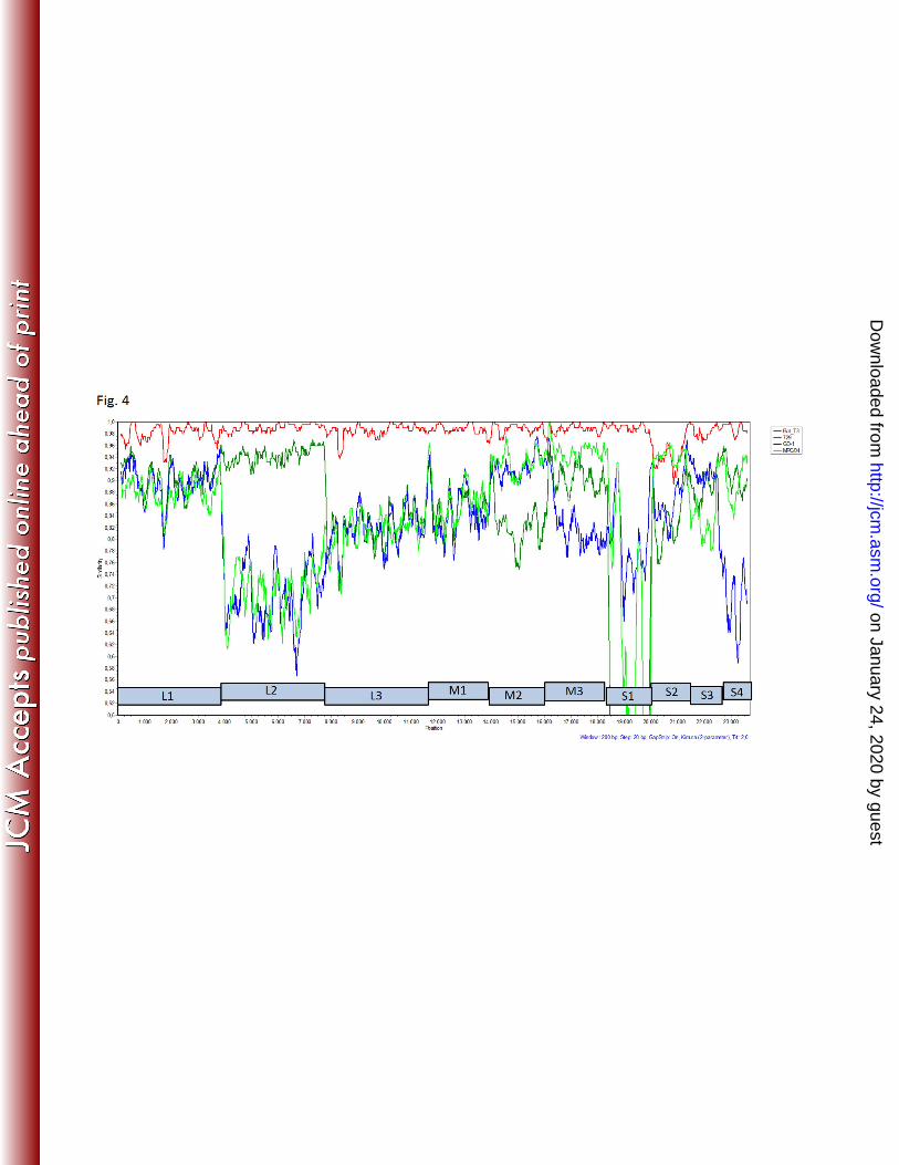
civet strain MPC/04 and bat strain 342/08. According to the results of the SimPlot analysis, 302
there are some indications of possible genome reassortments. Most indications of a 303
reassortment event were found for the L2 segment, comparing SI-MRV01 strain with porcine 304
GD-1, 729 and civet MPC/04 (Fig. 4). 305
The SI-MRV01 strain’s deduced amino acid sequence was analyzed for indicative amino acid 306
positions related to pathogenesis. In previous studies, two amino acid positions, 350D and 307
419E, in the σ1 protein (S1 gene) were shown to be indicative of neurotropism (31, 32). 308
However, in the SI-MRV01 strain, those sites were 350I and 419E. The significance of such 309
an amino acid composition is not known and needs to be examined. It was also shown that 310
249I is required for protease resistance of the σ1 protein, which enables efficient viral spread 311
on January 24, 2020 by guesthttp://jcm
.asm.org/
Dow
nloaded from

14
and replication (33) and was also found in the SI-MRV01 strain. The S1 segment is also 312
important in fusogenic reoviruses, since it encodes for the fusion protein FAST (fusion 313
associated transmembrane protein) (34). However, the Slovenian SI-MRV01 strain does not 314
have the FAST coding region. In the cell culture, no syncytia were observed, which is in 315
concordance with molecular findings of absent FAST coding region. 316
Discussion 317
The case described in this work was presented with acute gastroenteritis requiring 318
hospitalization in a 17-month old boy and MRV were found in a stool sample as the only 319
possible causative agent. There is a small possibility, though, of some other pathogen being 320
involved in the disease, i.e., some parasites that were not tested on this occasion. However, 321
the high concentration of MRV shedding in the stool of this child indicates acute infection and 322
efficient replication of MRV in the gut. No additional stool samples were available for 323
analysis to check the excretion dynamic and possible asymptomatic carriage of reoviruses. 324
Also, no acute serum sample was stored for additional tests to serologically confirm the 325
infection. As MRV infections are common in humans and the seroprevalence in early 326
childhood from 1-5 years of age was shown to be 8-50% (35), we believe that serum 327
reactivity to other MRV’s beside the isolate SI-MRV01 should also be tested to exclude 328
possible cross reactivity. Although there was no serological proof of MRV infection causing 329
gastroenteritis, we have presented data that demonstrate MRV as the possible causative agent 330
of the disease. 331
It is well known that the disadvantage of electron microscopy is its low sensitivity. For 332
successful detection of viruses with electron microscopy, the concentration of viral particles 333
should be higher than 106 per milliliter of suspension (36). Bearing in mind the dilution factor 334
of 10 (when preparing a 10% stool suspension), this means that the MRV concentration in the 335
on January 24, 2020 by guesthttp://jcm
.asm.org/
Dow
nloaded from

15
patient’s stool sample was at least of 107 viruses per millilitre or even higher. Moreover, the 336
viruses found in the stool sample were infective since they were successfully propagated in a 337
cell culture. High concentration of reoviruses in stool sample was confirmed also by 338
theoretically calculated TCID50/ml, which was as high as 2.26×10⁷. All these facts suggest 339
high replication activity of MRV in the patient’s gut. 340
After the MRV isolate was cultured and genotyped, the parents were contacted with regard to 341
the child's potential animal exposure. The family lived in a renovated house in a village close 342
to the city, where no bats had been observed. The only animal that had close contact with the 343
child was a dog at the grandparents' house but the animal was predominantly kept indoors. At 344
the time of child’s illness none of the family members experienced similar symptoms. The 345
parents stated that, at the time of the disease, the child was known to ingest non-food items 346
(e.g., mild form of pica). According to the reovirus inactivation studies, showing their 347
persistent stability in environment, indirect infection through contaminated surfaces could 348
also be possible (2, 17, 37). The source of infection therefore remains unknown. 349
Unfortunately, no bat stool samples from child’s residence were available for analysis to 350
investigate the possible source of infection. It would be interesting in the future to screen bats 351
in Slovenia for reoviruses, in order to have a clear picture of reovirus molecular epidemiology 352
and to link possible zoonotic transmissions, such as the one described in our case. 353
MRV were traditionally believed to be causative agents of mild respiratory and enteric 354
infections, without significant clinical impact (1). However, in the last decade or two, 355
increasing reports on severe human infections with reoviruses have been published, including 356
central nervous system involvement (10, 38, 39). The pathogenesis of reoviruses is not yet 357
elucidated in detail but some parameters influencing tissue tropism, efficient spread within 358
host and severe outcomes of infection are already known (31-33). It is thus important for 359
diagnostic personnel to be aware of a possible reovirus etiology and to have tools to prove this 360
on January 24, 2020 by guesthttp://jcm
.asm.org/
Dow
nloaded from

16
infection. However, any reovirus positive result should be interpreted carefully, with a broad 361
spectrum of co-tested pathogens and clinical picture of the patient. Whether or not zoonotic 362
transmission of reoviruses is one of the possible factors associated with a severe clinical 363
outcome, is not yet clear. Nevertheless, there are reports in the literature of bat reoviruses in 364
humans with severe clinical manifestations. Those reovirus isolates, though, all clustered to 365
Pteropine reovirus species and not to MRV species (6-8). This study is the first description of 366
bat MRV found in humans. Probably they are widely distributed among bat species, since 367
they have been found in insectivorous bats in Germany and in Italy (11, 12). 368
Whole genome sequence comparison of our strain to MRV’s available in GenBank clearly 369
shows that it is most closely related to MRV found in bats in Germany. This was supported by 370
high nucleotide and amino acid identities, phylogenetic analysis of separate genome segments 371
and whole genome sequences, including SimPlot analysis. Genome reassortment is indicated 372
with similarity plot of reovirus genome sequences most closely related to Slovenian SI-373
MRV01 strain (Fig. 4). Sharp decrease in nucleotide sequence identities for some of the 374
genome segments was observed, like L2 and S1 segments comparing SI-MRV01 to GD-1 and 375
MPC/04 strains and S2 segment comparing SI-MRV01 to 342/8 strain. Unfortunately, no 376
other strains with higher nucleotide identity were found in GenBank to explain this diversity 377
or possible reassortment events. 378
Finding MRV in the stool sample of a hospitalized child with diarrhea was surprising for us, 379
since no such case had previously been described in our laboratory. In the cell culture unit, no 380
previous work with reoviruses was performed and there was no reovirus nucleic acid isolated 381
in the molecular laboratory, neither reovirus genome amplification project performed. Thus, 382
the possibility for sample contamination is negligible. In addition to antigen detection tests 383
and real time RT-PCR assays for the detection of the most common viral causes of 384
gastroenteritis, electron microscopy is still an important diagnostic tool, routinely used in our 385
on January 24, 2020 by guesthttp://jcm
.asm.org/
Dow
nloaded from

17
laboratory. It is possible with electron microscope to detect various viruses associated with 386
rare cases, not included in specific antigen and/or molecular testing procedures. It is a »catch-387
all« method, providing a broad spectrum analysis of examined samples (40). For rapid and 388
accurate diagnostics, combination of classical and new techniques in diagnostic virology is 389
often needed. This study demonstrates a good case of such an interplay. Moreover, an 390
improvement in sample preparation for successful downstream application was introduced 391
with virus enrichment using CIM monolithic support. It was shown that gradient elution, used 392
in sample 3 without benzonase treatment, in principle allows more detailed separation 393
between the eluted molecules, including viruses and nucleic acids. In addition, virus particles 394
were concentrated in specific fractions after chromatographic separation. This was confirmed 395
also by sequencing results. In sample 3 the total number of reads was the lowest but the 396
percentage of orthoreovirus reads was the highest. 397
Summarizing the results of our study, there are two major outputs of this work. First, the 398
detection of a MRV with high identity to MRV isolates found in European bats is an 399
important indicator of bat-to-human interspecies transmission, as was already speculated by 400
German and Italian groups (11, 12). The real epidemiological situation regarding MRV in bats 401
and their transmission to other animal species should be further investigated in order to 402
understand the full zoonotic potential and pathogenesis of these viruses. The second output is 403
a tool for improving the sample pre-treatment, in search for enrichment in viral particles and 404
nucleic acid. The pre-treatment step is essential in NGS application, in which target nucleic 405
acid is expected to be present with high background. The described CIM chromatographic 406
approach demonstrates an excellent method, combining concentration and purification of the 407
target; a new potential shortcut for NGS application in diagnostic clinical virology. 408
409
410
on January 24, 2020 by guesthttp://jcm
.asm.org/
Dow
nloaded from

18
411
412
413
Acknowledgments 414
We thank Irena Šest for technical support in electron microscopy, diagnostics and sample 415
preparation and Snežana Kramar for assistance in cell culture propagation. This work was 416
financially supported by the Slovenian Research Agency (contracts no. L2-4314 and J3-417
4252). 418
419
420
421
422
423
424
425
426
427
428
429
430
431
432
433
434
435
on January 24, 2020 by guesthttp://jcm
.asm.org/
Dow
nloaded from

19
436
437
438
References 439
1. Schiff LA, Nibert, M.L., Tyler, K.l. 2007. Orthoreoviruses and their replication, p. 440
1854-1915. In Knipe D, Howley, P.M. (ed.), Fields Virology, 5th ed. Lippincott 441
Williams & Wilkins. 442
2. Katz BD, Margolin AB. 2007. Inactivation of hepatitis A HM-175/18f, reovirus T1 443
Lang and MS2 during alkaline stabilization of human biosolids. J Appl Microbiol 444
103:2225-2233. 445
3. Day JM. 2009. The diversity of the orthoreoviruses: molecular taxonomy and 446
phylogentic divides. Infection, genetics and evolution : journal of molecular 447
epidemiology and evolutionary genetics in infectious diseases 9:390-400. 448
4. Sabin AB. 1959. REOVIRUSES. Science 130:1387-1389. 449
5. Ouattara LA, Barin F, Barthez MA, Bonnaud B, Roingeard P, Goudeau A, 450
Castelnau P, Vernet G, Paranhos-Baccala G, Komurian-Pradel F. 2011. Novel 451
human reovirus isolated from children with acute necrotizing encephalopathy. Emerg 452
Infect Dis 17:1436-1444. 453
6. Chua KB, Voon K, Crameri G, Tan HS, Rosli J, McEachern JA, Suluraju S, Yu 454
M, Wang LF. 2008. Identification and characterization of a new orthoreovirus from 455
patients with acute respiratory infections. PLoS One 3:e3803. 456
7. Chua KB, Voon K, Yu M, Keniscope C, Abdul Rasid K, Wang LF. 2011. 457
Investigation of a potential zoonotic transmission of orthoreovirus associated with 458
acute influenza-like illness in an adult patient. PLoS One 6:e25434. 459
on January 24, 2020 by guesthttp://jcm
.asm.org/
Dow
nloaded from

20
8. Cheng P, Lau CS, Lai A, Ho E, Leung P, Chan F, Wong A, Lim W. 2009. A novel 460
reovirus isolated from a patient with acute respiratory disease. Journal of clinical 461
virology : the official publication of the Pan American Society for Clinical Virology 462
45:79-80. 463
9. Hermann L, Embree J, Hazelton P, Wells B, Coombs RT. 2004. Reovirus type 2 464
isolated from cerebrospinal fluid. Pediatr Infect Dis J 23:373-375. 465
10. Tyler KL, Barton ES, Ibach ML, Robinson C, Campbell JA, O'Donnell SM, 466
Valyi-Nagy T, Clarke P, Wetzel JD, Dermody TS. 2004. Isolation and molecular 467
characterization of a novel type 3 reovirus from a child with meningitis. The Journal 468
of infectious diseases 189:1664-1675. 469
11. Kohl C, Lesnik R, Brinkmann A, Ebinger A, Radonic A, Nitsche A, Muhldorfer 470
K, Wibbelt G, Kurth A. 2012. Isolation and characterization of three mammalian 471
orthoreoviruses from European bats. PLoS One 7:e43106. 472
12. Lelli D, Moreno A, Lavazza A, Bresaola M, Canelli E, Boniotti MB, Cordioli P. 473
2013. Identification of Mammalian orthoreovirus type 3 in Italian bats. Zoonoses 474
Public Health 60:84-92. 475
13. Decaro N, Campolo M, Desario C, Ricci D, Camero M, Lorusso E, Elia G, 476
Lavazza A, Martella V, Buonavoglia C. 2005. Virological and molecular 477
characterization of a mammalian orthoreovirus type 3 strain isolated from a dog in 478
Italy. Vet Microbiol 109:19-27. 479
14. Lodder WJ, de Roda Husman AM. 2005. Presence of noroviruses and other enteric 480
viruses in sewage and surface waters in The Netherlands. Appl Environ Microbiol 481
71:1453-1461. 482
on January 24, 2020 by guesthttp://jcm
.asm.org/
Dow
nloaded from

21
15. Lodder WJ, van den Berg HH, Rutjes SA, de Roda Husman AM. 2010. Presence 483
of enteric viruses in source waters for drinking water production in The Netherlands. 484
Appl Environ Microbiol 76:5965-5971. 485
16. Spinner ML, Di Giovanni GD. 2001. Detection and identification of mammalian 486
reoviruses in surface water by combined cell culture and reverse transcription-PCR. 487
Appl Environ Microbiol 67:3016-3020. 488
17. Irving LG, Smith FA. 1981. One-year survey of enteroviruses, adenoviruses, and 489
reoviruses isolated from effluent at an activated-sludge purification plant. Appl 490
Environ Microbiol 41:51-59. 491
18. Wong S, Lau S, Woo P, Yuen KY. 2007. Bats as a continuing source of emerging 492
infections in humans. Rev Med Virol 17:67-91. 493
19. Wang LF, Walker PJ, Poon LL. 2011. Mass extinctions, biodiversity and 494
mitochondrial function: are bats 'special' as reservoirs for emerging viruses? Curr Opin 495
Virol 1:649-657. 496
20. Lipkin WI. 2013. The changing face of pathogen discovery and surveillance. Nat Rev 497
Microbiol 11:133-141. 498
21. Capobianchi MR, Giombini E, Rozera G. 2013. Next-generation sequencing 499
technology in clinical virology. Clin Microbiol Infect 19:15-22. 500
22. Beerenwinkel N, Gunthard HF, Roth V, Metzner KJ. 2012. Challenges and 501
opportunities in estimating viral genetic diversity from next-generation sequencing 502
data. Front Microbiol 3:329. 503
23. Gutierrez-Aguirre I, Steyer A, Banjac M, Kramberger P, Poljsak-Prijatelj M, 504
Ravnikar M. 2011. On-site reverse transcription-quantitative polymerase chain 505
reaction detection of rotaviruses concentrated from environmental water samples using 506
methacrylate monolithic supports. J Chromatogr A 1218:2368-2373. 507
on January 24, 2020 by guesthttp://jcm
.asm.org/
Dow
nloaded from

22
24. Kovac K, Gutierrez-Aguirre I, Banjac M, Peterka M, Poljsak-Prijatelj M, 508
Ravnikar M, Mijovski JZ, Schultz AC, Raspor P. 2009. A novel method for 509
concentrating hepatitis A virus and caliciviruses from bottled water. J Virol Methods 510
162:272-275. 511
25. Kageyama T, Kojima S, Shinohara M, Uchida K, Fukushi S, Hoshino FB, 512
Takeda N, Katayama K. 2003. Broadly reactive and highly sensitive assay for 513
Norwalk-like viruses based on real-time quantitative reverse transcription-PCR. J Clin 514
Microbiol 41:1548-1557. 515
26. Svraka S, van der Veer B, Duizer E, Dekkers J, Koopmans M, Vennema H. 2009. 516
Novel approach for detection of enteric viruses to enable syndrome surveillance of 517
acute viral gastroenteritis. J Clin Microbiol 47:1674-1679. 518
27. Reed LJ, Muench, H. 1938. A simple method of estimating fifty per cent endpoints. 519
Am J Hyg 27:493-497. 520
28. Zheng YZ, Webb R, Greenfield PF, Reid S. 1996. Improved method for counting 521
virus and virus like particles. J Virol Methods 62:153-159. 522
29. Tamura K, Dudley J, Nei M, Kumar S. 2007. MEGA4: Molecular Evolutionary 523
Genetics Analysis (MEGA) software version 4.0. Mol Biol Evol 24:1596-1599. 524
30. Lole KS, Bollinger RC, Paranjape RS, Gadkari D, Kulkarni SS, Novak NG, 525
Ingersoll R, Sheppard HW, Ray SC. 1999. Full-length human immunodeficiency 526
virus type 1 genomes from subtype C-infected seroconverters in India, with evidence 527
of intersubtype recombination. J Virol 73:152-160. 528
31. Bassel-Duby R, Spriggs DR, Tyler KL, Fields BN. 1986. Identification of 529
attenuating mutations on the reovirus type 3 S1 double-stranded RNA segment with a 530
rapid sequencing technique. J Virol 60:64-67. 531
on January 24, 2020 by guesthttp://jcm
.asm.org/
Dow
nloaded from

23
32. Kaye KM, Spriggs DR, Bassel-Duby R, Fields BN, Tyler KL. 1986. Genetic basis 532
for altered pathogenesis of an immune-selected antigenic variant of reovirus type 3 533
(Dearing). J Virol 59:90-97. 534
33. Chappell JD, Barton ES, Smith TH, Baer GS, Duong DT, Nibert ML, Dermody 535
TS. 1998. Cleavage susceptibility of reovirus attachment protein sigma1 during 536
proteolytic disassembly of virions is determined by a sequence polymorphism in the 537
sigma1 neck. J Virol 72:8205-8213. 538
34. Shmulevitz M, Duncan R. 2000. A new class of fusion-associated small 539
transmembrane (FAST) proteins encoded by the non-enveloped fusogenic reoviruses. 540
Embo J 19:902-912. 541
35. Tai JH, Williams JV, Edwards KM, Wright PF, Crowe JE, Jr., Dermody TS. 542
2005. Prevalence of reovirus-specific antibodies in young children in Nashville, 543
Tennessee. The Journal of infectious diseases 191:1221-1224. 544
36. Hazelton PR, Gelderblom HR. 2003. Electron microscopy for rapid diagnosis of 545
infectious agents in emergent situations. Emerg Infect Dis 9:294-303. 546
37. Ward RL, Ashley CS. 1978. Heat inactivation of enteric viruses in dewatered 547
wastewater sludge. Appl Environ Microbiol 36:898-905. 548
38. Tyler KL. 1998. Pathogenesis of reovirus infections of the central nervous system. 549
Curr Top Microbiol Immunol 233:93-124. 550
39. Johansson PJ, Sveger T, Ahlfors K, Ekstrand J, Svensson L. 1996. Reovirus type 1 551
associated with meningitis. Scand J Infect Dis 28:117-120. 552
40. Biel SS, Gelderblom HR. 1999. Diagnostic electron microscopy is still a timely and 553
rewarding method. Journal of clinical virology : the official publication of the Pan 554
American Society for Clinical Virology 13:105-119. 555
556
557
on January 24, 2020 by guesthttp://jcm
.asm.org/
Dow
nloaded from

24
Table 1: Quality and quantity of nucleotide sequences obtained from Ion Torrent sequencing for the three samples tested. 558
Sample 1 (stool)
benzonase
CIM one-step elution
Sample 2 (cell culture)
benzonase
CIM one step elution
Sample 3 (cell culture)
no benzonase
CIM gradient elution
Number of reads 199,352 521,242 140,621
De novo assembly
N50 155 88 236
N of contigs 885 2556 371
N (and total length) of regions not
covered by contigsa
73 (4787 nt) 96 (6258 nt) 9 (89 nt)
Mapping to new Orthoreovirus consensus sequence
% of reads mapped 4.91 33.4 40.14
Average coverage 11.47x (SD: 5.49x) 151.77x (SD: 83.42x) 119.07x (SD: 46.06x)
Average coverage excluding duplicates 10.08x (SD: 4.35x) 39.34x (SD: 12.11x) 76.74x (SD: 21.17x)
N (and total length) of zero coverage regions (all)
6 (17 nt) 4 (5 nt) 4 (5 nt)
Mapping to Macaca mulattta
genomeb
% of reads mapped 0.03 30.83 2.54
a After mapping them to the new Orthoreovirus consensus sequence 559
b Only reads longer than 25 bp 560
on January 24, 2020 by guesthttp://jcm
.asm.org/
Dow
nloaded from

25
561
Fig. 1: Electron micrograph of reoviruses in stool suspension (A, magnification 100,000×) 562
cell culture supernatant (B, magnification 100,000×) and ultrathin section of LLC-MK2 cells 563
(C, magnification 10,000×) infected with SI-MRV01 orthoreovirus strain. Arrows in figure 564
1C are indicating reovirus particles. 565
566
Fig. 2: Elution peaks obtained in the chromatographic runs involving samples 1(stool, 567
Benzonase + CIM one-step elution), 2 (cell culture, Benzonase + CIM one-step elution) and 3 568
(cell culture, CIM gradient elution). Fraction analysis for 18S rRNA (as indicative of 569
eukaryiotic RNA presence) and reovirus EM detection is shown below the graphs. In the case 570
of sample 1, the presence of reovirus had to be confirmed by classic RT-PCR (shown left 571
below). NTC – negative template control; NT – not tested; undet - undetermined 572
573
Fig. 3: Phylogenetic analysis of L1, M1, S1 and S2 genome segments for the Slovenian strain 574
and most related whole genome strains from GenBank. Neighbor-joining algorithm was used 575
for the construction of phylogenetic tree with bootstrap values of 1000 replicates shown at the 576
branches. The scale bar represents the p-distance. 577
● Slovenian MRV isolate SI-MRV01; Δ Italian MRV strain identified in bat; ▲ German 578
MRV isolate 342/08 identified in bat 579
580
Fig. 4: Similarity plot analysis of the whole genome nucleotide sequence comparing the 581
Slovenian orthoreovirus strain with some of the most related strains from GenBank. 582
583
584
on January 24, 2020 by guesthttp://jcm
.asm.org/
Dow
nloaded from