novel molecular, cytotoxical, and immunological - biomed central
TRANSCRIPT

Hafidh et al. BMC Complementary and Alternative Medicine 2012, 12:208http://www.biomedcentral.com/1472-6882/12/208
RESEARCH ARTICLE Open Access
Novel molecular, cytotoxical, and immunologicalstudy on promising and selective anticanceractivity of Mung bean sproutsRand R Hafidh1,2†, Ahmed S Abdulamir3,2*†, Fatimah Abu Bakar2*, Farid Azizi Jalilian4,5, Faridah Abas6
and Zamberi Sekawi4
Abstract
Background: The anticancer and immunomodulatory activity of mung bean sprouts (MBS) and the underlyingmechanisms against human cervical and hepatocarcinoma cancer cells were explored.
Methods: MBS cytotoxicity and MBS-induced anticancer cytokines, TNF-α and IFN-β from cancer cells, andimmunological cytokines, IL-4, IFN-γ, and IL-10 from peripheral mononuclear cells (PMNC) were assessed by MTS andELISA assays. Apoptotic cells were investigated by flow cytometry. The expression level of apoptotic genes (Bax, BCL-2,Capsases 7–9) and cell cycle regulatory genes (cyclin D, E, and A) and tumor suppressor proteins (p27, p21, and p53) wasassessed by real-time qPCR in the cancer cells treated with extract IC50.
Results: The cytotoxicity on normal human cells was significantly different from HeLa and HepG2 cells, 163.97 ± 5.73,13.3 ± 0.89, and 14.04 ± 1.5 mg/ml, respectively. The selectivity index (SI) was 12.44 ± 0.83 for HeLa and 11.94 ± 1.2 forHepG2 cells. Increased levels of TNF-α and IFN-β were observed in the treated HeLa and HepG2 culture supernatantswhen compared with untreated cells. MBS extract was shown to be an immunopolarizing agent by inducing IFNγ andinhibiting IL-4 production by PBMC; this leads to triggering of CMI and cellular cytotoxicity. The extract inducedapoptosis, in a dose and time dependent manner, in treated HeLa and HepG2, but not in untreated, cells (P < 0.05). Thetreatment significantly induced cell cycle arrest in G0/G1 in HeLa cells. The percentage of cells in G0/G1 phase of thetreated HeLa cells increased from 62.87 ± 2.1%, in untreated cells, to 80.48 ± 2.97%. Interestingly, MBS IC50 induced theexpression of apoptosis and tumor suppressor related genes in both HeLa and HepG2 cells. MBS extract succeeded ininducing cdk-inhibitors, p21, p53, and p27 in HeLa cells while it induced only p53 in HepG2 cells (P < 0.05). This is a cluefor the cell type- specific interaction of the studied extract. These proteins inhibit the cyclin-cdk complexes apart fromthe presence of some other components that might stimulate some cyclins such as cyclin E, A, and D.
Conclusion: MBS extract was shown to be a potent anticancer agent granting new prospects of anticancer therapyusing natural products.
BackgroundMore attention has recently been given to the role ofplant-derived compounds as promising nutraceuticalsfor controlling various disorders such as cardiovascular,neurological, neoplastic and immunological diseases [1].
* Correspondence: [email protected]; [email protected]†Equal contributors2Institute of Bioscience, Universiti Putra Malaysia, 43400 UPM Serdang,Selangor Darul Ehsan, Malaysia3Department of Microbiology, College of Medicine, Al-Nahrain University,Baghdad, IraqFull list of author information is available at the end of the article
© 2012 Hafidh et al.; licensee BioMed CentralCommons Attribution License (http://creativecreproduction in any medium, provided the or
The phytochemical compounds are found to be integralcomponents of human diet. They are commonly presentas constituents of flowering plants, particularly of foodplants [2].Studies verified that phytochemicals are able to alter
the likelihood of carcinogenesis in every stage of cancerprocess in a way reducing the risk but usually in a favor-able direction [3]. Interestingly, the main activity ofthese compounds depends on the fact that the exposureof human cells to a wide variety of chemoprotectivecompounds confers resistance against a broad set of car-cinogens [4]. Much information exists nowadays on the
Ltd. This is an Open Access article distributed under the terms of the Creativeommons.org/licenses/by/2.0), which permits unrestricted use, distribution, andiginal work is properly cited.

Hafidh et al. BMC Complementary and Alternative Medicine 2012, 12:208 Page 2 of 24http://www.biomedcentral.com/1472-6882/12/208
antitumor action of plants, and many in vitro studieshave concentrated on the direct and indirect actions ofphytochemicals on tumor cells, and have found a varietyof anticancer effects such as cell growth [5], kinase activ-ity inhibition [6], apoptosis induction [7], and suppres-sion of the secretion of matrix metalloproteinases, andtumor invasive behavior [8]. Furthermore, some studiesreported the impairment of in vivo angiogenesis by diet-ary phytochemicals [9]. Therefore, the discovery of newanticancer agents from plant-derived substances is con-sidered to be a highly promising approach in order toenrich the pharmaceutical field with effective drugs oflower side effects.Besides, plants produce a vast number of natural pro-
ducts which have antimicrobial and immunomodulatingpotential as defense mechanisms for adapting to variousenvironmental insults [10]. Many natural compoundshave shown a significant ability to regulate immuneresponses [11]. Some of these phytochemicals withimmunomodulating effects are isoflavonoids, indoles,phytosterols, polysaccharides, sesquiterpenes, alkaloids,glucans, tannins, a variety of vitamins and trace mineralsthat function as antioxidants and co-enzymes, and manyother phytochemical substances [12].It is clear that human immune response is a highly com-
plex and extraordinarily sophisticated system involvingboth innate and adaptive mechanisms [13]. Immunomodu-lating activity refers to biological or pharmacological effectsof compounds on humoral or cellular aspects of the im-mune response. In other words, immunomodulation is theprocess of modifying an immune response in a positive ornegative manner by administration of a drug or a com-pound [11]. Although the field of study of immune enhan-cing compounds is relatively not new [14], naturalproducts from plants represent a rich and promisingsource of novel molecules with immunomodulating prop-erties that may augment a disease recovery alone or to-gether with commercially known drugs.For the first time in the field, the current study
investigated the in vitro selective cytotoxic and immu-nomodulatory effects of mung bean sprout (Vignaradiata L.) methanol crude extracts on human cancerand peripheral mononuclear cells (PMNC), respect-ively. The rationale behind testing the anticancer andimmunomodulatory effects of MBS extract is that first,MBS has not been assessed as anticancer or as immu-nomodulatory agent before, second, MBS is a germin-ating plant which usually possesses very high levels ofantioxidants that are well known to act as potent antic-ancer and immunomodulatory agents. More in depth,this study explored thoroughly the underlying mechan-isms of the newly discovered findings in the currentstudy, namely, the novel anticancer and immunomodu-latory effects of mung bean sprout (Vigna radiata L.)
methanol extract by using cytological and molecularassays for assessing and measuring anticancer cyto-kines, cell cycle phases, percentage of apoptotic cells,expression level of cell cycle genes, tumor suppressorgenes and apoptosis genes in MBS treated and un-treated cancer cells. Moreover, PBMC-secreted cyto-kines, in response MBS extract exposure, weremeasured as an indicator for the immunomodulatoryeffect of MBS. Therefore, the ultimate goal of thecurrent study is to find a natural anticancer product able toinhibit the proliferation of human cancer cell lines withhigh selectivity index, safe usage, and effective anticanceractivity. This study has been patented in Malaysian intellec-tual property (MyIPO) under Malaysian patent applicationnumber PI2011001617 on 12th April 2011 (http://www.rmc.upm.edu.my/upmip/index.php?content=getfaculty&ipid=683&ipdetailid=671&projectlead=151&cluster=3&fac=8)and the current patented research has been considered forcommercialization by University Putra Malaysia.
MethodsEthical approachThe current study was conducted in compliance withHelsinki Declaration for ethical approaches of conduct-ing scientific research. This study was approved by theethical committee of University Putra Malaysia in KualaLumpur.
Preparation of the MBS extractFresh mung bean sprouts (MBS), devoid of any preserva-tive antimicrobials was purchased from local markets inthe State of Selangor, Malaysia. The growth of mung beansand the germination of the sprouts were done in SelangorState. The mung bean sprouts were left to dry in dark areafor three days at room temperature 23-25°C. After the dry-ness of sprouts, they were ground to powder. The groundpowder was extracted 1:10 wt/v with 2.4 M HCl acidifiedmethanol (Merck, Darmstadt, Germany) to extract all, freeand conjugated, components of phenolic compounds [15];the ground powder was then soaked in dark area for threedays at room temperature. The supernatant was collectedafter filtration and the fresh solvent was added to the plantmaterial. The extraction procedure was repeated twice andthe collected extracts were evaporated to dryness undervacuum at 40°C by using rotary evaporator. To remove theeffect of the acidity from the crude extract, the crude ex-tract of MBS was neutralized to exclude any pH-related ef-fect. The pH for MBS extract was neutral ranging from 6.8to 7.0. The dried extracts were stored at −18°C in a desic-cant until further use.
Preparation of stock extractThe stock extract for MBS was prepared by redissolvingthe MBS extract powder in dimethyl sulfoxide (DMSO)
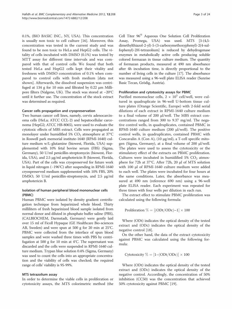
Hafidh et al. BMC Complementary and Alternative Medicine 2012, 12:208 Page 3 of 24http://www.biomedcentral.com/1472-6882/12/208
0.1%, (BIO BASIC INC., NY, USA). This concentrationis usually non toxic to cell culture [16]. Moreover, thisconcentration was tested in the current study and wasfound to be non toxic to HeLa and HepG2 cells. The vi-tality of cells incubated with DMSO (0.1%) was tested byMTT assay for different time intervals and was com-pared with that of control cells We found that bothtested HeLa and HepG2 cells kept their vitality andfreshness with DMSO concentration of 0.1% when com-pared to control cells with fresh medium [data notshown]. Afterwards, the dissolved suspension was centri-fuged at 134 g for 10 min and filtrated by 0.22 μm Milli-pore filters (Nalgene, UK). The stock was stored at −20°Cuntil it further use. The concentration of the stock extractwas determined as required.
Cancer cells propagation and cryopreservationTwo human cancer cell lines, namely, cervix adenocarcin-oma cells (HeLa; ATCC CCL-2) and hepatocellular carce-noma (HepG2; ATCC HB-8065), were used to evaluate thecytotoxic effects of MBS extract. Cells were propagated asmonolayer under humidified 5% CO2 atmosphere at 37°Cin Roswell park memorial institute-1640 (RPMI-1640) cul-ture medium w/L-glutamine (biowest, Florida, USA) sup-plemented with 10% fetal bovine serum (FBS) (Sigma,Germany), 50 U/ml penicillin-streptomycin (biowest, Flor-ida, USA), and 2.5 μg/ml amphotericin B (biowest, Florida,USA). Part of the cells was cryopreserved for future workin liquid nitrogen (−196°C) after suspending in RPMI-1640cryospreserved medium supplemented with 10% FBS, 20%DMSO, 50 U/ml penicillin-streptomycin, and 2.5 μg/mlamphotericin B.
Isolation of human peripheral blood mononuclear cells(PBMC)Human PBMC were isolated by density gradient centrifu-gation technique from heparinized whole blood. Thirtymilliliters of fresh heparinized blood sample isolated fromnormal donor and diluted in phosphate buffer saline (PBS),(CALBIOCHEM, Darmstadt, Germany) were gently laidover 15 ml of Ficoll Hypaque (GE Healthcare Bio-sciencesAB, Sweden) and were spun at 500 g for 20 min at 25°C.PBMC were collected from the interface of spun bloodsamples and were washed three times with PBS by centri-fugation at 500 g for 10 min at 4°C. The supernatant wasdiscarded and the cells were suspended in RPMI-1640 cul-ture medium. Trypan blue solution 0.4% (Sigma, Germany)was used to count the cells into an appropriate concentra-tion and the viability of cells was checked; the requiredrange of cells’ viability is 95-99%.
MTS tetrazolium assayIn order to determine the viable cells in proliferation orcytotoxicity assays, the MTS colorimetric method (the
Cell Titer 96® Aqueous One Solution Cell ProliferationAssay, Promega, USA) was used. MTS [3-(4,5-dimethylthiazol-2-yl)-5-(3-carboxymethoxyphenyl)-2(4-sul-fophenyl)-2H-tetrazolium] is reduced by dehydrogenaseenzymes in metabolically active cells producing solublecolored formazan in tissue culture medium. The quantityof formazan products, measured at 490 nm absorbanceafter 4h incubation time, is directly proportional to thenumber of living cells in the culture [17]. The absorbancewas measured using a 96-well plate ELISA reader (SunriseBasic Tecan, Grödig, Austria).
Proliferation and cytotoxicity assays for PBMCPurified mononuclear cells, 2 × 10⁵ cell/well, were cul-tured in quadruplicate in 96-well U-bottom tissue cul-ture plates (Orange Scientific, Europe) with 2-fold serialdilutions of each extract in RPMI-1640 culture mediumto a final volume of 200 μl/well. The MBS extract con-centrations ranged from 300 to 9.37 mg/ml. The nega-tive control wells, in quadruplicates, contained PBMC inRPMI-1640 culture medium (200 μl/well). The positivecontrol wells, in quadruplicates, contained PBMC withConcavalin A (Con A), (10 μg/ml), a T-cell specific mito-gen (Sigma, Germany), at a final volume of 200 μl/well.The plates were used to assess the cytotoxicity or thestimulatory effect of the extracts on PBMC proliferation.Cultures were incubated in humidified 5% CO2 atmos-phere for 72h at 37°C. After 72h, 20 μl of MTS solutionwith 100 μl of RPMI-1640 culture medium were addedto each well. The plates were incubated for four hours atthe same conditions. Later, the absorbance was mea-sured at 490 nm (reference 690 nm) using a 96-wellplate ELISA reader. Each experiment was repeated forthree times with four wells per dilution in each run.The extract effect to stimulate PBMC proliferation was
calculated using the following formula:
Proliferation % ¼ ODt=ODcð Þ–1½ � � 100
Where (ODt) indicates the optical density of the testedextract and (ODc) indicates the optical density of thenegative control [18].On the other hand, the data of the extract cytotoxicity
against PBMC was calculated using the following for-mula:
Cytotoxicity % ¼ 1– ODt=ODcð Þ½ � � 100
Where (ODt) indicates the optical density of the testedextract and (ODc) indicates the optical density of thenegative control. Accordingly, the concentration of 50%inhibition (CC50) was the concentration that achieved50% cytotoxicity against PBMC [19].

Hafidh et al. BMC Complementary and Alternative Medicine 2012, 12:208 Page 4 of 24http://www.biomedcentral.com/1472-6882/12/208
Cytotoxicity on human cancer cell linesCytotoxicity assay was performed according to the estab-lished method of Mena-Rejon et al. (2009), where 1 ×k 105
cell/well viable HeLa and HepG2 cells were grown inRPMI-1640 culture medium in 96-well flat-bottom tissueculture plates (Orange Scientific, Europe). The plates wereincubated in humidified 5% CO2 for 24h at 37°C. Whencells reached > 80% confluence, the medium was replacedwith 200 μl/well of 2-fold serial dilutions of MBS extractfrom 164 to 10.25 mg/ml prepared in RPMI-1640 mainten-ance medium (with 2% FBS). The concentration of MBSextract was 328 mg/ml. The negative control wells con-tained DMSO (0.1%) in RPMI-1640 maintenance mediumwith final volume of 200 μl/well. All the plates were incu-bated in humidified 5% CO2 for 24h at 37°C. Later, all thewells’ contents were removed and replaced with 200 μl/wellof RPMI-1640 maintenance medium. The plates were re-incubated for 48 h at the same conditions. Afterwards, 20μl of MTS solution with 100 μl of RPMI-1640 culturemedium were added to each well. The plates were incu-bated for four hours in the same conditions. The absorb-ance was measured at 490 nm (reference 690 nm) using a96-well plate ELISA reader. Each experiment was repeatedfor three times with four wells per dilution in each run [20].The concentration of the extract that killed 50% of the
cells (IC50) was calculated using the following formula:
Cytotoxicity % ¼ 1– ODt=ODcð Þ½ � � 100
Where (ODt) indicates the optical density of the testedextract and (ODc) indicates the optical density of thenegative control. The selectivity index (SI) was the ratioof CC50 (cytotoxicity on PBMC) to the IC50 (cytotox-icity on human cancer cells), [19].
Cytokine production by PBMCIn order to evaluate the immunomodulatory effect of MBSextract, the cytokine level produced from PBMC aftertreatment with MBS extract was investigated. PBMC at2×105 cell/well were cultured, in triplicates, in 96-well U-bottom tissue culture plates with 2-fold serial dilutions ofthe extract in RPMI-1640 culture medium to a final vol-ume of 200 μl/well. The extract concentrations rangedfrom 100 to 3.12 mg/ml. The negative control wells, in tri-plicates, contained 200 μl/well of PBMC in RPMI-1640culture medium. After 24 h of incubation at 37°C in hu-midified 5% CO2 atmosphere, the plates were centrifugedat 300 g for 10 min. The supernatant was collected andcentrifuged at 1000 g for 10 min to be ready to determinethe cytokine level produced into the medium [21].
Cytokine production by human cancer cell linesThe level of anticancer cytokines, IFN-β and TNF-α,was investigated in treated and untreated HeLa and
HepG2 cells. HeLa and HepG2 cells at 1 × 10⁵ cell/wellwere grown in RPMI-1640 culture medium in 96-wellflat-bottom tissue culture plates in a humidified 5% CO2
atmosphere for 24 h at 37°C. Later, serial 2-fold dilutionsof MBS 10 to 0.31 mg/ml prepared in RPMI-1640 main-tenance medium were added to the cells, in triplicates,to a final volume of 200 μl/well. The MBS extract stockconcentration was 20 mg/ml. The negative control wells,in triplicates, contained cells with RPMI-1640 mainten-ance medium only with final volume of 200 μl/well. Theplates were incubated for 48h at the same conditions.Afterwards the supernatants from all the wells wereremoved and centrifuged at 1000 g for 10min to beready for the ELISA technique [22].
Measuerment of cytokines produced by PBMC and cancercellsThe levels of immunomodulatory cytokines, IL-2, IL-4and IFN-γ, in the PBMC culture supernatant with andwithout extract treatment, and the levels of anticancercytokines, IFN-β and TNF-α, in the supernatant of cul-tured treated and non-treated cancer cells, were mea-sured. The procedures pursued were according to themanufacturers’ instructions of each kit. For IL-2 and IL-4, the used kit was human enzyme immunometric assay(EIA) kits (Cayman, USA), for IFN-γ, a human interferongamma ELISA kit (abcam, USA) was used, and for antic-ancer cytokines, human IFN-β ELISA (abcam, USA) andTNF-α human enzyme immunometric assay (EIA) kits(Cayman, USA) were used.Each microtiter plate was already coated with mono-
clonal antibodies specific for the corresponding cytokine.The standards of IL-2 and IL-4 were reconstituted in thesame matrix of the samples, namely RPMI-1640 mediumplus each 2-fold serial dilution of the extract; this step isneeded to match the color effect of unpurified samplesthat might persist even after the subsequent washingsteps. Accordingly, a standard curve was made for eachdilution of the extracts used. The IL-2 or IL-4 standardswere prepared as 2-fold serial dilutions ranging from250 to 3.9 pg/ml in eight tubes of 15 ml capacity (Or-ange Scientific, Europe); the eighth tube contained zeroconcentration of IL-2 or IL-4 standard.For IFN-γ, standards were freshly prepared by reconsti-
tuting in the standard diluents buffer to give a stock con-centration of 400 pg/ml. Two hundred microliter of thisstock was added, in triplicates, to the already coated micro-titer plates provided by the kit. Each plate was coated withmonoclonal IFN-γ specific antibodies. From stock wells,serial 2-fold dilutions of IFN-γ standard were prepared bydiluting the standard in a matrix similar to that of the sam-ples which is RPMI-1640 culture medium plus each dilu-tion of the extracts used. This step is needed to match thecolor effect of unpurified samples that might persist even

Hafidh et al. BMC Complementary and Alternative Medicine 2012, 12:208 Page 5 of 24http://www.biomedcentral.com/1472-6882/12/208
after the subsequent washing steps. Accordingly, a stand-ard curve was made for each dilution of the extracts used.IFN-γ standard concentrations ranged from 400 to 12.5pg/ml; and the last standard contained zero concentrationof IFN-γ standard in the sample matrix.For immunomodulatory cytokines, IL-2, IL-4, IFN-γ, and
anticancer cytokines, IFN-β and TNF-α, one hundred μl ofstandards, samples, and conjugates were all assayed as tri-plicates. For IL-2 and IL-4, the absorbance was measuredat 412 nm (reference 690 nm) using a 96-well plate ELISAreader. The assay was repeated for three times for eachsample. The concentrations of samples’ IL-2 and IL-4 weredetermined by extrapolating their OD values with that ofthe generated standard curves. For IFN-γ, the absorbancewas measured at 450 nm (reference 620 nm) by ELISAreader. The sample concentrations were determined by ex-trapolating OD values to IFN-γ concentrations using thegenerated linear standard curves (the average absorbanceon the vertical axis versus the corresponding IFN-γ stand-ard concentration on the horizontal axis). One hundred μlof sample, conjugates and substrate-chromogen were usedaccording to the guidelines of the kits manufacturer. Later,the absorbance was measured at 412 nm (reference 690nm) and at 450 nm (reference 620 nm) using a 96-wellplate ELISA reader for TNF-α and IFN-β plates, respect-ively. The assay was repeated for three times for each sam-ple. The concentrations of the cytokines were determinedby extrapolating their OD values with that of the generatedstandard curves.
Analysis of apoptosisFlow cytometry analysis for cell apoptosisFlow cytometry can rapidly quantify and evaluate theproperties of apoptotic cells. It can give information onthe ratio of apoptotic cells, based on the cellular size orDNA contents. It is well known that several differencesare present between apoptotic cells and normal cells.These differences can be utilized by flow cytometric tech-niques for apoptosis detection [23]. The cells (HeLa andHepG2) were seeded (1 × 10⁵ cell/well) in 6-wells tissueculture plates and were incubated in a humidified 5%CO2 atmosphere for 24 h at 37°C. The medium was thenreplaced with RPMI-1640 maintenance medium with orwithout MBS extract and was incubated for further 24 hat the same conditions. The cell treatment was dividedinto two groups. In the first group (dose-dependentgroup), the effect of three 2-fold serial dilutions of the ex-tract (concentration > IC50 > concentration) was investi-gated regarding the level of apoptosis, if any, after a fixedtime. The concentrations of MBS extract were 26.6, 13.3,and 6.65 mg/ml with HeLa cells and were 28.08, 14.04,and 7.02 mg/ml with HepG2 cells. In the second group(time-dependent group), the IC50 of MBS extract wasused to investigate the level of cells’ apoptosis as well as
cell cycle arrest after different time intervals of incuba-tion with the extract. For the first group, after 24 h, thecells were harvested and transferred to 15 ml tubes. Allof the tubes were centrifuged at 190 g for 10 min. Thesupernatants were discarded and the pellets were washedtwo times by cold PBS. Later, the pellets were resus-pended in 70% ice-cold ethanol with PBS, 1:10 v/v, andwere incubated for 2h at −20°C. Then, all the superna-tants were aspirated after centrifugation at 500 g for10 min. The washing step by PBS was repeated and thesupernatants were aspirated. The pellets were resus-pended in 500 μl of DNA staining solution containing25 μl of propidium iodide (PI) 1 mg/ml (MP Biomedicals,LLC, IIIKrick, France), a double-stranded nucleic acidintercalating agent, and 50 μl Ribonuclease A from bo-vine pancrease (1 mg/ml), (Sigma, Germany) in PBS. Allthe tubes were incubated on ice in dark area for 30min[24]. The assay was measured in duplicate for each sample.The propidium iodide fluorescence of individual nucleiwas measured using CyAn ADP apparatus (BECKMANCOULTER, USA). The software Summit (V4.3) was usedto analyze the flow cytometry results.For the second group of the flow cytometry analysis, the
stock extract was prepared and the same method men-tioned earlier for the first group was used except for thefollowing differences: the IC50 for MBS extract with HeLaand HepG2 cells was used to treat the cells. The IC50 ofMBS extract was 13.3 mg/ml with HeLa cells and14.04 mg/ml with HepG2 cells. The group was subdividedinto seven treatments (8, 12, 16, 20, 24, 48 and 72 h). Theassay was measured in duplicate for each sample in thespecific time of treatment.
Detection of apoptosis- and cell cycle arrest- related genesby real time quantitative PCR (qRT-PCR)The current trend of research uses advanced techniquesin molecular biology to study the apoptosis- and cellcycle arrest- related genes’ expression such as Bcl-2 fam-ily, members of caspase family, tumor suppressor pro-teins, and cyclins. Nowadays, by using the reversetranscription-polymerase chain reaction (RT-PCR), thescientists can demonstrate the level of mRNA expressionof apoptosis and cell cycle proteins even though they areexpressed only in a small cell population in tissues [25].
RNA extractionThe cells (HeLa and HepG2) were seeded (1 × 10⁵ cell/well) in 6-wells tissue culture plates and were incubated ina humidified 5% CO2 atmosphere for 24 h at 37°C. Themedium was then replaced with RPMI-1640 maintenancemedium either alone or with MBS extract, in duplicates,and the medium was incubated at the same conditions.The IC50 for MBS extract against each type of cells wasused. The IC50 of MBS extract was 13.3 mg/ml with HeLa

Hafidh et al. BMC Complementary and Alternative Medicine 2012, 12:208 Page 6 of 24http://www.biomedcentral.com/1472-6882/12/208
cells and 14.04mg/ml with HepG2 cells. After determiningthe best timing for studying the apoptosis and cell cycle ar-rest by flow cytometry, treatment of cells with extract for12, 16, and 20 h was conducted. At the end of 12, 16, or20h of extract treatment, the cells were harvested andtransferred to 15 ml tubes. After centrifuging the tubes at134 g for 5 min, the supernatants were discarded. The pel-lets were resuspended in PBS and were washed for fourtimes.It is noteworthy to mention that one of the most import-
ant steps preceding the synthesis of good quality cDNA isthe isolation of intact (undegraded) total RNA from cul-tured cells or tissues [25]. Total RNA was isolated usingGF-1 kit (Vivantis Technologies, Malaysia). According tothe manufacturer’s protocol for RNA isolation from cellculture, 1 × 10⁷ cells were precipitated in 1.5 ml microtubes(Eppendrof, Hamberg, Germany) at 1000 × g for 5 min.The cell pellets were resuspended in 700 μl of lyses buffer(Buffer TR) with vigorous mixing by vortexing. This bufferis specially formulated to inactivate cellular RNases to-gether with cell lysis. Later, the lysed cells were transferredto homogenization columns assembled in a collectiontubes. The columns were centrifuged at 10.000 × g for2 min. The flow-though was saved and equal volume of80% ethanol (700 μl) was added. The lysed cells were mixedthoroughly by pipetting and were transferred into RNAbinding columns assembled in collection tubes. RNA bind-ing columns were centrifuged at 10.000 × g for 1 min andthe flow-though was discarded. The columns were washedby adding 500 μl of washing buffer and were centrifuged at14.000 × g for 1 min. The flow-though was discarded andall of the DNA fragments were removed by DNase I treat-ment. Seventy microlitter of DNase I Digestion Mix wereadded to RNA binding columns and were incubated atroom temperature for 15 min. DNase I Digestion Mix wascomposed of DNase I (7 μl), digestion buffer (56 μl), and di-gestion enhancer (7 μl). Then, 500 μl of inhibitor removalbuffer were added to the columns which were centrifugedat 14.000 × g for 1 min. The columns were washed with500 μl washing buffer for two times with centrifugation at10.000 × g for 1 min for each run. Further centrifugation at10.000 × g for 1 min was done to remove any traces of buf-fer. Total RNA was collected by placing the columns intonew 1.5 ml microtubes with 60 μl RNase-free wateraddition and standing for 1 min. The microtubes were cen-trifuged at 10.000 × g for 1 min. RNA quality and quantitywere determined by Life Science UV/Vis Spectrophotom-eter, DU Series 700 (BECKMAN COULTER, USA).The iso-lated RNA was stored at −80°C and was ready for use indownstream application, namely, qRT-PCR.
Real time quantitative RT-PCROne microgram of the isolated RNA from each samplewas reverse-transcribed by iScript™ cDNA Synthesis Kit
(BIO-RAD, Hercules, Canada). According to the manufac-turer’s protocol, 4 μl of 5 × iScript reaction mix were mixedwith 1 μl iScript reverse transcriptase and 15 μl of RNAtemplate in 1.5 ml microtubes to give final volume of 20 μlper reaction. The complete reaction mix was incubated for5 min at 25°C then for 30 min at 42°C using Thermo Bath,ALB64 (FINEPCR, Seoul, Korea). The incubation temperaturewas increased to 85°C for 5 min. Finally, cDNA was stored at−80°C for qRT-PCR reaction.Real-time quantitative PCR reaction was conducted using
SsoFast™ EvaGreen® Supermix (BIO-RAD, Hercules,Canada). Depending on the manufacturer’s protocol, 10 μlof 1x SsoFast EvaGreen supermix were mixed with 7 μlRNase/DNase free water. One microlitter of forward primer(500 nM) and 1 μl of reverse primer (500 nM) were addedto the previous mix (Table 1). Finally, 1 μl of cDNA templatecorresponding to 50 ng of total RNA was added. The PCRreaction (20 μl) was run for 40 cycles using CFX96™ Real-Time System (BIO-RAD, Hercules, Canada). Cycling condi-tions were 95°C for 3 min, 95°C for 10 sec, 55-61°C for30 sec, and 72°C for 20 sec. PCR reaction for cDNAtemplates from untreated HeLa and HepG2 cells wereused as negative controls. The PCR reaction was run intriplicate for each target gene. PCR reaction mix withoutcDNA template was used to detect any contamination.At the end of the amplification, measurement of EvaGreen fluorescence was done continuously with the con-duction of the melting curve analysis by slow heating at0.5°Cs-1 increments from 70 to 95°C, with continuousfluorescence collection. Accordingly, a melting curve wasgenerated at the end of the PCR amplification for moni-toring the specificity of PCR reaction. Melting curve ana-lysis of the negative first derivative was pursued. Beta-actin was used as a housekeeping gene (reference gene)to normalize the mRNA expression of target genes. Be-cause PCR efficiency may vary among different primers,the calculation of PCR primers’ efficiency is essential forobtaining accurate measurements of the relative expres-sion of the mRNA of target genes [26]. For this reason, astandard curve was created by diluting template cDNA ofeach single primer used in this study. The cDNA tem-plate for each primer was serially diluted (10-1 to 10-7);each dilution serves as a standard. In this reaction, cDNAtemplate was used from samples with high expression tothe target of interest. The amplification efficiency can beobtained by analyzing the slope of the log-linear portionof the standard curve. When the logarithm of the initialtemplate concentration is plotted on the x axis and thethreshold cycle (Ct) is plotted on the y axis, PCR effi-ciency is calculated according to the following equation[26]:
PCR efficiency ¼ 10�1=slope–1

Table 1 Primers used in Real-Time quantitative PCR analysis (Vivantis Technologies, Malaysia)
Forward primer Reverse primer
Bax CAC CAG CTC TGA GCA GAT GCG AGG CGG TGA GCA CTC
BCL-2 TAC CTG AAC CGG CAC CTG GCC GTA CAG TTC CAC AAA GG
Caspase 7 GTC TCA CCT ATC CTG CCC TCA TTC TTC TTC TGC CTC ACT GTC
Caspase 8 GAA AAG CAA ACC TCG GGG ATA C CCA AGT GTG TTC CAT TCC TGT C
Caspase 9 CCA GAG ATT CGC AAA CCA GAG G GAG CAC CGA CAT CAC CAA ATC C
Cyclin D AGA CCT GCG CGC CCT CGG TG GTA GTA GGA CAG GAA GTT GTT C
Cyclin E CTC CAG GAA GAG GAA GGC AA TCG ATT TTG GCC ATT TCT TCA
Cyclin A GTC ACC ACA TAC TAT GGA CAT G AAG TTT TCC TCT CAG CAC TGA C
p21 GTG ATT GCG ATG CGC TCA TG TCT CTT GCA GAA GAC CAA TC
p27 GTC TAA CGG GAG CCC TAG CC CTA ACC CCG TCT GGC TGT CC
p53 TGT GGA GTA TTT GGA TGA CA GAA CAT GAG TTT TTT ATG GC
β-actin (reference gene) TCA CCC TGA AGT ACC CCA TC CCA TCT CTT GCT GCA AGT CC
Note: all primers are listed 5’-3’.
Hafidh et al. BMC Complementary and Alternative Medicine 2012, 12:208 Page 7 of 24http://www.biomedcentral.com/1472-6882/12/208
The software BIO-RAD CFX Manager (V 1.1.308) wasused to relatively quantify the target genes according tothe following equation [27]:
Ratio ¼ Etarget� �ΔCt target control�sampleð Þ
= Erefð ÞΔCtRef control–sampleð Þ
In which (E): represents the amount of fold change percycle per gene. Ref: represents the reference gene. Tar-get: represents the target gene.It has been well known that the ratio of Bax to Bcl-2
determines, in part, the susceptibility of cells to deathsignals [28]. Therefore, the Bax to Bcl-2 ratio was calcu-lated using the following equation:
Bax=Bcl� 2 ratio
¼ mean PCR efficiency for Bax and Bcl� 2ð Þ CtBcl2–CtBaxð Þ
Data analysisAll the data in the current study are shown as mean±2SE.The selectivity index (SI) was determined by using the ratioof CC50 to IC50. The data analysis was conducted by usingSPSS software version (12.0.0.2). The effect of the testedextract on the inhibition of cell growth was evaluated byusing 95% confidence intervals. IC50 and CC50 valueswere calculated using linear regression index equations.The statistically different effects of the extract on theability of PBMC, HeLa and HepG2 cells to synthesizeselected cytokines were compared with the control
Table 2 The cytotoxic effect of MBS extract on cancer cell line
Extract Cancer cell line aCC50 mg/ml bIC50
MBS HeLa 163.97 ± 5.73 13.3 ±
HepG2 163.97 ± 5.73 14.04 ±
Note: aThe results of CC50 (on peripheral blood mononuclear cells) are shown as mcSelectivity index represents the ratio of CC50/IC50. The results are shown as mean
groups using the Student’s t-test. For flow cytomtericanalysis, R2 fraction represented sub-G apoptotic cells;moreover, the percentage of cells at different cell cyclephases was calculated from the total cells minus apop-totic cells. For quantitative real time PCR, the up- ordown- regulation of mRNA expression of selected geneswas measured as expression fold changes in term ofmean±2SD. The significance of up- or down- regulationof the normalized mRNA expression of selected geneswas determined by comparing the mean ± 2SD of anyup- or down- regulation with the mean ± 2SD of con-trol (untreated cells), equal to 1 ± 2SD. P values lessthan 0.05 were considered significant.
ResultsCytotoxicity on human cancer cellsThe results of the current study revealed that the cyto-toxic effects of MBS extract on normal human cells(PBMC) was significantly different (P < 0.05) from thaton human cancer cells (Table 2). The cytotoxic effect ofMBS extract on PBMC, expressed as CC50, was 163.97mg/ml while its IC50 on HeLa cells was 13.3 mg/ml andon HepG2 cells was 14.04 mg/ml. These findingsrevealed that MBS extract required high concentrationsto be cytotoxic on normal human cells (Figure 1) whileonly low concentrations were enough to give the sameeffect on human cancer cells (Figure 2). These results
s
mg/ml P value P < 0.05 cSI
0.89 6.54539E-06 Significant 12.44 ± 0.83
1.5 7.25478E-06 Significant 11.94 ± 1.2
ean±2SE. bThe results of IC50 (on cancer cell lines) are shown as mean ± 2SE.± 2SE.

Figure 1 The percentage of PBMC death after treatment with2-fold serial dilutions of MBS extracts in term of mean ± 2SE(confidence interval CI 95%).
Hafidh et al. BMC Complementary and Alternative Medicine 2012, 12:208 Page 8 of 24http://www.biomedcentral.com/1472-6882/12/208
reflected the good selectivity and safety of these extractsas cytotoxic agents (Figure 3).The cytotoxicity of MBS extract on PBMC, HeLa and
HepG2 cells was dose dependent. In other words, the
Figure 2 The percentage of A) HeLa and B) HepG2 cells death after trmean ± 2SE (confidence interval CI 95%.
cytotoxicity of MBS extract decreased with higher dilu-tions, lower concentrations, of the extract. The signifi-cant differences of MBS cytotoxicity were supported bythe results of the selectivity index (SI) which is the ratioof the highest concentration that causes 50% death tonormal cells (CC50) to the lowest concentration thatcauses 50% death to cancer cells (IC50). The SI values ofMBS extract demonstrated effective SI values on HeLacells, 12.44, and HepG2 cells, 11.94 (Table 2). The cyto-toxic effect of MBS extract showed no significant differ-ence between HeLa and HepG2 cells.
Non-specific immune response by PBMCThe results of the proliferation assay of PBMC treatedwith MBS extract were not significant. According to theformula of the proliferative %, the data of the prolifera-tion % for PBMC were the same for that of the cytotox-icity % but in negative values (Figure 1). The resultsshowed that MBS extract has no proliferative effect onPBMC when compared to the mitogenic effect of Con A(proliferative % = 63.89 ± 4.7). Instead, MBS extractshowed a cytotoxic effect on these cells but this cytotoxiceffect is of far less impact than that on the cancer cells.
A
B
eatment with 2-fold serial dilutions of MBS extract in term of

a b
c
Figure 3 Figures demonstrate the level of the produced cytokines, in comparison with negative control (neg-ctl), in culturesupernatant of peripheral blood lymphocyte cell after treatment with mung bean sprout extract (MBS): a) IL-2, b) IFN-γ and c) IL-4.
Hafidh et al. BMC Complementary and Alternative Medicine 2012, 12:208 Page 9 of 24http://www.biomedcentral.com/1472-6882/12/208
Accordingly, PBMC were treated with extract concentra-tions less than that of the CC50 (Figure 3) to avoid any mi-nute cytotoxic effect by the extract and to allow cytokinesproduction, if any. The cytokine production assay showedthat there was no significant difference (P > 0.05) in thelevel of IL-2 production between the treated PBMC andthe control group, the untreated PBMC. Thus, PBMC trea-ted with MBS extract did not produce IL-2 in a significantamount when compared with untreated PBMC (Table 3).However, there was a significant difference (P < 0.05) inthe level of IFN-γ in PBMC culture supernatants of MBS-treated and MBS-untreated cells.. It was shown that IFN-γ
Table 3 The level of IL-2 produced by PBMC aftertreatment with different concentrations of MBS extract
IL-2 concentrations
Extractconcentrationsmg/ml
*Treated cellspg/ml
*Negativecontrolpg/ml
P value P < 0.05
100 90.6 ± 11.64 76.26 ± 12.3 0.41 Non-significant
50 88.47 ± 14.6 0.53
25 86.42 ± 8.47 0.5
12.5 86.11 ± 9.33 0.53
6.25 84.75 ± 10.18 0.6
3.12 83.63 ± 12.85 0.68
Note: *All the results are shown as mean ± 2SE.IL-2 level of each extract concentration was compared with that of thenegative control (PBMC without extract).
level in PBMC treated with MBS extract was high in cellstreated with high concentrations of the extract; on theother hand, the level of IFN-γ decreased in dose dependentmanner with decreasing concentrations of MBS extract in-dicating a stimulatory effect of MBS extract on the synthe-sis of IFN-γ by PBMC (Table 4). Moreover, the resultsindicated the dose dependent nature of IFN-γ synthesis byPBMC in response to MBS extract treatment.On the other hand, the level of Th2 cytokine, IL-4, in
culture supernatants of PBMC treated with MBS extractwas much lower than in untreated cells (P < 0.05). Thesefindings demonstrated a significant decrease of IL-4
Table 4 The level of IFN-γ produced by PBMC aftertreatment with different concentrations of MBS extract
IFN-γ concentrations
Extractconcentrationsmg/ml
*Treatedcells pg/ml
*Negativecontrolpg/ml
P value P < 0.05
100 288.63 ± 13.65 84.38 ± 11.5 < 0.0001 Significant
50 238.57 ± 20.5 < 0.0001 Significant
25 204.62 ± 12.64 < 0.0001 Non-significant
12.5 133.85 ± 10.53 0.006 Non-significant
6.25 102.49 ± 9.69 0.24 Non-significant
3.12 87.92 ± 11.22 0.83 Non-significant
Note: *All the results are shown as mean ± 2SE.IFN-γ level of each extract concentration was compared with that of thenegative control (PBMC without extract).

Hafidh et al. BMC Complementary and Alternative Medicine 2012, 12:208 Page 10 of 24http://www.biomedcentral.com/1472-6882/12/208
level, in dose dependent manner, with increasing con-centrations of MBS extracts used in the treatment ofPMBC. This reflects clearly an inhibitory effect of MBSextract on the production of IL-4 cytokine (Table 5).
Specific immune response by human cancer cellsThe human cancer cell lines were treated with MBS ex-tract concentrations less than the IC50 for each extract.These concentrations allowed the detection of the antic-ancer cytokines production in the culture supernatantsof HeLa and HepG2 cells (Figure 4). The IFN-β levels inculture supernatants of HeLa and HepG2 treated withMBS extract showed significant and dose-dependent in-crease (P < 0.05) when compared to that of untreatedcells (Tables 6 and 7). Accordingly, MBS extract revealeda clear stimulatory effect on the synthesis of IFN-β byboth HeLa and HepG2 cells. Similarly, TNF-α levelsshowed a remarkable increase in the culture superna-tants of HeLa and HepG2 cells treated with MBS extractwhen compared to that of untreated cells. Clearly, theMBS-driven increase of TNF-α levels was also in a dosedependent manner (Tables 8 and 9).
MBS extract induced apoptosis and cell cycle arrest inhuman cancer cellsThe flow cytometric analysis showed possibility of MBS ex-tract to induce apoptosis in the treated cells in comparisonto untreated cells (Figures 5 and 6). By testing the apop-tosis for fixed time interval, 24 h, and by using differentdoses of MBS extract, the percentage of the apoptotic cellswere directly correlated with the concentration of MBS ex-tract. MBS extract induced apoptosis, in a dose dependentmanner, in treated HeLa and HepG2 cells while noobserved apoptosis was found in untreated cells (P < 0.05).The IC50 of MBS extract induced apoptosis in 56.6 and55.4% of HeLa and HepG2 cells, respectively, after 24 h oftreatment (Figure 7).
Table 5 The level of IL-4 produced by PBMC aftertreatment with different concentrations of MBS extract
IL-4 concentrations
Extractconcentrationsmg/ml
*Treatedcells pg/ml
*Negativecontrolpg/ml
P value P < 0.05
100 21.63 ± 6.8 71.52 ± 9.94 0.001 Significant
50 37.5 ± 7.11 0.015 Significant
25 41.86 ± 10.4 0.022 Significant
12.5 58.9 ± 9.48 0.37 Non-significant
6.25 62.6 ± 12.56 0.62 Non-significant
3.12 68.19 ± 7.2 0.9 Non-significant
Note: *All the results are shown as mean ± 2SE.IL-4 level of each extract concentration was compared with that of thenegative control (PBMC without extract).
By testing apoptosis at different time intervals, the flowcytometric analysis of HeLa and HepG2 cells treated withthe IC50 of MBS extract showed that the extract provokedsignificant apoptosis (P < 0.05) in the treated HeLa andHepG2 cells in a time dependent manner when comparedwith untreated cells (Figure 8). There were no significantdifferences (P > 0.05) in the percentage of HeLa andHepG2 cells in different cell cycle phases when treatedwith MBS IC50 for different times (Figures 9A and 10A).However, MBS IC50 induced cell cycle arrest in G0/G1phase in the treated HeLa, but not HepG2 when com-pared to untreated cells. The mean percentage of HeLacells, treated with MBS extract for different times, inG0/G1 phase was higher than that in untreated cells(Figure 9B). The treatment with MBS IC50 increasedthe percentage of HeLa cells in G0/G1 phase from62.87 ± 2.1%, in untreated cells, to 80.48 ± 2.97%. Alter-natively, MBS IC50 did not increase significantly thepercentage of HepG2 cells in G0/G1 phase from 60.83± 3.6, in untreated cells, to 65.30 ± 3.25% (Figure 10B).
The apoptosis- and cell cycle- related genes in humancancer cells treated with MBS extractThe results disclosed that 12, 16, and 20 h were the besttimes to study the expression level of the apoptosis- andcell cycle- related genes using real-time quantitativePCR. The other treatments of 8, 24, 48, and 72 h wereignored because they either gave very low or very highpercentage of apoptotic cells. Studying the apoptosis-and cell cycle- related genes cannot be covered well dur-ing very early phase of extracts’ treatment during whichnot all apoptosis genes might be upregulated or downre-gulated; alike, during very late phase of apoptosis, mostcells already died which renders measuring the expres-sion of selected genes erroneous. A single peak at theexpected melting temperature of PCR product, meltingtemperature (Tm) 76-87°C, was observed while no sig-nificant premature peaks were found indicating that pri-mer dimers were minimal and providing furtherevidence on the specific detection of the target mRNAgenes (Figure 11). The PCR efficiency of the primersused was greater than 90% and the correlation coeffi-cients were greater than 0.99.MBS IC50 showed remarkable influence on the expres-
sion of the apoptosis-related genes in a positive and nega-tive manner on both HeLa and HepG2 cells (Figures 12and 13). MBS IC50 upregulated Bax gene expression inHeLa and HepG2 cells after 12, 16, and 20 h (P < 0.05).Bax upregulation at 16 h was not significantly differentfrom that at 12 and 20 h (P > 0.05). Incubating HeLaand HepG2 cells with MBS IC50 did not show any ef-fect on Bcl-2 gene expression after all the tested timesof incubation (P > 0.05). MBS IC50 upregulated Caspase7 gene expression after 12, 16, and 20 h of treatment of

a b
c d
Figure 4 Figures demonstrate the level of the produced cytokines, in comparison with negative control (neg-ctl), in culturesupernatant of HeLa cell (a and b) and HepG2 cells (c and d) after treatment with MBS extract.
Hafidh et al. BMC Complementary and Alternative Medicine 2012, 12:208 Page 11 of 24http://www.biomedcentral.com/1472-6882/12/208
HeLa cells (P < 0.05). Alternatively, MBS IC50 upregulatedCaspase 7 gene in the treated HepG2 cells only after 16 h(P < 0.05) with no significant difference from 12 h upregu-lation (P > 0.05). The expression of Caspase 8 gene in thetreated HeLa cells was upregulated after 12, 16, and 20 hof treatment (P < 0.05) with no significant differencesamong the expressions of all of them (P > 0.05). At thesame time, MBS IC50 upregulated Caspase 8 and 9genes’ expressions in HepG2 cells only after 16h (P <0.05) with no significant differences in their expressionsfrom 12 h treatment (P > 0.05). On the contrary, MBS
Table 6 The level of IFN-β produced by cancer cells, HeLa,after treatment with different concentrations of MBSextract
IFN-β concentrations
Extractconcentrationsmg/ml
*Treatedcells pg/ml
*Negativecontrolpg/ml
P value P < 0.05
10 142.85±13.18 19.54±8.23 <0.0001 Significant
5 116.83±11.52 <0.0001 Significant
2.5 94.8±10.32 <0.0001 Significant
1.25 51.67±11.47 0.04 Significant
0.625 31.67±11.3 0.4 Non-significant
0.31 27.31±8.47 0.73 Non-significant
Note: *All the results are shown as mean ± 2SE.IFN-β level of each extract concentration was compared with that of thenegative control (HeLa without extract).
IC50 upregulated Caspase 9 gene expression in the treatedHeLa cells after 12h (P < 0.05) with significant differences(P < 0.05) from that after 16 and 20h treatments.The ratio of Bax to Bcl-2 proteins influences the apop-
totic rate of cells; therefore, Bax/Bcl-2 ratio was calcu-lated in treated and untreated HeLa and HepG2 cells(Table 10). The ratio of Bax/Bcl-2 in the treated HeLaand HepG2 cells after 12, 16, and 20h with MBS IC50was higher than in the untreated cells (P < 0.05).Because flow cytometric analysis showed cell cycle ar-
rest by MBS extracts at G0/G1 phase, the regulatory
Table 7 The level of IFN-β produced by cancer cells,HepG2, after treatment with different concentrations ofMBS extract
IFN-β concentrations
Extractconcentrationsmg/ml
*Treatedcells pg/ml
*Negativecontrolpg/ml
P value P < 0.05
10 147.38±16.38 21.56±8.48 <0.0001 Significant
5 102.67±18.43 0.002 Significant
2.5 89.21±11.42 0.0001 Significant
1.25 71.68±12.73 0.006 Significant
0.625 37.5±9.58 0.23 Non-significant
0.31 30.17±7.34 0.6 Non-significant
Note: *All the results are shown as mean ± 2SE.IFN-β level of each extract concentration was compared with that of thenegative control (HepG2 without extract).

Table 8 The level of TNF-α produced by cancer cells,HeLa, after treatment with different concentrations ofMBS extract
TNF-α concentrations
Extractconcentrationsmg/ml
*Treatedcells pg/ml
*Negativecontrolpg/ml
P value P < 0.05
10 121.72 ± 16.83 42.74 ± 8.26 0.001 Significant
5 104.36 ± 15.28 0.004 Significant
2.5 89.42 ± 12.16 0.007 Significant
1.25 55.69 ± 8.28 0.28 Non-significant
0.625 41.3 ± 7.39 0.89 Non-significant
0.31 41.13 ± 6.71 0.87 Non-significant
Note: *All the results are shown as mean ± 2SE.TNF-α level of each extract concentration was compared with that of thenegative control (HeLa without extract).
Hafidh et al. BMC Complementary and Alternative Medicine 2012, 12:208 Page 12 of 24http://www.biomedcentral.com/1472-6882/12/208
proteins of G0/G1 phase in the mammalian cell cycle,cyclin D, E, and A were studied. These cyclins are re-sponsible for the activation of cyclin-dependent kinases(cdk) in G1 and S phases of the cell cycle of HeLa andHepG2 cells. Moreover, the mRNA expression of theproteins responsible for the inhibition of cyclin-cdk ac-tive complexes of the G1 and S phases of HeLa andHepG2 cells exposed to the IC50 of MBS extracts werestudied as well, namely tumor suppressor proteins p27,p21, and p53 (Figure 4.24 and Figure 4.25).The current findings of real-time quantitative PCR were
congruous with that of flow cytometry. In addition, theresults of real time PCR granted valuable details on someaspects and mechanisms that underlie the cell cycle arrestability of MBS extract. For MBS extract effect on HeLacells, it was found that the expression of both cyclin Dand E, cyclins of G0/G1 phase, was downregulated after12, 16, and 20h exposure of HeLa cells to MBS extract
Table 9 The level of TNF-α produced by cancer cells,HepG2, after treatment with different concentrations ofMBS extract
TNF-α concentrations
Extractconcentrationsmg/ml
*Treatedcells pg/ml
*Negativecontrolpg/ml
P value P < 0.05
10 118.47±18.3 37.21±9.98 0.002 Significant
5 95.28±12.89 0.003 Significant
2.5 70.14±14.22 0.08 Non-significant
1.25 43.85±12.63 0.68 Non-significant
0.625 31.56±8.91 0.67 Non-significant
0.31 33.16±6.38 0.74 Non-significant
Note: *All the results are shown as mean ± 2SE.TNF-α level of each extract concentration was compared with that of thenegative control (HepG2 without extract).
with no significant differences (P > 0.05) while the expres-sion of cyclin A, cyclin of late G1 and S phase, was notaffected by MBS extract (P > 0.05). However, the cdk-inhibitory protein, p27 was significantly upregulated after16 h (P < 0.05) while its upregulation after 12 and 20h wasnot significant (P > 0.05) indicating that p27 peak of upre-gulation was around 16h. For p21 and p53, both of themwere largely upregulated, especially p21, after 12 and 16 h(P < 0.05) but not after 20 h (P > 0.05). The upregulationof p21 reached 128 folds of expression after 12 h andabove 512 folds of expression after 16 h while the upregu-lation of p53 reached 16 folds after 12 h and more than 32after 16 h. Both p53 and p21 are expressed together forthe inhibition of cyclin-cdk complexes and peaked after16 h of cells exposure to MBS extract indicating a clearinhibiting role of MBS extract for the cell cycle at G0/G1through cdk-inhibitory proteins rather than affectingmuch the expression level of cyclins themselves.The effect of MBS extract on HepG2 cells was some-
how different from its effect on HeLa cells. MBS extractdid not show, via both flow cytometry and real timePCR, a remarkable inhibitory effect on the cell cycle ofHepG2 cells. The mRNA expression level of cyclin Dand A was not affected by MBS extract (P > 0.05) whilecyclin E was upregulated after 12 h (P < 0.05). Moreover,the expression of p27 and p21 proteins did not reach thesignificant level of upregulation (P > 0.05) indicating thatMBS extract has little inhibitory effect on the cell cycleof HepG2 cells. Nevertheless, p53 was the only proteinshown to be significantly upregulated after 12 and 20 h(P < 0.05) and borderline upregulation (P = 0.048) after16h. However, it is not well understood why p53 wasupregulated while other tumor suppressor proteins werenot. Collectively, the current results of the effect of MBSextract on HepG2 are in harmony with flow cytometryresults. Both assays revealed weak inhibitory effect ofMBS extract on HepG2, but not HeLa, cells indicating acell-specific activity of MBS extract on the cell cycle ofdifferent types of cells. The sole upregulation of p53 byMBS extract explains some aspects of the remarkableapoptotic activity of MBS extract towards HepG2 cells.
DiscussionThe cytotoxic effects of MBS extract was investigated ontwo of the most important types of cancer in Asian coun-tries. Depending on recent studies, hepatocellular carcin-oma, a liver cancer, is considered to be one of the mostcommon cancers worldwide with an extremely poor prog-nosis. Moreover, it ranks as the second leading cause ofcancer-related deaths in China and many Asian regions[29]. On the other hand, cervical cancer continues to bethe commonest cause of death among women in develop-ing countries. In addition, it is the second most frequentcancer among females worldwide [30].

(a)
(b)
(c)
(d)
Figure 5 DNA content frequency histograms representing HeLa cells after 24 h from (a) untreated cultures (b) cultures treated withMBS extract concentration <IC50 (c) cultures treated with MBS extract IC50 (d) cultures treated with MBS extract concentration >IC50.The treatment affected the cell cycle distribution and induce apoptosis. The cells were stained with PI. Fluorescence of the PI-stained cells wasmeasured using CyAn ADP appartus and Summit (V4.3) software. The software program provides the estimate of percentage of cells withfractional DNA content (apoptotic cells: R2) and cells in G0/G1 (R3), S (R4), and G2/M (R5) phases of the cycle. Total cell number (R1).
Hafidh et al. BMC Complementary and Alternative Medicine 2012, 12:208 Page 13 of 24http://www.biomedcentral.com/1472-6882/12/208

(a)
(b)
(c)
(d)
Figure 6 DNA content frequency histograms representing HepG2 cells after 24 h from (a) untreated cultures (b) cultures treated withMBS extract concentration <IC50 (c) cultures treated with MBS extract IC50 (d) cultures treated with MBS extract concentration >IC50.The treatment affected the cell cycle distribution and induce apoptosis. The cells were stained with PI. Fluorescence of the PI-stained cells wasmeasured using CyAn ADP appartus and Summit (V4.3) software. The software program provide the estimate of percentage of cells withfractional DNA content (apoptotic cells: R2) and cells in G0/G1 (R3), S (R4), and G2/M (R5) phases of the cycle. Total cell number (R1).
Hafidh et al. BMC Complementary and Alternative Medicine 2012, 12:208 Page 14 of 24http://www.biomedcentral.com/1472-6882/12/208

A
B
Figure 7 A graph of flow cytometric analysis shows the percentage of apoptotic cells after treatment with MBS extract for 24 h incomparison with untreated cells (Negative control). The extract’s concentrations represent 2-fold serial dilutions (concentration > IC50 >concentration). A) Apoptotic % of HeLa cells. B) Apoptotic % HepG2 cells. The increase in the percentage of the apoptotic cells was dosedependent.
Hafidh et al. BMC Complementary and Alternative Medicine 2012, 12:208 Page 15 of 24http://www.biomedcentral.com/1472-6882/12/208
The findings of the current study revealed effectivecytotoxic effects on HeLa and HepG2 cells by MBS ex-tract. No previous studies have investigated the cytotoxiceffect of MBS extract. Interestingly, in the current study,MBS extract showed selective cytotoxic effects againstboth HeLa and HepG2 cells with SI values of 12.44 and11.94, respectively. Taken into account the SI biologicalefficacy, or SI, ≥ 10 is considered not due to non-specificcytotoxicity, the SI values of MBS extract reflect remark-able selectivity. There is a need to find new chemicalagents able to differentiate between normal and cancer-ous cells. This is a necessary criterion to selectively killcancer cells and this such selective natural proidcts havebecome highly needed [31]. It has been proven that ger-mination of the mung bean causes a rise in the totalcontent of the antioxidant components like phenoliccompounds, α-tocopherol and vitamin C [32]. The ab-sence of significant differences in MBS cytotoxic effectson HeLa and HepG2 cells may highlight the commoncytotoxic mechanisms that are possibly used by the
extract to seize the cell growth of both cell lines. One ofthe possible mechanisms responsible for the cytotoxiceffect of MBS extract is the great possibility of flavonoidsand phenolics in reducing alkylperoxyl radical (ROO•)content which has a role in radical-mediated pathogen-esis such as carcinogenesis. It was found that the scaven-gers of alkylperoxyl radical (ROO•) may play animportant role in cancer prevention [33]. Another pos-sible mechanism that may explain the results of thisstudy is the synergistic effect of phenolic compoundsand α-tocopherol. It was observed that phenolic com-pounds indirectly increase the (ROO•)-scavenging cap-acity in vivo by increasing the level of α-tocopherol [33].It is known that α-tocopherol, one of vitamin E familymembers, has a potent antioxidant activity [34]. Inaddition, many studies proved the efficacy of α-tocoph-erol as antithrombotic, anticoagulant, neuroprotective,antiproliferative, immunomodulatory, cell membrane-stabilizing, and antiviral [35-38]. Thus, the clear cyto-toxic effect of MBS extract may be related to the efficacy

A
B
Figure 8 A graph of flow cytometric analysis showes the percentage of apoptotic cells after treatment with MBS extract comparisonwith untreated cells. The percentage of apoptotic cells increased with the time of treatment by extract IC50. A) Apoptotic % of HeLa cells.B) Apoptotic % of HepG2 cells. The increase in the percentage of the apoptotic cells was time dependent.
Hafidh et al. BMC Complementary and Alternative Medicine 2012, 12:208 Page 16 of 24http://www.biomedcentral.com/1472-6882/12/208
of α-tocopherol alone [39] or, more likely, together withphenolic compounds. Besides, the presence of anticarci-nogenic substances, such as vitamin C, [40] in the ger-minated mung bean sprouts may enhance the cytotoxiceffect of MBS extract.MBS extract was shown to be a good inducer for both
anticancer cytokines, i.e., TNF-α and IFN-β in culturesupernatants of HeLa and HepG2 cells. There was a sig-nificant increase in their levels in MBS-treated cells whencompared to untreated cells. The increase was dosedependent which reflected the capability of MBS extractas an inducer to the production of these two essentialanticancer cytokines. Both TNF-α and IFN-β are import-ant cytokines to regulate cell growth and death [41]. Arecent study found that TNF-α and IFN-β are major in-ducible cytokines that function to counteract cellulartransformation in a synergistic action [42]. Accordingly,the current study clarified that MBS extract induced theproduction of TNF-α and IFN-β from human cancercells, and this led to the inhibition of the growth of thesecells and this might lead to the death of treated humancancer cells. The cytokines synthesized by cancer cells
are released to culture medium and then bind to theirreceptors on the cell surface of cancer cells, leading tocell growth arrest and apoptosis via an autocrine pathway[22]. The anticancer activity of IFN-β has been wellrecognized [43], whereas TNF-α plays a paradoxical rolein carcinogenesis [44]. The ability of MBS extract to in-duce the production of anticancer cytokines was inagreement with some recent findings. Several studiesdemonstrated that α-tocopherol is considered as a potentantitumor agent which increases apoptosis and decreasesproliferation in tumor cells [45,46]. Similarly, vitamin Cproved to have cytotoxic action on human cancer cellsand induce apoptosis [47]. And, it was found that vitaminC is capable to induce TNF-α production in vivo [48].Thus, the cytotoxic effect of MBS extract could be partlyexplained by the induction of anticancer cytokines whichin turn induces cell death; moreover, all these actionsmay be induced by the two important components ofMBS extract, i.e., α-tocopherol and vitamin C.Besides inducing anticancer cytokines, there was a need
to explore the pro-apoptotoic effect of MBS extract oncancer cells. The flow cytometry analysis was done to

A
B
Figure 9 A) Cell cycle arrest of HeLa cells treated by MBS extract IC50 at different time intervals. B) The mean ± 2SE of the percentage ofcells at G0/G1, S, and G2/M phases of the cell cylce of HeLa cells treated with MBS extract for different times in comparison with that ofuntreated cells.
Hafidh et al. BMC Complementary and Alternative Medicine 2012, 12:208 Page 17 of 24http://www.biomedcentral.com/1472-6882/12/208
investigate the presence or absence of the apoptotic cells inthe treated HeLa and HepG2 cells. The results revealedthat MBS extract induced apoptosis in the treated cells in adose and time dependent manner with significant differ-ences from untreated cells. The cell cycle arrest and the in-duction of apoptosis in cancer cells have become majorindicators of anticancer effects [49]. Moreover, the antitu-mor effects could be attributed to altered biochemicalmechanisms, including inhibitions of proliferation, induc-tion of cell cycle arrest at various cell cycle checkpoints,and enhanced apoptosis [50]. The current study revealedthat treated HeLa and HepG2 cells with the IC50 of MBSextract induced cell cycle arrest significantly in G0/G1phase in HeLa but not in HepG2 cells. The mean percent-age of HeLa cells, treated with MBS extract for differenttimes, in G0/G1 phase was higher than in untreated cells(Figure 9). The treatment with MBS IC50 increased thepercentage of HeLa cells in G0/G1 phase from 62.87 ±2.1%, in untreated cells, to 80.48 ± 2.97%. Alternatively,MBS IC50 did not increase significantly the percentage of
HepG2 cells in G0/G1 phase from 60.83 ± 3.6, in untreatedcells, to 65.30 ± 3.25% (Figure 10). The results of this studywere in agreement with other previous studies whichfound that flavonoids, phenolic acids, and other antioxi-dants inhibit the cancer cell cycle progression, cell prolif-eration and tumor growth. In addition, they can preventtumor metastasis by inducing cell-cycle arrest and apop-tosis [51,52].Subsequently, real-time quantitative PCR was used to
confirm the deteted aoptotisis by flow cytometry and todetect the underlying mechanism(s) used by MBS ex-tract to induce apoptosis. To demonstrate these mech-anism(s), the expression of several apoptosis-relatedgenes was investigated in the current study. Apoptosis isa broad network of signals that act through two majorapoptotic pathways: the extrinsic death receptor pathway(via caspase 8), which triggers the activation of a caspasecascade, and the intrinsic mitochondrial pathway (viacaspase 9), which shifts the balance in the Bcl-2 familytowards the pro-apoptotic members and, consequently,

A
B
Figure 10 A) Cell cycle arrest of HepG2 cells treated by MBS extract IC50 at different time intervals. B) The mean ± 2SE of thepercentage of cells at G0/G1, S, and G2/M phases of the cell cylce of HepG2 cells treated with MBS extract for different times in comparison withthat of untreated cells.
Hafidh et al. BMC Complementary and Alternative Medicine 2012, 12:208 Page 18 of 24http://www.biomedcentral.com/1472-6882/12/208
toward caspase-mediated apoptosis [53]. Both caspase 8and 9 are considered as initiator caspases which in turncan activate the effector caspases, namely caspase 3 andcaspase 7, leading to dramatic morphologic changes ofapoptosis [54]. In the current study, MBS extract wasfound to be a potent inducer to the extrinsic pathway ofapoptosis via caspase 8. MBS extract upregulated thegene expression of caspase 8 after 12 h of treatment inboth HeLa and HepG2 cells. Caspase 8 upregulationcontinued till 20 h in the treated HeLa cells while it con-tinued maximally till 16 h in the treated HepG2 cells. Ithas been shown that the activation of caspase 8 requiresthe involvement of apoptotic ligands such as TNF-α andFas ligand [53]. As mentioned earlier, MBS extract sti-mulated TNF-α production in the culture supernatantsof the treated HeLa and HepG2 cells. Thus, we proposethat TNF-α which was produced by the treated cellsmight have an autocrine effect on the same producingcells. And it activated the extrinsic apoptosis pathway
via caspase 8 by binding to its receptors on the surfaceof cancer cells.The current results revealed that MBS extract upregu-
lated the expression of Bax gene after 12 h of treatmentand this upregulation continued till 20 h. It was statedthat cytochrome c release from mitochondria could becontrolled by Bax. And the translocation of Bax can alterthe outer mitochondrial membrane permeability, leadingto cytochrome c release from the mitochondria to thecytosol then activation of the intrinsic apoptosis pathway[55]. Accordingly, MBS extract promoted the intrinsicapoptosis pathway by its ability to upregulate Bax gene.Moreover, the results demonstrated the predominanceof Bax gene over Bcl-2 gene in all of the treated cells. Itwas proven that the ratio of Bax to Bcl-2 determines, inpart, the susceptibility of cells to death signals [28]. Forthat reason, Bcl-2 proteins have emerged as an attractivetarget for the development of novel anticancer drugs,and this could be one of the targets hit by MBS active

Figure 11 The melting curve analysis of the PCR products at the end of the amplification was done by measuring the Eva Greenfluorescence by slow heating at 0.5°Cs-1 increments from 70 to 95°C, with continuous fluorescence collection. Accordingly, a meltingcurve was generated at the end of the PCR amplification for monitoring the specificity of PCR reaction. It was found that a single peak (singleproduct) at the expected melting temperature of PCR product, Tm 76-87°C, was observed while no significant premature peaks were foundindicating that primer dimers artifacts or incorrect amplification products were minimal and providing further evidence on the specific detectionof the target mRNA genes.
Hafidh et al. BMC Complementary and Alternative Medicine 2012, 12:208 Page 19 of 24http://www.biomedcentral.com/1472-6882/12/208
compounds to induce apoptosis. Although, there was noeffect on the level of Bcl-2 expression in the treatedHeLa and HepG2 cells with MBS extract, the Bax/Bcl-2ratio in cells treated with MBS extract was high after16h, for HeLa cells, and was high after 20h, for HepG2.Hence, this ratio may explain in part the susceptibility ofthe MBS-treated HeLa and HepG2 cells to apoptosis.The upregulation of caspase 9 in HeLa and HepG2 cells
was clear after 12h of treatment with MBS extract. It is wellknown that caspase 9 can be activated by caspase 8 or canbe activated independently on binding of cytochrome c re-lease from the mitochondria [56]. We assumed that the in-trinsic apoptosis pathway induced by MBS extract in thetreated cancer cells might be provoked via direct upregula-tion of caspase 9 gene, via the activation of caspase 8 by theextrinsic pathway, or via the upregulation of Bax gene. Inaddition, MBS extract induced the expression of caspase 7gene in the treated HeLa and HepG2 cells after 12h. Cas-pase 7 can be activated by both extrinsic and intrinsic path-ways of apoptosis. Moreover, caspase 7-dependent pathwaywithout caspase-3 activation is recently considered ascaspase-independent apoptosis pathway. And caspase 7 canactivate caspase 12 which results in the induction of apop-tosis during endoplasmic reticulum stress [57]. Therefore,the results of the current study disclosed a fact that MBSextract might induce apoptosis by different pathways. Thereason behind these results is that we are dealing with acurde extract with a large number of different componentsthat could trigger different pathways of apoptosis. Accord-ingly, using MBS extract might have advantage orver singleactive componnents in triggering two or three pathways ofapoptosis simultaneously leading to vigorous induction of
apoptosis. The induction of multi-pathway apoptosis usu-ally leads to effective anticancer activity able to overcomeany resistance that might issue from cancer cells againstapoptotic signals or against one of the apopotitic pathways.For this reason, finding new natural anticancer productshas increasingly become a favorable trend of treating can-cer. In addition, the current results highlight the ability toisolate more than one effective cytotoxic component fromMBS extract.The regulatory proteins of cell cycle evaluated in the
current study were chosen carefully in order to give fur-ther image on the underlying mechanisms for theobserved G0/G1 arrest as well as apoptosis found inMBS-treated HeLa and HpeG2 cells. The studied mar-kers were the cdk-activating proteins, namely cyclinsand cdk-inhibitors, namely CKI or tumor suppressorproteins, such as p27, p21, and p53. Interestingly, MBSextract succeeded in inducing all the studied cdk-inhibi-tors, p21, p53, and p27 in HeLa cells while it inducedonly p53 in HepG2 cells. This is a clue for the cell type-specific interaction of MBS extract. This feature necessi-tates studying the cell growth- inhibiting activity ofplants’ extracts individually on different human cancercell lines. The peak time for the induction of tumor sup-pressor proteins was after 16 h. Therefore, after 20 h,most affected cells died due to either cell cycle arrest orapoptosis. Upon comparing the current results withthese of flow cytometry, it is obvious that both resultsare in harmony. Via flow cytometry, MBS extract causedslight and insignificant arrest in G1 phase of cell cycle ofHepG2 cells but not HeLa cells. This feature wasexplained by the results of real time PCR. MBS extract

A
B
Figure 12 Real-time quantitative PCR analysis illustrates the gene expression in (A) HeLa cells and (B) HepG2 cells after 12, 16, and20 h of treatment with the IC50 of MBS extract. The gene expression was normalized with the reference gene (β-actin). The relativequantification of the target genes, Bax, Bcl-2, Caspase 7 (Casp 7), Caspase 8 (Casp 8), and Caspase 9 (Casp 9), by the delta–delta–Ct method wasdone using the software BIO-RAD CFX Manager (V 1.1.308).
Hafidh et al. BMC Complementary and Alternative Medicine 2012, 12:208 Page 20 of 24http://www.biomedcentral.com/1472-6882/12/208
showed weak inducing capability to cdk-inhibitors, p21,p53, and p27 in HepG2 cells while it largely inducedp21, p53, and p27 in HeLa cells.The current findings of the cell cycle inhibitory effects
of MBS extract showed weak or absent influence on theexpression of cdk-activating cyclins. Instead, MBS ex-tract showed remarkable induction and upsurge of cdk-inhibitor proteins. Therefore, it is concluded that thesecdk-inhibitor proteins are the main mechanism pursuedby MBS extract to exert the G1 cell cycle arrest and ul-timately the final fate, death of cells.In addition, the current study revealed an interesting
finding on MBS extract; it induced synthesis of TNF-α
from cancerous cells. TNF-α can be the central link be-tween the extract and its remarkable ability to induce cdk-inhibitors and/or to downregulate cyclins. TNF-α wasfound to induce p21 (waf1) protein in tumor cells, and italso induces p21 binding to CDK 2/4 and 6 complexesresulting in the inhibition of their activities [58]. This in-hibition drives cells to G1 arrest. In addition, p27Kip1 wasreported to induce caspase -dependent and -independentphases of cell death through TNF-α signaling [59].In the current study, another antitumor cytokine was
found to be secreted by tumor cells in response to MBSextract, namely IFN-β. Like TNF-α, IFN-β is most prob-ably linked to the apoptotic and cell cycle slowing/

Figure 13 Real-time quantitative PCR analysis illustrates the gene expression in (A) HeLa cells and (B) HepG2 cells after 12, 16, and20 h of treatment with the IC50 of MBS extract. The gene expression was normalized with the reference gene (β-actin). The relativequantification of the target genes, Cyclin D (CYCD), Cyclin E (CYCE), Cyclin A (CYCA), p27, p21, and p53, by the delta–delta–Ct method was doneusing the software BIO-RAD CFX Manager (V 1.1.308).
Hafidh et al. BMC Complementary and Alternative Medicine 2012, 12:208 Page 21 of 24http://www.biomedcentral.com/1472-6882/12/208
arresting potential of MBS. Interferons inhibit thegrowth of tumor cells by blocking the progression oftheir cell cycle via the upregulation of the cyclin-dependent kinase inhibitor p21 (waf1), [60]. Moreover,
Table 10 A table summarizes the susceptibilty of the treateddepending on the ratio between the pro-apoptotic protein (B
Extract-cells Bax/Bcl-2 untreated Bax/Bcl-2 treated 12h P value
MBS-Hela 0.31 ± 0.03 2.35 ± 0.22 0.004
MBS-HepG2 0.64 ± 0.06 2.75 ± 0.25 0.005
Note: the ratio was calculated as mean ± 2SE.The ratio of the treated cells was calculated after 12, 16, and 20 h of treatment with
TNF and IFN molecules were shown to synergisticallyinduce a G1 arrest associated with reduced levels of cyc-lin D1 and cdk2, and increased expression of the cdkinhibitors p16INK4a, p21WAF1 and p27Kip1 [61]. In a
and untreated HeLa and HepG2 cells to apoptosisax) and the anti-apoptotic protein (Bcl-2)
Bax/Bcl-2 treated 16h P value Bax/Bcl-2 treated 20h P value
10.71 ± 2.75 0.023 2.82 ± 0.55 0.016
4.09 ± 1.01 0.027 4.22 ± 1.25 0.038
the IC50 of MBS extracts.

Hafidh et al. BMC Complementary and Alternative Medicine 2012, 12:208 Page 22 of 24http://www.biomedcentral.com/1472-6882/12/208
recent study, IFN-β signaling was shown to repress tel-omerase activity in ovarian cancer and this signaling wasfound to be mediated by p21(waf1) [62]. Interestingly,two previous studies proved the positive signaling path-way between IFN-β and p53 and p21 proteins in indu-cing cell cycle arrest and apoptosis [63,64].
Immunomodulatory activity of MBS extractThe reason behind studying the immunomodulatory ef-fect of MBS extract is that MBS is rich in flavonoids[65]. These compounds are able to stimulate CD4+ Tlymphocytes that represent the major source of the IL-2cytokine [66]. However, this study revealed a non-significant effect of MBS extract on IL-2 productionfrom human PBMC. These results were supported bythe findings of the proliferation assay which was per-formed on the same cells treated with MBS extract. Theresults showed negative effect of MBS extract on PBMCproliferation instead of a positive effect.Cell-mediated immune response is an important aspect
of host resistance to infection and cancer. It is thought tobe tightly regulated by balance between type 1 cytokines(Th1) including IL-2, IFN-γ, TNF-α, and IL-12 and thetype 2 cytokines (Th2) such as IL-4, IL-6, and IL-10 [13].Immunomodulators can be divided into main threegroups, i.e., immunostimulating, immunosuppressive, andimmunopolarizing agents, which all are useful for differenttherapeutic needs [67]. MBS extract was found to lack theimmunostimulatory effect. Nevertheless, MBS extract wasshown to shift the polarization of PBMC towards type 1(Th1) rather than type 2 (Th2); this polarization deter-mines the prognosis of many infectious diseases. And mostimportantly MBS-polarization can shift immune responsefrom humoral to cell-mediated immunity (CMI) where theanti-tumor immune cytotoxicity lies. The immunomodula-tory effect of MBS extract was evaluated by studying theproduction of IFN-γ and IL-4 by PBMC cultured in vitrowith MBS extract. IFN-γ and IL-4 are key cytokines for thedevelopment of type 1 and type 2 immune responses, re-spectively [68]. The current study showed that MBS extractincreased reamrakably IFN-γ and decreased IL-4 levels inthe supernatant of PBMC culture when compared with un-treated (control) cells. The increase and the decrease inIFN-γ and IL-4 levels, respectively, were dose-dependent;the highest increase in IFN-γ or the highest decrease in IL-4 levels were driven by the highest concentration used ofMBS extract, 100 mg/ml. The immunopolarizing effect ofMBS extract towards Th1 immune response could be inagreement with a previous study which found that α-toc-opherol induces high secretion of IFN-γ in vivo [69]. Therole of IFN-γ in enhancing antitumor immunity has beenwell proven by inducing CD8+ cells-based cellular cytotox-icity and by inducing abundant production of IL-12 whichstimulates natural killer (NK) cells that act together with
CD8+ cells as the main immunological cytotoxicity defenseline against tumors [70]. Moreover, ascorbic acid (vitaminC), the other major components of MBS extract, alsoinduces IFN-γ production by stimulating Th1 pathwaycytokines [71,72]. In addition, the immunopolarizing effectof MBS extract shown in this study could be supported bya previous study which found a remarkable hepatoprotec-tive role of mung bean aqueous extract in curing liver in-jury during hepatotoxicity [73]. The hypothesis behind thehepatoprotective action of mung bean aqueous extractcould be due to its ability to induce the production of IFN-γ. Interferon gamma is produced by certain subsets ofnatural killer T cells in the liver which are the primarymediators of antitumor responses [74,75]. Accordingly, inaddition to the direct antitumor cytotoxicity and the induc-tion of antitumor cytokines by MBS extract, the third anti-tumor mechanism of MBS could be attributed to theinduction of IFN-γ production and immunoplorization ofimmune resposne towards CMI and cellular cytotoxicity.
ConclusionThe cytotoxic effects of MBS extract were investigatedthoroughly. The current study showed significant cyto-toxic effects exerted by MBS extract against human cer-vical cancer cells and human hepatocarcinoma cells. Thecytotoxicity of MBS extract to HeLa and HepG2 cellswas shown to be hgihly selective. Moreover, MBS extractwas found to act as a potent inducer for apoptosis in thetreated human cancer cells via caspase-dependent, bothextrinsic and intrinsic pathways, and may be caspase-independent pathway. MBS extract induced apoptosisand cell cycle arrest in the treated human cancer cellsvia cell-type specific interactions. Cdk-inhibitor proteins(p21, p27, and p53) were the main mechanisms used byMBS extract to exert G1 cell cycle arrest and ultimatelythe final fate of cells, apoptosis.In addition, MBS extract induced synthesis of TNF-α and
IFN-β from cancerous cells and this might be associatedwith the apoptotic and cell cycle slowing/arresting capabil-ity of MBS extract. Interestingly, MBS extract was shown tobe an immunopolarizing agent by inducing IFN-γ and inhi-biting IL-4 production by PBMC. This triggers CMI andcellular cytotoxicity which are critical defense mechanismsagainst cancer, allergy, and different pathogens.Taken together, the findings of the current study indi-
cated that MBS extract is a highly promising cytotoxicagent by affecting either known or new targets in the antic-ancer chemotherapy. And these anticancer activities werethought to be driven by more than one component givingchance for MBS extract to exert strong, multi-mechanism,and synergistic anticancer and/or immunomodulatoryeffects. As the current study is unpredecented in exploringthe anticancer activitieis and underlying mechanisms ofMBS extract, further study is needed urgently to identify

Hafidh et al. BMC Complementary and Alternative Medicine 2012, 12:208 Page 23 of 24http://www.biomedcentral.com/1472-6882/12/208
the bioactive compounds responsible for the discoveredanticancer and immonumodaltory powerful and selectiveactivities of MBS extract.
Competing interestsAuthors declare that there are no competing interests associated with thecurrent study.
Authors’ contributionsRRH and ASA contributed equally to this work; RRH and ASA designed thework; FAJ provided vital reagents and analytical tools; ASA and FAB involvedin editing the manuscript; ASA, ZS, and FA analyzed the data; FAB providedthe financial support for this work; RRH wrote the manuscript. All authorshave read and approved the final manuscript.
AcknowledgementsThis study was supported by University Putra Malaysia which offered allrequired support whether financial or scientific.
Author details1Department of Microbiology, College of Medicine, Baghdad University,Baghdad, Iraq. 2Institute of Bioscience, Universiti Putra Malaysia, 43400 UPMSerdang, Selangor Darul Ehsan, Malaysia. 3Department of Microbiology,College of Medicine, Al-Nahrain University, Baghdad, Iraq. 4Department ofMedical Microbiology and Parasitology, Faculty of Medicine and HealthSciences, Universiti Putra Malaysia, 43400 UPM Serdang, Selangor DarulEhsan, Malaysia. 5Clinical Microbiology Research Center, Ilam University ofMedical Sciences, Ilam, Iran. 6Department of Food Science, Faculty of FoodScience and Technology and Institute of Bioscience, Universiti Putra Malaysia,43400 UPM Serdang, Selangor Darul Ehsan, Malaysia.
Received: 14 April 2012 Accepted: 31 October 2012Published: 5 November 2012
References1. Ullah MF, Khan MW: Food as medicine: potential therapeutic tendencies
of plant derived polyphenolic compounds. Asian Pac J Cancer Prev 2008,9(2):187–196.
2. Miean KH, Mohamed S: Flavonoid (Myricetin, Quercetin, Kaempferol,Luteolin, and Apigenin) Content of Edible Tropical Plants. J Agric FoodChem 2001, 49:3106–3112.
3. Win NN, Awale S, Esumi H, Tezuka Y, Kadota S: Novel anticancer agents,kayeassamins C-I from the flower of Kayea assamica of Myanmar. BioorgMed Chem 2008, 16(18):8653–8660.
4. Youn U, Chen QC, Lee IS, Kim H, Yoo JK, Lee J, Na M, Min BS, Bae K: Twonew lignans from the stem bark of Magnolia obovata and their cytotoxicactivity. Chem Pharm Bull (Tokyo) 2008, 56(1):115–117.
5. Weng MS, Liao CH, Chen CN, Wu CL, Lin JK: Propolin H from Taiwanesepropolis induces G1 arrest in human lung carcinoma cells. J Agric FoodChem 2007, 55(13):5289–5298.
6. Yagura T, Motomiya T, Ito M, Honda G, Iida A, Kiuchi F, Tokuda H, Nishino H:Anticarcinogenic compounds in the Uzbek medicinal plant, Helichrysummaracandicum. Nat Med (Tokyo) 2008, 62(2):174–178.
7. Eum HA, Lee WY, Kim SH, Kim JY, Park SW, Lee JS, Choi SM, Pyo S, Kang MJ,Min YD, et al: Anti-inflammatory activity of CML-1: an herbal formulation.Am J Chin Med 2005, 33(1):29–40.
8. Ha KT, Lee TK, Kwak KH, Kim JK, Kim DI, Choi DY, Kim CH: Inhibitory effectof Cho-Deung-San on human aortic smooth muscle cell migrationinduced by TNF-alpha through inhibition of matrix metalloproteinase-2and −9 activity. Vascul Pharmacol 2004, 41(3):83–90.
9. Zhou JR, Yu L, Zhong Y, Blackburn GL: Soy phytochemicals and teabioactive components synergistically inhibit androgen-sensitive humanprostate tumors in mice. J Nutr 2003, 133(2):516–521.
10. Dixon RA: Progress natural products and plant disease resistance. Nature2001, 411:843–847.
11. Lawson BR, Belkowski SM, Whitesides JF, Davis P, Lawson JW:Immunomodulation of murine collagen-induced arthritis by N. N-dimethylglycine and a preparation of Perna canaliculus. BMC ComplementAltern Med 2007, 7:20.
12. Waterhouse PM, Wang MB, Lough T: Gene silencing as an adaptivedefence against viruses. Nature 2001, 411(6839):834–842.
13. Roitt IM, Delves PJ: Roitt’s essential immunology. 10th edition. Massachusetts,USA: Blackwell Science Ltd; 2001.
14. Williams LAD, Reese PB: Biologically active natural products for the 21stcentury, 2006. Trivandrum, Kerala, India: Research Signpost; 2006.
15. Stratil P, Klejdus B, Kuban V: Determination of total content of phenoliccompounds and their antioxidant activity in vegetables–evaluation ofspectrophotometric methods. J Agric Food Chem 2006, 54(3):607–616.
16. Piazza GA, Rahm AL, Krutzsch M, Sperl G, Paranka NS, Gross PH, Brendel K,Burt RW, Alberts DS, Pamukcu R, et al: Antineoplastic drugs sulindacsulfide and sulfone inhibit cell growth by inducing apoptosis. Cancer Res1995, 55(14):3110–3116.
17. Cory AH, Owen TC, Barltrop JA, Cory JG: Use of an aqueous solubletetrazolium/formazan assay for cell growth assays in culture. CancerCommun 1991, 3(7):207–212.
18. Sriwanthana B, Chavalittumrong P: In vitro effect of Derris scandens onnormal lymphocyte proliferation and its activities on natural killer cells innormals and HIV-1 infected patients. J Ethnopharmacol 2001, 76(1):125–129.
19. Lin CC, Ng LT, Hsu FF, Shieh DE, Chiang LC: Cytotoxic effects of Coptischinensis and Epimedium sagittatum extracts and their majorconstituents (berberine, coptisine and icarin) on hepatoma andleukaemia cell growth. Clin Exp Pharmacol Physiol 2004, 31:65–69.
20. Mena-Rejon G, Caamal-Fuentes E, Cantillo-Ciau Z, Cedillo-Rivera R, Flores-Guido J, Moo-Puc R: In vitro cytotoxic activity of nine plants used inMayan traditional medicine. J Ethnopharmacol 2009, 121(3):462–465.
21. Tanabe S, Kinuta Y, Yasumatsu H, Takayanagi M, Kobayashi S, Takido N,Sugiyama M: Effects of Citrus unshiu powder on the cytokine balance inperipheral blood mononuclear cells of patients with seasonal allergicrhinitis to pollen. Biosci Biotechnol Biochem 2007, 71(11):2852–2855.
22. Kang JX, Liu J, Wang J, He C, Li FP: The extract of huanglian, a medicinalherb, induces cell growth arrest and apoptosis by upregulation ofinterferon-beta and TNF-alpha in human breast cancer cells.Carcinogenesis 2005, 26(11):1934–1939.
23. Buolamwini JK, Adjei AA: Novel anticancer drug protocols. Totowa, NJ:Humana Press; 2003.
24. Siddik ZH: Checkpoint controls and targets in cancer therapy. Totowa, N.J.;London: Humana; 2009.
25. Otsuki Y, Li Z, Shibata MA: Apoptotic detection methods–frommorphology to gene. Prog Histochem Cytochem 2003, 38(3):275–339.
26. Bustin SA, Benes V, Garson JA, Hellemans J, Huggett J, Kubista M, Mueller R,Nolan T, Pfaffl MW, Shipley GL, et al: The MIQE guidelines: minimuminformation for publication of quantitative real-time PCR experiments.Clin Chem 2009, 55(4):611–622.
27. Pfaffl MW: A new mathematical model for relative quantification in real-time RT-PCR. Nucleic Acids Res 2001, 29(9):e45.
28. Marzo I, Naval J: Bcl-2 family members as molecular targets in cancertherapy. Biochem Pharmacol 2008, 76(8):939–946.
29. Wild CP, Montesano R: A model of interaction: aflatoxins and hepatitisviruses in liver cancer aetiology and prevention. Cancer Lett 2009,286(1):22–28.
30. Bhatla N, Joseph E: Cervical cancer prevention & the role of humanpapillomavirus vaccines in India. Indian J Med Res 2009, 130(3):334–340.
31. Cseke LJ: Natural products from plants. 2nd edition. Boca Raton, FL: CRC/Taylor & Francis; 2006.
32. Fernandez-Orozco R, Frias J, Zielinski H, Piskula MK, Kozlowska H, Vidal-Valverde C: Kinetic study of the antioxidant compounds andantioxidant capacity during germination of Vigna radiata cv.emmerald, Glycine max cv. jutro and Glycine max cv. merit. FoodChem 2008, 111:622–630.
33. Sawa T, Nakao M, Akaike T, Ono K, Maeda H: Alkylperoxyl radical-scavenging activity of various flavonoids and other phenoliccompounds: implications for the anti-tumor-promoter effect ofvegetables. J Agric Food Chem 1999, 47(2):397–402.
34. Ricciarelli R, Zingg JM, Azzi A: The 80th anniversary of vitamin E: beyondits antioxidant properties. Biol Chem 2002, 383(3–4):457–465.
35. Nagibina MV, Neifakh EA, Krylov VF, Braginskii DM, Kulagina MG: Thetreatment of pneumonias in influenza using antioxidants. Ter Arkh 1996,68(11):33–35.
36. Pertseva NG, Ananenko AA, Malinovskaia VV, Klembovskii AI, Burova V,Meshkova EN, Kleimenova NV: The effect of reaferon and alpha-tocopherol on lipid peroxidation in experimental influenza. Vopr Virusol1995, 40(2):59–62.

Hafidh et al. BMC Complementary and Alternative Medicine 2012, 12:208 Page 24 of 24http://www.biomedcentral.com/1472-6882/12/208
37. Lee E, Choi MK, Lee YJ, Ku JL, Kim KH, Choi JS, Lim SJ: Alpha-tocopherylsuccinate, in contrast to alpha-tocopherol and alpha-tocopheryl acetate,inhibits prostaglandin E2 production in human lung epithelial cells.Carcinogenesis 2006, 27(11):2308–2315.
38. Xu WH, Dai Q, Xiang YB, Zhao GM, Ruan ZX, Cheng JR, Zheng W, Shu XO:Nutritional factors in relation to endometrial cancer: a report from apopulation-based case–control study in Shanghai, China. Int J Cancer2007, 120(8):1776–1781.
39. Ramanathapuram LV, Kobie JJ, Bearss D, Payne CM, Trevor KT, Akporiaye ET:alpha-Tocopheryl succinate sensitizes established tumors to vaccinationwith nonmatured dendritic cells. Cancer Immunol Immunother 2004,53(7):580–588.
40. Huang JP, Zhang M, Holman CD, Xie X: Dietary carotenoids and risk ofbreast cancer in Chinese women. Asia Pac J Clin Nutr 2007, 16:437–442.
41. Iqbal Ahmed CM, Johnson HM: Interferon gene therapy for the treatmentof cancer and viral infections. Drugs Today (Barc) 2003, 39(10):763–766.
42. Bartee E, McFadden G: Human cancer cells have specifically lost theability to induce the synergistic state caused by tumor necrosis factorplus interferon-beta. Cytokine 2009, 47(3):199–205.
43. Brierley MM, Fish EN: Review: IFN-alpha/beta receptor interactions tobiologic outcomes: understanding the circuitry. J Interferon Cytokine Res2002, 22(8):835–845.
44. Rosfjord EC, Dickson RB: Growth factors, apoptosis, and survival of mammaryepithelial cells. J Mammary Gland Biol Neoplasia 1999, 4(2):229–237.
45. Yu W, Jia L, Park SK, Li J, Gopalan A, Simmons-Menchaca M, Sanders BG,Kline K: Anticancer actions of natural and synthetic vitamin E forms: RRR-alpha-tocopherol blocks the anticancer actions of gamma-tocopherol.Mol Nutr Food Res 2009, 53(12):1573–1581.
46. Kanai K, Kikuchi E, Mikami S, Suzuki E, Uchida Y, Kodaira K, Miyajima A,Ohigashi T, Nakashima J, Oya M: Vitamin E succinate induced apoptosisand enhanced chemosensitivity to paclitaxel in human bladder cancercells in vitro and in vivo. Cancer Sci 2010, 101(1):216–223.
47. Belin S, Kaya F, Duisit G, Giacometti S, Ciccolini J, Fontes M:Antiproliferative effect of ascorbic acid is associated with the inhibitionof genes necessary to cell cycle progression. PLoS One 2009, 4(2):e4409.
48. Scarfi S, Magnone M, Ferraris C, Pozzolini M, Benvenuto F, Benatti U, GiovineM: Ascorbic acid pre-treated quartz stimulates TNF-alpha release in RAW264.7 murine macrophages through ROS production and membranelipid peroxidation. Respir Res 2009, 10:25.
49. Kummalue T, Oc P, Jiratchariyakul W, Chanchai M, Pattanapanyasat K,Sukapirom K, Iemsri S: Antiproliferative effect of Erycibe elliptilimba onhuman breast cancer cell lines. J Ethnopharmacol 2007, 110(3):439–443.
50. Swanton C: Cell-cycle targeted therapies. Lancet Oncol 2004, 5(1):27–36.51. Kandaswami C, Lee LT, Lee PP, Hwang JJ, Ke FC, Huang YT, Lee MT: The
antitumor activities of flavonoids. In Vivo 2005, 19(5):895–909.52. Tsuda H, Ohshima Y, Nomoto H, Fujita K, Matsuda E, Iigo M, Takasuka N,
Moore MA: Cancer prevention by natural compounds. Drug MetabPharmacokinet 2004, 19(4):245–263.
53. Haupt S, Berger M, Goldberg Z, Haupt Y: Apoptosis - the p53 network.J Cell Sci 2003, 116(Pt 20):4077–4085.
54. Erhardt P: Apoptosis. New York, NY: Humana Press; 2009.55. Saito M, Korsmeyer SJ, Schlesinger PH: BAX-dependent transport of
cytochrome c reconstituted in pure liposomes. Nat Cell Biol 2000,2(8):553–555.
56. Green DR, Reed JC: Mitochondria and apoptosis. Science 1998,281(5381):1309–1312.
57. Rao RV, Hermel E, Castro-Obregon S, del Rio G, Ellerby LM, Ellerby HM,Bredesen DE: Coupling endoplasmic reticulum stress to the cell deathprogram. Mechanism of caspase activation. J Biol Chem 2001,276(36):33869–33874.
58. Moneo V, del Valle Guijarro M, Link W, Carnero A: Overexpression of cyclinD1 inhibits TNF-induced growth arrest. J Cell Biochem 2003, 89(3):484–499.
59. Jaruga-Killeen E, Rayford W: TNF receptor 1 is involved in the induction ofapoptosis by the cyclin dependent kinase inhibitor p27Kip1 in theprostate cancer cell line PC-3. FASEB J 2005, 19(1):139–141.
60. Hobeika AC, Etienne W, Torres BA, Johnson HM, Subramaniam PS: IFN-gamma induction of p21(WAF1) is required for cell cycle inhibition andsuppression of apoptosis. J Interferon Cytokine Res 1999, 19(12):1351–1361.
61. Dormond O, Lejeune FJ, Ruegg C: Modulation of cdk2, cyclin D1,p16INK4a, p21WAF and p27Kip1 expression in endothelial cells by TNF/IFN gamma. Anticancer Res 2002, 22(6A):3159–3163.
62. Lee J, Kim SS: The function of p27 KIP1 during tumor development. ExpMol Med 2009, 41(11):765–771.
63. Hamner JB, Sims TL, Cutshaw A, Dickson PV, Rosati S, McGee M, Ng CY,Davidoff AM: The efficacy of combination therapy using adeno-associated virus--interferon beta and trichostatin A in vitro and in amurine model of neuroblastoma. J Pediatr Surg 2008, 43(1):177–182.discussion 182–173.
64. Sasaki M, Ikeda H, Sato Y, Nakanuma Y: Proinflammatory cytokine-inducedcellular senescence of biliary epithelial cells is mediated via oxidativestress and activation of ATM pathway: a culture study. Free Radic Res2008, 42(7):625–632.
65. Arapitsas P, Sjoberg PJR, Turner C: Characterisation of anthocyanins in redcabbage using high resolution liquid chromatography coupled withphotodiode array detection and electrospray ionization-linear ion trapmass spectrometry. Food Chem 2008, 109:219–226.
66. Maghraby AS, Shalaby N, Abd-Alla HI, Ahmed SA, Khaled HM, Bahgat MM:Immunostimulatory effects of extract of Pulicaria crispa before and afterSchistosoma mansoni infection. Acta Pol Pharm 2010, 67(1):75–79.
67. Werner GH, Jolles P: Immunostimulating agents: what next? A review oftheir present and potential medical applications. Eur J Biochem 1996,242(1):1–19.
68. Kobayashi H, Nagasawa T, Aramaki M, Mahanonda R, Ishikawa I: Individualdiversities in interferon gamma production by human peripheral bloodmononuclear cells stimulated with periodontopathic bacteria.J Periodontal Res 2000, 35(6):319–328.
69. Koizumi T, Bando N, Terao J, Yamanishi R: Feeding with both beta-carotene and supplemental alpha-tocopherol enhances type 1 helper Tcell activity among splenocytes isolated from DO11.10 mice. BiosciBiotechnol Biochem 2006, 70(12):3042–3045.
70. Xu Z, Hurchla MA, Deng H, Uluckan O, Bu F, Berdy A, Eagleton MC, HellerEA, Floyd DH, Dirksen WP, et al: Interferon-gamma targets cancer cellsand osteoclasts to prevent tumor-associated bone loss and bonemetastases. J Biol Chem 2009, 284(7):4658–4666.
71. Fukuzawa K, Kogure K, Morita M, Hama S, Manabe S, Tokumura A:Enhancement of nitric oxide and superoxide generations by alpha-tocopheryl succinate and its apoptotic and anticancer effects.Biochemistry (Mosc) 2004, 69(1):50–57.
72. Chang HH, Chen CS, Lin JY: High dose vitamin C supplementation increasesthe Th1/Th2 cytokine secretion ratio, but decreases eosinophilic infiltrationin bronchoalveolar lavage fluid of ovalbumin-sensitized and challengedmice. J Agric Food Chem 2009, 57(21):10471–10476.
73. Wu SJ, Wang JS, Lin CC, Chang CH: Evaluation of hepatoprotective activityof legumes. Phytomedicine 2001, 8(3):213–219.
74. Subleski JJ, Wiltrout RH, Weiss JM: Application of tissue-specific NK and NKTcell activity for tumor immunotherapy. J Autoimmun 2009, 33(3–4):275–281.
75. Uemura A, Takehara T, Miyagi T, Suzuki T, Tatsumi T, Ohkawa K, Kanto T,Hiramatsu N, Hayashi N: Natural killer cell is a major producer ofinterferon gamma that is critical for the IL-12-induced anti-tumor effectin mice. Cancer Immunol Immunother 2010, 59(3):453–463.
doi:10.1186/1472-6882-12-208Cite this article as: Hafidh et al.: Novel molecular, cytotoxical, andimmunological study on promising and selective anticancer activity ofMung bean sprouts. BMC Complementary and Alternative Medicine 201212:208.
Submit your next manuscript to BioMed Centraland take full advantage of:
• Convenient online submission
• Thorough peer review
• No space constraints or color figure charges
• Immediate publication on acceptance
• Inclusion in PubMed, CAS, Scopus and Google Scholar
• Research which is freely available for redistribution
Submit your manuscript at www.biomedcentral.com/submit