novel elements of immune suppression within the tumor ...novel "elements" of immune...
TRANSCRIPT
Cancer Immunology at the Crossroads
Novel "Elements" of Immune Suppression withinthe Tumor MicroenvironmentDevikala Gurusamy1,2, David Clever3, Robert Eil4, and Nicholas P. Restifo1,2
Abstract
Adaptive evolution has prompted immune cells to use a widevariety of inhibitory signals, many of which are usurped bytumor cells to evade immune surveillance. Although tumorimmunologists often focus on genes and proteins as mediatorsof immune function, here we highlight two elements from theperiodic table—oxygen and potassium—that suppress theimmune system in previously unappreciated ways. While both
are key to the maintenance of T-cell function and tissuehomeostasis, they are exploited by tumors to suppressimmuno-surveillance and promote metastatic spread. Wediscuss the temporal and spatial roles of these elements withinthe tumor microenvironment and explore possible therapeuticinterventions for effective and promising anticancer therapies.Cancer Immunol Res; 5(6); 426–33. �2017 AACR.
IntroductionMutations in the genomes of tumor cells generate neoantigens
that can be recognized by T cells (1, 2). Despite this immunerecognition, growing tumor cells evade immune-mediateddestruction to establish primary lesions and to colonize distantand diverse metastatic environments (3–5). Tumors can be tar-getedwith checkpointmodulators or the transfer of T cells againstmutated antigens, potentially mediating the complete and dura-ble destruction of tumors (6–8). However, a critical challenge fornovel cancer therapies is elucidating mechanisms of immuneescape from these therapeutic interventions.
Great strides have been made to uncover the mechanisms ofimmune suppression that support tumor growth. Such mechan-isms include cellular components such as regulatory T cells,immunosuppressive cytokines, intratumoral nutrient availability,and engagement of "checkpoint" molecules such as PD-1 andCTLA-4 (4, 9). Understanding these immune suppressionmechanisms has led to the development of new therapeuticagents that show promise in the treatment of multiple cancerhistologies. However, it is becoming apparent that both primaryand metastatic tumors use multiple resistance mechanisms thatvary by tissue type and stage. Also, the spatial distributionof T cellswithin a tumor influences their function and thus antitumorproperties (10). Greater insight into these temporal and spatialregulatory factorsmay enable the development of new and potenttherapeutic strategies for combating tumors.
We and others have undertaken a more fundamental explora-tion into the elements found in abundance in metazoans. Ourresearch efforts have elucidated how two elements—oxygen andpotassium—influence T-cell function, especially within the tumormicroenvironment.
Oxygen Is Immunosuppressive for T CellsOxygenmakes up 65% bymass of the human body. Metastatic
tumor cells can colonize healthy tissues that are often very welloxygenated, such as the lungs (5). Sites of metastasis are inti-mately involved with local vasculature (4). Although this rela-tionship provides a developing metastatic nodule access to nutri-ent delivery, it also enriches the tumor microenvironment withoxygen.We set out to explore the impact of this abundant elementon immunity, hypothesizing that oxygen might inhibit the anti-tumor T-cell immune response (11).
Like most cells, T cells have an intrinsic capacity to senseoxygen. There are three functionally redundant oxygen sensorsin mammals that are members of the prolyl hydroxylasedomain containing (PHD) family of proteins. These proteins,which are encoded by homologs of the C. elegans egg-laying-abnormal 9 gene (Egl9), contain iron (Fe), which binds todioxygen (O2) with great facility (11–13). Cells use theseenzymes to signal the presence of O2 by hydroxylating specificproline residues on the hypoxia-inducible factor-a transcrip-tion factors (HIFa). The PHD proteins catalyze the addition ofone oxygen atom from O2 to proline to create 4-hyroxyproline,whereas the other atom is catalyzed to react with 2-oxoglutarateto form succinate (14).
In the presence of oxygen, hydroxylated proline residues onthe a subunits of the HIFs enable them to bind to the vonHippel–Lindau tumor suppressor protein (pVHL), which thendirects HIF1a's polyubiquitylation and degradation (15). Inthe absence of O2 (hypoxia), HIF1a is not degraded andtranslocates to the nucleus where it forms heterodimers withthe constitutively expressed HIF1b. This complex binds to siteson DNA called HIF-responsive elements (HRE), which exist ingene promoters that contain the sequence NCGTG (where N iseither A or G). Binding of the HIF1 heterodimer to DNA,complexed with p300, triggers transcription of a variety of
1Surgery Branch, National Cancer Institute (NCI), National Institutes of Health(NIH), Bethesda, Maryland. 2Center for Cell-Based Therapy, National CancerInstitute (NCI), National Institutes of Health (NIH), Bethesda, MD. 3MedicalScientist Training Program, The Ohio State University College of Medicine,Columbus, Ohio. 4Department of Surgery, Oregon Health and Sciences Univer-sity, Portland, Oregon.
Note: All authors contributed equally to the writing of this article.
Corresponding Author: Nicholas P. Restifo, National Institutes of Health, Bldg10/CRC, Room 3-5762, Bethesda, MD 20892. Phone: 301-496-4904; E-mail:[email protected]
doi: 10.1158/2326-6066.CIR-17-0117
�2017 American Association for Cancer Research.
CancerImmunologyResearch
Cancer Immunol Res; 5(6) June 2017426
on June 1, 2020. © 2017 American Association for Cancer Research. cancerimmunolres.aacrjournals.org Downloaded from

genes, many of which control energy metabolism. The func-tional effects of this oxygen sensing vary by cell type, includingamong different subsets of T cells (16).
One of the difficulties of studying the PHD oxygen sensors istheir redundancy in both mice and humans. In humans, thegenes encoding the PHD proteins are on different chromo-somes: PHD1 (407 amino acids in length) is encoded byEGLN2, located on chromosome 19q13.2, PHD2 (EGLN1)(426 amino acids) is on chromosome 1q42.1, and PHD3(EGLN3) (239 amino acids) is on chromosome 14q13.1. Thisorganization is similar in mice, so eliminating PHD proteinfunction in T lymphocytes required a complex breedingscheme. To elucidate the cell-intrinsic role that oxygen sensingplays in influencing T-cell fate and function, we selectivelyeliminated all three PHD proteins only within the T-cell com-partment using the CD4-Cre transgenic mouse model.
By conditionally deleting PHD proteins in the T cells of mice,we found that oxygen sensing by T cells is an important cell-intrinsic mechanism of establishing immunological tolerancein the lungs and other well-oxygenated tissues. Knockout of allthree PHD proteins in T lymphocytes (tKO) resulted in spon-taneous pulmonary inflammation and made both CD4þ andCD8þ T cells prone to producing the type 1 effector cytokineinterferon-g (IFNg ; Fig. 1; ref. 11). In addition, we observed thatthe PHD proteins maintain pulmonary-induced T regulatorycells (Treg), which are marked by low levels of the neuropilin-1(Nrp-1) coreceptor (17–19). Thus, tKOmice have high ratios ofcells that produce IFNg to the Tregs that limit their function(Fig. 1; ref. 11).
Just as immune cells in the gut must not overreact to thenovel antigenic material to which they are continually exposed(20), there is an evolutionary imperative to be able to breathein innocuous materials such as smoke, dust, and pollen,which have been present in the environment throughout ourevolutionary history. It is also important that immune cells inthe lungs and airways are not hyperresponsive to infectious
agents such as bacteria and viruses (21). The requirementfor moderation of pulmonary immune response derives fromthe structure of the airways, which tolerate very limitedswelling and inflammation before they become obstructed.Bronchiolitis and asthma vividly illustrate the consequencesof airway hyper-reactivity (22, 23). Several studies have out-lined T-cell–extrinsic mechanisms in which specialized lungcell subsets prime T cells in a manner that limits hyperin-flammation, especially in response to innocuous environmen-tal antigens (21, 24, 25). We found that T cells limit theirinflammatory response and support the local induction ofimmunosuppressive Tregs by sensing a highly oxygenatedenvironment through the PHD proteins. The PHD proteinsdirectly connect environmental oxygen with T-cell differenti-ation and function to support appropriate physiology in well-oxygenated tissues.
The suppression of T-cell effector function by oxygen isevolutionarily adaptive, in that it restrains T-cell inflammationagainst innocuous environmental antigens in the lung. Thissame mechanism may also limit beneficial inflammatoryresponses against colonizing tumor cells, establishing an envi-ronment that is favorable for tumor growth and metastasis. Ashypothesized, knocking out all three PHD oxygen sensors in Tcells significantly reduced pulmonary metastasis. Followingintravenous injection of B16 melanoma tumors in tKO mice,the animals developed 3- to 4-fold fewer pulmonary metastasesthan wild-type mice. Metastatic colonization of the lungs byB16 melanoma tumors incites a local increase in immunosup-pressive regulatory T cells that was absent in the tKO mice. Incontrast, the growth of subcutaneous tumors was not affectedby the loss of PHD sensors (11), suggesting that non-oxygenmechanisms of immunosuppression are dominant in support-ing tumor growth in these environments.
Pharmaceutical companies are actively working on inhibitorsof PHD proteins to treat anemia. Some of these, includingroxadustat, vadadustat, and molidustat, are currently in clinical
Figure 1.
Mechanism of oxygen-mediated T-cellimmunosuppression and therapeuticinterventions to restore the antitumoreffector functions of T cells. Undernormoxic conditions, PHD proteinsfunction redundantly as oxygen sensorsand promote the downregulation of HIF1alevels within T cells. This oxygen-sensingfunction of the PHD proteins subdues Th1cell differentiation (T-betþ cells) andeffector activity, concomitantly promotinginduced Treg differentiation (Foxp3þ
cells). Under hypoxic conditions, T celleffector functions and Th1 differentiationare increased at the expense of decreasediTreg differentiation. The oxygen-mediated immunosuppression of T-cellfunction under normoxic conditions can bereversed by using small moleculeinhibitors like DMOG to block PHD proteinactivity or through genetic deletion of PHDproteins in T cells using the CRISPR/Cas9technology.
Oxygen and Potassium Axis of Tumor Immunosuppression
www.aacrjournals.org Cancer Immunol Res; 5(6) June 2017 427
on June 1, 2020. © 2017 American Association for Cancer Research. cancerimmunolres.aacrjournals.org Downloaded from
trials (26, 27). It might be possible to systemically modify oreliminate PHD genes using CRISPR/Cas9 or other emergingtechnologies, but the risks of eliminating PHD function in all Tcells to increase antitumor immune surveillance is likely to behigh. Normal mice react to antigens that are innocuous in mostpeople, such as house dust mite (28), by producing a mutedimmune response that is predominantly characterized by IL4 andIL5 and IL13, a "type-2" or Th2 response. In sharp contrast, tKOmice react to house dust mite antigens with a profound Th1response characterized by the release of IFNg in the lungs andproduce bloody bronchoalveolar lavage fluid. For tKO mice,challenge with innocuous antigens can be fatal (11).
To summarize, the problem is that T cells with properlyfunctioning PHD proteins permit tumor colonization in thelungs, but the absence of PHD proteins makes T cells hyperre-sponsive to innocuous antigens. Thus, the global inhibition ofPHD proteins using systemic small molecule inhibitors couldresult in unacceptable immune-mediated toxicities. Additionally,systemically delivered PHD inhibitors could act intrinsically ontumor cells to potentially support their proliferation, as unop-posed HIF activity can promote tumor angiogenesis and prolif-eration (26). One solution to this conundrum is to "drug" PHDproteins only in T cells specific for tumor antigens, while leavingall other T cells intact.
To block PHD activity in tumor-specific T cells, we treated T-cell receptor (TCR) transgenic T cells in vitro with an inhibitorydrug called dimethyloxalylglycine (DMOG) that structurallyresembles one of the main substrates of the PHD enzymes, 2-oxoglutarate (11). Gene set enrichment analysis (GSEA)revealed that DMOG's effect on gene expression is similar thatseen in tKO mice (11), indicating that the immunologicaleffects of DMOG are based on its ability to inhibit the oxy-gen-sensing PHD proteins. Inhibition of PHD proteins withDMOG prior to adoptive cell transfer–based immunotherapydramatically changed the functionality of antitumor T cells in aCD4þ model of antitumor immunity, enabling them to pro-duce more IFNg . DMOG-treated cells also resisted acquiringexpression of Foxp3, which encodes a transcription factor thatestablishes regulatory T-cell ontogeny and coordinates immu-noregulatory functional programs (Fig. 1; ref. 29). We havepreviously shown that regulatory T cells can diminish thefunctionality of effector T cells in adoptive cell transfer immu-notherapy (30, 31).
DMOG-cultured antitumor T cells are significantly better atcontrolling pulmonary metastases and clearing large, establishedsubcutaneous tumors, prolonging the survival of tumor-bearingmice. Inhibiting the function of the PHD proteins with DMOGalso prevents Foxp3 expression in human T cells cultured instandard conditions of 20%oxygen (room air; ref. 11), suggestingthat this strategy may improve the effectiveness of adoptive celltransfer for cancer immunotherapy.
Thus, T-cell oxygen-sensing sustains normal pulmonaryimmune homeostasis in the healthy state, but oxygen sensingalso enables tumor metastasis. This is a clear example oftumors "hijacking" normal immunological physiology to sup-port their needs. We predict that this oxygen-driven immuno-suppression program supports tumor growth during the ear-liest stages of tumor development and metastatic establish-ment, when oxygen is abundantly available in the tumormicroenvironment. As tumors grow, however, they becomeincreasingly hypoxic, effectively losing this oxygen-mediated
immune evasion. In fact, hypoxia can enhance the prolifera-tion and function of effector memory T cells (32). At this stage,tumors use other mechanisms to suppress T cell–driven anti-tumor responses. When tumors reach this stage, they experi-ence changes in local concentrations of another element potas-sium (Kþ), which has an unexpectedly profound ability toregulate immunity.
Potassium Suppresses T-cell Function inTumors
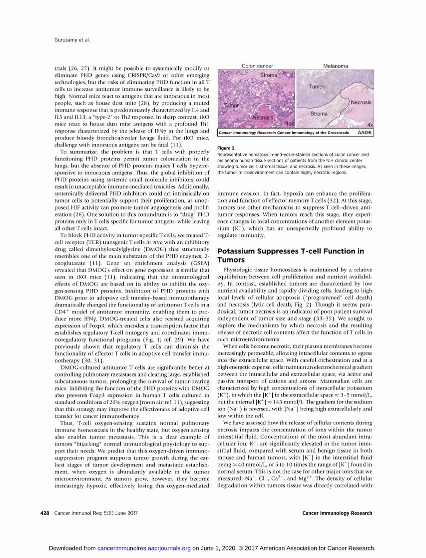
Physiologic tissue homeostasis is maintained by a relativeequilibrium between cell proliferation and nutrient availabil-ity. In contrast, established tumors are characterized by lownutrient availability and rapidly dividing cells, leading to highlocal levels of cellular apoptosis ("programmed" cell death)and necrosis (lytic cell death; Fig. 2). Though it seems para-doxical, tumor necrosis is an indicator of poor patient survivalindependent of tumor size and stage (33–35). We sought toexplore the mechanisms by which necrosis and the resultingrelease of necrotic cell contents affect the function of T cells insuch microenvironments.
When cells become necrotic, their plasma membranes becomeincreasingly permeable, allowing intracellular contents to egressinto the extracellular space. With careful orchestration and at ahigh energetic expense, cellsmaintain an electrochemical gradientbetween the intracellular and extracellular space, via active andpassive transport of cations and anions. Mammalian cells arecharacterized by high concentrations of intracellular potassium(Kþ), in which the [Kþ] in the extracellular space � 3–5 mmol/L,but the internal [Kþ]� 145mmol/L. The gradient for the sodiumion (Naþ) is reversed, with [Naþ] being high extracellularly andlow within the cell.
We have assessed how the release of cellular contents duringnecrosis impacts the concentration of ions within the tumorinterstitial fluid. Concentrations of the most abundant intra-cellular ion, Kþ, are significantly elevated in the tumor inter-stitial fluid, compared with serum and benign tissue in bothmouse and human tumors, with [Kþ] in the interstitial fluidbeing� 40 mmol/L, or 5 to 10 times the range of [Kþ] found innormal serum. This is not the case for other major ions that wemeasured: Naþ, Cl�, Ca2þ, and Mg2þ. The density of cellulardegradation within tumors tissue was directly correlated with
10x
Colon cancer
Tumor
Necrosis
Stroma
4x
Melanoma
Necrosis
Stroma
Tumor
Figure 2.
Representative hematoxylin-and-eosin-stained sections of colon cancer andmelanoma human tissue sections of patients from the NIH clinical centershowing tumor cells, stromal tissue, and necrosis. As seen in these images,the tumor microenvironment can contain highly necrotic regions.
Gurusamy et al.
Cancer Immunol Res; 5(6) June 2017 Cancer Immunology Research428
on June 1, 2020. © 2017 American Association for Cancer Research. cancerimmunolres.aacrjournals.org Downloaded from

the [Kþ] of the interstitial fluid. Specific experimental induc-tion of either apoptosis or necrosis similarly led to the releaseof intracellular Kþ into the extracellular space (36–38). Sim-ilarly, rapid chemotherapy-induced lysis of hematologicmalignancies in humans leads to a "tumor lysis syndrome"with associated elevations in serum potassium (39). "Solidtumor lysis syndrome" has also been noted to produce potas-sium release, with an appreciable elevation of the ion inpatient serum (40).
Given that mouse and human tumors contain dense areas ofcell necrosis that produce elevations in [Kþ] within the extra-cellular space, we investigated how this increase in [Kþ]in tumor interstitial fluid impacts T-cell function. The addi-tional 40 mmol/L of Kþ acutely inhibited TCR-induced pro-duction of effector cytokines by T cells. This Kþ-inducedimmunosuppression is nonredundant to the coinhibitory sig-naling of CTLA-4 and PD-1 receptor ligation. A broader char-acterization of this phenomenon via whole transcriptomeRNA-sequencing revealed that elevated extracellular [Kþ]broadly represses TCR-induced effector programs. Gene-setenrichment analysis (GSEA) showed that genes induced byNF-kB activation or involved in escape from anergy, the adap-tive immune response, or cytokine pathways are suppressed byhigh extracellular Kþ.
As elevated extracellular [Kþ] acutely suppresses TCR-driventranscriptional events, we explored whether [Kþ] could affectTCR-induced signal transduction pathways. TCR ligation rapidlyinduces signaling cascades that lead to both tyrosine- and serine/threonine phosphorylation, along with influx of divalent ionsCa2þ and Mg2þ. Suppression of any of these signal transductionprocesses can lead to a blunted effector T-cell response. ProximalTCR activation result in a localized cascade of phosphorylationon activating tyrosine residues within a cascade of tyrosinekinases, including Zap70, and leading to activation of the enzymePLCg1, producing store-operated Ca2þ efflux (SOCE) as well asactivation of the MAPK/Erk pathway. Additionally, TCR ligation,most notably when coordinated with CD28 costimulation,induces phosphoinositol-3-kinase (PI3K) activity with subse-quent activation of the serine/threonine kinase Akt and themammalian target of rapamycin (mTOR), which act to induce
protein synthesis, anabolic metabolism, and a program of T-celleffector function via dynamic regulation of the transcriptionfactors HIF1a, c-Myc, Foxo1, and Bach2 among others. We couldnot detect any changes in TCR-induced Ca2þ flux in the presenceof 40 mmol/L isotonic hyperkalemia. Additionally, elevatedKþ did not affect TCR-associated tyrosine phosphorylation ofZap70, Erk1/2, or PLCg-1 (36).
Although TCR-induced Ca2þ flux and tyrosine phosphory-lation are normal in the presence of high Kþ levels, activitywithin the Akt–mTOR cascade is profoundly inhibited. Elevat-ed extracellular [Kþ] suppresses TCR induced Akt phosphory-lation, subsequently silencing phosphorylation of the serine/threonine residues targeted by Akt on mTOR and the ribosomalprotein S6 (36). The activity of TCR-induced cytokine produc-tion and Akt signaling can be restored with pharmacologic orgenetic inactivation of protein phosphatase 2A (PP2A), a ser-ine/threonine phosphatase known to dephosphorylate, andthereby inactivate Akt (Fig. 3; ref. 36).
Mechanistically, we found that Kþ-induced T-cell suppressionis primarily dependent on intracellular [Kþ]. Pharmacologic inter-ventions that lower the intracellular [Kþ], augmenting TCR-induced cytokine production, implicate T-cell–intrinsic ion trans-port as critical to effector functions, antitumor immunity, andimmunotherapy. Endogenous control of ion transport in T cells isa complex and relatively understudied field of investigation. Chiefamong the channels found to have an identified role in T-cellfunction is the channel Kv1.3 (encoded by the gene Kcna3), avoltage-gated potassium channel linked to T-cell lineage spec-ification and antitumor functions (41).
Under specific conditions, local tumor-induced nutrient restric-tions reduce the expression and function of Kv1.3 in locallyinfiltrating T cells, leading to blocked T-cell activation. The func-tion of Kv1.3 channels in tumor-infiltrating T cells from head andneck tumors is reduced by 70% compared with their function inperipheral blood T cells from the same patients (42–45). Com-plete loss of Kcna3 can cause CD4þ T cells to take on properties ofregulatory cells and reduce production of IFNg and IL17 cytokines(46). In T cells isolated from head and neck tumors, low Kv1.3expression is associated with low secretion of the cytotoxic pro-tease granzyme B (47).
Figure 3.
Mechanism of potassium-mediated T-cell immunosuppression and therapeutic interventions to restore the antitumor effector functions of T cells. Tumor cellsundergo necrotic cell death and release intracellular Kþ ions into the tumor interstitial space; T cells in these environments have more Kþ entering the cells. Highintracellular potassium within the cell might affect the protein phosphatase PP2A, resulting in the inhibition of Akt and the suppression of T-cell effector functions.Decreased T-cell functions due to raised intracellular [Kþ] can be reversed through the overexpression of the voltage gated Kþ channel Kv1.3 (marked in red).
Oxygen and Potassium Axis of Tumor Immunosuppression
www.aacrjournals.org Cancer Immunol Res; 5(6) June 2017 429
on June 1, 2020. © 2017 American Association for Cancer Research. cancerimmunolres.aacrjournals.org Downloaded from

When extracellular [Kþ] increased, we measured an accom-panying rise in intracellular [Kþ] within T cells. To counteractthe high [Kþ] coupled with the downregulation of Kv1.3 func-tion that may occur in large hypoxic tumors, we enforced theexpression of Kcna3 in T cells, using a retrovirus. In line withour pharmacologic findings, we found that genetic meansto ectopically overexpress Kv1.3 reduced intracellular Kþ andaugmented effector function. Adoptive transfer of T cells withhigh enforced expression of Kcna3 into mice with B16 mela-noma tumors results in highly phosphorylated Akt and S6kinases and improved release of IFNg in the tumor microen-vironment. Most importantly, enforced expression of Kcna3improved T-cell–mediated clearance of large, establishedtumors (Fig. 3; ref. 36).
PP2A activity suppresses T-cell function in the presence of high[Kþ], but direct exposure of purified PP2A to high [Kþ] had noeffect on its activity. This may indicate the involvement of otherintermediate cell intrinsic molecules. For example, several post-translationalmodifications and endogenous smallmolecules and
metabolites, such as ceramides, bimetallic cations, sphingoids,phenols, and polyamines, affect PP2A's localization, targeting,and activity (48, 49).
These findings may also shed light on prior observations thatchanges in [Kþ] regulate inflammasome activation in macro-phages and can control cellular peptide and phospholipidprocessing (50, 51). More than a decade ago, elegant experi-ments revealed that the phagocytic activity of neutrophils ismediated through activation of proteases by intra-organellepotassium flux (52). However, a comprehensive understandingof how immune cells, including T cells, delicately regulate theirintracellular potassium concentration to tune signaling cas-cades is still lacking.
An Elemental View of Tumor-InducedImmunosuppression
Tumor immunologists often draw byzantine diagrams involv-ingmultiple cytokines,metabolites, and cell types as being potent
Figure 4.
The model illustrates the spatial impact onT cells' antitumor functions by the elementsoxygen (O2) and potassium (Kþ) within thetumor microenvironment. At the time ofinitial tumor growth and metastases,tumors may experience normoxia.However, as tumor cells grow, vasculaturemay be inadequately developed, resultingin regions of insufficient oxygen deliveryand tissue hypoxia, and cellular necrosis,which results in the release of potassiumions (Kþ). The inset shows the diffusiongradient of oxygen and potassium withinthe tumor microenvironment. The tumorcontains a limited number of tumorinfiltrating lymphocytes, and these T cellsare differentially regulated by theirimmediate microenvironment. Withinnormoxic regions, T-cellimmunosuppression occurs through thework of the PHD oxygen-sensing proteinsthrough the downregulation of HIF1a. Tcells immunosuppressed by oxygenare marked blue. Under more hypoxic andhostile necrotic tumor microenvironments,the immunosuppression restricted by theelement oxygen is lifted, and in suchregions, immunosuppression of T cellsoccurs through the effect of anotherelement Kþ, which is a byproduct of tumorcell necrosis. T cells immunosuppressed bypotassium are marked green. Whereasthe effects of Kþ can be counteracted byKv1.3 expression in T cells, the hypoxictumor microenvironment downregulatesthe expression of Kv1.3 in these cells,stranding their effector functions.
Gurusamy et al.
Cancer Immunol Res; 5(6) June 2017 Cancer Immunology Research430
on June 1, 2020. © 2017 American Association for Cancer Research. cancerimmunolres.aacrjournals.org Downloaded from

suppressors of antitumor immune function. Although much ofthis tumor immunobiology is undoubtedly true, we have yet toexamine the influence on immunity by the elements of theperiodic table, which comprise a significant portion of the mam-malian tissues. We are often said to be carbon-based organisms,but carbon makes up 18.5% of a person by weight, whereas65% of any individual is oxygen. Although most elementaloxygen is present in the form of water, fats, carbohydrates,proteins, and nucleic acids, we also breathe in O2, which isessential for cellular respiration. The most abundant cationwithin cells is potassium, which by weight is twice as plentifulin the body as sodium. Both oxygen and potassium play majorroles in maintaining cellular and tissue homeostasis, and ourfindings reveal that tumors exploit these functions to facilitatetheir growth and progression as well as promote distant seedingand metastasis.
Although various immune cells participate in tumor immu-nosurveillance and promote tumor cell arrest, tumor-infiltrat-ing lymphocytes recognizing mutated antigens are likely to bethe final common pathway for the immune eradication oftumor cells (53). It is an unfortunate reality for patients thatT cells often fail, or fall short, in mediating tumor destruction.This can be due to what has come to be known as adaptiveresistance (54), but can also be caused by advantageous muta-tions in any of a variety of genes involved in antigen processingand presentation (55–59), or by defects in pathways involvedin interferon signaling (60) within cancer cells. In addition,localized factors in the tumor microenvironment—such assuppressive immune cells, cytokines, metabolites, nutrients,and even elements—can silence the tumoricidal effector activ-ity of intratumoral T cells, allowing tumors to persist andprogress (9, 61–65).
We have highlighted two novel mechanisms of adaptivetissue-specific immune suppression, but multiple mechanismsof immune suppression occur simultaneously and/or progres-sively within the same tissue, including a myriad of mechan-isms not discussed here. Within the Kþ/O2 immune suppressiveaxis, oxygen-mediated immune suppression promotes seedingof the disseminated tumor cells to distant well-vascularizedtissues like the lungs, but as tumors progress, hypoxic regions
emerge as tumor cell growth exceeds its vascular supply.Although hypoxia has been reported to promote tissue necro-sis, it has also been shown to promote both acute and long-term inhibition of Kv1.3 in T lymphocytes (Fig. 4). Conse-quently, this may render intratumoral T cells less fit in accom-modating the local increase in potassium concentration thatoccurs secondary to tumor necrosis (42).
This brief review is focused substantially on the impact ofoxygen and potassiumdirectly on T cells. However, it is importantto note that a considerable bodyofwork indicates that oxygen andpotassium can also influencemany other immune cells, includingtumor-associatedmacrophages, neutrophils, NK cells, B cells, andmany other cell types (51, 52, 66, 67).
Tumor immunotherapy hasmade great strides, but it is curativein only small fractions of patients who have a limited number oftumor histologies, albeit the list is growing. The immunosup-pressive tumor immune compartment challenges basic and trans-lational scientists to devise strategies to enhance anticancer T-cellfunction, tumor clearance, and subsequent patient survival. Wehope and expect that knowledge of "Immunology of the ele-ments" will lead to increases in the efficacy of immunotherapiesfor patients with cancer.
Disclosure of Potential Conflicts of InterestNo potential conflicts of interest were disclosed.
Authors' ContributionsConception and design: D. Gurusamy, D. Clever, R. Eil, N.P. RestifoWriting, review, and/or revision of the manuscript: D. Gurusamy, D. Clever,R. Eil, N.P. Restifo
AcknowledgmentsThis work was supported by the Intramural Research Program of the Center
for Cancer Research,NCI,NIH.We thank SumanK. Vodnala andPing-Hsien Leefor helpful discussions and JenniferMichalowski for excellent editorial input onthis article. We are grateful to Richard Lee for histologic images of tumors andErina He and Alan Hoofring for illustrations.
Received March 9, 2017; revised March 30, 2017; accepted April 19, 2017;published OnlineFirst May 3, 2017.
References1. Dunn GP, Bruce AT, Ikeda H, Old LJ, Schreiber RD. Cancer immunoe-
diting: from immunosurveillance to tumor escape. Nat Immunol 2002;3:991–8.
2. Prehn RT, Main JM. Immunity to methylcholanthrene-induced sarcomas.J Natl Cancer Inst 1957;18:769–78.
3. Schreiber RD, Old LJ, Smyth MJ. Cancer immunoediting: integratingimmunity's roles in cancer suppression and promotion. Science 2011;331:1565–70.
4. Hanahan D, Weinberg RA. Hallmarks of cancer: the next generation. Cell2011;144:646–74.
5. Massague J, Obenauf AC. Metastatic colonization by circulating tumourcells. Nature 2016;529:298–306.
6. Restifo NP, Dudley ME, Rosenberg SA. Adoptive immunotherapyfor cancer: harnessing the T cell response. Nat Rev Immunol 2012;12:269–81.
7. Rosenberg SA, Restifo NP. Adoptive cell transfer as personalized immu-notherapy for human cancer. Science 2015;348:62–8.
8. Sharma P, Allison JP. The future of immune checkpoint therapy. Science2015;348:56–61.
9. Rabinovich GA, Gabrilovich D, Sotomayor EM. Immunosuppressivestrategies that are mediated by tumor cells. Annu Rev Immunol2007;25:267–96.
10. Joyce JA, Fearon DT. T cell exclusion, immune privilege, and the tumormicroenvironment. Science 2015;348:74–80.
11. Clever D, Roychoudhuri R, Constantinides MG, Askenase MH, SukumarM, Klebanoff CA, et al. Oxygen sensing by T cells establishesan immunologically tolerant metastatic niche. Cell 2016;166:1117–31e14.
12. Bruick RK, McKnight SL. A conserved family of prolyl-4-hydroxylases thatmodify HIF. Science 2001;294:1337–40.
13. Epstein AC, Gleadle JM, McNeill LA, Hewitson KS, O'Rourke J, Mole DR,et al. C. elegans EGL-9 and mammalian homologs define a family ofdioxygenases that regulate HIF by prolyl hydroxylation. Cell 2001;107:43–54.
14. Kaelin WG Jr., Ratcliffe PJ. Oxygen sensing by metazoans: the central roleof the HIF hydroxylase pathway. Mol Cell 2008;30:393–402.
15. Jaakkola P, Mole DR, Tian YM, Wilson MI, Gielbert J, Gaskell SJ, et al.Targeting of HIF-alpha to the von Hippel-Lindau ubiquitylation
Oxygen and Potassium Axis of Tumor Immunosuppression
www.aacrjournals.org Cancer Immunol Res; 5(6) June 2017 431
on June 1, 2020. © 2017 American Association for Cancer Research. cancerimmunolres.aacrjournals.org Downloaded from

complex by O2-regulated prolyl hydroxylation. Science 2001;292:468–72.
16. Xu Y, Chaudhury A, Zhang M, Savoldo B, Metelitsa LS, Rodgers J, et al.Glycolysis determines dichotomous regulation of T cell subsets in hypoxia.J Clin Invest 2016;126:2678–88.
17. Weiss JM, Bilate AM, Gobert M, Ding Y, Curotto de Lafaille MA, ParkhurstCN, et al. Neuropilin 1 is expressed on thymus-derived natural regulatory Tcells, but not mucosa-generated induced Foxp3þ T reg cells. J Exp Med2012;209:1723–42, S1.
18. Bilate AM, Lafaille JJ. Induced CD4þFoxp3þ regulatory T cells in immunetolerance. Annu Rev Immunol 2012;30:733–58.
19. Yadav M, Louvet C, Davini D, Gardner JM, Martinez-Llordella M, Bailey-Bucktrout S, et al. Neuropilin-1 distinguishes natural and inducible reg-ulatory T cells among regulatory T cell subsets in vivo. J Exp Med2012;209:1713–22, S1–19.
20. Rooks MG, Garrett WS. Gut microbiota, metabolites and host immunity.Nat Rev Immunol 2016;16:341–52.
21. Holt PG, Strickland DH, Wikstrom ME, Jahnsen FL. Regulation of immu-nological homeostasis in the respiratory tract. Nat Rev Immunol 2008;8:142–52.
22. Smyth RL, Openshaw PJ. Bronchiolitis. Lancet 2006;368:312–22.23. Fanta CH. Asthma. N Engl J Med 2009;360:1002–14.24. Condon TV, Sawyer RT, Fenton MJ, Riches DW. Lung dendritic cells
at the innate-adaptive immune interface. J Leukoc Biol 2011;90:883–95.
25. Stumbles PA, Thomas JA, Pimm CL, Lee PT, Venaille TJ, Proksch S,et al. Resting respiratory tract dendritic cells preferentially stimulateT helper cell type 2 (Th2) responses and require obligatorycytokine signals for induction of Th1 immunity. J Exp Med 1998;188:2019–31.
26. Warnecke C, GrietheW,Weidemann A, Jurgensen JS,WillamC, BachmannS, et al. Activation of the hypoxia-inducible factor-pathway and stimula-tion of angiogenesis by application of prolyl hydroxylase inhibitors. FASEBJ 2003;17:1186–8.
27. NangakuM, Izuhara Y, TakizawaS, Yamashita T, Fujii-KuriyamaY,OhnedaO, et al. A novel class of prolyl hydroxylase inhibitors induces angiogenesisand exerts organ protection against ischemia. Arterioscler Thromb VascBiol 2007;27:2548–54.
28. Sporik R, Holgate ST, Platts-Mills TA, Cogswell JJ. Exposure to house-dustmite allergen (Der p I) and the development of asthma in childhood. Aprospective study. N Engl J Med 1990;323:502–7.
29. Fontenot JD, Gavin MA, Rudensky AY. Foxp3 programs the developmentand function of CD4þCD25þ regulatory T cells. Nat Immunol 2003;4:330–6.
30. Antony PA, Piccirillo CA, Akpinarli A, Finkelstein SE, Speiss PJ, SurmanDR,et al. CD8þ T cell immunity against a tumor/self-antigen is augmented byCD4þ T helper cells and hindered by naturally occurring T regulatory cells.J Immunol 2005;174:2591–601.
31. Antony PA, Restifo NP. CD4þCD25þ T regulatory cells, immu-notherapy of cancer, and interleukin-2. J Immunother 2005;28:120–8.
32. Xu X, Araki K, Li S, Han JH, Ye L, Tan WG, et al. Autophagy is essential foreffector CD8(þ) T cell survival and memory formation. Nat Immunol2014;15:1152–61.
33. RichardsCH,RoxburghCS,Anderson JH,McKeeRF, Foulis AK,Horgan PG,et al. Prognostic value of tumour necrosis andhost inflammatory responsesin colorectal cancer. Br J Surg 2012;99:287–94.
34. Richards CH, Mohammed Z, Qayyum T, Horgan PG, McMillanDC. The prognostic value of histological tumor necrosis in solidorgan malignant disease: a systematic review. Future Oncol 2011;7:1223–35.
35. Sengupta S, Lohse CM, Leibovich BC, Frank I, Thompson RH,Webster WS,et al. Histologic coagulative tumor necrosis as a prognostic indicator ofrenal cell carcinoma aggressiveness. Cancer 2005;104:511–20.
36. Eil R, Vodnala SK, Clever D, Klebanoff CA, Sukumar M, Pan JH, et al. Ionicimmune suppression within the tumour microenvironment limits T celleffector function. Nature 2016;537:539–43.
37. Parham WA, Mehdirad AA, Biermann KM, Fredman CS. Hyperkalemiarevisited. Tex Heart Inst J 2006;33:40–7.
38. Davidson MB, Thakkar S, Hix JK, Bhandarkar ND, Wong A, Schreiber MJ.Pathophysiology, clinical consequences, and treatment of tumor lysissyndrome. Am J Med 2004;116:546–54.
39. Wilson D, Stewart A, Szwed J, Einhorn LH. Cardiac arrest due to hyperka-lemia following therapy for acute lymphoblastic leukemia. Cancer1977;39:2290–3.
40. Mirrakhimov AE, Ali AM, Khan M, Barbaryan A. Tumor lysis syndrome insolid tumors: an up to date review of the literature. Rare Tumors2014;6:5389.
41. Cahalan MD, Chandy KG. The functional network of ion channels in Tlymphocytes. Immunol Rev 2009;231:59–87.
42. Conforti L, Petrovic M, Mohammad D, Lee S, Ma Q, Barone S, et al.Hypoxia regulates expression and activity of Kv1.3 channels in Tlymphocytes: a possible role in T cell proliferation. J Immunol2003;170:695–702.
43. Robbins JR, Lee SM, Filipovich AH, Szigligeti P, Neumeier L, Petrovic M,et al. Hypoxia modulates early events in T cell receptor-mediatedactivation in human T lymphocytes via Kv1.3 channels. J Physiol2005;564:131–43.
44. Szigligeti P, Neumeier L, Duke E, Chougnet C, Takimoto K, Lee SM, et al.Signalling during hypoxia in human T lymphocytes–critical role of the srcprotein tyrosine kinase p56Lck in the O2 sensitivity of Kv1.3 channels. JPhysiol 2006;573:357–70.
45. Chimote AA, Hajdu P, Kottyan LC, Harley JB, Yun Y, Conforti L.Nanovesicle-targeted Kv1.3 knockdown in memory T cells suppressesCD40L expression and memory phenotype. J Autoimmun 2016;69:86–93.
46. Gocke AR, Lebson LA,Grishkan IV,Hu L,NguyenHM,WhartenbyKA, et al.Kv1.3 deletion biases T cells toward an immunoregulatory phenotype andrenders mice resistant to autoimmune encephalomyelitis. J Immunol2012;188:5877–86.
47. Chimote AA, Hajdu P, Sfyris AM, Gleich BN, Wise-Draper T, CasperKA, et al. Kv1.3 channels mark functionally competent CD8þ tumor-infiltrating lymphocytes in head and neck cancer. Cancer Res 2017;77:53–61.
48. Sutter BM, Wu X, Laxman S, Tu BP. Methionine inhibits autophagy andpromotes growth by inducing the SAM-responsive methylation of PP2A.Cell 2013;154:403–15.
49. Voronkov M, Braithwaite SP, Stock JB. Phosphoprotein phosphatase 2A: anovel druggable target for Alzheimer's disease. Future Med Chem 2011;3:821–33.
50. Wolfs JL, Comfurius P, Bekers O, Zwaal RF, Balasubramanian K,Schroit AJ, et al. Direct inhibition of phospholipid scramblingactivity in erythrocytes by potassium ions. Cell Mol Life Sci 2009;66:314–23.
51. Munoz-Planillo R, Kuffa P, Martinez-Colon G, Smith BL, Rajendiran TM,Nunez G. K(þ) efflux is the common trigger of NLRP3 inflammasomeactivation by bacterial toxins and particulate matter. Immunity2013;38:1142–53.
52. Reeves EP, LuH, JacobsHL,MessinaCG,Bolsover S,GabellaG, et al. Killingactivity of neutrophils is mediated through activation of proteases by Kþflux. Nature 2002;416:291–7.
53. Braumuller H,Wieder T, Brenner E, Assmann S, HahnM, AlkhaledM, et al.T-helper-1-cell cytokines drive cancer into senescence. Nature 2013;494:361–5.
54. Restifo NP, Smyth MJ, Snyder A. Acquired resistance to immunotherapyand future challenges. Nat Rev Cancer 2016;16:121–6.
55. Restifo NP, Kawakami Y, Marincola F, Shamamian P, Taggarse A, EsquivelF, et al. Molecular mechanisms used by tumors to escape immune recog-nition: immunogenetherapy and the cell biology of major histocompat-ibility complex class I. J Immunother Emphasis Tumor Immunol1993;14:182–90.
56. Restifo NP, Esquivel F, Kawakami Y, Yewdell JW, Mule JJ, Rosenberg SA,et al. Identification of human cancers deficient in antigen processing. J ExpMed 1993;177:265–72.
57. Restifo NP, Esquivel F, Asher AL, Stotter H, Barth RJ, Bennink JR, et al.Defective presentation of endogenous antigens by a murine sarcoma.Implications for the failure of an anti-tumor immune response. J Immunol1991;147:1453–9.
Gurusamy et al.
Cancer Immunol Res; 5(6) June 2017 Cancer Immunology Research432
on June 1, 2020. © 2017 American Association for Cancer Research. cancerimmunolres.aacrjournals.org Downloaded from

58. Khong HTRestifo NP. Natural selection of tumor variants in thegeneration of "tumor escape" phenotypes. Nat Immunol 2002;3:999–1005.
59. Restifo NP, Marincola FM, Kawakami Y, Taubenberger J, Yannelli JR,Rosenberg SA. Loss of functional beta 2-microglobulin in metastaticmelanomas from five patients receiving immunotherapy. J Natl CancerInst 1996;88:100–8.
60. Zaretsky JM, Garcia-Diaz A, Shin DS, Escuin-Ordinas H, Hugo W, Hu-Lieskovan S, et al. Mutations associated with acquired resistance to PD-1blockade in melanoma. N Engl J Med 2016;375:819–29.
61. Chang CH, Qiu J, O'Sullivan D, Buck MD, Noguchi T, Curtis JD, et al.Metabolic competition in the tumormicroenvironment is a driver of cancerprogression. Cell 2015;162:1229–41.
62. Singer M, Wang C, Cong L, Marjanovic ND, Kowalczyk MS, Zhang H, et al.A distinct gene module for dysfunction uncoupled from activation intumor-infiltrating T cells. Cell 2016;166:1500–11e9.
63. Ho PC, Bihuniak JD, Macintyre AN, Staron M, Liu X, Amezquita R, et al.Phosphoenolpyruvate is a metabolic checkpoint of anti-tumor T cellresponses. Cell 2015;162:1217–28.
64. Chang CH, Curtis JD, Maggi LB Jr., Faubert B, Villarino AV, O'Sullivan D,et al. Posttranscriptional control of T cell effector function by aerobicglycolysis. Cell 2013;153:1239–51.
65. Sitkovsky M, Lukashev D. Regulation of immune cells by local-tissueoxygen tension: HIF1 alpha and adenosine receptors. Nat Rev Immunol2005;5:712–21.
66. Cho SH, Raybuck AL, Stengel K, Wei M, Beck TC, Volanakis E, et al.Germinal centre hypoxia and regulation of antibody qualities by a hypoxiaresponse system. Nature 2016;537:234–8.
67. Doedens AL, Stockmann C, Rubinstein MP, Liao D, Zhang N, DeNardoDG, et al. Macrophage expression of hypoxia-inducible factor-1 alphasuppresses T-cell function and promotes tumor progression. Cancer Res2010;70:7465–75.
www.aacrjournals.org Cancer Immunol Res; 5(6) June 2017 433
Oxygen and Potassium Axis of Tumor Immunosuppression
on June 1, 2020. © 2017 American Association for Cancer Research. cancerimmunolres.aacrjournals.org Downloaded from
2017;5:426-433. Cancer Immunol Res Devikala Gurusamy, David Clever, Robert Eil, et al. MicroenvironmentNovel ''Elements'' of Immune Suppression within the Tumor
Updated version
http://cancerimmunolres.aacrjournals.org/content/5/6/426
Access the most recent version of this article at:
Cited articles
http://cancerimmunolres.aacrjournals.org/content/5/6/426.full#ref-list-1
This article cites 67 articles, 17 of which you can access for free at:
Citing articles
http://cancerimmunolres.aacrjournals.org/content/5/6/426.full#related-urls
This article has been cited by 3 HighWire-hosted articles. Access the articles at:
E-mail alerts related to this article or journal.Sign up to receive free email-alerts
Subscriptions
Reprints and
To order reprints of this article or to subscribe to the journal, contact the AACR Publications Department
Permissions
Rightslink site. Click on "Request Permissions" which will take you to the Copyright Clearance Center's (CCC)
.http://cancerimmunolres.aacrjournals.org/content/5/6/426To request permission to re-use all or part of this article, use this link
on June 1, 2020. © 2017 American Association for Cancer Research. cancerimmunolres.aacrjournals.org Downloaded from