nov-dec 2017 | vol. 3 | issue 6 €¦ · - human pelvic anatomy trough the laparoscope, with live...
TRANSCRIPT
+
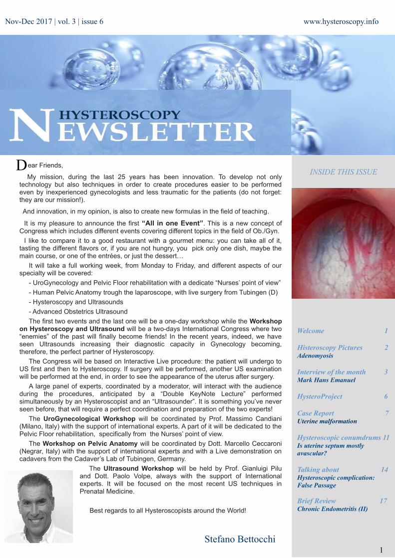
ear Friends,
My mission, during the last 25 years has been innovation. To develop not only technology but also techniques in order to create procedures easier to be performed even by inexperienced gynecologists and less traumatic for the patients (do not forget: they are our mission!).
And innovation, in my opinion, is also to create new formulas in the field of teaching.
It is my pleasure to announce the first “All in one Event”. This is a new concept of Congress which includes different events covering different topics in the field of Ob./Gyn.
I like to compare it to a good restaurant with a gourmet menu: you can take all of it, tasting the different flavors or, if you are not hungry, you pick only one dish, maybe the main course, or one of the entrées, or just the dessert…
It will take a full working week, from Monday to Friday, and different aspects of our specialty will be covered:
- UroGynecology and Pelvic Floor rehabilitation with a dedicate “Nurses’ point of view”
- Human Pelvic Anatomy trough the laparoscope, with live surgery from Tubingen (D)
- Hysteroscopy and Ultrasounds
- Advanced Obstetrics Ultrasound
The first two events and the last one will be a one-day workshop while the Workshopon Hysteroscopy and Ultrasound will be a two-days International Congress where two “enemies” of the past will finally become friends! In the recent years, indeed, we have seen Ultrasounds increasing their diagnostic capacity in Gynecology becoming, therefore, the perfect partner of Hysteroscopy.
The Congress will be based on Interactive Live procedure: the patient will undergo to US first and then to Hysteroscopy. If surgery will be performed, another US examination will be performed at the end, in order to see the appearance of the uterus after surgery.
A large panel of experts, coordinated by a moderator, will interact with the audience during the procedures, anticipated by a “Double KeyNote Lecture” performed simultaneously by an Hysteroscopist and an “Ultrasounder”. It is something you’ve never seen before, that will require a perfect coordination and preparation of the two experts!
The UroGynecological Workshop will be coordinated by Prof. Massimo Candiani (Milano, Italy) with the support of international experts. A part of it will be dedicated to the Pelvic Floor rehabilitation, specifically from the Nurses’ point of view.
The Workshop on Pelvic Anatomy will be coordinated by Dott. Marcello Ceccaroni (Negrar, Italy) with the support of international experts and with a Live demonstration on cadavers from the Cadaver’s Lab of Tubingen, Germany.
The Ultrasound Workshop will be held by Prof. Gianluigi Pilu and Dott. Paolo Volpe, always with the support of International experts. It will be focused on the most recent US techniques in Prenatal Medicine.
Best regards to all Hysteroscopists around the World!
Welcome 1
Histeroscopy Pictures 2Adenomyosis
Interview of the month 3Mark Hans Emanuel
HysteroProject 6
Case Report 7Uterine malformation
Hysteroscopic conumdrums 11Is uterine septum mostlyavascular?
Talking about 14 Hysteroscopic complication:False Passage
Brief Review 17Chronic Endometritis (II)
www.hysteroscopy.info
1
INSIDE THIS ISSUED
Stefano Bettocchi
Nov-Dec 2017 | vol. 3 | issue 6
HYSTEROSCOPY
PICTURES
www.hysteroscopy.info
2
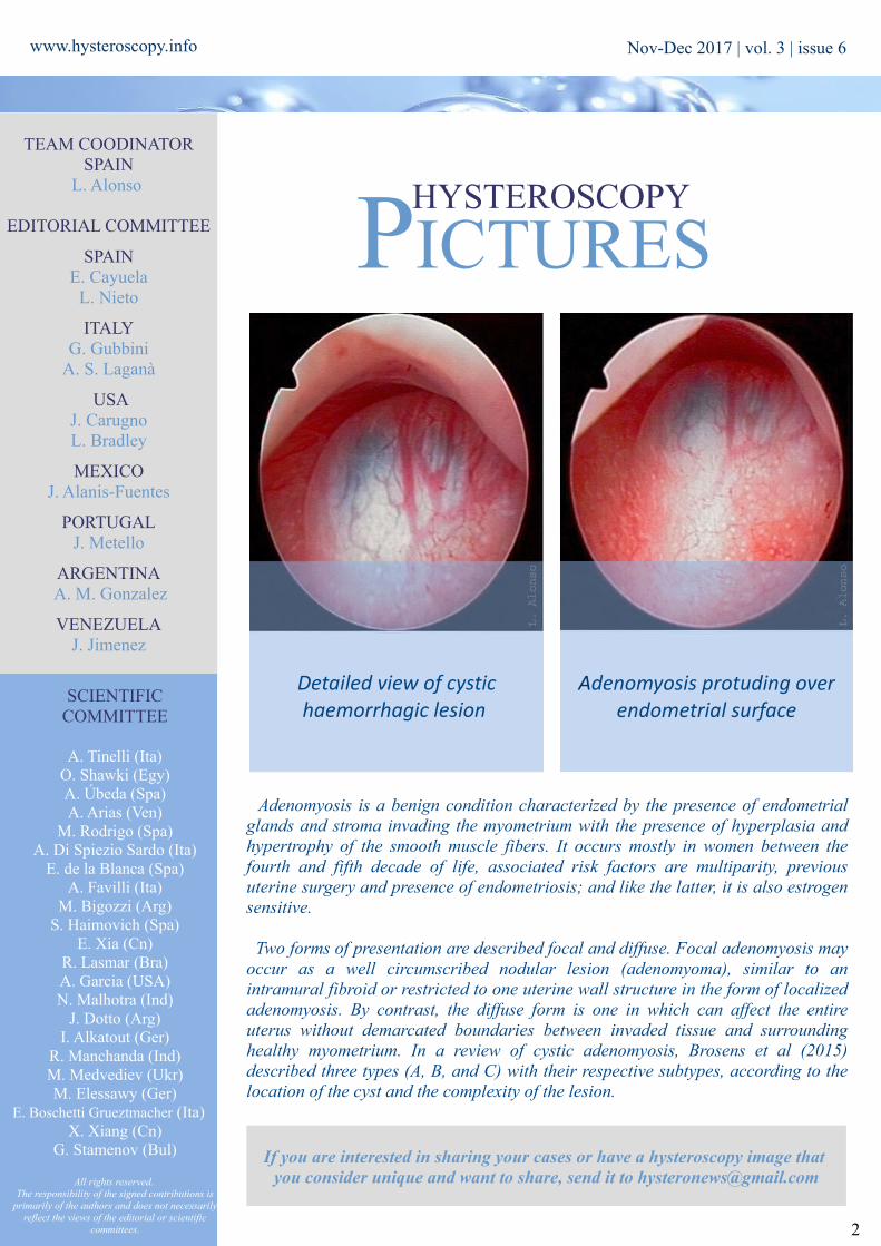
Adenomyosis is a benign condition characterized by the presence of endometrial glands and stroma invading the myometrium with the presence of hyperplasia and hypertrophy of the smooth muscle fibers. It occurs mostly in women between the fourth and fifth decade of life, associated risk factors are multiparity, previous uterine surgery and presence of endometriosis; and like the latter, it is also estrogen sensitive.
Two forms of presentation are described focal and diffuse. Focal adenomyosis may occur as a well circumscribed nodular lesion (adenomyoma), similar to an intramural fibroid or restricted to one uterine wall structure in the form of localized adenomyosis. By contrast, the diffuse form is one in which can affect the entire uterus without demarcated boundaries between invaded tissue and surrounding healthy myometrium. In a review of cystic adenomyosis, Brosens et al (2015) described three types (A, B, and C) with their respective subtypes, according to the location of the cyst and the complexity of the lesion.
If you are interested in sharing your cases or have a hysteroscopy image that you consider unique and want to share, send it to [email protected]
Superficial vaginal endometriotic implant
Adenomyosis protuding over endometrial surface
TEAM COODINATORSPAIN
L. Alonso
EDITORIAL COMMITTEE
SPAINE. Cayuela
L. Nieto
ITALYG. Gubbini
A. S. Laganà
USAJ. CarugnoL. Bradley
MEXICOJ. Alanis-Fuentes
PORTUGALJ. Metello
ARGENTINAA. M. Gonzalez
VENEZUELAJ. Jimenez
SCIENTIFIC COMMITTEE
A. Tinelli (Ita)O. Shawki (Egy)A. Úbeda (Spa)A. Arias (Ven)
M. Rodrigo (Spa)A. Di Spiezio Sardo (Ita)
E. de la Blanca (Spa)A. Favilli (Ita)
M. Bigozzi (Arg)S. Haimovich (Spa)
E. Xia (Cn)R. Lasmar (Bra)A. Garcia (USA)N. Malhotra (Ind)
J. Dotto (Arg)I. Alkatout (Ger)
R. Manchanda (Ind)M. Medvediev (Ukr)M. Elessawy (Ger)
E. Boschetti Grueztmacher (Ita)X. Xiang (Cn)
G. Stamenov (Bul)
All rights reserved. The responsibility of the signed contributions is
primarily of the authors and does not necessarily reflect the views of the editorial or scientific
committees.
Detailed view of cystic haemorrhagic lesion
Nov-Dec 2017 | vol. 3 | issue 6

www.hysteroscopy.info
Mark Hans Emanuel
Professor (dept. gynaecology and
reproductive medicine) at the University Medical
Center Utrecht and at the University Hospital Ghent
Belgium
INTERVIEW WITH... Prof. Mark Hans Emanuel. A recognized gentleman of the hysteroscopy world. He is always looking for a way to improve this technique with great creativity and a very inquiring mind
The amount of hysteroscopic procedures is increasing worldwide. In your opinion, what can be done to promote hysteroscopy?
The best promotion for hysteroscopy is the stimulation of industries/start-ups to further improve techniques that enable us to treat more and more patient in an office setting for an increasing number of abnormalities. Gynecologist should be in the lead in this cooperative process with engineers. Due to such recent technical improvements procedures have become less painful, faster to perform and more effective. Therefore ambulatory hysteroscopy is offered more often by the gynaecologistst and more accepted by patients. An increasing number of patients do not want to be treated in operating rooms with general or regional anaeshesia, they prefer conscious sedation, local anaesthesia or no anaesthesia at all in an office setting. Furthermore, such a shift safes tremendous amount of time and money.
In 2009, the launch of the Truclear system was announced. How was this innovative device born?
Actually the first launch was in 2006 before it was given the name Truclear. The first patents date from the late nineties already. At that time the only acceptable techniques for operative hysteroscopy were the use of conventional forceps and scissors and monopolar resectoscopy. Both were difficult techniques to learn with a rather long learning curve. The removal of tissue was cumbersome, often leading to bad visualization and perforation or fluid overload with non-physiologic fluids were not uncommon. If the gynaecologist were not extremely experienced, failures were frequent and the techniques never became popular in the hands of the average gynaecologist. Less than 5% of gynaecoloigsts were performing resectoscopy. So there was an immense need for alternatives. In the search for other ways of removing tissue out of the human body under direct visualization, I started to talk with and observe colleagues in other fields of medicine. Often we do have similarities in practical problems and solutions. To be honest resectoscopy started in urology and was never designed as a technique for gynaecologists.
3
”Gynecologists should be in the lead in this cooperative process with engineers”
Intrauterine morcellator patent images
Nov-Dec 2017 | vol. 3 | issue 6

4
www.hysteroscopy.info
Tissue removal, which was tiring for the surgeon and more complicated and in the small uterine cavity, compared to the urinary bladder, had to be solved. A non-electrical technique based on saline distention, using conventional cutting and aspiration was the most logical option. Besides, for the optimal visualization and fluid management my personal opinion was that continuous flow was the best. A full range of sizes of scopes for use in the office and in the operating room with different types of surgical blades for different types of tissue completed the new technology. Therefore the Truclear system still has all these options.
Hysterosalpingo-foam sonography (HyFoSy) as a new technique to check tubal patency. How would you explain this technique to our readers?
When we introduced Gel Instillation Sonography (GIS) as an alternative for Saline Infusion Sonography (SIS) (easier to perform and more convenient for patient and examiner) to improve the observation of the uterine cavity and endometrial lining, colleagues asked us whether it was possible to observe the gel passing through the tube. The gel is too viscous and a very thin black line is difficult to distinguish. Therefore we had to dilute the gel to make it less viscous and shift the contrast from echolucent to echodense (from black to white). Creating Foam during the process of diluting was the most pragmatic solution and a new technique was born. The most common tubal patency check is still Hysterososalpingography (HSG). HSG has some remarkable disadvantages: the need for X-ray, re-scheduling and referral to a radiology department, it is painful and expensive. HyFoSy can be performed with a standard vaginal ultrasound probe, it is less painful (Dreyer K et al. Hysterosalpingo-foam sonography, a less painful procedure for tubal patency testing during fertility workup compared with (serial) hysterosalpingography: a randomized controlled trial. Fertil Steril. 2014 Sep;102(3):821-5) and cheaper. The technique is very easy to learn for those who have basic experience in transvaginal ulytrasound scanning. The discordance between the results of HSG and HyFoSy is low and usually approximately 5%. After a vaginal examination with a speculum and visualizing the cervix a small IUI-like catheter is placed intracervically (a balloon catheter is typically not necessary but can be used if desired). A few milliliters of foam is injected through the catheter and the flow of the foam is observed on the monitor in a standard longitudinal and transversal plane. In our first report about HyFoSy a pregnancy rate of 19% was found after three months. (Emanuel MH et al. First experiences with hysterosalpingo-foam sonography (HyFoSy) for office tubal patency testing. Hum Reprod. 2012 Jan;27(1):114-7.)
You're an expert in Asherman's syndrome. Do you think that this surgery should be centralized?
Last year we saw approximately 150 new patients with hysteroscopically diagnosed intra-uterine adhaesions (IUAs). All patients had serious menstrual disorders like amenorrhea or hypomenrohea. Almost all patients became symptomatic after a pregnancy related intra-uterine procedure for retained products of conception (RPOC) after a miscarriage or delivery. We see an increasing number of RPOC that need curettage or (preferably) hysteroscopic treatment. A cohort of 638 patients that were treated for Asherman Syndrome (AS) between 2003 and 2013 are systematically followed up in a prospective study. All patients were treated hysteroscopically with or without fluoroscopic guidance, all had an IUD without copper or Levonorgestrel as adhesion barrier for 6 weeks and all patients had a control hysteroscopy 2 months after initial surgery.
”I think that it is important to meet younger and older colleagues with different levels of experience to exchange opinions,
strategies and tips and tricks of daily practices”
Nov-Dec 2017 | vol. 3 | issue 6

www.hysteroscopy.info
5
The results of this treatment were reported (Hanstede et al. Results of centralized Asherman surgery, 2003-2013. Fertil Steril. 2015 Dec;104(6):1561-8). In 95% of women with Asherman syndrome, a healthy uterine cavity was restored with hysteroscopic adhesiolysis, in 1-3 sessions, with a 28.7% recurrence rate of spontaneous IUAs. Without centralization it would have never been possible to achieve such encouraging results and to collect these data. Yes, I am a very strong advocator of centralization of AS surgery; without centralization it would not have been possible to collect such data and to perform prospective trials. We are currently working on the reproductive results of the largest cohort of AS patients ever described. Prelimenary data show that the baby take home rate in this group is approximately 70%. These patients are definitively at a higher risk for postpartum problems like retained placenta, haemorrhage and puerperal RPOC. Another problem is that we are confronted with is the high number of spontaneous recurrences. Adjuvant hormonal medication is often prescribed to stimulate endometrial growth, however we recently finished a randomised controlled trial in which we the effect on recurrence of adhaesions wil be evaluated.
What is your opinion about the use of anti-adhesion barrier gels following operative hysteroscopy?
In general I believe that the risk for intra-uterine adhaesions after proper and well performed hysteroscopic surgery, either conventional, resectoscopic or with tissue removal systems (morcellation) is very low. Classical high risk procedures as the removal of opposing submucous myomas should be prevented. Adhaesion prevention is most needed after procedures for RPOC to prevent AS. Until now there is insufficient evidence to determine the best adhesion barrier is and how it should be used. Further trials are needed to find the optimal strategy to prevent adhaesions.
Do you have any advice for the young physician who is starting out in the world of gynecologic minimally invasive surgery?
Training, training, training. Courses are organized for different types of surgery and for different levels of experience. Many procedures can be trained by computer simulation nowadays. I think that it is important to meet younger and older colleagues with different levels of experience to exchange opinions, strategies and tips and tricks of daily practices. Furthermore (international) meetings are also the place to hear about new technologies and to meet representatives of the industries.
Nov-Dec 2017 | vol. 3 | issue 6

www.hysteroscopy.info
6
ProjectsHY
ST
ER
OHysteroscopy Newsletter New App
We're proud to announce that we've created a new APP that will be available soon on iOS and android . All the information
about hysteroscopy available at your fingertips. Stay tuned!!
Coordinators: L. Nieto, J. Carugno, L. ALonso
Nov-Dec 2017 | vol. 3 | issue 6

www.hysteroscopy.info
77
A common challenge in Hysteroscopy is the therapeutic approach to uterine malformations.
Currently, in many countries of North Europe, for example, Germany, the gold standard for the diagnosis of uterine anomalies is laparoscopy combined hysteroscopy, although many noninvasive methods are available such as two-three dimensional ultrasound, hysterosalpingography or magnetic resonance imaging (MRI).
The Literature estimates that about 5% of women in the general population have a type of deviation from normal anatomy, especially patient with recurrent pregnancy losses. For this reason, the noninvasive techniques for the diagnosis (office hysteroscopy and two or three-dimensional ultrasound) and minimally invasive surgery for the treatment of this condition are very important.
In my experience, certainly the introduction of new surgical instruments like the mini resectoscope (Gubbini’s System) has increased the surgical approach with very low cost and very fast and safe surgical outcomes.
In the office Hysteroscopy Center of Charite’, University of Berlin, uterine malformations are treated only with the Gubbini’s system using monopolar energy. Without dilation of the cervix, it preserves the anatomy of the uterus better, especially the anatomy of uterine anomalies. Moreover, the mini resectoscope is very easy to use (the diameter is 5 mm) in a small cavity like T-shaped uterus. Compared to the 26 Fr resectoscope, the Gubbini system have the same accessories but smaller diameter, as a loop, a needle and other that they could be changed depending on the surgical needs.
Our last case of T-shaped uterus treated with the Gubbini’s System was in a 34 year old healthy patient, with the unfortunate history of 5 miscarriages at 5/6 weeks gestation.
CASE REPORTUterine Malformation Dr. Eleonora Boschetti Grueztmacher. Head of Hysteroscopy Centre.
Charité, University of Berlin
Nov-Dec 2017 | vol. 3 | issue 6

www.hysteroscopy.info
8
T-shaped Uterus is an abnormal shape of the uterine cavity associated with infertility.
For the systematic categorization of anomalies of the ESHRE and ESGE classification system the T-Shaped Uterus is in Class U1. It is defined as any uterus having normal uterine outline but with an abnormal shape of the uterine cavity excluding septa.
Class U1 is further subdivided into three categories:
a- Class U1a or T- Shaped uterus, having normal correlation 2/3 uterine corpus and 1/3 cervix,and characterized by a narrow uterine cavity due to thickened lateral walls (giving to it thecharacteristic T-shaped).
b- Class U1b or uterus infantile, having an inverse correlation 1/3 uterine body and 2/3 cervix,and characterized also by a narrow uterine cavity but without lateral wall thickening.
c- Class U1c or others, including all minor deformities of the uterine cavity and incorporatingalso those with an inner indentation at the fundal midline level of <50% of the uterine wallthickness.
The aim of the surgery for every uterine morphologic abnormality is to create a normal shape uterine anatomy. The concept of the surgical approach with the Gubbini’s System (monopolar or bipolar) introducing at first, the use of the mini loop to cut the fibrotic lateral wall and after introducing the 5 mm needle to cut all the walls.
Surgical Technique
No cervical preparation or antibiotic therapy are needed before the metroplasty. Vaginoscopic approach without dilatation of the cervical canal is utilized. It is necessary, before starting with the surgical technique to measure the fundal myometrium thickness and the lateral wall thickness near the tubal ostium and near the isthmus in order to understand the classification of anomalies and in order to understand the surgical approach.
Firstly, the resectoscopy with the mini loop is used on the right and left lateral wall near the tubal ostium. A second step is cutting with the 5 Fr needle of the Gubbini System the anterior, posterior and lateral wall following the line is from the tubal ostium to the isthmus uteri. The depth of the incision is no more 3 mm in all the uterine wall.
Before operation: Tubal ostium are impossible to visualize. Without dilatation, the shape of anomalie is preserved.
Nov-Dec 2017 | vol. 3 | issue 6

www.hysteroscopy.info
9
Summary:
- The diagnostic investigation of uterine abnormalities was with two/three dimensional ultrasound combined with the office hysteroscopy.
- The Surgical approach was with the Gubbini’s System in monopolar use and we used the mini loop and mini needle.
- No general anesthesia but only a paracervical block was used with levobupivacaine HCL 100 mg.
- After 4 hours the patient was resigned from the hospital without bleeding and pain.
- A second look hysteroscopy is usually performed after 40 days or two menstrual bleeding cycles in
- order to decrease the intrauterine adhesions after the surgery and to evaluate the result of surgery.
Mini Needle on the lateral wallResection with the mini loop
Cutting the posterior wall Panoramic view
Nov-Dec 2017 | vol. 3 | issue 6

www.hysteroscopy.info
10
The Accardi Harpoon complements the Advanced Hysteroscopy Instruments with their ergonomic, lightweight handle, special shaped rings and finger rest.
The rotating mechanism allows an optimal position of the jaw while the integrated automatic closing system facilitates grasping by the use of a spring system.
The lotus-effect of the surface enables an increase of lifetime.
It has been developed due to the needs of hysteroscopic surgeons by Dr. Accardi. The instrument facilitates catching big polyps, myoma fragments and biopsies. Easier grasping and holding of tissue let the daily work of the
surgeon become more secure and faster.
https://www.rz-medizintechnik.com/
DEVICESHYSTEROSCOPY
Nov-Dec 2017 | vol. 3 | issue 6

www.hysteroscopy.info
11
Loo
k fo
r us
: hys
tero
scop
y gr
oup
in L
inke
d In
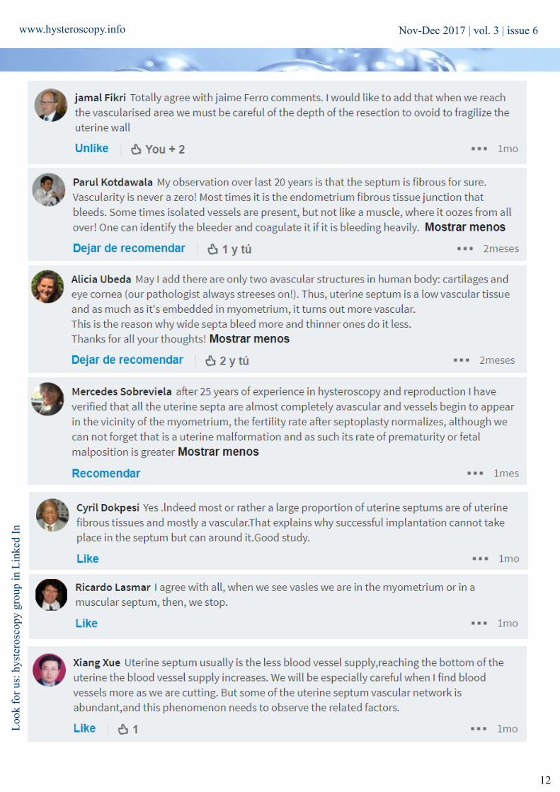
Hysteroscopy ConundrumsIs the uterine septum "mostly" avascular?
We can read in different informative media that the tissue of a uterine septum tends to be fibrous and mostly avascular it does not have the same rich blood supply as the rest of the uterus. Is uterine septum "mostly" avascular? What do you think about? what's your experience?
https://drive.google.com/file/d/0B9_0fZ2JnKsOMmI5TXVZSWcxT1E/view?usp=sharing
I hope you find it interesting
Nov-Dec 2017 | vol. 3 | issue 6
www.hysteroscopy.info
12
Loo
k fo
r us
: hys
tero
scop
y gr
oup
in L
inke
d In
Nov-Dec 2017 | vol. 3 | issue 6

www.hysteroscopy.info
13
Chirugia isteroscopica
oggi. Tecnologia e realtà
Roberto Liguori Giovanni Battista
This volume presents techniques, indications, outcomes and
complications of hysteroscopic surgery.
Authors and Contributors are among the leading Italian experts in the
field.
The discussion of every topics is updated and represents the best
possible synthesis of the personal experience of authors and obtained
from Evidence Based Medicine.
The aim of the volume is to be a useful reference tool not only for colleagues who already perform
hysteroscopy but also for those who desire to start incorporating
hysteroscopy in their practice
Answer to the previous issue:Endometrial Cancer
Sometimes, when performing hysteroscopy, it is important to pay attention to every corner of the uterus, as Vasari stated «cerca trova», «he who
seeks finds»
WHAT'S YOUR DIAGNOSIS?
Hysteroscopy Newsletter
Nov-Dec 2017 | vol. 3 | issue 6

14
www.hysteroscopy.info
Brief Review Hysteroscopy complication: false passage
Dr. Amal Drizi; Dr. Adel Sedrati Consultants in obstetrics and Gynecology. Algeria.
Hysteroscopy is an endoscopic procedure that enables to evaluate the cervix and the uterine cavity, as well as the vagina through vaginoscopy. It also allows to perform intra-uterine surgical procedures. It is a minimally invasive gynecological procedure, yet, not risk-free. In fact, many complications may occur during or after hysteroscopy, hopefully not frequently.
One of these complications is the false passage. It is a rare complication encountered during diagnostic or operative hysteroscopy. How could we define it? How does it occur? What are the consequences? What are the alarm signs? And, how should it be managed?
Definition
During hysteroscopy, the telescope is guided inside the cervical canal then into the uterine cavity. This is the “right passage”. Sometimes, there are difficulties to get through the right canal, which might lead to forcing the passage and “digging a tunnel” in the cervix, or in the uterine wall.
Therefore, while misguidedly inserting the scope, or even dilators, a false passage could be created by tunneling a new passageway in the cervix wall, or the uterus, between endometrium and myometrium or in the myometrium . (Pictures 1 - 2).
Risk factors
Any condition that obstructs the cervical canal could lead to a false passage, because then, the operator might use force to overcome the resistance.
This could be anything from a tight cervix to a cervical stenosis, a deviated canal or an acute angulation of the uterus (Extreme anteversion or retroversion on the uterus). In these cases, the risk of making a false passage is higher, especially if the operator lacks experience and tends to use force.
This could also happen during an operative hysteroscopy, while forcefully performing cervical dilation before insertion of the resectoscope. Surgical treatment of complex synechiae or severe Asherman’s syndrome is another high risk condition for the creation of false passage.
Consequences
A false passage increases absorption of the distending medium, especially when tunneled inside the myometrium , opening bigger vascular channels. This could lead to high fluid deficit and major complications, depending on the medium used and the length of surgery after identification (or not) of the problem.
Moreover, if unrecognized, inserting the scope further could cause uterine perforation with all the complications related to it, such us bowel injury or copious bleeding.
Nov-Dec 2017 | vol. 3 | issue 6
Picture 1
Picture 2

For these reasons, diagnosing a false passage during hysteroscopy could lead, depending on the circumstances, to terminate the procedure. Furthermore, a false passage could also lead to obstetrical complications during a future pregnancy. For all these reasons, it is very important to recognize a false passage and most importantly, to prevent it.
Diagnosis
The major clue that should make a gynecologist suspect the creation of a false passage is the unusual appearance of the uterine cavity. The tubal ostia cannot be visualized. And when the passage is created between myometrium and endometrium, we can see this latter floating in the unusual uterine cavity (Picture3 ; video). The surface of the muscular fibers of the myometrium are clearly visualized. (picture 4; video). When removing the hysteroscope, we can have a panoramic view over the newly created cervical canal. (picture5).
Management
First, the hysteroscope should be slowly removed, so as to identify the external os of the cervical canal. The movements should be very gentle and slow, to allow the evaluation of the neoformed canal. Once the “right passage” recognized, it becomes possible to catheterize it reaching the uterine cavity. If the false passage was created during the treatment of complex synechiae, concomitant ultrasound guidance could be useful.
In all cases, pursuing or terminating the procedure depends on the size and location of the defect and the length of surgery while constantly keeping in mind the fluid deficit. In addition to continuous monitoring the patient. The related video is a good illustration of the procedure and the typical images encountered in this case. Yet, the best way to manage this complication is to prevent it.
Prevention
First, before any hysteroscopic procedure, it is recommended to perform a bimanual pelvic exam, as well as an ultrasound exam to assess position and shape of the uterus, including the cervix. If predisposing factors for false passage are identified, this will lead to caution.
Cervical dilation must be performed slowly, progressively and constantly under visualization, or ultrasound guidance in case of complex synechiae or severe Asherman’s syndrome. The use of small diameter instruments is of a great help in preventing these complications, since it enables inserting the hysteroscope without the need of cervical dilation in most cases. If difficulties are encountered, a single tooth tenaculum could be used to correct the uterine angulation.
Conclusion
False passage is a rare complication encountered during diagnostic and operative hysteroscopy. It is very important to create awareness of risk factors and the means of prevention, as well as its management when it occurs in order to minimize the complications.
15
www.hysteroscopy.infoNov-Dec 2017 | vol. 3 | issue 6
Picture 3 Picture 4 Picture 5

www.hysteroscopy.info
16
Hysteroscopy has the advantages of quick recovery, early return to normal activities, reduced hospital stay and increased satisfactory
for the patient
Abnormal uterine bleeding is the most common gynecological problem comprising more than 30-50% of gynecological OPD
patientshttp://dx.doi.org/10.18203/2320-1770.ijrcog20174653
DID YOU KNOW...?
Nov-Dec 2017 | vol. 3 | issue 6
www.twitter.com/hysteronews
HYSTEROscopy group
Hysteroscopy newsletter
Hysteroscopy newsletter
www.facebook.com/hysteronews
17
www.hysteroscopy.info
17
CE and reproductive success
The implantation process consists of a physiological process of inflammation involving mediators of inflammation such as leukocytes, cytokines, chemokins and other endometrial factors. All these cells and their mediators play a fundamental role in the regulation of the immunoresponse and growth of the trophoblast. The presence of CE can alter the receptivity of the endometrium creating an inadequate microenvironment for normal implantation.
In fact, Kitaya K et al has observed that the endometrium of one third of infertile patients presenting with CE expresses high levels of estrogen receptors, progesterone and Ki-67 nuclear marker cell proliferation in cells of the epithelium as well as in the stroma, and the expression of anti-apoptosis genes such as BCL2 and BAX is increased, all of which are changes representing a proliferative phenotype of the endometrium even in the secretory phase. On the contrary, other genes of local inflammatory response associated with decreased embryonic receptivity like IL11 and CCL4, preventing adequate implantation.
This increase in expression levels of estrogen and progesterone receptors has also been confirmed by the Di Wu et al, who postulates that CE modifies stromal cells by altering the function of hormonal receptors, which are found overexpressed.
On the other hand, it has been described that CE causes a change in the pattern of uterine contractility in both the periovulatory phase and the middle luteal phase. In the proliferative phase, there is anterograde contractility from the fundus to the cervix that facilitates the flow of the menstrual products; in the periovulatory phase there is predominance of retrograde contraction from the cervix to the fundus, which favors the arrival of the spermatozoa to the fallopian tubes and in luteal phase, there is absence of uterine contractility.
Pinto et al performed a study on a total of 45 women with hysteroscopic diagnosis of CE confirmed by endometrial biopsy and 45 controls without evidence of CE, and observed 3.3 times lower occurrence of retrograde contractility, and an increase in anterograde and retrograde contractility in the middle luteal phase, this abnormal peritalsis induced by the presence of CE could play a role in the etiology of infertility, and explain some of the symptoms in patients with CE such as dysmenorrhea.
CE and implementation failure (IF)
There is no consensus on the definition of implantation failure, according to Coughlan et al in a review published in 2013, it was defined as failure to achieve pregnancy after the transfer of at least 4 good quality embryos in a minimum of three cycles in a woman up to 40 years old.
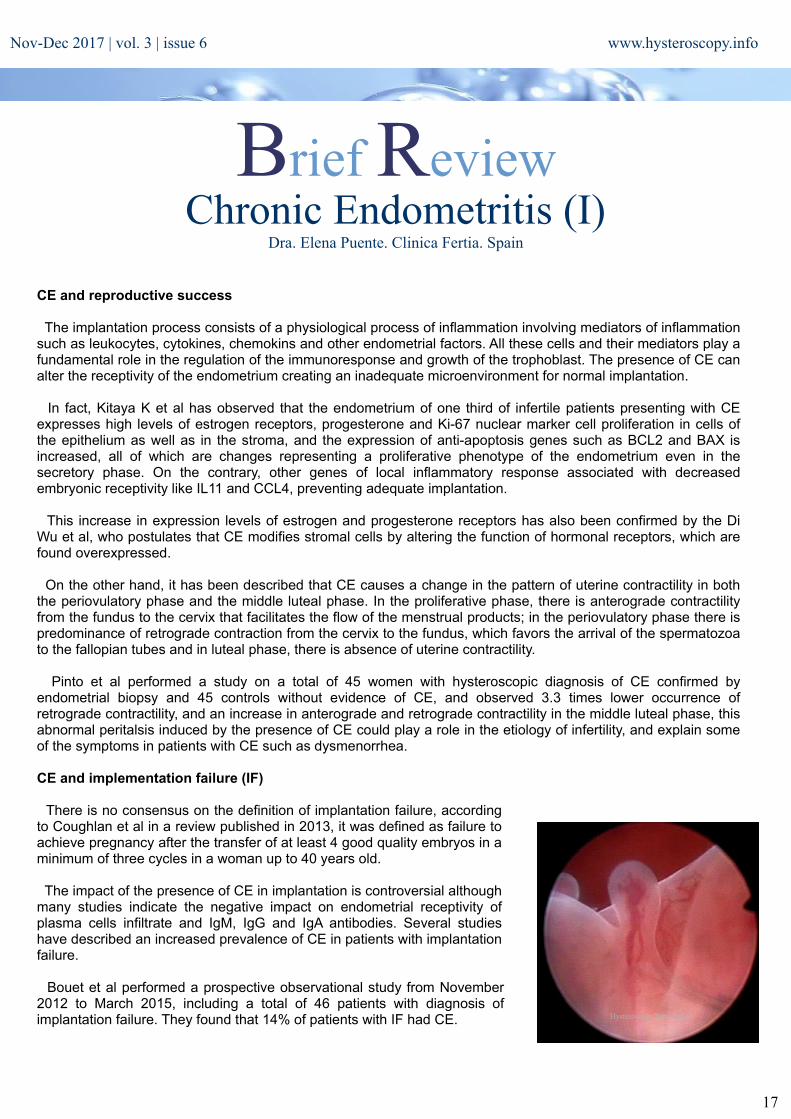
The impact of the presence of CE in implantation is controversial although many studies indicate the negative impact on endometrial receptivity of plasma cells infiltrate and IgM, IgG and IgA antibodies. Several studies have described an increased prevalence of CE in patients with implantation failure.
Bouet et al performed a prospective observational study from November 2012 to March 2015, including a total of 46 patients with diagnosis of implantation failure. They found that 14% of patients with IF had CE. Hysteroscopy Newsletter
Chronic Endometritis (I)Dra. Elena Puente. Clinica Fertia. Spain
Brief Review
Nov-Dec 2017 | vol. 3 | issue 6

They performed an initial study in all patients to rule out other causes of IF and established the diagnosis of CE by immunohistochemistry dilution, defining a positive result in the presence of 5 or more plasma cells in 10 large (x400) fields. They found a diagnostic coincidence of hysteroscopic and histological CE of 80%.
Johnston MacAnanny et al, presented a retrospective study from 2001 to 2007 on 33 women. This study defined IF as absence of pregnancy after two cycles of IVF with embryo transfer of at least one good quality embryo. They performed biopsy and immunohistochemistry considering it negative when plasma cells were absent. This study found a prevalence of 30.3% of CE in patients with IF.
Cicinelli et al performed a retrospective study from January 2009 to June 2012, including a total of 106 patients under the age of 40 with embryo transfer of at least 6 good-quality embryos in three or more previous IVF / ICSI cycles, with one response of 6 mocytes with standard protocol and standard karyotype. They discard women with FSH greater than 10 on day 3, and BMI greater than 30. Endometriosis, Recurrent Pregnancy Loss, autoimmune conditions, antiphospholipid Syndrome, thrombophilia, antispermatozoidal Antibodies. Perform hysteroscopy and endometrial biopsy in the follicular phase of the cycle following hysteroscopy tissue evaluation and culture. They made the diagnosis of CE in hysteroscopy in 66% of cases and CE in tissue samples 57.5%, and positive culture in 45%. It represent double the incidence reported by Mcaannany et al, which can be explained by its strict selection criteria as well as the experience of its group in the histological and hysteroscopic diagnosis of the CE. In addition to a possible selection bias determined by the fact that they are a referral center for patients with suspected CE. Cicinelli observed a diagnostic coincidence between hysteroscopy and histology of 87%.
Both MacAnanny and Cicinelli treated the patients with antibiotics, the first with Doxicillin 100 mg for two weeks, and if the patient continued with a positive after a posttreatment culture, a second cycle with Ciprofloxacin and Metronidazole 500mg twice daily for two more weeks. And Cicinelli with cirpofloxacin 500mg twice a day 10 days for gram negative and amoxicillin-clavulanic acid 1g twice daily for 8 days in gram positive and if the culture persisted positive after the initial treatment, then they repeated the same protocol up to three times. In cases of negative culture, they administer Ceftriaxone 250mg Im single dose, Doxycycline 100 mg twice daily 14 days and metronidazole 500 mg twice daily 14 days. For the MacAnanny group although it improves the clinical embryo after treatment, it remains in the group of CE rates of gestation lower than in the group that does not present CE despite a good response to antibiotic treatment.
Chronic Endometritis and Recurrent Pregnancy Loss
According to ESHRE, recurent pregnancy loss (RPL) is defined as three or more consecutive spontaneous abortions before 20 weeks of gestation, but the American Association of Reproductive Medicine guidelines (ASRM) requires two or more non-consecutive abortions to define RPL. According to the Spanish Fertility Society (SEF), the recommendation is to start the workup of RPL after two pregnancy losses, since the likelihood of a subsequent abortion is similar than after three abortions (24-30% vs. 30-33%).
As in implantation failure, the aberrant endometrial microenvironment resulting from an anomalous lymphocyte population pattern present in CE has been associated to RPL.
Kitaya el al reported a study of a total of 58 women with RPL (three or more abortions) in 2011, they detected syndecan 1 in 9.3% of cases. Zolghadri et al also in 2011 published in a cohort of 142 women with 3 or more abortions a prevalence of CE of 42.9%.
Dana McQueen in 2013 on a total of 395 women with two or more spontaneous abortions by week 10 or at least one pregnancy loss in a pregnancy over 10 weeks gestation, after excluding other causes of RPL found a prevalence of 9% of CE in endometrial biopsy. Patients were then treated with antibiotics. After the first course of antibiotics there was a 94% and 100% response after two courses of antibiotics and an increase of the live birth rate of 7% before treatment to 56% after two weeks of antibiotic treatment.
18
www.hysteroscopy.info
Hysteroscopy Newsletter
Hysteroscopy Newsletter
Nov-Dec 2017 | vol. 3 | issue 6

19
www.hysteroscopy.info
The Cicinelli group performed a retrospective analysis of 360 women under the age of 40 years with 3 or more pregnancy losses by 20 week of gestation, 57.8% had findings of CE in hysteroscopy, of which 91.3% were confirmed by pathology, and 68% had positive culture. They observed that in patients after antibiotic treatment with good response the live birth rate was higher compared to the group of patient not responding to antibiotics. They conclude that the presence of the infectious agent in the uterine cavity is the factor affecting the endometrial environment, which ultimately determines the presence of pathology.
Dana B McQueen published an observational case control study of 107 women with two or more pregnancy losses before week 20 of gestation, using hematoxylin eosin and CD 138, defined CE as the presence of 1 to 5 plasma cells in immunohistochemistry (Cd 138), the prevalence of CE increases from 13% to 56%. They noted that there is a trend towards a higher pregnancy loss rate in those women with untreated CE when compared to the non-CE recurrent pregnancy loss group.
And finally Bouet in 2015 performed a prospective observational study from November 2012 to March 2015, in 53 women with 2 or more pregnancy losses before 14 weeks of gestation, of unexplained cause. They performed hysteroscopy and endometrial biopsy, using syndecan 1 (considered positive if more 5 plasma cells in 10 fields), they reported a prevalence of CE of 27%.
Treatment
Dana McQueen treated with antibiotics and after the first course of antibiotics they reported a response rate of 94% and 100% after two courses of antibiotics, aiming for an increase in Live Birth Rate from 7% before treatment to 56% after two weeks of antibiotic treatment. Similar data is reported by Cicinelli with a live birth rate prior to evaluation of 15% that increased to 59% after treatment.
UTERINE MICROBIOMA
Historically the uterine cavity has been considered a sterile cavity and the endocervix a barrier to the rise of bacteria from the vagina, due to the viscoelastic properties of the cervical mucus and the presence of cytokines, immunoglobulins and peptides with antimicrobial properties. But as early as 1997 Kunz performed an experiment by placing labeled albumin macrospheres the size of a spermatozoid in the external os and documenting the presence of them in the uterine cavity after two minutes. There are more than 20 studies showing the existence of a small but active microbiome in the uterine cavity, many of these studies have sampled through the uterine fundus avoiding contamination in transit through the vagina or cervical canal.
In 2001, when the human genome was published, another project called "second human genome project" was started to investigate the microbiome colonies at different locations and to understand the synergistic relationships between the microbiome and its host. The metagenomic allows us to sequence the hypervariable regions of the 16S rRNA, which serves as the molecular imprint of the genus and species.
It is increasingly evident that the microbiome is not simply a collection of freely floating bacteria, it is organized into three-dimensional biofilms with one or two layers, an outer layer composed of polysaccharides, nucleic acids and proteins that can inhibit its detection by the immune system and reduce the effectiveness of antimicrobial treatment, these biofilms are found in the vagina but extend through the uterine cavity and the fallopian tubes. The interaction between these biofilms and the uterine cavity and their impact on reproductive success are the subject of research at the present time.
Nov-Dec 2017 | vol. 3 | issue 6

20
www.hysteroscopy.info
Hysteroscopy NewsletterHysteroscopy NewsletterHysteroscopy Newsletter
It is known that the vaginal microbiota is different in pregnant and non-pregnant women. Bacterial vaginosis is associated with obstetric complications that include early and late pregnancy loss and preterm delivery. The presence of bacteria at the tip of the transference catheter in IVF patients negatively affects implantation and pregnancy rates. Several groups have applied the metagenomic to see the impact of the uterine microbioma and its variations on the achievement of pregnancy.
The Mitchelll group published a study of 58 patients undergoing hysterectomy, they perfomed PCR finding a total of 12 specific bacterial species, that were sampled vaginally prior to hysterectomy and transfundal during the hysterectomy. The authors confirmed bacterial colonization by at least one species in 95% of the cases, the most frequent Lactobacillus and Prevotella.
The Franasiak group studied 33 patients at the time of transfer, the internal sheath of the transfer catheter was sampled, analyzed the 16s fraction of the bacterial ribosomal DNA by means of metagenomic amplifying the hypervariable regions V2-4,8 and V3-6,7-9, through mass sequencing, found 278 different types, the most frequent Lactobacillus and Flavobacterium, but they did not find differences in the two patients who were pregnant and in those who did not achieved pregnancy.
More recently the group of Dr. Carlos Simon has carried out a study to test the presence of an endometrial microbiota that differs from the vaginal enviroment, to analyze its hormonal regulation and its impact on the reproductive outcome of patients undergoing IVF. Performing endometrial and vaginal aspirate and study the hypervariable regions V3-V5, of the 16SrRNA gene, a different microbiota in utero and in the vagina. They obtained endometrial fluid sample in Lh +2 and Lh + 7, without showing significant changes in the microbiome.
They found in infertile patients three dominant microbiota group Lactobacillus (greater than 90%), or non-dominant less than 90% Lactobacillus and 10% other bacteria. The group with a dominant non-Lactobacillu microbiota (lower 90%) was associated with a lower implantation rate (60.7% vs 23.1%), gestation (70.6% vs. 33.3% and ongoing pregnancy rate (58, 8% vs 13.3%), live birth (58.8% vs 6.7%)
CONCLUSIONS
Chronic endometritis is associated with poor reproductive outcome, implantation failure and recurrent pregnancy loss. Hysteroscopy in expert hands is a good tool for the diagnosis of CE.It is necessary to establish strict criteria for its diagnosis by immunohistochemistry to be combined with tissue diagnosis.
Antibiotic treatment of this entity seems to improve implantation rates and it diminishes the rate of pregnancy loss although well-designed prospective studies that confirm this are lacking
The metagenomics and the better understanding of the microbioma of the reproductive tract will allow us in the future to develop therapies aimed not at the elimination of pathogenic flora but at the establishment of a favorable flora for reproductive success.
Nov-Dec 2017 | vol. 3 | issue 6

www.hysteroscopy.info
21
Nov-Dec 2017 | vol. 3 | issue 6

www.hysteroscopy.info
22
Hysteroscopy Newsletter is an opened forum to all professionals who want to contribute with their
knowledge and even share their doubts with a word-wide gynecological
community
HYSTEROSCOPY
The use of hysteroscopy is increasing throughout the world and is currently considered the gold standard for the diagnosis and treatment of pathologies of the uterine cavity and more recently for some pathologies of the cervix and vagina. Technological progress is opening new horizons for hysteroscopy allowing the gynecologist to perform many surgical procedures safely and effectively in an outpatient setting without significant inconvenience to the patient based on the concept of "see and treat". Today, a greater number of gynecologists show interest in learning to perform hysteroscopic procedures, motivating the experts to teach, which has increased the number of hysteroscopists worldwide. Now we acknowledge hysteroscopy as a low cost very efficient technique providing immediate diagnosis to some uterine pathology allowing us to properly plan uterine surgery, reducing the number of hysterectomies, eliminating the use of blind curettage as a diagnostic modality and bringing complex procedures historically performed in an operating room to the office with similar or better outcomes.
We must recognize that hysteroscopy is a technique that when indicated, produce excellent result, both diagnostic and therapeutic, decreasing the economic and social costs of surgery in terms of treatment and recovery. For the patient, this implies greater comfort when treated on an outpatient basis and with the benefit of reduced recovery time. There is a need for gynecologists who incorporate hysteroscopy into their daily work, since it is estimated that only 1 in 10 gynecologist incorporates hysteroscopy in their practice.
It is important to note that a great contribution of hysteroscopy is the ability to diagnose and treat cases of abnormal uterine bleeding in patients of any age, as well as to perform procedures such as hysterocopic metroplasty, replacing historically complex surgeries such as the Jones or Tompkins procedures. In addition, under the principle of "see and treat", giving the option to treat complex pathologies in one session, for which previously hysterectomy was the only treatment available. It is important to highlight also that hysteroscopy has allowed conservative treatment preserving the uterus and the treatment of fertility problems.
As the technology progress and hysteroscopic techniques improve, different classifications have been proposed for pathologies such as endometrial cancer and uterine malformations, which are now more detailed. This is supported a high correlation between the hysteroscopic diagnoses and the pathology reports, especially in cancer cases. Moreover, the hysteroscopic management of retained products of conception, has resolved a big problems of bleeding and infection. Functional, inflammatory and anatomical diagnoses must be performed in order to better explain the etiology of the uterine pathology of each patient, and thus, provide a better diagnosis and management of our patients.
Jose Alanis Fuentes Head of Hysteroscopy unit
Dr Manuel Gea Gonzalez General HospitalMexico
Editorial teaM
FIND US ON www.facebook.com/hysteronews
www.twitter.com/hysteronews
HYSTEROscopy group
Hysteroscopy newsletter
Hysteroscopy newsletter
www.medtube.net
Nov-Dec 2017 | vol. 3 | issue 6