note: this lecture is located in the museum lecture hall.... research in the identification and...
TRANSCRIPT

UNDERSTANDING 20TH CENTURY PHOTOGRAPHS:THE BARYTA LAYER SYMPOSIUM
Organized by the Getty Conservation Institute and Paul Messier Inc., Boston
Tuesday, January 24, 2006The Getty Center
Los Angeles, California

UNDERSTANDING 20TH CENTURY PHOTOGRAPHS:THE BARYTA LAYER RESEARCH SYMPOSIUM
8:45am Continental Breakfast: Herculaneum, L3 East Building
9:30am Opening remarksGiacomo Chiari, chief scientist, GCI
9:45am Photographic Paper Exposed: New Meaning and Possibilities for the Understanding of 20th Century PhotographsPaul Messier, Paul Messier Ltd. Boston
10:30am Break
10:50am Research in the Identification and Provenancing of 20th Century Photographs Dusan Stulik, senior scientist, GCI
11:35am Minor and Trace Elements in Photographic Material: Analysis and MeaningDavid Miller, professor of chemistry, California State University Northridge
12:00-1:30pm Lunch (not provided)
1:30pm Structural Measurements of DOP Photographic Paper and Particle Size Analysis of Baryta CoatingRenaud Duverne, Centre de Recherches sur la Conservation des Documents Graphiques, Paris
1:55pm Photographic Paper Musical Chairs: Where Does Each Element Sit?Art Kaplan, research lab associate, GCI
2:20pm Practical Demonstration of the Identification of Fiber-Based Black & White PhotographsTram Vo, Tram Vo Conservation, Los Angeles

3:30pm Break
4:00pm Round Table Discussion of the Next Phases of the Project and Problems of Provenancing and Authentication of Photographs Dusan Stulik and Paul Messier, moderators
4:45pm Tours of the GCI’s Conservation Science Laboratories/Visit to the Getty Museum
5:30pm Dinner (provided): Private Dining Room (Food Services Bldg, 2nd Floor)
7:00pm Preservation Strategies in a World of AccessAnne Cartier-Bresson, Director, Atelier for the Restoration and Conservation of Photographs of the City of Paris (ARCP)NOTE: this lecture is located in the Museum Lecture Hall.
For more information on the Symposium, please visit this website:
http://www.getty.edu/conservation/science/photocon/index.html

RESEARCH IN THE IDENTIFICATION AND PROVENANCING OF
20TH CENTURY PHOTOGRAPHS
Dr. Dusan Stulik , Getty Conservation Institute
Silver gelatin black and white (B&W) photographic paper was by far the most commonly used photographic printing material during the 20th century. Available in manyvarieties and different grades and produced by a great number of large and small manufacturersin many countries of the world, this type of paper was the medium for millions of art, technical, and documentary photographs now preserved in museums, historical collections andarchives as well as in countless private and family collections.
Very few 20th century photographers left detailed information about their darkroomtechniques, specifically recording the manufacturer and type of photographic paper used whenprinting and reprinting photographs. However, even if there is no documentation of darkroomtechniques or materials used, there remains the photograph itself. As a very complex materialobject, a photograph might harbor some important material clues to support its provenancingand authentication.
Scientific investigations conducted independently at the GCI and by Paul Messier haveidentified a number of chemical and physical markers, or signatures, of baryta-coated B&Wphotographic paper that could be used in provenancing, authenticating, and in some cases evendating of photographic material and photographs.
Chemical analysis of a large number of 20th century photographs and photographicpaper from the GCI’s and Paul Messier’s reference collections has shown that photographs andphotographic papers contain—in addition to silver—several other chemical elements such asbarium, strontium, calcium, and very often chromium. These elements are introduced in different stages of the photographic paper manufacturing process as part of the paper substrate(calcium), baryta layer coating (barium and strontium) or gelatin-hardening agent (chromium).
Analytical investigation has further shown that the concentration of these elements isvery uniform for any given emulsion run; and this concentration depends on production technology and the purity of raw materials used in the manufacture of photographic paper.Some important physical parameters, such as the overall thickness of photographic paper andpartial thicknesses of their individual layers, are also dependent on the technological

parameters and starting materials. At the same time, our investigation has shown that actualconcentrations of key chemical elements found in photographic paper and many individualphysical parameters of photographic paper differ enough from one type of photographic paperto another and vary enough between different manufacturers to provide the rationale for thedevelopment of a scientifically based provenancing methodology for both photographic paperand photographs.
The success of determining a new provenancing methodology and its widespreadapplication depends on both the quality of the analytical and scientific methodologies used forquantitative determination of all key chemical and physical parameters applied in the provenancing methodology and on a quality of a photographic paper database available forparameter matching.
Working closely with scientists at California State University Northridge and theNuclear Reactor Research facility at the University of California Irvine, we prepared and characterized a series of high quality barium, strontium, calcium, and chromium thin film analytical standards that allow for calibration not only for GCI’s analytical instruments butalso for any X-ray fluorescence spectrometer that might be now or in the future used in provenancing studies of photographs. These standards will be available to all potential users ofthis new provenancing methodology to facilitate an easy comparison of data, regardless of typeof XRF used or of experimental parameters.
We have also started a systematic analysis and characterization of all samples of photographic paper from the GCI’s Reference Collection, and we have already completedanalysis and characterization of about 580 well provenanced samples from Messier’s collection of historical photographic papers.
The chemical and structural complexity of photographic paper gives us an opportunityto expand a number of measured parameters of each photographic paper; and in turn allows tofine-tune matching an unknown photograph to the photographic paper database. The use ofmultidimensional data fields calls for the use of advanced “chemometrics” methods to conductdata analysis and aid data interpretation. This part of our research is still in its initial phase butour preliminary results are quite promising and we hope to report on our findings shortly.
With more than one hundred years of widespread use, silver gelatin photographicpaper was one of the most longlasting photographic printing mediums, well surpassing thealbumen printing process of the previous century. This longevity comes with a great variety ofdifferent types of photographic papers from manufacturing companies such as Defender,

Ansco or Orwo that are now known only by their historical names to leading companies suchas Kodak. But in fact, Kodak just announced its closure of their B&W photographic papermanufacturing facilities. Ilford has pledged to be the “last man standing” in the B&W silverhalide photographic paper business. Our investigation indicates that there is no comprehensive collection of photographic papers in existence that would allow us to successfully create a comprehensive database of silver gelatin photographic paper.
There is another experimental approach that might be used to build a database for theprovenancing of photographs. This approach would be based on the fact that no photographeror printer has had, obviously, the opportunity to experiment with the whole range of photographic papers available throughout the history of the medium. The direct opposite wasmuch more common.
Historically, many photographers and printers tested a limited variety of photographicpapers before selecting an even smaller range of papers to satisfy their needs and darkroomtechnique. Photographers changed their preferences only when their favorite photographicpaper became unavailable, or when new technologies delivered substantially better printingmaterials. Based on this fact, it should be possible to build a series of very specific “individual”database segments of photographic papers used by a particular photographer (based on an in-depth study of his or hers archives); or when archives are not available, on a scientific surveyof their well-provenanced photographs.
Dealing with all these various aspects of photographic research, we are convinced thatthe above-described provenancing methodologies should be used both independently andtogether to assemble a highly usable reference database of various photographic papers usedthroughout the history of the silver gelatin photographic printing process.
Of course, the task of building such a database is well beyond the abilities of a singleresearcher or institution without a broad network of collaborators. At the same time, thepotential impact of having a fully developed and scientifically based provenancing and authentication methodology for many aspects of photographic research is too important toleave it untapped.
Dr. Dusan Stulik
Dr. Dusan Stulik received his B.S. and M.S. degrees in chemistry at Charles Universityin Prague and his Ph.D. degree in physics from the Czechoslovak Academy of Sciences.

His early research was focused on the application of surface analytical techniques in theinvestigation of surface chemistry and physics of materials. He was the first chairman of theSurface Analysis Group of the Czechoslovak Spectroscopic Society. After moving to the UnitedStates, he taught analytical and nuclear chemistry at Washington State University. After joining the Getty Conservation Institute in 1988, for a number of years he was also a visiting professor of material science at Cornell University, teaching an interdisciplinary course on artand science. His research at the GCI is focused on the application of modern analytical techniques in art conservation and on the development of technical art history as an academicdiscipline.
For his research relating to the conservation of the St. Vitus Catherdral’s 14th-centurymosaic in Prague, he was awarded the Medal of the President of the Czech Republic, andtogether with the project team won the very prestigious Engineering Academy Prize in 2000,presented by the Engineering Academy of the Czech Republic. He is an author and co-author ofmore than fifty scientific and conservation science articles and several books related to art conservation.
His current research is focused on research on photographic conservation and he is aleader of the GCI’s Conservation of Photographic Collections project.

info
rmat
ion
cont
ent
NEG
ATI
VE
POSI
TIVE
INFO
.
EXH
IBIT
ION
NO
TES
TITL
E
SIG
NA
TUR
E, S
TAM
P
DA
TE
PHO
TOG
RA
PHER
0
Phot
oPa
per
Dat
abas
e
GC
IR
efer
ence
Col
lect
ion
ofPh
otog
raph
ic
Mat
eria
ls
Paul
Mes
sier
’sPh
otog
raph
icPa
per
Col
lect
ion
Che
mic
al a
naly
sis
Phys
ical
mea
sure
men
tsC
ross
sec
tion
mea
sure
.C
hem
omet
rics
Opt
ical
brig
hten
ers
Phys
ical
mea
sure
men
tsFi
ber a
naly
sis
Met
hodo
logy
Dev
elop
men
t
Cas
e st
udy
Test
ing
Cro
ss-S
ectio
n of
FB
Pho
togr
aphi
c Pa
per
hard
en g
elat
inA
g +
gela
tin
BaS
O4,
(SrS
O4,
fille
rs) +
gel
atin
cellu
lose
+ fi
llers
+ si
zing
( )
Fille
rs =
CaC
O3,
kaol
in e
tc.
supe
rcoa
ting
emul
sion
laye
r(s)
bary
ta la
yer
pape
r sub
stra
te
How
muc
h is
the
Ba /
Sr ra
tio=
f p
oliti
cs, e
cono
my,
reso
urce
s?
Ba/
Sr18
8019
1419
1819
3919
45
???
??
??
WW
IW
WII

Ba
and
Sr C
onte
nt o
f Var
ious
Pap
ers
0
1000
2000
3000
4000
5000
6000
0.0
10.0
20.0
30.0
40.0
50.0
Sr (u
g/cm
2 )
Ba (ug/cm2)
Ans
co, 1
919
Koda
k, 1
940
Ans
co
1919
K
odak
1940
Expa
nsio
n of
the
Dat
a Fi
eld
Ba
Sr Ba/
Sr
chem
ical
com
posi
tion
phys
ical
para
met
ers
sing
ular
ities
othe
r par
amet
ers
VISU
ALI
ZATI
ON
2-3
-Dim
ensi
ons
GR
APH
ICA
Ln
-Dim
ensi
ons
CH
EMO
MET
RIC
S
artis
tin
terv
iew
s(a
rchi
ves)
art
hist
oric
alre
sear
ch
phot
ogra
phic
pape
r“C
atal
og
Rai
sonn
é”
scie
ntifi
can
alys
is
phot
ogra
phic
pape
rda
ta b
ase
bette
r und
erst
andi
ngof
pho
togr
aphi
cpr
oven
ance
and
auth
entic
ity
Hum
an G
enom
e Pr
ojec
tB
aryt
ome
Proj
ect?
Ba3
330
Sr10
.3 Ba1
110
Sr7.
9B
a35.
5Sr
1.7 B
a623
5Sr
67.3
PHOTOGRAPHIC PAPER EXPOSED: NEW MEANING AND POSSIBILITIES
FOR UNDERSTANDING OF 20TH CENTURY PHOTOGRAPHS
Paul Messier, Conservator of Photographs in Private Practice, Paul Messier LLC, Boston
The samples that form the basis of the Getty Conservation Institute’s baryta projectwere selected from a reference collection of photographic papers amassed over the past sevenyears. The GCI’s reference collection contains over 3,000 samples of 20th century photographic paper, and most have been catalogued by manufacturer, brand, surface texture,reflectance, base color, and emulsion grade. The samples fall into two broad categories: booksof specimen prints; and packages of unexposed photographic paper. The collection currentlycontains samples from 57 manufactures covering 369 brands. The collection is particularlystrong between 1935 and 1965 with over 1,400 samples included. In fact, of the nearly 900samples contributed to the GCI as part of this collaboration, most date from this period.Earlier material is much scarcer and is consequentially the highest collection priority. At present the collection contains a limited amount of material pre-dating 1935 (roughly 200samples). Paper marketed and sold in the United States is most heavily represented. There ismoderate western European representation, some papers from Eastern Europe and almostnothing from Russia, East Asia and elsewhere.
The collection began as a direct result of the Lewis Hine authenticity scandal thatemerged in the Fall of 1999. The questioned Hine prints exposed the critical lack of objectivecriteria to categorize, describe and date photographic paper. Early work on the project alsoexposed the fact that collections of well-characterized photographic paper either did not existor were not readily accessible to scholars.
Conservators and art historians working with other media have long benefited fromthe study of reference collections of pigments, resins, gums, paper and textile fibers. In largepart, the contemporary field of art conservation, from the standpoint of training, treatment,and scientific research, is based on the methodical study and characterization of these referencecollections. The apparent lack of such resources in the field of photograph conservation, coupled with the rapid obsolescence of chemical photography, heightened the perceived needto collect as much as possible as quickly as possible.
Considering the demise of silver-based printing, the collection also serves an intrinsically valuable preservation function, providing a tangible connection to the methodsand materials of 20th century photographers.
One of the first research applications for the collection was providing an authoritativebaseline documenting the use of optical brightening agents in 20th century photographicpapers (Messier, P., V. Baas, D. Tafilowski and L. Varga., 2005. Optical brightening agents inphotographic paper. Journal of the American Institute for Conservation). The project involveda surveyed of 2,076 black and white, fiber-based papers from the collection for optical brighteners. Very few incidences of brightened paper dating prior to 1955 were found, with noexamples pre-dating 1950. The scarce, early incidences of brightened paper were not preciselydated, but packaging, related graphics and image content indicate the papers were manufactured somewhere between 1950 and 1955.
During this early transitional period, the commercial availability of brightened paperwas apparently quite limited. The same survey established that the sustained use of brighteners, with widespread commercial availability, began in the latter part of the 1950’s,with roughly 33% of all papers from this period showing optical brighteners. The survey foundpeak use of brighteners in the periods 1960-1964 and post-1980. In the former time fame55% of papers contained brighteners. In the latter period 78% of fiber-based papers showedbrighteners. The survey also concluded that brighteners were found predominantly in theemulsion side of papers produced prior to 1960. After 1960 brighteners were mostly foundon both the emulsion side and paper base.
Relatively simple in design and execution, the optical brightener survey neverthelessprovides a useful illustration of the purpose and promise of the collection by addressing openquestions regarding the material history of photography through objective observation. The“baryta project” is a much more complex elaboration of this same goal.
Baryta papers were introduced in the late 19th century for use with collodion silverprinting out papers. Since that time baryta coatings have consisted of predominantly bariumsulfate, a white pigment, in a gelatin binder. The surface texture and highlight color of photographic paper are primarily attributable to manipulations made to this coating. On adeeper level, baryta is also a mix of elements, some benign contaminants and some intention-ally added to achieve certain functional and aesthetic goals. The hypotheses driving the collaboration with the GCI is that precise measurement of these elements in conjunction witha well-characterized reference collection could provide an important tool to support studiesinto the origins of significant 20th Century photographic prints, as well as the materials andtechniques of the past century’s master photographers.
Unlike the optical brightener study, proving this hypothesis required the advancedresearch capabilities of GCI thus illustrating the importance of collaboration for making

optimal use of the collection. Other collaborative projects, dealing with the permanence ofoptical brightening agents, paper fiber analysis and surface texture characterization, are currently underway or under consideration Ideally work on these and other future projectsshould not only be collaborative but coordinated; involving data sets and samples sharedacross multiple collecting institutions. To the extent possible, current and future effortsshould be complemented with a drive toward building permanent, peer-reviewed, literaturededicated to the material history of 20th century photographic printing.
As developing a shared body of knowledge emerges as a priority, there remains a needfor effective forums where the issues of print dating, provenance, technical analysis and catalograisonné studies can be discussed in a broader context among diverse constituencies includingconservators, conservation scientists, dealers, collectors, curators and art historians. And all thewhile, weaknesses and gaps in the current reference collection should be filled as aggressivelyas possible through continued active collecting and through the development, and possibleintegration, of other reference collections of photographic paper.
Paul Messier
Paul Messier holds an A.B. cum laude in art history from Vassar College (1984) andan M.A. and C.A.S. in the conservation of works of art on paper from the State UniversityCollege at Buffalo (1990). Other training includes an apprenticeship in New York City, aninternship in Paris, and an advanced research fellowship at the Smithsonian Institutions’Conservation Analytical Laboratory.
In 1995 Mr. Messier established his private conservation practice in Boston. Amonghis clients are some of the leading museums, private collections, galleries and auctions housesfrom around the world. He has published widely on the topic of photograph conservation, andhas lectured nationally and internationally at venues located in Taipei, Melbourne, Moscow,and St. Petersburg. Recent research projects include the assembly and cataloguing of a reference collection of 20th century photographic papers and the development of an authentication methodology for photographs attributed to Lewis Hine.
This project resulted in a method useful for dating 20th century photographic papers,receiving coverage from publications including The New York Times, the Wall Street Journal,ArtNews, The Economist and The Atlantic Monthly. Mr. Messier is an elected member of theboard of the American Institute for Conservation and has served this organization in variouscapacities, including chairing the Publications Committee and founding the Electronic MediaGroup.

Sam
ple
s o
f p
aper
in
th
e co
llec
tio
n a
re o
rgan
ized
by
dat
e, m
anu
fact
ure
r, b
ran
d a
nd
su
rfac
e fi
nis
h.
Sam
ple
s co
me
fro
m p
ack
ages
of
un
exp
ose
d p
aper
(li
ke
tho
se i
n t
he
illu
stra
tio
n)
and
fro
m m
anu
fact
ure
r sa
mp
le b
oo
ks.
Pau
l M
essi
er,
GC
I p
rese
nta
tio
n.
Jan
uar
y
24
, 2
00
6
A p
ack
age
of
un
exp
ose
d p
aper
. E
astm
an K
od
ak C
om
pan
y,
Vel
ox
, ex
pir
ed
Jun
e, 1
90
8
Pau
l M
essi
er,
GC
I p
rese
nta
tio
n.
Jan
uar
y
24
, 2
00
6
Un
bri
gh
ten
edp
aper
; u
nb
rig
hte
ned
pap
er c
on
tam
inat
ed w
ith
op
tica
l b
rig
hte
ner
; o
pti
call
y b
rig
hte
ne
d p
aper
, p
ho
tog
rap
hed
usi
ng
nea
r
ult
rav
iole
t ra
dia
tio
n.
Pau
l M
essi
er,
GC
I p
rese
nta
tio
n.
Jan
uar
y
24
, 2
00
6
Pau
l M
essi
er,
GC
I p
rese
nta
tio
n.
Jan
uar
y
24
, 2
00
6

Gev
aert
, N
ovag
as, S
ilk, ea
rly 1
940's
Ansc
o, C
ykora
, S
ilk, ea
rly 1
950's
Kodak
, P
oly
lure
, Y
,
1966
Dupont,
Vel
our
Bla
ck, S
ilk Y
, ea
rly
1960's
Sim
ilar
tex
ture
s ap
pli
ed b
y v
ario
us
man
ufa
ctu
rers
to
bar
yta
co
atin
gs.
Pau
l M
essi
er,
GC
I p
rese
nta
tio
n.
Jan
uar
y
24
, 2
00
6
Pau
l M
essi
er,
GC
I p
rese
nta
tio
n.
Jan
uar
y
24
, 2
00
6
Pau
l M
essi
er,
GC
I p
rese
nta
tio
n.
Jan
uar
y
24
, 2
00
6

STRUCTURAL MEASUREMENTS OF DOP PHOTOGRAPHIC PAPER AND
PARTICLE SIZE ANALYSIS OF BRYTA COATING
Renaud Duverne , (GCI graduate intern / CRCDG / Université de Paris I – SorbonnePh.D. program)
Besides specific chemical markers such as concentrations of Barium (Br), Strontium(Sr) or concentrations of other major chemical elements in baryta-coated fiber-based (FB) photographic paper, several physical parameters of photographic paper can also be used incharacterizing, provenancing and authenticating photographic paper.
An early development of the scientific methodology to determine certain physicalparameters related to photographic paper and preliminary tests of this methodology on a smallsample set of different photographic papers were conducted at the Centre de Recherches sur laConservation des Documents Graphiques (CRCDG) in Paris beginning in 2001. This studywas continued during my first stay at the GCI in 2002; and elements of this research are thetopic of my Ph.D. thesis at the Sorbonne.
A research task for my graduate internship at the GCI is to finalize the development ofa working methodology for a) the determination of selected physical markers of baryta-coatedphotographic papers; b) the measurement of some of these parameters for 550 samples ofphotographic paper from Paul Messier’s Collection of Historical Photographic Paper and thecollection of photographic paper in the GCI’s Reference Collection; and c) the integration ofthese measured physical parameters into the existing GCI/Messier Photographic PaperCollection database.
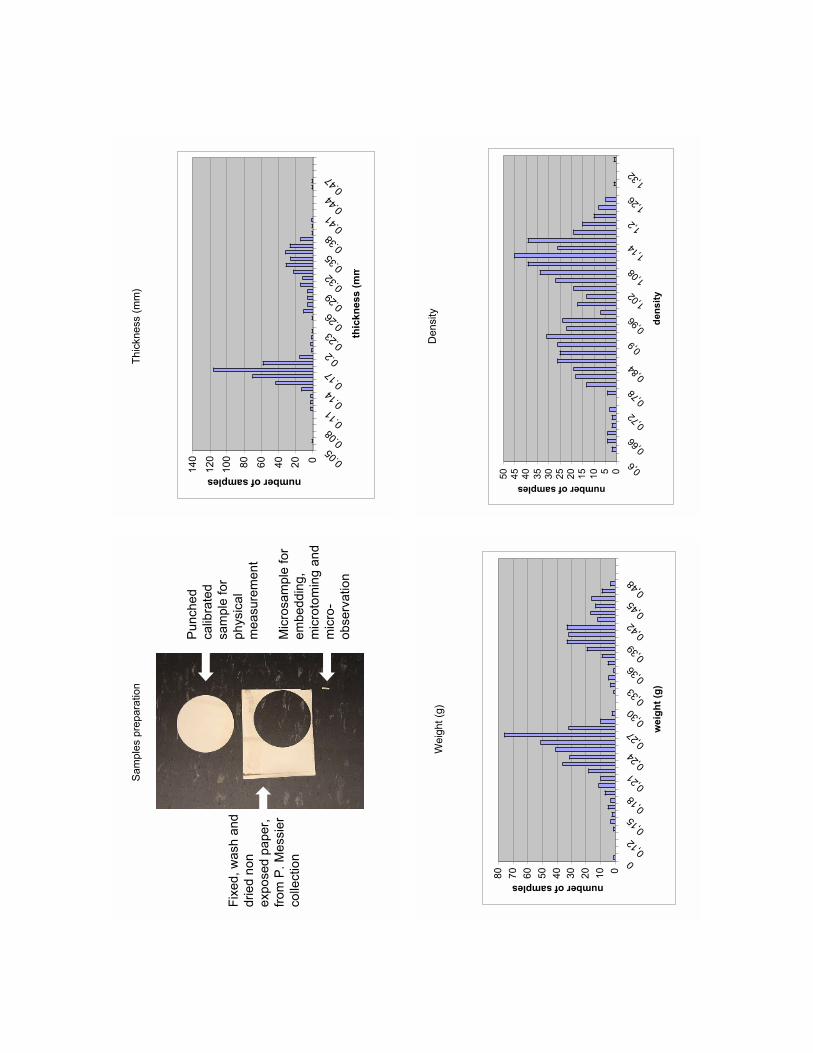
All photographic papers available for our experiments were unexposed photographicpapers from original enclosures, or boxes with full manufacturer description and annotation ofpaper type, surface, thickness (single/double weight) and year of production and/or expirationdate. Silver halide components of photographic paper were removed using a standard fixingprocedure; and photographic paper was washed, dried and permanently labeled with a databaseidentification number.
Two primary physical markers available for the characterization of photographic paperare the overall thickness (OT) of the photographic paper and its apparent density (AD). Bothof these macroscopic physical parameters can be easily determined for all unmounted photographic papers and photographs. In our study the thickness of well temperature and

humidity equilibrated samples of photographic paper were measured using a digital micrometer. The thicknesses of photographic paper in this study of 550 different photographic papers made by 25 different manufacturers varied between 0.066 mm and0.465 mm. The statistical treatment of thickness data shows a presence of two major groupsnear 0.17 and 0.36 mm, which, in general, correspond well to the manufacturer’s descriptionof the papers as “single weight” or “double weight” photographic papers, respectively.
To obtain a set of samples of calibrated surface area as needed for determining theapparent density of the photographic papers, a 1 5/8” circle of paper was cut of each papersample using a sharp circular steel punch. Each of the resulting paper discs has an identicalsurface area of 1338 sq. mm. The exact weight of each disc was determined using an analyticalmicrobalance. The apparent density of all studied photographic papers varied between 0.625and 1.341 g/sq. mm, showing two major groups with about 0.9 and 1.12 average density,each with no relation to the two thickness groups described above.
To study physical parameters of individual layers of photographic papers, it requiresphysical sampling. The thicknesses of paper base (TP), baryta layer (TB), emulsion layer (TE)and supercoating layer (TS) were determined after imbedding small samples of photographicpapers into Technivit resin and microtoming resulting resin blocks using glass or diamondknives on the precision MT-7 RMC microtome. The thicknesses of individual layers of photographic paper were determined on each 0.6–10 micrometer thick slice of glass slidemounted cross section of photographic paper using a high power optical microscope.Calibrated image-processing software aided the determination of the measured parameters ofpaper cross sections. There is no simple relationship between the thickness of paper base orthickness of baryta layer and the overall thickness of photographic paper. The thickness ofpaper base determined so far in this study ranged between 0.065 and 0.45 mm. The thickness of baryta layer for the same set of photographic paper samples ranged between0.006 and 0.045 mm. Emulsion layer and supercoating are about 0.015 and 0.005 mmthick, respectively.
Our Transmission Electron Microscope (TEM) investigation conducted at the CRCDGin Paris also showed that measurements of particle size and particle size distribution, as well asstudies of morphology of barium sulfate particles and the determination of the presence orabsence of inorganic fillers in the baryta layer, might provide some additional physical parameters that might be later incorporated into the current version of the photographic paperdatabase to obtain even more precise provenancing and authentication of the 20th centuryphotographic papers and photographs.
Renaud Duverne
After completing his Master’s degree in paper conservation at the University Paris I-Pantheon-Sorbonne (Paris) in 1996, Renaud worked for nine years as a private conservatorfor some famous French institutions including Bibliothèque Nationale de France, CentreHistorique des Archives Nationales, Université Sorbonne, Institut National du Patroimoine).At these prestigious entities he performed treatments on paper works of art as a consultant inpreventive conservation; and he also organized academic and professional training.
Renaud joined the Centre de Recherche sur la Conservation des DocumentsGraphiques in 1999, focusing on photographic material research. His background in conser-vation of cellulosic material oriented him towards photographic support analysis (e.g., cellulose acetate decomposition survey and DOP identification research). Four years ago, whileat CRCDG, he became part of the GCI’s baryta analysis project, developing micro-samplingtechniques for the study of multi-layer photographic material structure and performing analytical techniques such as chemical spot testing, Micro-XRF, IR spectrometry and atomicabsorbption spectrometry or PIXE.
Currently at the Getty Conservation Institute as a graduate intern, Renaud is completing his Ph.D. in art techniques history at the University Paris I-Pantheon-Sorbonne.
Sam
ple
s p
repara
tion
2 c
m
Punched
calib
rate
d
sam
ple
for
physic
al
measure
ment
Mic
rosa
mp
le f
or
em
beddin
g,
mic
roto
min
g a
nd
mic
ro-
observ
ation
Fix
ed,
wash a
nd
dried n
on
exposed p
aper,
from
P.
Messie
r
colle
ction
Thickness(mm)
0
20
40
60
80
100
120
140 0.
050.080.110.140.17
0.2
0.230.260.290.320.350.380.410.440.47
thic
kn
ess (
mm
number of samples
Weight(g)
0
10
20
30
40
50
60
70
80
0
0,12
0,150,18
0,210,240,27
0,300,33
0,360,39
0,420,450,48
we
igh
t (g
)
number of samples
Density
0510
15
2025
30
35
4045
50 0,
6
0,66
0,72
0,78
0,84
0,9
0,96
1,02
1,08
1,14
1,2
1,26
1,32
den
sit
y
number of samples

thic
kn
ess/d
en
sit
y K
od
ak s
am
p
0.0
0
0.1
0
0.2
0
0.3
0
0.4
0
0.5
0 0.6
00.7
00.8
00.9
01.0
01.1
01.2
01.3
0
de
ns
ity
thickness (mm)
# 2
35
# 2
010
# 1
444
# 7
2
Thic
knesses
and a
ppare
nt
density
com
parison
Overa
l T
hic
kn
ess
(OT
) 0.2
6 m
m
Em
uls
ion
layer
(TE
)
0.0
14 m
m
Pap
er
base (
TP
)
0.2
18 m
m
Su
per
co
ati
ng
layer
(TS
) 0.0
05 m
m
Bary
ta layer
(TB
)
0.0
44 m
m
Sam
ple
# 1
444
Thic
kness m
easure
ment
# s
am
ple
2010
72
235
1444
thic
kness
0.1
50.1
60.3
0.2
6
density
0.9
41.1
40.9
11.1
4
0.1
mm
Op
tica
l
Mic
rophoto
.,
X 2
00
Thic
kness a
nd s
pecific
gra
vity c
om
parison
Magnific
ation :
x45.0
00 /
Cre
dit :
C.
R.
C.
D.
G.
/ M
icro
gra
ph :
SC
ME
Jussie
u
silv
er
deposit
silv
er
gra
in
gela
tin
bary
ta g
rain
gela
tin
Tra
nsm
issio
n E
lectr
o P
ho
tog
rap
hy
Bary
ta a
nd im
age layer

MINOR AND TRACE ELEMENTS IN PHOTOGRAPHIC MATERIAL:ANALYSIS AND MEANING
David Miller, Department of Chemistry, California State University Northridge
A common method of analysis for objects of art is X-ray fluorescence spectrometry(XRF). The widespread appeal of XRF stems from the fact that it requires no physical samplingof the object, its relative ease of use and the ability to detect many elements simultaneously.However, the sensitivity of this method is limited, so that it is often difficult to detect minorand trace elements in a sample.
When applied to the analysis of photographs and photographic material, XRF hassome specific limitations. First, it is prone to a number of spectral interferences. These interferences result from the overlap of the analytical signals for certain elements and make theinterpretation of the results quite difficult. A specific example is the spectral overlap of the X-ray peaks of barium (Ba) and titanium (Ti) that makes it very difficult to detect a low concen-tration of titanium in a baryta-coated photographic paper. The X-ray peaks of lead (Pb) andarsenic (As) also overlap. Both elements may be present in photographic material, yet it is difficult to determine whether just one or both elements are present in an analyzed photographbased on XRF data alone. A second concern is associated with the quality of quantitative measurements made when analyzing photographs by XRF. The precise measurement of theconcentration of different elements in a photograph requires appropriate analytical standardsthat have been carefully analyzed by methods that are not subject to the limitations of XRF. Across-calibration approach is an excellent means of validating XRF measurements.
In order to confirm and extend the XRF analyses of photographs and photographicmaterial we have used two additional multi-element techniques: inductively coupled plasma-mass spectrometry (ICP-MS) and neutron activation analysis (NAA). ICP-MS is amodern analytical technique based on introduction of a sample solution into inductively heated argon plasma. In the high temperature of the plasma, compounds are broken downinto individual atoms which are then ionized. The resulting ions are extracted into a mass analyzer where they are separated by mass and detected. ICP-MS has the advantages of a general applicability to almost all elements of the periodic table and an extreme sensitivity formost elements, down to parts-per-billion (ppb) or parts-per-trillion (ppt) concentrations.Also, ICP-MS allows for many different modes of chemical analysis, ranging from a simple,rapid semi-quantitative screening of more than 70 chemical elements to a high precisionquantitative analysis of selected elements or a measurement of isotopic ratios. QuantitativeICP-MS is subject to errors due to the variation of the sample matrix and great care must be

exercised when preparing standards and samples for analysis. A major disadvantage of ICP-MS when applied to the analysis of photographs and photographic material is that it cannot beperformed without physical sampling or micro-sampling. Consequently, ICP-MS is best suited to support all phases of XRF methodology development when: (1) dealing with testmaterials; (2) screening the presence of different elements in samples of unexposed,unprocessed or fully processed photographic material; or (3) analyzing collections of photographic reference material or samples from collections of study and sacrificial photo-graphs. The application of ICP-MS for the analysis of photographs from a museum or anarchival collection of photographs will be always limited to very special cases where there is anurgent need to understand the details of the chemical composition of a photograph in question(due to conservation or authentication reasons) and where the conventional XRF techniquedoes not provide a satisfactory answer.
NAA is also a very sensitive analytical technique. This method has been used to analyzevarious forms of art including glass, marble and metal objects and paintings. Its major advantages, in comparison with many other analytical techniques, are that it is generally free ofspectral interferences, the physics of the process is well understood and little or no samplepreparation is required. Consequently, NAA can provide results of high precision and highaccuracy that are useful for the development and characterization of analytical standards usedto calibrate other techniques such as XRF. A major disadvantage of NAA is that it requiresaccess to a nuclear reactor set up for irradiating samples. All of our NAA measurements weredone in collaboration with the Nuclear Reactor Facility at the University of California, Irvine.As with ICP-MS, this method also requires sampling or micro-sampling.
Both ICP-MS and NAA were used to address the question: “In baryta-coated photographic papers, are there any minor and trace elements that can be used as markers oranalytical signatures in provenancing and authentication studies of photographs?”.
In the quest for an answer to this rather difficult analytical question, we have analyzeda series of photographic papers from the GCI Reference Collection and Paul Messier’sCollection of Historical Photographic Papers that exhibit an unusual elemental compositionwhen analyzed using XRF. We have also analyzed a series of raw materials used in the manufacture of baryta-coated photographic paper and samples of well characterized photographs from the GCI Photograph Study Collection.
This presentation focuses on a very basic introduction to the techniques of ICP-MSand NAA as needed by potential end users of analytical findings (conservators, museum curators and photographic collection managers) who might be new to these methods. The development of a basic methodology for applying ICP-MS and NAA to the analysis of photographic materials will be discussed, together with the first round of our analytical findings. The future direction of our collaborative research will also be presented.

David Miller
David Miller earned his B.A. University of California, San Diego and his Ph.D. inChemistry from the University of California, Irvine. He is currently professor of chemistry andbiochemistry at California State University Northridge.
His research areas include radiochemistry and analytical chemistry. Related experiencesinclude serving as a technical expert for the International Atomic Energy Agency; as anAlexander von Humboldt Fellow at the Technical University of Munich; and as a visiting scien-tist at the Los Alamos National Laboratory in New Mexico.
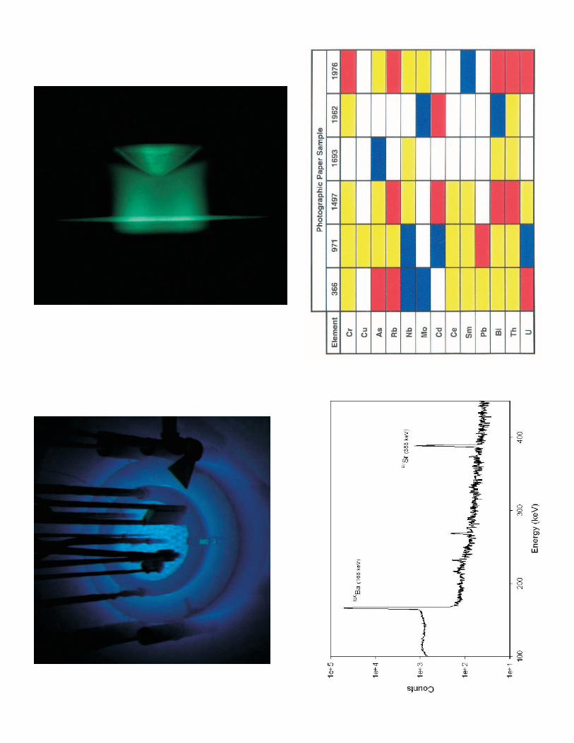

Trace Element Signatures
Photographic Paper Sample
Element 366 971 1497 1693 1962 1976
Cr
Cu
As
Rb
Nb
Mo
Cd
Ce
Sm
Pb
Bi
Th
U
Color Key
Concentration in Sample
same as blank
3 - 5 x blank
5 - 50 x blank
> 50 x blank

BARYTA PAPER MUSICAL CHAIRS, WHERE DOES EACH ELEMENT SIT?
Art Kaplan, Getty Conservation Institute
One of the major advantages of using X-ray fluorescence analysis (XRF) when analyzing photographs is its ability to provide for both qualitative and quantitative analysis ofmost important inorganic elements that participate directly in the formation of the photographic image or that are, in a supportive role, responsible for the mechanical, chemicaland optical properties of photographs. XRF also allows for completely non-destructive andoften even non-contact chemical analysis of photographs, making it a favorite method of collection curators and museum directors. Conversely, there are some disadvantages or limitations to consider when using XRF.
If there is a need to confirm some XRF findings or if the XRF is not providing a clearindication of the presence or absence of certain elements, we often use Inductively CoupledPlasma Mass Spectrometry (ICP-MS) to provide additional information. ICP-MS is capable ofrapid multi-element analysis of samples coupled with the ability to detect many elements at theparts-per-billion (ppb) level and some elements down to the parts-per-trillion level (ppt). Butof course, there are drawbacks to using ICP-MS, including needing to remove a sample fromthe material to be analyzed that must then be dissolved in solution. When dealing with photographic paper samples containing high levels of barium sulfate, getting the sample intosolution is a difficult process because of the high insolubility of barium sulfate. Both ICP-MSand XRF analysis are very powerful tools in providing information on the chemical elementspresent in an analyzed photograph, but neither of them allow for an easy determination ofwhere, in a complex internal structure of the photograph, the detected elements are located.
A typical analysis of a 20th century black and white fiber based photographs shows thepresence of Aluminum (Al), Silicon (Si), Sulfur (S), Potassium (K), Calcium (Ca), Chromium(Cr), Manganese (Mn), Iron (Fe), Copper (Cu), Zinc (Zn), Selenium (Se), Strontium (Sr),Silver (Ag) and Barium (Ba). The location of some of these elements in a photograph’s internalstructure can be easily deducted based on a general knowledge of photographic chemistry andtechnology. Most 20th century B&W photographs are based on silver halide technology, so itis not surprising to find a relatively large concentration of silver in the emulsion layer of darkareas of a photographic image. Many older contact photographic papers were made using silverchloride or silver chloro-bromide emulsion. Most enlarging photographic papers up to thepresent were produced using silver bromide, silver bromo-iodide or bromo-chloro-iodideemulsions. The photographic gelatin of the emulsion layer acts as a potential sink for halogens(chlorine, bromine, and iodine) produced during the development of the photographic image,and these elements might be detected even in well-processed and fixed photographs.

The baryta layer of most photographic papers contains barium sulfate, with smallamounts of strontium sulfate impurities. These two elements together with the sulfate are themain inorganic elements expected in the baryta layer of photographs.
Since the beginning of the 20th century photographers also used a number of differentprocedures to modify the color of a photographic image or to improve the stability of a silver-based image using both sulfur-based or heavy metal (e.g., Se, Au) toners. The location of theseelements in the emulsion layer together with developed silver is also easy to predict.
Locating other elements in photographs can be much more difficult. Chromium alumwas sometimes used to harden gelatin; and it can also be found in the gelatin binder of thebaryta layer, the emulsion layer and/or in a super-coating layer applied to a top of the emulsionlayer to provide protection against mechanical damage. Inorganic fillers of many differentkinds were used as additives to the baryta layer, and the same or different fillers were often alsoadded to cellulose mass to modify the properties of paper used as a base for photographicpaper. Small amounts of Mn, Fe,Cu as impurities might be present both in the baryta layer orpaper substrate.
For provenancing and identification of photographic paper, it is important to understand the location of all major elements detected in photographs. Even if two photographic papers contain identical sets of chemical elements based on both qualitative andquantitative analyses, the distribution of elements within the internal structure of a photograph might be quite different. In some cases it would be very important to understandthe location of different elements for interpreting data obtained during analysis. For example,results of quantitative analysis of calcium in the paper base of a photographic image would bedifferent when analyzed from verso or recto side due to the presence of the X-ray absorbingbaryta layer.
A study of the locations of different components in a multi-layer structure of a photograph without sampling is theoretically possible, but it would require very special instru-mentation, certainly not a practical solution for everyday use by conservation scientists andphotographs conservators. At this point of time it seems that the best and most accessible wayto study the distribution of different elements is to remove a small sample from a photograph,prepare a cross section and then study the distribution using an Electron Microprobe Analyzer(EMPA) or a combination of a Scanning Electron Microscope (SEM) along with an Energy-Dispersive X-ray Analyzer (EDX) and ICP-MS. These analytical methods provide an opportunity not only for a point analysis of selected areas of the analyzed cross-section butalso for obtaining elemental maps of the whole or partially selected area of a cross-section that

show the presence or absence of selected chemical elements in different layers.
These analytical methods require the removal of a small sample. Any removal of a physical sample from photographs should be done only with the approval of a collection curator and only when it is crucial to obtaining information about the internal composition ofa photograph and cannot be obtained by any other nondestructive means. Potential damage toa photograph can be minimized by using well-developed micro-sampling procedures that allowthe removal of a sample that is not visible to naked eye.
The analysis of photograph cross-sections from the GCI’s Reference Collection of photographs, and cross-sections of different B&W fiber-based photographic papers from theGCI’s and Paul Messier’s collection of photographic paper and an interpretation of analyticalresults in the light of understanding the internal chemical structure of 20th century photographic material, will be discussed in this presentation.
Art Kaplan
Art Kaplan was born in Bobruysk, Byelorussia. Moving with his parents to LosAngeles in 1980, he graduated from high school in Santa Monica and then enrolled at SantaMonica College. After earning his A.A. degree from Santa Monica College, Art joined theUnited States Marine Corps where he served a field radio operator for an artillery battalion forseveral years.
After leaving the Marines, Art returned to school and enrolled at California StateUniversity Northridge in the biochemistry program. During his junior and senior years, heworked with Dr. David Miller and the GCI performing Neutron Activation and InductivelyCoupled Plasma Mass Spectrometry analysis on glass samples for the GCI’s Conservation ofthe St. Vitus Mosaic project in Prague.
Upon graduating from California State University, Northridge with a B.S. in biochemistry, Art took a position with the Science Department of the GCI. His work centersaround the Conservation of Photographs project, while also maintaining the GCI’s extensiveReference Collection of artist’s materials.

Pape
rs S
ampl
ed
1937
Indi
aton
eAg
fa-A
nsco
1976
1952
Opa
lKo
dak
DS3
019
1896
Aris
toAm
eric
anAr
isto
type
Co.
366
1976
Ekt
alur
eKo
dak
1962
1950
Brov
iraAn
sco
1693
1959
Varig
ram
Dup
ont
1497
1918
Cyko
Ansc
o97
1
Year
Bran
dM
anuf
actu
rer
Sam
ple
#
XR
F Sp
ectra
of S
ampl
e #3
66

1
2
3
800°
C
HN
O3
Sam
ple
#366
0.0E
+00
4.0E
+06
8.0E
+06
1.2E
+07
1.6E
+07
724
4353
6072
8593
103
115
127
140
153
165
175
185
197
209
M/Z
CPS
ICP-
MS
Spec
tra o
f Sam
ple
#366
Sam
ple
#366
0.0E
+00
4.0E
+05
8.0E
+05
1.2E
+06
724
4353
6072
8593
103
115
127
140
153
165
175
185
197
209
M/Z
CPS
Na
Mn
Pb
Ba
Sr
Ba
K
Al
Mg
Ca
K
Si
Al
Cr
Mg
Na
Zn
Sr
Ba
FeP
b
Ba
Ag
Em
bedd
ing
Mic
roto
min
g
ESE
M
DS3
019