normal and histopathological organization of the opercular
TRANSCRIPT

AQUATIC BIOLOGYAquat Biol
Vol. 21: 67–84, 2014doi: 10.3354/ab00568
Published online June 18
INTRODUCTION
During the last decades, the finfish larvicultureindustry has considerably improved its rearing meth-ods under intensive conditions, but larval qualityremains one of the main problems for the proper sus-
tainable success of this productive sector (Kou moun -douros 2010, Boglione et al. 2013a). The quality offarmed fish is directly related to the quality of the fry,and depends on both organoleptic and morphologi-cal characteristics that should be as similar as possi-ble to that of wild fish, which is considered to be the
© The authors 2014. Open Access under Creative Commons byAttribution Licence. Use, distribution and reproduction are un -restricted. Authors and original publication must be credited.
Publisher: Inter-Research · www.int-res.com
*Corresponding author: [email protected]
Normal and histopathological organization of theopercular bone and vertebrae in gilthead sea
bream Sparus aurata
Juan B. Ortiz-Delgado1,*, Ignacio Fernández2,3, Carmen Sarasquete1, Enric Gisbert2
1Instituto de Ciencias Marinas de Andalucía-ICMAN/CSIC, Campus Universitario Río San Pedro, Apdo. Oficial, 11510, Puerto Real, Cádiz, Spain
2IRTA, Centre de Sant Carles de la Ràpita (IRTA-SCR), Unitat de Cultius Experimentals, Crta. del Poble Nou s/n, 43540 Sant Carles de la Ràpita, Spain
3Centre of Marine Sciences (CCMAR), University of Algarve, Campus de Gambelas, 8005-139 Faro, Portugal
ABSTRACT: This study provides a comprehensive description of the tissue organization of non-deformed and deformed opercula and vertebrae from gilthead sea bream Sparus aurata juvenilesby means of histological, histochemical and immunohistochemical approaches. Two types of oper-cular anomalies are described: the folding of the opercle and subopercle into the gill chamber,starting at the upper corner of the branchial cleft and extending down to its lower third; and thepartial lack of the operculum (opercle, subopercle, interopercle and preopercle underdeveloped)with a regression of the loose edge extending down to its lower third. Histological observationsrevealed a rare type of bone remodelling process in the opercular structure, which consisted of thecoalescence of contacting bone tissues (presumably from the preopercle and opercle), resulting inskeletal tissue with a trabecular aspect filled by a single-cell epithelium of cubic osteoblastic-likecells. Differences in collagen fiber thickness and its 3-dimensional arrangement between normaland deformed opercula were also found. Lordotic vertebrae were characterized by the formationof fibrous cartilage in the haemal and/or neural sides, indicating that a metaplastic shift occurredduring the process of lordosis. Another major histomorphological change found in lordotic verte-brae was the complete loss of notochordal sheath integrity. Histological alterations were coupledwith an imbalance of cell death and cell proliferation processes in lordotic vertebrae as well as thatof bone formation/resorption, and extracellular matrix deposition activity differences which mighthave resulted from the remodelling process occurring in lordotic vertebrae. Altogether, theseresults provide an increase in our basic knowledge of bone disorders that contribute to our under-standing of the mechanisms by which these skeletal anomalies appear in this fish species andwhich hamper its production efficiency.
KEY WORDS: Bone · Extracellular matrix · Histology · Histochemistry · Immunohistochemistry ·Skeletal deformities
OPENPEN ACCESSCCESS
Aquat Biol 21: 67–84, 201468
quality reference by the consumer. In this sense, ananomalous external morphology (even of a few fish)could substantially decrease the consumers’ overallperception of aquaculture products (Boglione et al.2013a). Skeletal deformities have been reported innatural environments (Gavaia et al. 2009, Diggles2013), but with lower prevalence than in intensiveaquaculture. The high incidence of skeletal deformi-ties found in farmed fish (affecting up to 30% of pro-duction) might be due to (1) the absence of predatorsand the constant and high availability of food, whichmaximizes fish survival, or (2) that industrial hatch-eries still lack the proper knowledge of how to pro-duce high-quality and healthy fry under intensiverearing conditions in order to reduce the causativefactors by which skeletal deformities are induced(Boglione et al. 2013a,b). However, since in all rearedbatches the skeletal deformity incidences are largelydifferent, whether the fish come from the same orfrom different industrial hatcheries, the second hypo -thesis seems more plausible. Nevertheless, skeletaldeformities are one of the most significant biologicaland recurrent problems affecting worldwide finfishaquaculture (see reviews in Koumoundouros 2010,Boglione et al. 2013a, Cobcroft & Battaglene 2013).
Fish skeletogenesis involves the activity of 3 maincell types: chondrocytes, osteoblasts and osteoclasts(see review in Boglione et al. 2013b). The first 2 celltypes secrete the extracellular matrix proteins of thecartilage and bone, respectively, while the third typeproduces cathepsins, matrix metalloproteinases andtartrate-resistant acid phosphatase (TRAP), providingan acidic environment in which the mineralized ma-trix is broken down (Ytteborg et al. 2010). In addition,skeletogenesis involves a fourth type of cell: the os-teocytes, which are involved in maintaining bone ma-trix and acting as mechanical load receptors. Inteleosts, the lack of osteocytes (acellular bone, a fea-ture of the Sparidae family) implies that induced boneremodelling by mechanical load is triggered by celltypes other than osteocytes (reviewed in Boglione etal. 2013a). Bone formation is brought about by a com-plex set of tightly regulated molecular pathways, in -volving extracellular matrix (ECM) constituents (e.g.alkaline phosphatase, ALP) or non-collagenous pro-teins such as the matrix Gla protein (MGP) or osteo-calcin (OC), signalling molecules (e.g. hedgehogsand bone morphogenetic proteins) and transcriptionfactors (Karsenty 2001, Boglione et al. 2013b). Hence,the perturbation of some of the above-mentioned factors in controlling bone development and home-ostasis, as well as the incapacity of homeorheticmechanisms to compensate for stressful environmen-
tal conditions, may disrupt skeletogenesis, ultimatelyleading to the appearance of skeletal deformities(Boglione et al. 2013a). With the exception of environ-mental contaminants and pathogens (which are gen-erally controlled under rearing conditions), differentstudies suggest that unfavourable environmental bi-otic and abiotic conditions, inappropriate nutritionand/ or genetic factors are the most probable causativeagents of skeletal abnormalities in reared fish (re-viewed in Boglione et al. 2013b). Previous studies haveindicated that skeletal anomalies are mainly inducedin early stages of development (i.e. during embryonicand larval periods), occurring long before osteologicaldeformities are externally visible (Daoulas et al. 1991,Koumoun douros et al. 1997a,b, Gavaia et al. 2009,Boglione et al. 2013b). Nevertheless, skeletal defor-mities could also be in duced at later stages during thenursery phase (Boglione et al. 2013a). Regardless ofwhen skeletal deformities are diagnosed, at juvenileor later stages of development (on-growing phase), itis difficult to gather informa tion about their onset andaetiology since this is considered a multifactorialproblem (Witten et al. 2005, 2006, Boglione et al.2013a).
Although gilthead sea bream Sparus aurata L. isone of the most important species in European aqua-culture, cranial and spinal abnormalities are still oneof the major problems that hinder the efficiency ofthe production cycle of this sparid species. Vertebraldeformities related to the non-inflation of the swim-bladder (mostly lordosis) were the most frequentabnormalities recorded at the beginning of giltheadsea bream intensive culture (1980s and early 1990s)(Chatain 1994, Andrades et al. 1996). Although theimprovement and refinement of rearing techniqueshave considerably reduced the frequency of thesetypes of deformities (Boglione et al. 2013a), lordosisand kyphosis are still 2 of the most relevant osteo -logical abnormalities observed in the axial skeletonof reared gilthead sea bream, while a partial and/ortotal lack of operculum is the most frequent anomalyaffecting its cranial region (Koumoundouros et al.1997b, Boglione et al. 2001, 2013b, Beraldo et al.2003, Verhaegen et al. 2007, Castro et al. 2008, Kou -moundouros 2010, Prestinicola et al. 2013). Severalstudies have described skeletogenesis in gilthead seabream (Faustino & Power 1998, 1999, 2001) and thetypology of fish skeletal deformities (Witten et al.2009, Boglione et al. 2013b, Prestinicola et al. 2013,among others) in order to better understand thephases of skeletogenesis and the effects of biotic andabiotic factors on the proper development of boneand the appearance of skeletal anomalies. Various
Ortiz-Delgado et al.: Normal and deformed S. aurata vertebrae and operculum
procedures have been used as simple and rapid diag-nostic tools for studying skeletal deformities in fish,such as X-rays, double staining or even computertomography; allowing the identification of abnormalgrowth in different skeletal structures (see reviews inWitten et al. 2009, Koumoundouros 2010, Boglioneet al. 2013b). Although there is a great variety of re -search in fish skeletal histology (reviewed in Meu-nier 2011), there is limited information about the his-tological organization of skeletal tissues in giltheadsea bream (Galeotti et al. 2000, Beraldo et al. 2003,Fernández et al. 2012). The use of histological proce-dures is considered a valuable tool for providing basicknowledge on bone formation, as well as providingnew insights into the structural changes oc curring indeformed skeletal structures (Ytteborg et al. 2012).
The aim of this study was to perform a comparativeanalysis of the tissue organization and distribution,as well as changes in ECM composition, betweennormal and deformed opercular bones and vertebraefrom hatchery-reared gilthead sea bream juveniles.This description was conducted by means of histo -logical, histochemical and immunohistochemical ap -proaches in order to unveil the organization of theseskeletal structures and the possible mechanisms bywhich the above-mentioned skeletal disorders develop.
MATERIALS AND METHODS
Animals
Gilthead sea bream larvae were reared under stan-dard conditions in two 500 l cylindroconical tanks(initial density of 100 larvae l−1) up to 60 d post hatch(dph) as described in Fernández et al. (2008). At thatage (ca. 80 to 100 mg wet body weight), fry werereared in a 2000 l tank connected to a recircula -tion unit (IRTAmar™) until they weighted ca. 2 g(90 dph). During this period, fish were fed commer-cial diets following standard procedures according toOrtega (2008). Water conditions were as follows: 18to 19°C; 35 ppt salinity; pH between 7.8 and 8.2; dailywater renewal in the rearing tank was 20%, and thetank had gentle aeration (oxygen levels = 6.2 ±1.1 mg l−1). Photoperiod was 12:12 h light:dark, andthe light intensity at water surface was 500 lux. From90 dph, fish were kept in an outdoor open-flow 14 000 ltank until they were 10 mo old (35.5 ± 15 g bodyweight, 10.5 ± 1.5 cm standard length). During thisperiod, fish were subjected to natural conditions interms of thermo- and photoperiod regimes. The over-all survival rate of the complete rearing process wasestimated at ca. 15 to 20%.
After a visual examination of 1064 live specimensin sorting tasks, normal and deformed specimenswith underdeveloped operculum and lordosis weresampled (7 specimens for each morphotype), sacrificedwith an overdose of anaesthetic (tricaine me thane -sulfonate, MS-222; Sigma-Aldrich) and dissected.
Histological, histochemical and immunohistochemical procedures
Normal and deformed opercula and vertebral cen-tra were fixed in 4% buffered paraformaldehyde (inphosphate buffer saline, PBS; pH 7.4). After fixa-tion, samples were washed in PTW (1% PBS, 0.1%Tween-20) 3 times for 30 min each, then either pre-served in methanol at −20°C or immersed for decalci-fication in a 10% EDTA/2% formaldehyde solution at4°C for 7 d, followed by washes in PTW as describedabove. After fixation and decalcification, sampleswere washed, embedded in paraffin and serially sec-tioned at 5 to 6 µm thickness as previously described(Ortiz-Delgado et al. 2005). Undecalcified sampleswere subjected to the von Kossa staining method: aprecipitation process in which silver ions react withphosphate in the presence of acidic medium (Clark1981). The von Kossa staining method alone may notbe sufficient to confirm mineralization, since it doesnot necessarily imply the presence of calcium or ofhydroxyapatite, the mineral phase of bone (Bone -wald et al. 2003). Although further evidence of bonemineralization may be needed using alternative pro-cedures (Bonewald et al. 2003), in the present study,and considering morphological and molecular datafrom Tiago et al. (2011), we used von Kossa stainingas a first approximation for whether structures weremineralized or not.
For studying ECM components of bone tissue andcartilage, adjacent sections were stained with the fol-lowing techniques: haematoxylin and eosin (H/E) forhistomorphological observation, orcein for elasticfibers, van Gieson trichrome and picrosirius red stain-ing (PRS) for collagen, Mallory and haematoxylin/VOF (H/VOF) trichrome (Gutiérrez 1967) for con -nective tissue and Alcian Blue (AB) 8GX (Sigma) atpH 2.5/periodic acid-Schiff (PAS) for acidic and neu-tral mucopolysaccharides. Dye affinity or intensity ofthe above-mentioned staining techniques from dif-ferent regions of studied tissues was ranked by visualscoring by at least 2 trained observers (10 slices pertechnique and 5 sections per slice) as: negative (−),very weak (+−), weak (+), moderate (++) and strong(+++). Values were scored considering the accumula-
69
Aquat Biol 21: 67–84, 2014
tion levels of the dye for each considered vertebralpart, and ranged from the absence of staining (nega-tive) to the highest stain intensity (strong), whereasintermediate scoring staining categories were estab-lished as intermediate ranges among the above-mentioned extreme scores. These protocols were con -ducted according to monographs by Martoja et al.(1970), Pearse (1985), Bancroft & Stevens (1990) andSarasquete & Gutiérrez (2005). Additionally, PRS sec -tions were observed under polarized light accordingto Junqueira et al. (1979) and Kiernan (2008). Briefly,after PRS and under bright field microscopy, collagenappears red on a pale yellow background. A BX41Olympus light microscope equipped with 2 kinds offilters (a polarizer and an analyzer) was used to pro-vide linearly polarized illumination. Digital imageswere obtained with a C3030 Olympus digital camera.
Bone remodelling/homeostasis, a dynamic processthat relies on the correct balance between boneresorption by osteoclasts and bone deposition byosteoblasts (Boglione et al. 2013b) was described bymeans of staining for TRAP and ALP activities,respectively. Both enzyme activities were visualizedwith the acid and alkaline phosphatase leukocyte kits(Sigma-Aldrich) according to the instructions of themanufacturers.
Immunohistochemistry for ECM proteins was per-formed using a rabbit polyclonal antibody developedagainst OC and MGP from meagre (ArgyrosomusregiusAsso,1801)asprimaryantibodies.Anti meagre-OC and -MGP antibodies presented a specific crossreactivity for gilthead sea bream-OC and -MGP(Simes et al. 2004). Immunohistochemical stainingwas performed with peroxidase-conjugated anti-rabbit IgG (Sigma) as secondary antibody, using themethodology proposed by Simes et al. (2003, 2004).Briefly, serial sections were incubated overnight withanti-OC and anti-MGP primary antibodies at appro-priate dilutions (1:500 for OC and 1:250 for MGP).After several washes in PBS, sections were incu-bated for 1 h with goat anti-rabbit IgG peroxidase-conjugated, and stained with 3-3’-diaminobenzidine-DAB (Sigma). The endogenous peroxidase activitywas blocked with 3% H2O2 before staining. Sectionsincubated with normal fish serum instead of the pri-mary antibody were used as negative controls.
Cell proliferation and apoptosis processes were alsoassessed by immunohistochemical detection of pro-liferating cell nuclear antigen (PCNA) and cleavedcaspase-3, respectively. The former assays were per-formed as described by Bakke-McKellep et al. (2007)for PCNA and Sanden et al. (2005) for caspase-3,using a mouse anti-PCNA (Santa Cruz Biotechno -
logy) and cleaved caspase-3 antibody (Cell Signalling),respectively. Sections incubated with normal fishserum instead of the primary antibody or withoutsubstrate incubation were used as negative controls.After staining, all sections were dehydrated by airdrying and mounted with EUKITT (Labolan).
RESULTS
Macroscopically, we detected 2 types of opercularanomalies according to the classification previouslydescribed by Beraldo et al. (2003): (1) the foldingof the operculum (opercle and subopercle) into thegill chamber, starting at the upper corner of thebranchial cleft and extending down to its lower third(Type I; Fig. 1a), and (2) the partial lack of the oper-culum (lack of development of the opercle, suboper-cle, interopercle and preopercle) with a regressionof the loose edge extending down to its lower third(Type II; Fig. 1b). The overall incidence of fish withdeformed operculum was 6.3%. These opercular ab -normalities unilaterally affected both sides of the head:the right (3.3%) and left (3.0%) equally; where as thebilateral abnormality in the operculum only affected1.1% of the reared fish.
Fish with a severe external body shape deformity(Fig. 1c) showed internal lesions characterised by anaccentuated ventral curvature (lordosis) of the verte-bral column (Fig. 1d). The degree of these pathologi-cal symptoms varied along the vertebral column axisand mainly affected vertebrae located between thelimit of pre-haemal and haemal areas of the vertebralcolumn (position 11 to 15 from urostyle upwards).The incidence of fish affected by lordosis was 10.1%.
Histological organization of normal and deformedoperculum
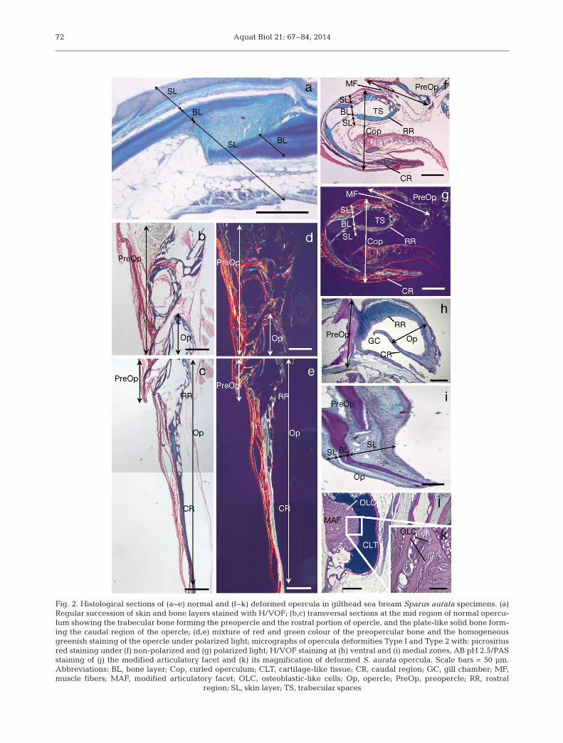
The operculum is a complex structure composed of4 distinct and articulated bony plates: the opercle,preopercle, interopercle and subopercle. At a histo-logical level, the opercular complex of gilthead seabream shows a regular succession of tissue layers:outer and inner skin layers, and an intermediatelayer of bone (Fig. 2a). The posterior rim of the oper-culum is a flexible ribbed structure, which closes thegill cavity like a lid.
Transversal sections at the mid region of normaloperculum stained with the PRS technique showed apreopercle mainly composed of highly trabeculatedbony tissue, and a plate-like opercle with solid bone
70

Ortiz-Delgado et al.: Normal and deformed S. aurata vertebrae and operculum
and homogeneous mineralisation levels (Fig. 2b−e,Table 1). The anterior rim showed medially directedtrabecular projections at the level of the articula -tory facet that were absent in the caudal direction(Fig. 2b,c). In addition, the PRS evidenced some dif-ferences in composition of both preopercle and oper-cle bones. While the preopercle had a predominantgreen colour with scarce areas of red (orange underpolarized light), the opercle showed reticular fiberswith a homogeneous greenish staining (green underpolarized light) (Fig. 2d,e).
The 2 different patterns of opercular deformitiesreported in this study showed some differences withregards to the tissue layers involved in the osteo -logical anomaly. The opercular Type I deformitycor re sponded to a folded operculum, directed to -wards the inside of the gill cavity. Although thedeformed operculum affected all tissue layers, asevere ly curled opercle and/or subopercle concomi-tant with a proliferation of epithelial tissue withinthe folded area were detected (Fig. 2f,g). Addition-ally, hypo plasia of muscular fibers and enlargementof trabecular spaces in the rostral portion of theopercle were also observed. Polarized light revealedsome differences in bone composition of normal anddeformed opercula (e.g. changes in colour stain);the opercle and subopercle from deformed fish pre-sented a brighter green colour than normal ones(Fig. 2g). In contrast, the opercular Type II deformity(Fig. 2h−k) corresponded to a hypoplasic operculumin which some bone structures had disappeared de -pending on the dissected region (articulatory facet,medial and ventral zones; see Fig. 1b). For instance,the dissected operculum at the ventral zone showeda regression of the subopercle and a complete lackof the opercle. In this case, fibrous and epithelialcells proliferated and filled the empty space; thus,as a result of a lack of bone support, the outer andinner skin layers of the opercle folded into the gillcavity (Fig. 2h). At the medial opercular zone, thefor mation of an outward folding of the opercle wasdetected (H/VOF; Fig. 2i), as well as a thickening ofthe skin epithelium. Finally, the dissection of thedorsal area of the operculum revealed a modifiedarticulatory facet in which a coalescence of contact-ing bony tissues was detected, as well as the forma-tion of a chondroid bone-like structure. The tra -becular structure of the opercle and subopercle wasalso modified; the trabecular spaces were filled witha single-cell epithelium (presumably osteoblasts)(Fig. 2j,k). All skeletal tissue involved in this defor-mity was ossified, as revealed by von Kossa staining(data not shown).
71
Fig. 1. Macroscopical appearance of deformed operculumand vertebra in gilthead sea bream Sparus aurata speci-mens. (a) Type I and (b) Type II opercular malformations and(c) external and (d) internal appearance of a lordotic fish.Note the accentuated ventral curvature and the upwardsbending of vertebral column. Bars indicate the level of trans-versal dissected regions. Abbreviations: AF, articulatory
facet; MZ, medial zone; VZ, ventral zone
Aquat Biol 21: 67–84, 201472
Fig. 2. Histological sections of (a−e) normal and (f−k) deformed opercula in gilthead sea bream Sparus aurata specimens. (a)Regular succession of skin and bone layers stained with H/VOF; (b,c) transversal sections at the mid region of normal opercu-lum showing the trabecular bone forming the preopercle and the rostral portion of opercle, and the plate-like solid bone form-ing the caudal region of the opercle; (d,e) mixture of red and green colour of the preopercular bone and the homogeneousgreenish staining of the opercle under polarized light; micrographs of opercula deformities Type I and Type 2 with: picrosiriusred staining under (f) non-polarized and (g) polarized light; H/VOF staining at (h) ventral and (i) medial zones, AB pH 2.5/PASstaining of (j) the modified articulatory facet and (k) its magnification of deformed S. aurata opercula. Scale bars = 50 µm. Abbreviations: BL, bone layer; Cop, curled operculum; CLT, cartilage-like tissue; CR, caudal region; GC, gill chamber; MF,muscle fibers; MAF, modified articulatory facet; OLC, osteoblastic-like cells; Op, opercle; PreOp, preopercle; RR, rostral
region; SL, skin layer; TS, trabecular spaces

Ortiz-Delgado et al.: Normal and deformed S. aurata vertebrae and operculum
Histological organization of normal and deformedvertebrae
The normal histological structure of transversalsections of the vertebral centra of gilthead sea breamstained with Mallory, van Gieson, H/VOF trichromeand PRS techniques showed a succession of tissuelayers from the inner to the outer region. A centralnotochordal tissue (composed of chordoblasts andchordocytes) is surrounded of 2 fibrous layers (noto-chordal sheath), one composed of elastic fibers — theprimary chorda sheath (Mallory trichrome-positive),and the other with mucopolysaccharides (AB-positive) and sparse collagen fibers—the second-ary chorda sheath (van Gieson trichrome-positive)
(Table 2, Fig. 3a−c). Thenotochord sheath is en -closed by 2 different bonelayers: a laminar and com-pact layer (internal; lightgreen under polarized light)and a cancellous layer (ex -ternal; green with largeorange areas under polar-ized light). An osteogene -ous tissue layer (osteo blastlayer) and the peri o steumcomplete the skeletal struc-ture of the vertebral body(Fig. 3a−c). Neural andhemal arches are composedof chondral bone. Thesearches end in a spine com-posed of fibrous bone thatprojects into the myosepta(Fig. 3d). PRS stainings oftransversal sections of nor-mal vertebra visualized un -der polarized light exhibitedsome orange surfaces em -bedded in a bright greencolour. The fibrous layersurrounding the bony struc-ture also contained fine andsparse green colour regions(Fig. 3e).
Transversal sections ofdeformed vertebrae showedan alteration of the layereddisposition with fibro/cell-rich cartilage displacing can -cellous and compact bone.Enlargement of trabecular
spaces were also detected in lordotic vertebrae(Fig. 3f,g). Moreover, the notochordal lumen was re -duced, with distorted chordocytes showing com -pression with a clear reduction in size (Mallory stain-ing; Fig. 3f). Additionally, the PRS staining revealedsome differences in the composition of the bonematrix between normal and deformed vertebrae. Ingeneral, and under a bright microscope, compactbone of deformed vertebra showed a red colour(immature bone) in contrast to normal vertebrae, inwhich compact bone stained green (mature bone)(Fig. 3d,e for normal vertebrae; Fig. 3h,i for de -formed vertebrae). Moreover, under polarized light,deformed vertebra showed weak greenish staining(Fig. 3i).
73
H/VOF van PAS AB von Mallory OrceinGieson Kossa
PreoperculumOuter layer Light green Pink −/+− − − Red BrownIntermediate layer Purple Deep red −/+− − +++ Blue PinkInner layer Ligth green Pink +− ++ − Red Brown
OperculumOuter layer Light green Pink − − − Red BrownIntemediate layerRostral portion Purple Deep red + +− ++/+++ Blue PinkCaudal portion Light purple Red + +− ++ Blue PinkInner layer Light green Pink − − − Red Brown
Ribbed structure Green Light red − − − Orange Brown
Table 1. Staining properties of each part of the operculum in gilthead sea bream Sparusaurata. Staining intensity: (−) negative, (+−) very weak, (+) weak, (++) moderate, (+++)
strong. See ‘Materials and methods’ for details
H/VOF van PAS AB von Mallory OrceinGieson Kossa
Notochord Light green Pink −/+− − − Red −
Surrounding notochordal Green Light red −/+− − − Red −epithelial layer
Secondary chordal sheath Light Green Pink −/+− − +/++ Blue Brown
Primary chordal sheath Green Red +− ++ − Red −
Centrum Purple Deep red +−/+ − +++ Deep blue Pink
Intervertebral ligamentNotochordal sheath Light green Pink + + − Pink −Elastic membrane Purple Pink − − − Blue BrownCollagenous ligament Light green Red − − − Pink/red Pink
Osteoblasts Purple Red − − − Red −
Periosteum Light green Pink − +− − Red Brown
Arches Blue Deep red + + ++ Deep blue Pink
Table 2. Staining properties of each part of the vertebra in gilthead sea bream Sparusaurata. Staining intensity: (−) negative, (+−) very weak, (+) weak, (++) moderate, (+++)
strong. See ‘Materials and methods’ for details

Aquat Biol 21: 67–84, 201474
Fig. 3. Transversal sections of (a−e)normal and (f−i) deformed structure ofvertebral tissue of gilthead sea breamSparus aurata specimens. (a) Detailedvisualization of the succession of tissuelayers surrounding the notochord bypricrosirius red staining under non-polarized light; (b) vertebral tissuestained with pricrosirius red and visu-alized under polarized light showinginternal (compact bone, light green)and external bony layers (trabecularbone, green with big orange areas); (c)detail of the layered distribution of thechorda sheath surrounding the noto-chord with H/VOF staining and (d) ofthe trabecular bone forming the verte-bral arches stained with pricrosiriusred and visualized under non-polarizedlight. Note (e) the presence of some or-ange surfaces embedded in a brightgreen colour staining in vertebralarches with pricrosirius red techniquevisualized under polarized light. (f,g)Transversal section of deformed verte-bra with Mallory staining. Note thepresence of a fibrous cartilage replac-ing both cancellous and compact bone.Picrosirius red staining of deformedvertebra showing a weak greenishstaining visualized under (h) non-po-larized and (i) polarized light. Scalebars = 50 µm. Abbreviations: C, chor-doblasts; CB, compact bone; CL, col-lagenous ligament; EL, external layer;FCRC, fibro/cell-rich cartilage; IL, in-ternal layer; N, notochord; NS, noto-chordal sheath; OL, osteogenic layer;PO, periosteum; PCS, primary chordasheath; SCS, secondary chorda sheath;TB, trabecular bone; TBE, trabecular
space enlargement

Ortiz-Delgado et al.: Normal and deformed S. aurata vertebrae and operculum
Mediosagittal sections of normal vertebral centrastained with Mallory, van Gieson, H/VOF, orceinand PRS techniques (Fig. 4a−f) showed a centralpart of cancellous bone with a mesh of longitudinaland transverse trabecula, funnel-shaped vertebralend-plates and the so-called intervertebral ligaments
(Fig. 4a). The vertebral centrum was composed of abone matrix with osteoblasts within cancellous lacu-nae, a periosteum consisting of an external layer ofconnective tissue rich in fibrillar elements, and aninternal layer rich in cellular elements (osteogeneustissue composed of osteoblasts; Fig. 4b). The inter-
75
Fig. 4. Mediosagittal sections of (a−f) normal and (g−m) deformed vertebra of Sparus aurata. (a) Central part of cancellousbone and vertebral end plates stained with van Gieson; (b) detail of the vertebral body comprising the bone matrix with os-teoblasts within cancellous lacunae and the periosteum stained with H/VOF; detailed pictures of the intervertebral regionshowing its different layers stained with picrosirius red and visualized under (c) non-polarized and (d) polarized light (note theheterogeneous staining of the trabecular bone in contrast with the compact one). Detailed pictures of the fibrous and spongylayers of the notochord stained with picrosirius red and visualized under (e) non-polarized and (f) polarized light (note thepresence of orange stained fibers within notochordal fibrous and spongy layers). Detail of the altered vertebral core with en-larged trabecular spaces and the presence of a fibrous connective tissue in the intervertebral region with (g) van Gieson and(h,i) H/VOF stainings. Detailed micrograph of the intervertebral ligament of deformed vertebra showing a modification of theintervertebral ligament (double arrow) and of the fibrous layer stained with picrosirius red and visualized under (j) non-polar-ized and (k) polarized light. Note (l) the lesser proportion of the notochordal spongy layer in deformed vertebrae in comparisonwith normal ones with picrosirius red staining and visualized under non-polarized light and (m) the lesser orange staining withpicrosirius red technique and visualized under polarized light. Scale bars = 50 µm. Abbreviations: CB, compact bone; CL, col-lagenous ligament; EM, elastic membrane; F, notochordal fibrous layer; FCRC, fibro/cell-rich cartilage; FL, fibrous layer; IL,intervertebral ligament; N, notochord; NS, notochordal sheath; OL, osteogenic layer; PO, periosteum; S, notochordal spongy
layer; TB, trabecular bone; TBE, trabecular space enlargement; VEP, vertebral end-plates

Aquat Biol 21: 67–84, 2014
vertebral ligament was composed of 3 acellular structural components: an inner notochordal sheath,a medial elastic membrane (orcein positive; Table 2)and an external sclerotome-derived collagenous lig-ament (Fig. 4c,d). Between each vertebra, the noto-chord tissue was composed of fibrous and spongylayers (Figs. 4e,f). Trabecular bone samples stainedwith PRS and visualized under polarized light hada heterogeneous composition consisting of a mixtureof orange and green stainings, whereas compactbone exhibited a homogeneous composition (Fig. 4d).Notochordal fibrous and spongy layers also showedorange staining (Fig. 4f).
In comparison with normal vertebrae, the histolog-ical analysis of deformed vertebrae allowed the iden-tification of a different tissue. Mediosagittal sectionsof deformed vertebrae showed an altered centralpart with enlarged trabecular spaces compared tonormal vertebra. Moreover, affected vertebrae showeda pathological formation of fibro/cell-rich cartilage(stained pink with van Gieson’s technique; Fig. 4g)replacing the notochordal and the cancellous acel -lular bone in the haemal or neural sides (H/VOF;Fig. 4h). In the opposite area of the same deformedvertebrae, a loss of the typical funnel-shaped boneend-plates, and a thickening of the intervertebral lig-ament and the external elastic membrane were alsodetected (H/VOF; Fig. 4i). In the intervertebral liga-ment of deformed vertebrae, an additional layerrich in orange staining was also visible filling thenotochordal space. Moreover, abnormal vertebraeshowed a lower orange staining surface on thefibrous layer of the growth zone of the bone end-plates in comparison to normal vertebrae (Fig. 4j,k),and a lesser proportion of the notochordal spongylayer was detected in deformed vertebrae in com -parison with normal ones (Fig. 4l,m).
Histochemical and immunohistochemical analysisof OC and MGP proteins
Immunohistochemical analyses revealed that OCand MGP proteins showed similar distribution inmineralized bone matrix in the normal (Fig. 5a) anddeformed Type I (Fig. 5b,c) and Type II (Fig. 5d−f)opercula. Additionally, MGP was found in both mineralized bone matrix and cellular elements of themodified articulatory facet from the Type II deformity(Fig. 5e).
Considering the vertebral tissue, OC and MGPprotein distribution presented some differences be -tween normal and deformed structures both in the
mineralized matrix and the notochordal tissue (OC orMGP immunostaining counterstained with AB pH 2.5and H/VOF; Fig. 6a−p). In normal vertebrae, OC washomogeneously accumulated in the compact and can -cellous bone matrix from the periosteal zone of thevertebral centra and in the arches, as well as in someosteoblasts located in the growth zone (Fig. 6a−c).In contrast, MGP preferentially accumulated in thenotochordal cells, with minimal presence in the different bone layers (Fig. 6d). Deformed vertebraeshowed a preferential deposition of OC in the com-pact bone layer compared with the cancellous one(Fig. 6e), as well as in the notochord (Fig. 6f), whichcoincided with von Kossa positive staining (Fig. 6g).In addition, a replacement of bone by cartilaginoustissue, showing glycosamineglycans instead of OCdeposition, was detected in the lordotic compressedregion (Fig. 6h). Regarding the vertebral bone end-plates, some differences in OC distribution were alsodetected between normal and deformed vertebrae:OC was deposited in the intervertebral ligament andin the ring-like structure of non-deformed vertebrae,whereas it was undetectable in the same areas fromdeformed vertebrae. In addition to the thickening ofthe interverbebral ligament and the external elasticmembrane, an accumulation of glycosamineglycans(AB positive) was also detected in these areas. Addi-tionally, some differences in OC deposition were alsodetected in the fibrous layer of the growth zone andin the trabecular bone, being lower in OC content inde formed vertebra than in normal ones (Fig. 6i,j).Concerning MGP distribution in deformed vertebrae,a slight increase of staining intensity was detected innotochordal cells of deformed vertebrae (Fig. 6k,l).
Finally, histological observations revealed that os -seous tissue was replaced by cartilaginous tissue richin glycosaminoglycans in the intervertebral space oflordotic vertebrae (Fig. 6m,n). In a more advancedstage, the recently formed cartilaginous tissue accu-mulated OC, and a new calcified matrix was formed(incipient fusion stage) (Fig. 6o,p).
Bone homeostasis markers
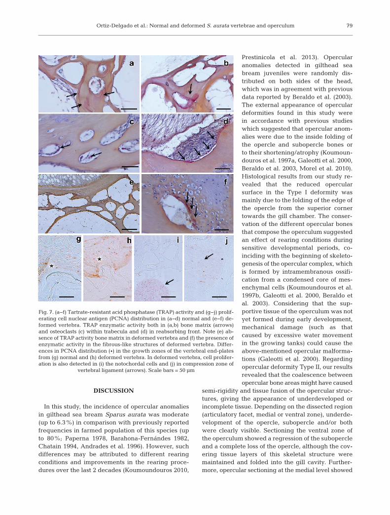
We analysed ALP and TRAP activities in both nor-mal and deformed bone structures in order to identifycells responsible for bone deposition and resorption,respectively. However, our results did not show cleardifferences between non-deformed and deformedskeletal structures with regards to ALP activity (datanot shown). TRAP activity in non-deformed verte-brae was distributed in the bone matrix of the trabec-
76

Ortiz-Delgado et al.: Normal and deformed S. aurata vertebrae and operculum
ular region (Fig. 7a,b), as well as the osteoclastswithin the trabecula lacunae and bone-reabsorbingfronts of normal vertebrae (Fig. 7c,d), but not in de -formed ones (Fig. 7e). Moreover, an intense reactionfor TRAP activity was exclusively detected in thechondrocytic areas of deformed vertebrae (Fig. 7f).
To determine whether the skeletal anomaliesfound in gilthead sea bream could be linked to animbalance in cell cycling rather than to unbalancedcell activity, PCNA immunostaining was performedto measure cell proliferation, and a caspase-3 assay
was conducted to detect processes related to cel l -ular apoptosis. Immunohistochemistry of PCNAshowed differential distribution between normaland deformed vertebral bodies and operculum, witha marked increase of immunopositive cells in thegrowth zone of bone end-plates (Fig. 7g,h) in thenotochordal cells (Fig. 7i) and the compression zoneof vertebral ligaments (Fig. 7j) from deformed verte-brae. However, caspase-3 as an immunomarker ofcell apoptosis did not show such differences (data notshown).
77
Fig. 5. Osteocalcin (OC) and matrix Gla protein (MGP) immunoreactivity counterstained with AB pH 2.5 in normal and de-formed Sparus aurata opercular structures. (a) OC immunostaining in non-deformed operculum; (b) OC and (c) MGP im-munostaining in bone opercular structures from Type I deformity. Note that no differences in OC or MGP protein distributionwithin the bone matrix could be detected. (d) OC and (e) MGP immunostaining in modified articulatory facet in an opercularType II deformity. Note the presence of MGP protein both in cellular elements (osteoblastic-like cells from the articulatoryfacet and cells within cartilage-like tissue, *) and in calcified matrix from the modified articulatory facet. (f) OC immunostain-ing in an opercular Type II deformity at level of ventral zone. Scale bars = 50 µm. Abbreviations: CLT, cartilage-like tissue;Cop, curled operculum; CR, caudal region; MAF, modified articulatory facet; OLC, osteoblastic−like cells; Op, operculum;
PreOp, preoperculum; RS, ribbed structure

Aquat Biol 21: 67–84, 201478
Fig. 6. Osteocalcin (OC) and matrix Gla protein (MGP) distribution counterstained with AB pH 2.5 in control and deformedvertebra of Sparus aurata. Homogeneous OC distribution both in compact and trabecular bone from the periosteal zones of (a)the vertebral centra and from (b) neural and (c) haemal arches. (d) Preferential accumulation of MGP in notochord with mini-mal presence in the different bone layers. Preferential accumulation of OC in (e) compact bone and (f) in notochordal cellsfrom deformed fish coinciding with (g) a calcium deposition (von Kossa staining) in the same area. (h) Fibrous-like structure indeformed vertebra showing glycosaminoglycans (*) instead of OC deposition. (i) OC accumulation in the zone of vertebralend-plates from un-deformed fish and (j) OC deposition in the fibrous layer of the growth zone and notochordal sheath in de-formed fish. (k) MGP distribution in deformed vertebra with a scarce presence of MGP in bone matrix and (l) a slight increasein staining intensity in altered notochordal cells. Detail of a compression area in the vertebral end-plates from a deformed ver-tebra, showing (m) initial and (n) more advanced stages in which osseous tissue is gradually replaced by cartilaginous. OC ac-cumulation in (1) the periphery of (2) the newly formed cartilaginous tissue from the compression area (incipient vertebral fu-sion) stained with (o) H/VOF or (p) not. Scale bars = 50 µm. Abbreviations: C, chordoblasts; CB, compact bone; CD, calciumdeposition; CL, collagenous ligament; FL, fibrous layer; HArch, haemal arch, N, notochord; NArch, neural arch; NS, noto-chordal sheath; OC, osteocalcin; SC, spinal cord; TB, trabecular bone, VCIS, vertebral compression initial stage; VCAS verte-
bral compression in an advanced stage
Ortiz-Delgado et al.: Normal and deformed S. aurata vertebrae and operculum
DISCUSSION
In this study, the incidence of opercular anomaliesin gilthead sea bream Sparus aurata was moderate(up to 6.3%) in comparison with previously reportedfrequencies in farmed population of this species (upto 80%; Paperna 1978, Barahona-Fernándes 1982,Chatain 1994, Andrades et al. 1996). However, suchdifferences may be attributed to different rearingconditions and improvements in the rearing proce-dures over the last 2 decades (Koumoundouros 2010,
Prestinicola et al. 2013). Opercularanomalies detected in gilthead seabream juveniles were randomly dis-tributed on both sides of the head,which was in agreement with previousdata reported by Beraldo et al. (2003).The external appearance of operculardeformities found in this study werein accordance with previous studieswhich suggested that opercular anom-alies were due to the inside folding ofthe opercle and subopercle bones orto their shortening/atrophy (Koumoun -douros et al. 1997a, Galeotti et al. 2000,Beraldo et al. 2003, Morel et al. 2010).Histological results from our study re -vealed that the reduced opercular surface in the Type I deformity wasmainly due to the folding of the edge ofthe opercle from the superior cornertowards the gill chamber. The conser-vation of the different opercular bonesthat compose the operculum suggestedan effect of rearing conditions duringsensitive developmental periods, co -inciding with the beginning of skeleto-genesis of the opercular complex, whichis formed by intramembranous ossifi-cation from a condensed core of mes-enchymal cells (Koumoundouros et al.1997b, Galeotti et al. 2000, Beraldo etal. 2003). Considering that the sup -portive tissue of the operculum was notyet formed during early development,mechanical damage (such as thatcaused by excessive water movementin the growing tanks) could cause theabove-mentioned opercular malforma-tions (Galeotti et al. 2000). Regardingopercular deformity Type II, our resultsrevealed that the coalescence betweenopercular bone areas might have caused
semi-rigidity and tissue fusion of the opercular struc-tures, giving the appearance of underdeveloped orincomplete tissue. Depending on the dissected region(articulatory facet, medial or ventral zone), underde-velopment of the opercle, subopercle and/or bothwere clearly visible. Sectioning the ventral zone ofthe operculum showed a regression of the subopercleand a complete loss of the opercle, although the cov-ering tissue layers of this skeletal structure weremaintained and folded into the gill cavity. Further-more, opercular sectioning at the medial level showed
79
Fig. 7. (a−f) Tartrate-resistant acid phosphatase (TRAP) activity and (g−j) prolif-erating cell nuclear antigen (PCNA) distribution in (a−d) normal and (e−f) de-formed vertebra. TRAP enzymatic activity both in (a,b) bone matrix (arrows)and osteoclasts (c) within trabecula and (d) in reabsorbing front. Note (e) ab-sence of TRAP activity bone matrix in deformed vertebra and (f) the presence ofenzymatic activity in the fibrous-like structures of deformed vertebra. Differ-ences in PCNA distribution (*) in the growth zones of the vertebral end-platesfrom (g) normal and (h) deformed vertebra. In deformed vertebra, cell prolifer-ation is also detected in (i) the notochordal cells and (j) in compression zone of
vertebral ligament (arrows). Scale bars = 50 µm
Aquat Biol 21: 67–84, 201480
an outward folding of the opercle and a thickening ofthe epithelial cell layers. According to Galeotti et al.(2000), the above-mentioned opercular disordersmight be mechanically-induced by forced opercularmovements occurring during ventilation or foodingestion processes, although the putative involve-ment of nutritional factors and/or environmental pollutants should not be neglected (Boglione etal. 2013a).
Specimens displaying the opercular Type II defor-mity also showed a novel type of opercular anomalythat consisted of a coalescence of contacting bone tissues, presumably from the preopercle and opercle.The above-mentioned anomaly had a trabecular as-pect filled by a single cell epithelium of cubic os-teoblastic-like cells. The origin of this anomaly mightbe a disorganization of the blastema (aggregate ofmesenchymal cells), the precursor of the opercularelements formed by intramembranous ossification atearly developmental stages, brought about as aresult of inadequate rearing conditions such as un-balanced diet, inadequate aeration or unfavourabletemperature during the embryonic and larval stage(Loizides et al. 2013). Regardless of the cause of thesetypes of deformities, there is evidence that opercularabnormalities compromise the welfare (Noble et al.2012) and product quality (Cobcroft & Battaglene2013) of the fish stock, as well as the biological per-formance of the fish, reducing their resistance to oxy-gen stress and predisposing the gills to pathologicalinfections (Koumoundouros et al. 1997b). Moreover,a clear connection has been established between op-ercular or vertebral abnormalities and high mortalityrates (Andrades et al. 1996, Loizides et al. 2013).
Opercular bones stained with picrosirius red werevisualized under polarized light, which enhances thenormal birefringence of collagen fibers. The birefrin-gence colour appears to be a measure of the thick-ness of the collagen fibers, the density of their pack-ing and their spatial arrangement. As fiber thicknessincreases, their colour changes from green (thinnercollagen fibers) to bright orange/red (larger collagenfibers; Junqueira et al. 1982, Hiss et al. 1988, Dayanet al. 1989). Thus, we were able to determine po -tential differences in ECM composition. In non-deformed opercula, this technique showed 2 popula-tions of collagen fibers: one group of orange, stronglybirefringent fibres (thick collagen fibers) and anothergroup of green, weakly birefringent fibers (thinfibers). A comparison between normal and deformedopercula revealed differences in collagen fiber thick-ness and in their 3-dimensional arrangement, con-sisting of reduced and scattered areas of orange dye
(fewer big collagen fibers) in deformed opercularbones (opercle and subopercle). This fact has beensuggested as a bone remodelling process linked tochanges in bone composition and degradation of col-lagen fibers (Fernández et al. 2012). Considering thatthe mechanical properties of the opercular bone de -pend on the composition and structure of its matrix(Totland et al. 2011), the thickness of collagen fibersin deformed opercula might indicate a weakness andvulnerability to mechanical disturbances of thisskeletal structure, since collagen content confers cer-tain tissue elasticity (Prades et al. 2010), and there-fore changes in its proportion could reduce tissueflexibility and plasticity (Canavese & Colitti 1996,Ytteborg et al. 2012).
Vertebral column malformations may occur fre-quently in the wild, but are rarely detected (Gavaiaet al. 2009) whereas they are quite commonlydetected under intensive aquaculture conditions(Kranenbarg et al. 2005, Koumoundouros 2010, Pres-tinicola et al. 2013, among others). In this study, thefrequency of lordotic vertebrae (10.1%) was withinthe normal range found in other studies and hatch-eries (Andrades et al. 1996, Koumoundouros et al.2002, Prestinicola et al. 2013). The aetiology of thisskeletal disorder might vary depending on the rear-ing and environmental conditions (see reviews inKoumoundouros 2010, Boglione et al. 2013a). Inde-pendent of the causative factor, lordosis may occur atdifferent developmental stages. For instance, someauthors have reported that vertebral deformities mayappear during the notochord segmentation and ver-tebral centrum differentiation processes (Fernándezet al. 2008, Haga et al. 2009). Others have suggestedthat they are a consequence of dysfunctions in colla-gen metabolism at notochordal and perinotochordalcollagen sheets during early development (Sanata-maría et al. 1994). Moreover, vertebral deformitiesmay also occur later in ontogeny (i.e. during the on-growing period), at which point they are generallyinduced by mechanical overloads (Kranenbarg et al.2005) or by the curvature of the vertebral axis (Gor-man et al. 2010), or a combination of both (Gavaia etal. 2002, Cardeira et al. 2012). Irrespective of thecause of vertebral anomalies, they can compromisethe biological performance of fish. For example, inEuropean sea bass, pre-haemal kyphosis was shownto induce lethargic behaviour and subsequent heavymortality during vertebral axis osteogenesis, as aresult of the compression of the neural tube by thedeformed vertebrae (Koumoundouros et al. 2002).Furthermore, Basaran et al. (2007) showed that lor-dosis significantly decreased the endurance and crit-
Ortiz-Delgado et al.: Normal and deformed S. aurata vertebrae and operculum
ical swimming speed in European sea bass juveniles.In the present study, gilthead sea bream lordotic ver-tebrae showed the formation of fibrous cartilage inthe haemal and/or neural sides of the vertebral cen-trum. This chondroid tissue might meet the demandfor an accelerated growth rate and/or the demand fora shear-resistant support (Huysseune 2000, Cardeiraet al. 2012). Moreover, in the central notochord ofaffected vertebrae, regions of densely packed chor-docytes lacking vacuoles were also observed, whereascalcium deposition within de-vacuolated chordocyteswas detected as reported in salmonids (Ytteborg etal. 2012). Thus, the change in chordocyte morpho -logy from vacuolated to hyperdense, and the increasein calcium deposition in the affected region of gilt-head sea bream lordotic vertebrae might indicatethat a metaplastic shift was involved, as it has beenpreviously described in spinal fusions in fish (Wittenet al. 2005, Ytteborg et al. 2012, Boglione et al.2013b). Another major histomorphological change inlordotic gilthead sea bream vertebrae was the disor-ganization of the intervertebral region, which leadto a complete loss of notochordal sheath integrity.These results were in concordance with those re -ported by other authors for Atlantic salmon Salmonsalar reared at high temperatures (Ytteborg et al.2010), gilthead sea bream fed hypervitaminosis A(Fernández et al. 2012) and guppy Poecilia reticulatadisplaying a curveback syndrome (Gorman et al.2010). Major histomorphological changes of lordoticvertebrae in gilthead sea bream specimens from thisstudy were linked to the loss of the integrity of noto-chordal cells, which was associated with the pres-ence of modified chordocytes in which calcium depo-sition was noticeable. Similarly, Loizides et al. (2013)pointed out the ectopic presence of altered chordo-cytes as well as increased notochordal sheath pro-duction as the causes of the vertebral compressionand fusion (VCF) syndrome in gilthead sea bream.
PCNA immunohistochemistry indicated that osteo -blasts in the growth zone of the bone end-platesshowed a marked increase in cell proliferation indeformed vertebral centra, which was not counter-acted by an increase in cell death as revealed by caspase-3 immunostaining. In addition, a markedincrease in cell proliferation (PCNA staining), but notof cell apoptosis was also detected in notochordalcells and in the compression zone of vertebral liga-ment in lordotic vertebrae. Several studies in highervertebrates have suggested that changes in the bal-ance between cell death and cell proliferation areinvolved in bone and cartilage defects, which couldultimately lead to skeletal deformities (Cockroft
& New 1978, Miura et al. 2004). In this sense, theabove-mentioned imbalanced cell cycling detectedin lordotic vertebrae might also explain the presenceof dense packaged chordocytes, occupying most ofthe intervertebral space without vacuolation, as wasdescribed by Ytteborg et al. (2010) in Atlantic salmonvertebral fusions. Regarding the bone homeostasismeasured by ALP and TRAP immunostaining, al -though this procedure may not be considered to bea quantitative assessment of bone formation andresorption, the present data might indicate that lor-dotic vertebrae were subjected to a more localizedbone resorption process than non-deformed ones,since TRAP activity was only found in the chondro-cytic areas of deformed vertebrae but was homoge-neously distributed in the trabecular spaces of nor-mal vertebrae. Local and intense TRAP activity indeformed vertebrae is in agreement with the re -duced thickness of collagen fibers and the previouslysuggested bone remodelling process occurring inthis deformed skeletal structure. These results differfrom those previously described in spinal fusions byYtteborg et al. (2012) and Boglione et al. (2013b).
In the vertebral growth zones of the teleosts, ECM-producing cells (osteoblasts and bone lining cells)express a combination of proteins having distinctfunctions in mineralisation and/or deposition of theosteoid (Krossøy et al. 2009, Boglione et al. 2013b).Among these proteins, OC and MGP have beenreported to be involved in the mineralization of theECM. In this sense, OC has been proposed as a regulator of bone maturation (Krossøy et al. 2009),whereas MGP acts as an inhibitor of calcification(Luo et al. 1997). In this study, a preferential accumu-lation of OC in compact bone and notochordal cells oflordotic vertebrae was detected, which might beattributed to the remodelling process occurring inlordotic vertebrae (Boglione et al. 2013b) as well asthe above-mentioned cartilage formation by meta-plasic transformation of bone, forming cells thatwould be latter mineralized and remodelled intobone (Witten et al. 2005). Concerning MGP, a prefer-ential deposition was detected in notochordal cells ofnon-deformed vertebrae compared with deformedones. Higher levels of OC and MGP deposition innotochordal tissue of deformed vertebrae were simi-lar to those reported by Fernández et al. (2012) ingilthead sea bream juveniles fed hypervitaminosis A.These results are in agreement with previous studiesin which an increased co-transcription of both chon-drogenic and osteogenic markers were found in thenotochord of Atlantic salmon displaying spinal fusions(Ytteborg et al. 2010, 2012). Furthermore, bone tissue
81

Aquat Biol 21: 67–84, 2014
with lower levels of OC might demineralize moreeasily than that with higher OC content (Krossøy etal. 2009), suggesting that lordotic vertebrae may bemore fragile than non-deformed ones. However, thishypothesis requires further corroboration by addi-tional mineral content analyses.
CONCLUSIONS
This study provided a comprehensive descriptionof the main morphological and histological featuresof normal and deformed opercula and vertebrae fromgilthead sea bream in order to increase basic knowl-edge of bone disorders in this species. Our data re -vealed that important histological, histochemical andimmunohistochemical differences were found be -tween non-deformed and deformed opercula andvertebrae. A rare type of tissue remodelling processwas described in fish displaying the Type II operculardeformity, which consisted of the coalescence of con-tacting bone tissues, presumably from the preopercleand opercle, resulting in skeletal tissue with a trabec-ular aspect filled by a single cell epithelium of cubicosteoblastic-like cells. Additionally, there were dif-ferences in collagen fiber thickness and 3-dimen-sional arrangement between normal and deformedopercula, as well as hypoplasia of muscle fibers,which might affect the flexibility of deformed oper-cula. Lordotic gilthead sea bream vertebrae showedthe formation of fibrous cartilage in their haemaland/or neural sides, indicating that a metaplasticshift occurred during the process of lordosis. Anothermajor histomorphological change in lordotic verte-brae was the complete loss of notochordal sheathintegrity. These alterations were coupled with animbalance between cell death and cell proliferationprocesses in lordotic vertebrae, as well as in bone formation/resorption and ECM deposition activity,which might have resulted from the remodelling pro-cess occurring in lordotic vertebrae. Altogether,these results provide an increase in the basic knowl-edge of bone disorders that will add to our under-standing of the mechanisms by which these skeletaldisorders appear in this fish species and which ham-per its production efficiency.
Acknowledgements. The authors express their gratitude toI. Viaña for providing technical assistance and to Dr. D.Simes for providing the OC and MGP antibodies. This workwas funded by Ministry of Science and Innovation (MICIIN)of the Spanish government (projects AGL2008-03897-C01/C04 and AGL2010-15951). J.B.O-D. was supported by thePrograma Ramón y Cajal (MICINN, Spain). I.F. was sup-
ported by a predoctoral Spanish MICINN fellowship (refer-ence, BES- 2006-12650) and a postdoctoral fellowship(SFRH/BDP/82049/2011) from Fundação para a Ciência eTecnologia (FCT), Portugal.
LITERATURE CITED
Andrades JA, Becerra J, Fernández-Llebrez P (1996) Skele-tal deformities in larval, juvenile and adult stages of cul-tured gilthead sea bream (Sparus aurata L.). Aquaculture141: 1−11
Bakke-McKellep AM, Froystad MK, Lilleeng E, Dapra F,Refstie S, Krogdahl A, Landsverk T (2007) Response tosoy: T-cell-like reactivity in the intestine of Atlanticsalmon, Salmo salar L. J Fish Dis 30: 13−25
Bancroft JD, Stevens A (1990) Theory and practice of histo-logical techniques. Churchill Livingstone, London
Barahona-Fernándes MH (1982) Body deformation in hatch-ery reared European sea bass Dicentrachus labrax (L).Types, prevalence and effect on fish survival. J Fish Biol21: 239−249
Basaran F, Ozbilgin H, Ozbilgin YD (2007) Effect of lordosison the swimming performance of juvenile sea bass(Dicentrarchus labrax L.). Aquacult Res 38: 870−876
Beraldo P, Pinosa M, Tibaldi E, Canavese B (2003) Abnor-malities of the operculum in gilthead sea bream (Sparusaurata): morphological description. Aquaculture 220: 89−99
Boglione C, Gagliardi F, Scardi M, Cataudella S (2001)Skeletal descriptors and quality assessment in larvae andpost-larvae of wild-caught and hatchery-reared giltheadsea bream (Sparus aurata L. 1758). Aquaculture 192: 1−22
Boglione C, Gisbert E, Gavaia P, Witten PE, Moren M,Fontagné S, Koumoundouros G (2013a) A review onskeletal anomalies in reared European larvae and juve-niles. Part 2: Main typologies, occurrences and causativefactors. Rev Aquac 5: S121−S167
Boglione C, Gavaia P, Koumoundouros G, Gisbert E, MorenM, Fontagné S, Witten PE (2013b) A review on skeletalanomalies in reared European fishes. Part 1: Normaland anomalous skeletogenic processes. Rev Aquac 5: S99−S120
Bonewald LF, Harris SE, Rosser J, Dallas MR and others(2003) Von Kossa staining alone is not sufficient to con-firm that mineralization in vitro represents bone for -mation. Calcif Tissue Int 72: 537−547
Canavese B, Colitti M (1996) Observations under LM andSEM of opercle malformations in sea bream (Sparusaurata). Teratology 53: 27A (Abstract)
Cardeira J, Bensimon-Brito A, Pousão-Ferreira P, Cancela ML,Gavaia PJ (2012) Lordotic-kyphotic vertebrae developectopic cartilage-like tissue in Senegalese sole (Soleasenegalensis). J Appl Ichthyology 28: 460−463
Castro J, Pino-Querido A, Hermida M, Chavarrias D andothers (2008) Heritability of skeleton abnormalities (lor-dosis, lack of operculum) in gilthead seabream (Sparusaurata) supported by microsatellite family data. Aqua-culture 279: 18−22
Chatain B (1994) Abnormal swimbladder development andlordosis in sea bass (Dicentrachus labrax) and sea bream(Sparus aurata). Aquaculture 119: 371−379
Clark G (1981) Staining procedures. Williams & Wilkins,Baltimore, MD
Cobcroft JM, Battaglene SC (2013) Skeletal malformations
82

Ortiz-Delgado et al.: Normal and deformed S. aurata vertebrae and operculum
in Australian marine finfish hatcheries. Aquaculture 396-399: 51−58
Cockroft DL, New DAT (1978) Abnormalities induced in cultured rat embryos by hypertermia. Teratology 17: 277−283
Daoulas C, Economou AN, Bantavas I (1991) Osteologicalabnormalities in laboratory reared sea-bass (Dicentrar-chus labrax) fingerlings. Aquaculture 97: 169−180
Dayan D, Hiss Y, Hirshberg A, Bubis JJ, Wolman M (1989)Are the polarization colors of Picrosirius red-stained collagen determined only by the diameter of the fibers?Histochemistry 93: 27−29
Diggles BK (2013) Saddleback deformities in yellowfinbream, Acanthopagrus australis (Günther), from SouthEast Queensland. J Fish Dis 36: 521−527
Faustino M, Power DM (1998) Development of osteologicalstructures in the sea bream: vertebral column and caudalfin complex. J Fish Biol 52: 11−22
Faustino M, Power DM (1999) Development of the pectoral,pelvic, dorsal and anal fins in cultured sea bream. J FishBiol 54: 1094−1110
Faustino M, Power DM (2001) Osteologic development ofthe viscerocranial skeleton in sea bream: alternative ossi -fication strategies in teleost fish. J Fish Biol 58: 537−572
Fernández I, Hontoria F, Ortiz-Delgado JB, Kotzamanis Y,Estévez A, Zambonino-Infante J, Gisbert E (2008) Larvalperformance and skeletal deformities in farmed giltheadsea bream (Sparus aurata) fed with graded levels of Vitamin A enriched rotifers (Brachionus plicatilis). Aqua-culture 283: 102−115
Fernández I, Ortiz-Delgado JB, Sarasquete C, Gisbert E(2012) Vitamin A effects on vertebral bone tissue home-ostasis in gilthead sea bream (Sparus aurata) juveniles.J Appl Ichthyology 28: 419−426
Galeotti M, Beraldo P, de Dominis S, D’Angelo L and others(2000) A preliminary histological and ultrastructuralstudy of opercular anomalies in gilthead sea bream larvae (Sparus aurata). Fish Physiol Biochem 22: 151−157
Gavaia PJ, Dinis MT, Cancela ML (2002) Osteological devel-opment and abnormalities of the vertebral column andcaudal skeleton in larval and juvenile stages of hatchery-reared Senegal sole (Solea senegalensis). Aquaculture211: 305−323
Gavaia PJ, Domingues S, Engrola S, Drake P, Sarasquete C,Dinis MT, Cancela ML (2009) Comparing skeletal devel-opment of wild and hatchery-reared Senegalese sole(Solea senegalensis, Kaup 1985): evaluation in larval andpostlarval stages. Aquacult Res 40: 1585−1593
Gorman KF, Handrigan GR, Jin G, Wallis R, Breden F (2010)Structural and micro-anatomical changes in vertebraeassociated with idiopathic-type spinal curvature in thecurveback guppy model. Scoliosis 5: 10
Gutiérrez M (1967) Coloración histológical para ovarios depeces, crustáceos y moluscos. Inv Pesq 31: 265−271
Haga Y, Dominique VJ, Du SJ (2009) Analyzing notochordsegmentation and intervertebral disc formation using thetwhh: gfp transgenic zebrafish model. Transgenic Res 18: 669−683
Hiss J, Hirshberg A, Fundoiano-Dayan D, Bubis JJ, WolmanH (1988) Aging of wound healing in an experimentalmodel in mice. Am J Forensic Med Pathol 9: 310−312
Huysseune A (2000) Skeletal system. In: Ostrander GK (ed)The laboratory fish. Academic Press, San Diego, CA,p 307−317
Junqueira LCU, Bignolas G, Brentani RR (1979) Picrosirius
staining plus polarization microscopy, a specific methodfor collagen detection in tissue sections. Histochem J 11: 447−455
Junqueira LCU, Montes GS, Sanchez EM (1982) The influ-ence of tissue sections thickness on the study of collagenby the picrosirius-polarization method. Histochemistry74: 153−156
Karsenty G (2001) Transcriptional control of osteoblast differentiation. Endocrinology 142: 2731−2733
Kiernan JA (2008) Methods for connective tissue. In: Kiernan JA (ed) Histological and histochemical methods: theory and practice, 4th edn. Scion, Bloxham, p 190−213
Koumoundouros G (2010) Morpho-anatomical abnormalitiesin Mediterranean marine aquaculture. In: Koumoun -douros G (ed) Recent advances in aquaculture research.Transworld Research Network, Kerala, p 125−148
Koumoundouros G, Gagliardi F, Divanach P, Boglione D,Cataudella S, Kentouri M (1997a) Normal and abnormalosteological development of caudal fin in Sparus aurataL. fry. Aquaculture 149: 215−226
Koumoundouros G, Oran G, Divanach P, Stefanakis S, Ken-touri M (1997b) The opercular complex deformity inintensive gilthead sea bream (Sparus aurata L.) larvicul-ture. Moment of apparition and description. Aquaculture156: 165−177
Koumoundouros G, Maingot E, Divanach P, Kentouri M(2002) Kyphosis in reared sea bass (Dicentrarchus labraxL.): ontogeny and effects on mortality. Aquaculture 209: 49−58
Kranenbarg S, Waarsing JH, Muller M, Weinans H, vanLeeuwen JL (2005) Lordotic vertebrae in sea bass(Dicentrarchus labrax L.) are adapted to increased loads.J Biomech 38: 1239−1246
Krossøy C, Ornsrud R, Wargelius A (2009) Differential geneexpression of bgp and mgp in trabecular and compactbone of Atlantic salmon (Salmo salar L.) vertebrae.J Anat 215: 663−672
Loizides M, Georgiou AN, Somarakis S, Witten PE,Koumoundouros G (2013) A new type of lordosis and ver-tebral body compression in Gilthead seabream (Sparusaurata Linnaeus, 1758): aetiology, anatomy and conse-quences for survival. J Fish Dis, doi: 10.1111/jfd.12189
Luo G, Ducy P, Mckee MD, Pinenro GJ, Loyer E, BehringerRR, Karsenty G (1997) Spontaneous calcification of arteries and cartilage in mice lacking matrix GLA protein.Nature 386: 78−81
Martoja R, Martoja-Pierson M (1970) Técnicas de histologíaanimal. Toray-Masson, Barcelona
Meunier FJ (2011) The osteichtyes, from the paleozoic to theextant time, through histology and palaeohistology ofbony tissues. C R Pal 10: 347−355
Miura M, Chen XD, Allen MR, Bi YM, Gronthos S, Seo BM(2004) A crucial role of caspase-3 in osteogenic differen-tiation of bone marrow stromal stem cells. J Clin Invest114: 1704−1713
Morel C, Adriaens D, Boone M, De Wolf T, Van HoorebekeL, Sorgeloos P (2010) Visualizing mineralization in de -formed opercular bones of larval gilthead sea bream(Sparus aurata). J Appl Ichthyology 26: 278−279
Noble C, Cañon Jones H, Damsgård B, Flood M and others(2012) Injuries and deformities in fish: their potentialimpacts upon aquacultural production and welfare. FishPhysiol Biochem 38: 61−83
Ortega A (2008) Cultivo de Dorada (Sparus aurata). In: Espinosa de los Monteros J (ed) Cuadernos de Acuicul-
83

Aquat Biol 21: 67–84, 2014
tura. Fundación Observatorio Español de Acuicultura,Madrid
Ortiz-Delgado JB, Simes DC, Gavaia P, Sarasquete C, Can-cela ML (2005) Osteocalcin and matrix GLA protein indeveloping teleost teeth: identification of sites of mRNAand protein accumulation at single cell resolution. His-tochem Cell Biol 124: 123−130
Paperna I (1978) Swimbladder and skeletal deformation inhatchery bred Sparus aurata. J Fish Biol 12: 109−114
Pearse AGE (1985) Histochemistry: theoretical and applied,4th edn. Vol 2: analytical technology. Churchill Livin-stone, Edinburgh
Prades JM, Dumollard JM, Duband S, Timoshenko A andothers (2010) Lamina propria of the human vocal fold: histomorphometric study of collagen fibers. Surg RadiolAnat 32: 377−382
Prestinicola L, Boglione C, Makridis P, Spanò A and others(2013) Environmental conditioning of skeletal anomaliestypology and frequency in gilthead seabream (Sparusaurata L., 1758) juveniles. PLoS ONE 8: e55736
Sanden M, Berntssen MHG, Krogdahl A, Hemre GI, Bakke-McKellep AM (2005) An examination of the intestinaltract of Atlantic salmon, Salmo salar L., parr fed differentvarieties of soy and maize. J Fish Dis 28: 317−330
Sanatamaría JA, Andrades JA, Herráez P, Fernández-Llebrez P, Becerra J (1994) Perinotochordal connectivesheet of gilthead sea bream larvae (Sparus aurata, L.)affected by axial malformations: a histochemical andimmunocytochemical study. Anat Rec 240: 248−254
Sarasquete C, Gutiérrez M (2005) New tetrachromic VOFstain (Type III-G.S) for normal and pathological fish tissues. Eur J Histochem 49: 105−114
Simes DC, Williamson M, Ortiz-Delgado JB, Viegas SCB,Price PA, Cancela ML (2003) Purification of matrix Glaprotein from a marine teleost fish, Argyrosomus regius: calcified cartilage and not bone as the primary site ofMGP accumulation in fish. J Bone Miner Res 18: 244−259
Simes DC, Williamson M, Schaff BJ, Gavaia PJ, Ingleton
PM, Price PA, Cancela ML (2004) Charaterization ofosteocalcin (BGP) and matrix Gla protein (MGP) fish specific antibodies: validation for immunodetection studies in lower vertebrates. Calcif Tissue Int 74: 170−180
Tiago DM, Laizé V, Bargelloni L, Ferraresso S, Romualdi C,Cancela ML (2011) Global analysis of gene expressionin mineralizing fish vertebra-derived cell lines: newinsights into anti-mineralogenic effect of vanadate. BMCGenomics 12: 310
Totland GK, Fjelldal PG, Kryvi H, Løkka G and others (2011)Sustained swimming increases the mineral content andosteocyte density of salmon vertebral bone. J Anat 219: 490−501
Verhaegen Y, Adriaens D, De Wolf T, Dhert P, Sorgeloos P(2007) Deformities in larval gilthead sea bream (Sparusaurata): a qualitative and quantitative analysis usinggeometric morphometrics. Aquaculture 268: 156−168
Witten PE, Gil-Martens L, Hall BK, Huysseune A, Obach A(2005) Compressed vertebrae in Atlantic salmon Salmosalar: evidence for metaplastic chondrogenesis as askeletogenic response late in ontogeny. Dis Aquat Org64: 237−246
Witten PE, Obach A, Huyseune A, Baeverfjord G (2006) Ver-tebrae fusion in Atlantic salmon (Salmo salar): develop-ment aggravation and pathways of containment. Aqua-culture 258: 164−172
Witten PE, Gil-Martens L, Huysseune A, Takle H, Hjelde K(2009) Towards a classification and an understanding ofdevelopmental relationships of vertebral body malforma-tions in Atlantic salmon (Salmo salar L.). Aquaculture295: 6−14
Ytteborg E, Baeverfjord G, Torgersen J, Hjelde K, Takle H(2010) Molecular pathology of vertebral deformities inhyperthermic Atlantic salmon (Salmo salar). BMC Physiol10: 12
Ytteborg E, Baeverfjord G, Takle H (2012) Four stages char-acterizing vertebral fusions in Atlantic salmon. J ApplIchthyology 28: 453−459
84
Editorial responsibility: Christine Paetzold, Oldendorf/Luhe, Germany
Submitted: September 16, 2013; Accepted: April 14, 2014Proofs received from author(s): June 5, 2014