nonhealing vulvar ulcer - home | american academy … · lichen sclerosus is an eruption of...
TRANSCRIPT
August 1, 2011 ◆ Volume 84, Number 3 www.aafp.org/afp American Family Physician 311
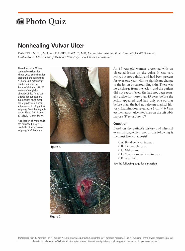
An 89-year-old woman presented with an ulcerated lesion on the vulva. It was very itchy, but not painful, and had been present for over one year with no significant change to the lesion or surrounding skin. There was no discharge from the lesion, and the patient did not report fever. She had not been sexu-ally active for more than 15 years before the lesion appeared, and had only one partner before that. She had no relevant medical his-tory. Examination revealed a 1 cm × 0.5 cm erythematous, ulcerated area on the left labia majora (Figures 1 and 2).
QuestionBased on the patient’s history and physical examination, which one of the following is the most likely diagnosis?
❑ A. Basal cell carcinoma. ❑ B. Lichen sclerosus. ❑ C. Melanoma. ❑ D. Squamous cell carcinoma. ❑ E. Syphilis.
See the following page for discussion.
Nonhealing Vulvar UlcerDANETTE NULL, MD, and DANIELLE WALZ, MD, Memorial/Louisiana State University Health Sciences Center–New Orleans Family Medicine Residency, Lake Charles, Louisiana
The editors of AFP wel-come submissions for Photo Quiz. Guidelines for preparing and submitting a Photo Quiz manuscript can be found in the Authors’ Guide at http://www.aafp.org/afp/ photoquizinfo. To be con-sidered for publication, submissions must meet these guidelines. E-mail submissions to [email protected]. Contributing edi-tor for Photo Quiz is John E. Delzell, Jr., MD, MSPH.
A collection of Photo Quiz-zes published in AFP is available at http://www.aafp.org/afp/photoquiz.
Figure 2.
Photo Quiz
Figure 1.
Downloaded from the American Family Physician Web site at www.aafp.org/afp. Copyright © 2011 American Academy of Family Physicians. For the private, noncommercial use of one individual user of the Web site. All other rights reserved. Contact [email protected] for copyright questions and/or permission requests.

Photo Quiz
312 American Family Physician www.aafp.org/afp Volume 84, Number 3 ◆ August 1, 2011
DiscussionThe correct answer is D: squamous cell carcinoma. Biopsy confirmed the diagnosis of basaloid squamous carcinoma, a type of squamous cell carcinoma. More than 90 percent of vulvar malignancies are squamous cell carcinomas.1 Vulvar squamous cell carcinoma typi-cally presents as a unifocal plaque, ulcer, or mass on the labia majora1 and is most common in postmenopausal white women.1,2 Patients often report a pruritic area that becomes a red, nonhealing wound.1 Vulvar bleeding, discharge, dysuria, and enlarged lymph nodes are less common symptoms.1,3 Many patients are asymptomatic at the time of diagnosis. The signs and symptoms of vul-var malignancies are similar, so it is essential to biopsy all lesions to establish a diagnosis.
Squamous cell carcinoma is strongly associated with human papillomavirus types 16 and 33 or chronic inflammation from an autoimmune process.1,4 Human papillomavirus is responsible for approximately 60 per-cent of vulvar cancers.1,4 Risk factors for vulvar cancers include smoking, vulvar dystrophy (lichen sclerosus), vulvar or cervical intraepithelial neoplasia, immuno-deficiency syndromes, history of cervical cancer, and Northern European ancestry.1,4
Treatment options for squamous cell carcinoma include surgical excision, radiation therapy, and
chemotherapy. Each of the treatments may be used alone or in combination, depending on the stage of the lesion.1,3 The prognosis is determined by the size of the lesion, lymph node involvement, and presence or absence of distant metastasis. Squamous cell carcinoma that is found early, before it spreads into deeper tissues, has a 90 percent cure rate.1,3
Basal cell carcinoma tends to affect the face, but comprises approximately 2 percent of vulvar cancers.2 It may present as a locally invasive rodent, or Jacob, ulcer with rolled edges and a central ulceration. The lesions may be pigmented or pearly gray and typically have a granular appearance.
Lichen sclerosus is an eruption of pruritic, white, atrophic papules and plaques that usually occur in post-menopausal women. The lesions are either discrete or confluent and contain a central depression.
Melanoma is the second most common vulvar malig-nancy, accounting for 5 to 10 percent of primary vulvar neoplasms.1,2 The disease predominantly affects non-Hispanic white, postmenopausal women.2 Melanomas often present as a mass or nodule with bleeding, dis-charge, dysuria, and pain.2 The disease rarely affects genitalia, but lesions are more common on the clitoris or the labia minora. Melanomas are usually pigmented, although amelanotic lesions occur. Melanoma rarely presents as an ulcer.
Syphilis is an infectious disease caused by Treponema pallidum that is transmitted via direct sexual contact. A chancre, which develops after an incubation period of 12 to 30 days, starts as a painless papule or an area of infiltration that is a dull red color and hard. The center then erodes and becomes ulcerated. Unlike a squamous cell carcinoma lesion, this chancre will heal slowly over four to six weeks followed by a generalized rash.
Address correspondence to Danette Null, MD, at [email protected]. Reprints are not available from the authors.
Author disclosure: No relevant financial affiliations to disclose.
REFERENCES
1.CanavanTP,CohenD.Vulvar cancer.Am Fam Physician.2002;66(7):1269-1274.
2.FinanMA,BarreG.Bartholin’sglandcarcinoma,malignantmelanomaandotherraretumoursofthevulva.Best Pract Res Clin Obstet Gynae-col.2003;17(4):609-633.
3.Ansink A. Vulvar squamous cell carcinoma. Semin Dermatol. 1996;15(1):51-59.
4.MonkBJ,BurgerRA,LinF,ParhamG,VasilevSA,WilczynskiSP.Prog-nosticsignificanceofhumanpapillomavirusDNAinvulvarcarcinoma.Obstet Gynecol.1995;85(5pt1):709-715.■
Summary Table
Condition Characteristics
Basalcellcarcinoma
Locallyinvasiverodent,orJacob,ulcerwithrollededgesandcentralulceration;pigmentedorpearlygraywithgranularappearance;tendstoaffecttheface
Lichensclerosus
Pruritic,white,atrophicpapulesandplaques;discreteorconfluentwithcentraldepression
Melanoma Massornodulewithbleeding,discharge,dysuria,andpain;usuallypigmented;rarelyaffectsgenitalia,butmorecommononclitorisorlabiaminora
Squamouscellcarcinoma
Unifocalplaque,ulcer,ormass;pruriticareabecomesred,nonhealingwound
Syphilis
Chancreappearsaspainlesspapuleorareaofinfiltrationthatisadullredcolorandhard,thencentererodesandbecomesulcerated;chancrehealsonitsown,followedbyageneralizedrash