nonconvulsive seizures: developing a rational … j, et al. nonconvulsive... · nonconvulsive...
TRANSCRIPT

www.elsevier.com/locate/clinph
Clinical Neurophysiology 118 (2007) 1660–1670
Invited review
Nonconvulsive seizures: Developing a rational approachto the diagnosis and management in the critically ill population
J. Jirsch1, L.J. Hirsch *
Comprehensive Epilepsy Center, Columbia University Medical Center, New York, NY, USA
See Editorial, pages 1653–1654
Abstract
Originally described in patients with chronic epilepsy, nonconvulsive seizures (NCSs) are being recognized with increasing frequency,both in ambulatory patients with cognitive change, and even more so in the critically ill. In fact, the majority of seizures that occurin the critically ill are nonconvulsive and can only be diagnosed with EEG monitoring. The semiology of NCSs and the associatedEEG findings are quite variable. There are a number of periodic, rhythmic or stimulation-related EEG patterns in the critically ill ofunclear significance and even less clear treatment implications. The field struggles to develop useful diagnostic criteria for NCSs, to stan-dardize nomenclature for the numerous equivocal patterns, and to devise studies that will help determine which patterns should be trea-ted and how aggressively. This review surveys the evidence for and against NCSs causing neuronal injury, and attempts to develop arational approach to the diagnosis and management of these seizures, particularly in the encephalopathic population.� 2007 International Federation of Clinical Neurophysiology. Published by Elsevier Ireland Ltd. All rights reserved.
Keywords: Nonconvulsive seizure; Nonconvulsive status epilepticus; Critically ill; Intensive care unit; EEG; Periodic discharges
1. Introduction
Early descriptions of convulsive seizures are found inBabylonian tablets from 2000 BC that emphasize thesupernatural nature of these events. Centuries later, Hippo-crates wrote on the ‘‘sacred disease’’ and argued that epi-leptic convulsions were manifestations of a diseased brainand recommended physical treatments. Detailed accountsof less overt epilepsies began in the 18th and 19th centurywith the French description of ‘‘petit mal’’ seizures andHughlings Jackson’s ‘‘uncinate fits’’ (Kaplan, 2002). Theadvent of EEG in the 20th century allowed physicians todiscover unequivocal seizures in previously unsuspectedcircumstances, yet the field still struggles to define theextent of EEG patterns that should be considered ictaland to determine when these events should be treated. Thisreview examines nonconvulsive seizures in the critically ill
1388-2457/$32.00 � 2007 International Federation of Clinical Neurophysiolo
doi:10.1016/j.clinph.2006.11.312
* Corresponding author. Tel.: +1 212 305 1742; fax: +1 212 305 1450.E-mail address: [email protected] (L.J. Hirsch).
1 Current address: Department of Neurology and Neurosurgery, McGillUniversity Health Center, Montreal, Canada.
population, particularly emphasizing controversies as towhich EEG patterns should be considered ictal, andexploring the evidence whether such seizures are intrinsi-cally harmful. Whenever possible the American Academyof Neurology classification of evidence scheme is used(Appendix, for example, see Armon and Evans, 2005).
2. Nonconvulsive seizures in encephalopathic patients
The clinical spectrum in nonconvulsive seizures (NCSs)is protean (Table 1). In the community, NCSs present mostfrequently in patients who are confused but remain ambu-latory, while in the hospital NCSs are most frequently diag-nosed in the intensive care unit (ICU). We focus on theobtunded or comatose patients for this article, and in thesecases the clinical diagnosis is often challenging as manifes-tations are often absent or may consist of only subtle myo-clonic limb, facial or ocular movements. In fact,approximately 90% of critically ill patients with seizuresrecorded have purely nonconvulsive seizures that areunrecognized at the bedside and can only be diagnosed
gy. Published by Elsevier Ireland Ltd. All rights reserved.
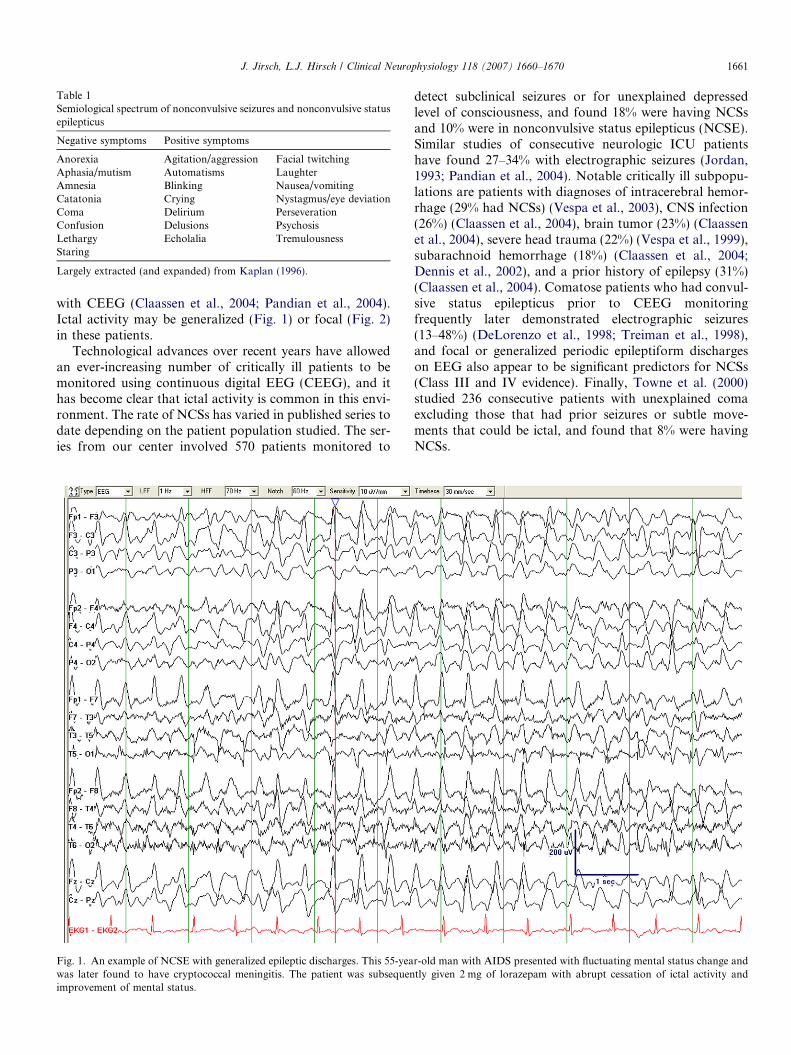
Table 1Semiological spectrum of nonconvulsive seizures and nonconvulsive statusepilepticus
Negative symptoms Positive symptoms
Anorexia Agitation/aggression Facial twitchingAphasia/mutism Automatisms LaughterAmnesia Blinking Nausea/vomitingCatatonia Crying Nystagmus/eye deviationComa Delirium PerseverationConfusion Delusions PsychosisLethargy Echolalia TremulousnessStaring
Largely extracted (and expanded) from Kaplan (1996).
J. Jirsch, L.J. Hirsch / Clinical Neurophysiology 118 (2007) 1660–1670 1661
with CEEG (Claassen et al., 2004; Pandian et al., 2004).Ictal activity may be generalized (Fig. 1) or focal (Fig. 2)in these patients.
Technological advances over recent years have allowedan ever-increasing number of critically ill patients to bemonitored using continuous digital EEG (CEEG), and ithas become clear that ictal activity is common in this envi-ronment. The rate of NCSs has varied in published series todate depending on the patient population studied. The ser-ies from our center involved 570 patients monitored to
Fig. 1. An example of NCSE with generalized epileptic discharges. This 55-yeawas later found to have cryptococcal meningitis. The patient was subsequenimprovement of mental status.
detect subclinical seizures or for unexplained depressedlevel of consciousness, and found 18% were having NCSsand 10% were in nonconvulsive status epilepticus (NCSE).Similar studies of consecutive neurologic ICU patientshave found 27–34% with electrographic seizures (Jordan,1993; Pandian et al., 2004). Notable critically ill subpopu-lations are patients with diagnoses of intracerebral hemor-rhage (29% had NCSs) (Vespa et al., 2003), CNS infection(26%) (Claassen et al., 2004), brain tumor (23%) (Claassenet al., 2004), severe head trauma (22%) (Vespa et al., 1999),subarachnoid hemorrhage (18%) (Claassen et al., 2004;Dennis et al., 2002), and a prior history of epilepsy (31%)(Claassen et al., 2004). Comatose patients who had convul-sive status epilepticus prior to CEEG monitoringfrequently later demonstrated electrographic seizures(13–48%) (DeLorenzo et al., 1998; Treiman et al., 1998),and focal or generalized periodic epileptiform dischargeson EEG also appear to be significant predictors for NCSs(Class III and IV evidence). Finally, Towne et al. (2000)studied 236 consecutive patients with unexplained comaexcluding those that had prior seizures or subtle move-ments that could be ictal, and found that 8% were havingNCSs.
r-old man with AIDS presented with fluctuating mental status change andtly given 2 mg of lorazepam with abrupt cessation of ictal activity and

Fig. 2. An example of NCSE with focal ictal activity. This 39-year-old woman with primary CNS lymphoma presented with a cluster of generalizedconvulsions, but several hours later was poorly responsive. A right parieto-temporal hemorrhage was present on imaging with associated right hemisphereictal activity.
1662 J. Jirsch, L.J. Hirsch / Clinical Neurophysiology 118 (2007) 1660–1670
Clearly, NCSs are common in neurocritical care patientsand it has become evident that the majority of these eventswould be missed without prolonged EEG studies. The seriesby Pandian et al. (2004) illustrates this point well where allpatients in their series had 30 min routine EEGs at the begin-ning of their monitoring. The authors found that more thanhalf of all seizure cases would have been missed without pro-longed recording (routine EEG found seizures in 11% ofpatients vs. 27% with cEEG; median duration of CEEGwas 2.9 days). Similarly, our institution found seizure activ-ity present at the beginning of the EEG record in 16 of 110(15%) patients who would eventually develop ictal activityduring monitoring, and in about half of patients by 1 h.
The duration of monitoring required to exclude thepresence of seizures in critically ill patients remains some-what unclear. Claassen et al. (2004) found that of 110patients with seizures during CEEG, 95% of noncomatosepatients had their first seizure identified within 24 h ofrecording, but the first seizure had been recorded in only80% of the comatose patients. Furthermore, 13% of thecomatose patients with seizures did not have their first sei-zure until more than 48 h of recording. The presence of cer-tain interictal patterns may help predict a delayed time tofirst seizure, and a finding of periodic lateralized epileptic
discharges (PLEDs) should prompt the clinician to con-tinue EEG monitoring beyond 24 h as 21% of thesepatients had their first seizure after the first full day ofcEEG compared with 8% in those without PLEDs (ClassIII evidence). There are almost certainly other importantEEG monitoring variables to help improve the sensitivityof NCS detection in the ICU, and to date little is knownabout the optimal number of channels or electrodesrequired in these patients.
The specificity and accuracy of CEEG monitoring forseizure detection is also incompletely understood. Largecenters that review abundant EEG monitoring records incritically ill patients recognize that a clear division ofEEG patterns as either ictal or interictal is elusive or non-existent, and interpretation varies considerably among dif-ferent electroencephalographers. Indeed the strength ofevidence suggesting various periodic EEG patterns are pre-dictive of NCSs is weakened in many studies by a lack ofindependent definitions of the two. EEG patterns that havehistorically been considered non-ictal are on occasionreportedly ictal. A case in point is triphasic waves, whichcommonly occur in metabolic encephalopathies but alsoin cortical degenerative diseases, CNS infections, and sepsis(Sundaram and Blume, 1987; Young et al., 1990). They are

J. Jirsch, L.J. Hirsch / Clinical Neurophysiology 118 (2007) 1660–1670 1663
classically characterized by generalized, frontally dominantperiodic discharges at 1–2 Hz (but occasionally faster thanthis), often with fronto-occipital lag. Triphasic waves, how-ever, can be difficult to distinguish from NCSE becausewith both patterns patients typically have altered con-sciousness, and discharges may fluctuate at greater than1 Hz (Sheridan and Sato, 1986). Phase two amplitude pre-dominance and antero-posterior lag on referential montageare features of triphasic waves that are absent in NCSEaccording to some authors (Boulanger et al., 2006) butnot most others (Drake and Erwin, 1984; Hormes et al.,1988; Kaplan, 2004; Chong and Hirsch, 2005). Moreover,even the response to benzodiazepines, which has tradition-ally helped guide epileptologists in identifying ictal pat-terns, can eliminate or markedly attenuate classictriphasic waves (Fig. 3; Fountain and Waldman, 2001).
Many other periodic EEG patterns in the critically ill areequally ambiguous. Generalized periodic epileptic dis-charges (GPEDs) have been divided according to inter-dis-charge interval into periodic short interval diffusedischarges (PSIDDs with intervals of 0.5–4 s), and periodiclong interval diffuse discharges (PLIDDs, 4–30 s). PLIDDsare very rare and classically occur with subacute sclerosingpanencephalitis. Less rare are PSIDDs, which occur mostoften with anoxia–ischemia, Creutzfeld–Jacob disease orafter convulsive status epilepticus (Fig. 4). Yemisci et al.(2003) found GPEDs with a variety of etiologies to be asso-ciated with convulsive seizures (Class IV evidence). Husainet al. (1999) and others (Brenner and Schaul, 1990; Neiet al., 1999) argued that many instances could be consid-ered ictal with evolving discharge patterns and/or clinicalimprovement in response to antiepileptic drugs. We
Fig. 3. Typical triphasic waves are shown from a patient with hepatic encephastatus did not improve after benzodiazepine injection. Calibration is 1 s (horpermission.]
recently presented preliminary data from our center com-paring 98 consecutive patients with generalized periodicdischarges (GPDs) to matched controls also undergoingCEEG monitoring. Patients with GPDs were more likelyto have seizures of any kind (52% vs. 41%), NCSs (29%vs. 13%), and poor outcome (83% vs. 63%) (Abou Khaledet al., 2006; Class III evidence).
Another EEG pattern in the critically ill is periodic later-ized epileptic discharges (PLEDs). PLEDs most commonlyoccur following acute focal destructive lesions (e.g. infarct,infection or tumor) or with a combined structural lesionand metabolic insult (Fig. 5). The pattern has traditionallybeen considered an interictal epileptiform disturbance,however the discharges have occasionally been describedtime-locked to focal motor movements and in these casesare therefore ictal. Moreover, PLEDs have also been asso-ciated with negative symptoms sometimes seen with NCSs(e.g. aphasia, cognitive changes, even hemiparesis) andthese may improve immediately following AED treatment(Kuroiwa and Celesia, 1980; Snodgrass et al., 1989; Onoet al., 1997; Brussiere et al., 2005). Reiher et al. (1991) rec-ognized a continuum of periodic discharges from so-called‘‘PLEDs-proper’’ through ‘‘PLEDs-plus’’ and finally ictalactivity. PLEDs-plus were distinguished morphologicallyby having low-voltage rhythmic discharges associated intime and spatial distribution with the epileptiform dis-charges (Fig. 6), and these complexes were even morehighly associated with seizures than PLEDs-proper. Thissubcategorization would appear to be useful but remainsto be confirmed. In prolonged recordings, we rarely see arecording with PLEDs-proper without also seeingPLEDs-plus or definite electrographic seizures.
lopathy before (a) and after (b) intravenous injection of diazepam. Mentalizontal) and 50 lV (vertical). [From Fountain and Waldman, 2001, with

Fig. 4. An example of GPEDs in a patient with Creutzfeldt–Jakob disease. The EEG was performed in this 62-year-old man 3 months after the onset ofcognitive symptoms. [From Chong and Hirsch, 2005 , with permission.]
1664 J. Jirsch, L.J. Hirsch / Clinical Neurophysiology 118 (2007) 1660–1670
A recent meeting of The American Clinical Neurophys-iology Society proposed standardized terminology todescribe rhythmic and periodic EEG patterns in the criti-cally ill patient (Hirsch et al., 2005). Generic terminologywas emphasized that avoids using clinical terminology suchas ‘‘ictal’’, ‘‘epileptiform’’ or ‘‘triphasic waves’’, becausethere is no consensus regarding whether many of these pat-terns are related to seizures, when they are associated withongoing neuronal injury, or even the neurophysiologicalmechanisms underlying their generation. Moreover, crite-ria for NCSs are evolving and may also help generate uni-formity in the community. The criteria of Young et al.(1996), more recently modified by Chong and Hirsch(2005), are shown in Table 2. The 3 Hz cutoff used as a cri-terion for the frequency of repetitive epileptic discharges inNCSs is somewhat arbitrary. It is important to note thatwhen these criteria are not fulfilled, NCSE has not beenexcluded, it simply cannot be ruled in definitively. In otherwords, the criteria in the Table are specific but not sensi-tive. In many real-world situations the ultimate interpreta-tion of ictal vs. non-ictal remains unclear even with expertEEG’ers and proper trials of benzodiazepines.
Our suggested method for performing a benzodiazepinetrial is listed in Table 3. At our center this is performed bythe neurologist or epileptologist with proper nursing sup-port and monitoring of vitals (i.e. ECG, blood pressure,pulse oximetry). Midazolam is our drug of choice in diag-nostic cases because of its rapidly acting pharmacodynamicproperties, as well as its short half-life. Sequential 1 mgdoses are slowly infused intravenously while monitoringthe EEG as well as the patient’s clinical state, EKG, bloodpressure, respirations, and oxygen saturation, until a max-imum dose of approximately 0.2 mg/kg. After each dose, anew EEG and neurological examination assessment is initi-ated, and the trial is stopped if there has been definiteimprovement in either of these two variables, or if thereis evidence of respiratory depression or hypotension. Thismultiple small-step approach is stressed in order to avoidover-sedation and consequently the window to make thediagnosis. NCSE cannot be definitively diagnosed simplybased on the resolution of an EEG pattern without clinicalimprovement in the typical scenario if one large bolus ofbenzodiazepine is given. The test is only considered diag-nostic of NCSE if there has been an improvement in the
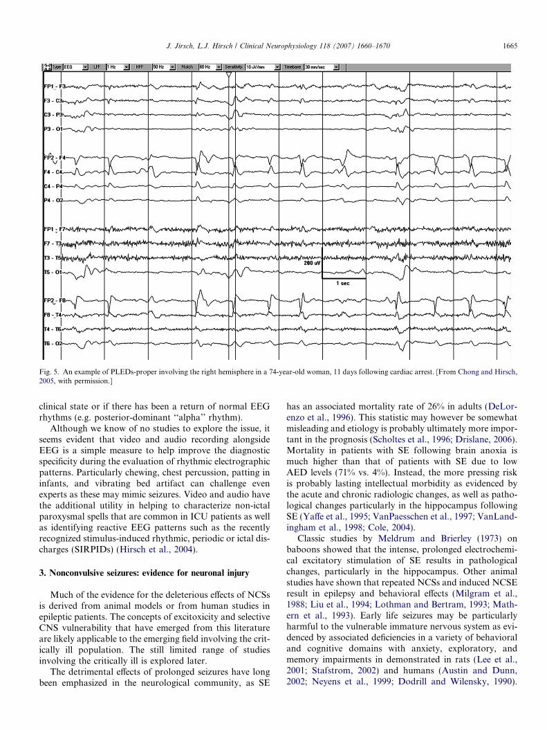
Fig. 5. An example of PLEDs-proper involving the right hemisphere in a 74-year-old woman, 11 days following cardiac arrest. [From Chong and Hirsch,2005, with permission.]
J. Jirsch, L.J. Hirsch / Clinical Neurophysiology 118 (2007) 1660–1670 1665
clinical state or if there has been a return of normal EEGrhythms (e.g. posterior-dominant ‘‘alpha’’ rhythm).
Although we know of no studies to explore the issue, itseems evident that video and audio recording alongsideEEG is a simple measure to help improve the diagnosticspecificity during the evaluation of rhythmic electrographicpatterns. Particularly chewing, chest percussion, patting ininfants, and vibrating bed artifact can challenge evenexperts as these may mimic seizures. Video and audio havethe additional utility in helping to characterize non-ictalparoxysmal spells that are common in ICU patients as wellas identifying reactive EEG patterns such as the recentlyrecognized stimulus-induced rhythmic, periodic or ictal dis-charges (SIRPIDs) (Hirsch et al., 2004).
3. Nonconvulsive seizures: evidence for neuronal injury
Much of the evidence for the deleterious effects of NCSsis derived from animal models or from human studies inepileptic patients. The concepts of excitoxicity and selectiveCNS vulnerability that have emerged from this literatureare likely applicable to the emerging field involving the crit-ically ill population. The still limited range of studiesinvolving the critically ill is explored later.
The detrimental effects of prolonged seizures have longbeen emphasized in the neurological community, as SE
has an associated mortality rate of 26% in adults (DeLor-enzo et al., 1996). This statistic may however be somewhatmisleading and etiology is probably ultimately more impor-tant in the prognosis (Scholtes et al., 1996; Drislane, 2006).Mortality in patients with SE following brain anoxia ismuch higher than that of patients with SE due to lowAED levels (71% vs. 4%). Instead, the more pressing riskis probably lasting intellectual morbidity as evidenced bythe acute and chronic radiologic changes, as well as patho-logical changes particularly in the hippocampus followingSE (Yaffe et al., 1995; VanPaesschen et al., 1997; VanLand-ingham et al., 1998; Cole, 2004).
Classic studies by Meldrum and Brierley (1973) onbaboons showed that the intense, prolonged electrochemi-cal excitatory stimulation of SE results in pathologicalchanges, particularly in the hippocampus. Other animalstudies have shown that repeated NCSs and induced NCSEresult in epilepsy and behavioral effects (Milgram et al.,1988; Liu et al., 1994; Lothman and Bertram, 1993; Math-ern et al., 1993). Early life seizures may be particularlyharmful to the vulnerable immature nervous system as evi-denced by associated deficiencies in a variety of behavioraland cognitive domains with anxiety, exploratory, andmemory impairments in demonstrated in rats (Lee et al.,2001; Stafstrom, 2002) and humans (Austin and Dunn,2002; Neyens et al., 1999; Dodrill and Wilensky, 1990).
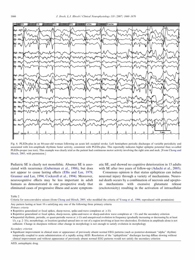
Fig. 6. PLEDs-plus in an 84-year-old woman following an acute left occipital stroke. Left hemisphere periodic discharges of variable periodicity andassociated with low-amplitude rhythmic faster activity, consistent with PLEDs-plus. This reportedly indicates higher epileptic potential than so-calledPLEDs-proper (see text). This example was clearly ictal as the patient had continuous motor activity involving the right arm and neck. [From Chong andHirsch, 2005, with permission.]
1666 J. Jirsch, L.J. Hirsch / Clinical Neurophysiology 118 (2007) 1660–1670
Pediatric SE is clearly not monolithic. Absence SE is asso-ciated with recurrence (Guberman et al., 1986), but doesnot appear to cause lasting effects (Ellis and Lee, 1978;Granner and Lee, 1994; Cockerell et al., 1994). Moreover,neurocognitive effects may be less important in adulthumans as demonstrated in one prospective study thateliminated cases of progressive illness and acute symptom-
Table 2Criteria for non-convulsive seizure (from Chong and Hirsch, 2005, who modi
Any pattern lasting at least 10 s satisfying any one of the following three primPrimary criteria
• Repetitive generalized or focal spikes, sharp-waves, spike-and-wave complex• Repetitive generalized or focal spikes, sharp-waves, spike-and-wave or sharp• Sequential rhythmic, periodic, or quasi-periodic waves at P1/s and unequivoc
1/s, e.g. 2–3/s), morphology, or location (gradual spread into or out of a regiosufficient. Change in sharpness without other change in morphology is not e
Secondary criterion
• Significant improvement in clinical state or appearance of previously absenttemporally coupled to acute administration of a rapidly acting AED. Resoluclinical improvement and without appearance of previously absent normal E
AED, antiepileptic drug.
atic SE, and showed no cognitive deterioration in 15 adultswith SE after two years of follow-up (Adachi et al., 2005).
Consensus opinion is that status epilepticus can induceneuronal injury through a variety of mechanisms. Neuro-nal death occurs by a combination of necrosis and apopto-sis mechanisms with excessive glutamate release(excitotoxicity) resulting in the activation of intracellular
fied the criteria of Young et al., 1996; reproduced with permission)
ary criteria
es at P3/s-and-slow wave complexes at <3/s and the secondary criterional evolution in frequency (gradually increasing or decreasing by at leastn involving at least two electrodes). Evolution in amplitude alone is notnough to satisfy evolution in morphology
normal EEG patterns (such as posterior-dominant ‘‘alpha’’ rhythm)tion of the ‘‘epileptiform’’ discharges leaving diffuse slowing withoutEG patterns would not satisfy the secondary criterion
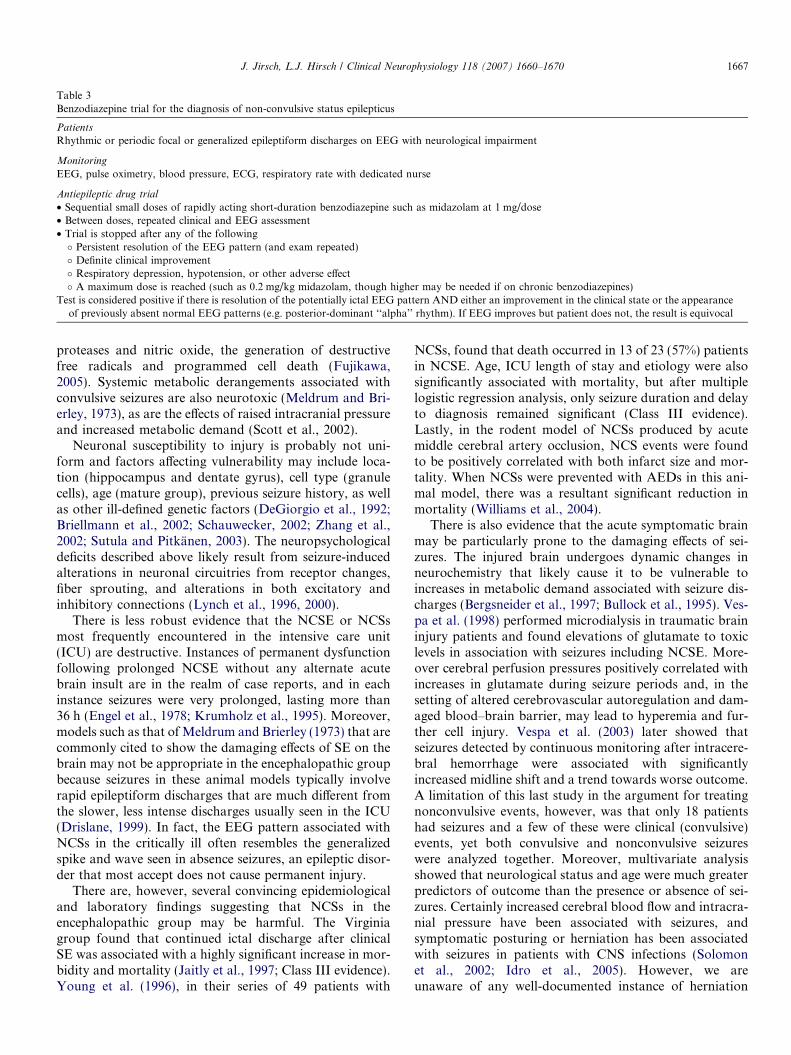
Table 3Benzodiazepine trial for the diagnosis of non-convulsive status epilepticus
Patients
Rhythmic or periodic focal or generalized epileptiform discharges on EEG with neurological impairment
Monitoring
EEG, pulse oximetry, blood pressure, ECG, respiratory rate with dedicated nurse
Antiepileptic drug trial
• Sequential small doses of rapidly acting short-duration benzodiazepine such as midazolam at 1 mg/dose• Between doses, repeated clinical and EEG assessment• Trial is stopped after any of the following� Persistent resolution of the EEG pattern (and exam repeated)� Definite clinical improvement� Respiratory depression, hypotension, or other adverse effect� A maximum dose is reached (such as 0.2 mg/kg midazolam, though higher may be needed if on chronic benzodiazepines)
Test is considered positive if there is resolution of the potentially ictal EEG pattern AND either an improvement in the clinical state or the appearanceof previously absent normal EEG patterns (e.g. posterior-dominant ‘‘alpha’’ rhythm). If EEG improves but patient does not, the result is equivocal
J. Jirsch, L.J. Hirsch / Clinical Neurophysiology 118 (2007) 1660–1670 1667
proteases and nitric oxide, the generation of destructivefree radicals and programmed cell death (Fujikawa,2005). Systemic metabolic derangements associated withconvulsive seizures are also neurotoxic (Meldrum and Bri-erley, 1973), as are the effects of raised intracranial pressureand increased metabolic demand (Scott et al., 2002).
Neuronal susceptibility to injury is probably not uni-form and factors affecting vulnerability may include loca-tion (hippocampus and dentate gyrus), cell type (granulecells), age (mature group), previous seizure history, as wellas other ill-defined genetic factors (DeGiorgio et al., 1992;Briellmann et al., 2002; Schauwecker, 2002; Zhang et al.,2002; Sutula and Pitkanen, 2003). The neuropsychologicaldeficits described above likely result from seizure-inducedalterations in neuronal circuitries from receptor changes,fiber sprouting, and alterations in both excitatory andinhibitory connections (Lynch et al., 1996, 2000).
There is less robust evidence that the NCSE or NCSsmost frequently encountered in the intensive care unit(ICU) are destructive. Instances of permanent dysfunctionfollowing prolonged NCSE without any alternate acutebrain insult are in the realm of case reports, and in eachinstance seizures were very prolonged, lasting more than36 h (Engel et al., 1978; Krumholz et al., 1995). Moreover,models such as that of Meldrum and Brierley (1973) that arecommonly cited to show the damaging effects of SE on thebrain may not be appropriate in the encephalopathic groupbecause seizures in these animal models typically involverapid epileptiform discharges that are much different fromthe slower, less intense discharges usually seen in the ICU(Drislane, 1999). In fact, the EEG pattern associated withNCSs in the critically ill often resembles the generalizedspike and wave seen in absence seizures, an epileptic disor-der that most accept does not cause permanent injury.
There are, however, several convincing epidemiologicaland laboratory findings suggesting that NCSs in theencephalopathic group may be harmful. The Virginiagroup found that continued ictal discharge after clinicalSE was associated with a highly significant increase in mor-bidity and mortality (Jaitly et al., 1997; Class III evidence).Young et al. (1996), in their series of 49 patients with
NCSs, found that death occurred in 13 of 23 (57%) patientsin NCSE. Age, ICU length of stay and etiology were alsosignificantly associated with mortality, but after multiplelogistic regression analysis, only seizure duration and delayto diagnosis remained significant (Class III evidence).Lastly, in the rodent model of NCSs produced by acutemiddle cerebral artery occlusion, NCS events were foundto be positively correlated with both infarct size and mor-tality. When NCSs were prevented with AEDs in this ani-mal model, there was a resultant significant reduction inmortality (Williams et al., 2004).
There is also evidence that the acute symptomatic brainmay be particularly prone to the damaging effects of sei-zures. The injured brain undergoes dynamic changes inneurochemistry that likely cause it to be vulnerable toincreases in metabolic demand associated with seizure dis-charges (Bergsneider et al., 1997; Bullock et al., 1995). Ves-pa et al. (1998) performed microdialysis in traumatic braininjury patients and found elevations of glutamate to toxiclevels in association with seizures including NCSE. More-over cerebral perfusion pressures positively correlated withincreases in glutamate during seizure periods and, in thesetting of altered cerebrovascular autoregulation and dam-aged blood–brain barrier, may lead to hyperemia and fur-ther cell injury. Vespa et al. (2003) later showed thatseizures detected by continuous monitoring after intracere-bral hemorrhage were associated with significantlyincreased midline shift and a trend towards worse outcome.A limitation of this last study in the argument for treatingnonconvulsive events, however, was that only 18 patientshad seizures and a few of these were clinical (convulsive)events, yet both convulsive and nonconvulsive seizureswere analyzed together. Moreover, multivariate analysisshowed that neurological status and age were much greaterpredictors of outcome than the presence or absence of sei-zures. Certainly increased cerebral blood flow and intracra-nial pressure have been associated with seizures, andsymptomatic posturing or herniation has been associatedwith seizures in patients with CNS infections (Solomonet al., 2002; Idro et al., 2005). However, we areunaware of any well-documented instance of herniation
1668 J. Jirsch, L.J. Hirsch / Clinical Neurophysiology 118 (2007) 1660–1670
convincingly associated with NCSs alone. Although a cau-sal relationship between seizures (including NCSs), cere-bral edema and progressive midline shift is certainlypossible and perhaps likely, this has not yet been proven.
One final line of evidence suggesting that NCSs may bedeleterious involves a biomarker. Neuron specific enolase(NSE) is a key enzyme for energy metabolism and is a mar-ker of acute brain injury as well as damage to the blood–brain barrier (Correale et al., 1998). DeGiorgio et al.(1999) prospectively measured levels of NSE in consecutivepatients with SE and found significant elevations in CPSEand subclinical generalized SE patients, even when therewere no other causes for acute brain injury beyond sei-zures. Moreover, this study found that absolute levels ofNSE correlated with the duration of SE, suggesting thatearly treatment of NCSs is desirable.
4. Treatment of nonconvulsive seizures and status epilepticus
While it would appear that NCSs are damaging to thebrain, it is far less obvious that single NCSs or NCSE needsto be treated as aggressively as convulsive varieties for rea-sons outlined above. Claassen et al. (2002) performed a sys-tematic review comparing various continuous IV infusionAEDs and pentobarbital in patients treated for refractoryconvulsive and nonconvulsive status epilepticus, and founda poor outcome overall (50% mortality) regardless of theagent utilized or the titration goal (i.e. seizure suppressionor background suppression). Pentobarbital treatmentappeared to be more effective in preventing breakthroughseizures than other medications, even in cases of NCSEwhich are known to be more refractory to treatment thanGCSE (Mayer et al., 2002), however it is frequently compli-cated by hypotension, and ultimate outcome was the same.While propofol has a shorter half-life than pentobarbitaland would seem to be safer than long acting barbiturates,it has become clear that propofol infusion syndrome is animportant complication. This serious complication is char-acterized by refractory metabolic acidosis, cardiac failure,rhabdomyolysis and renal failure, and occurs most oftenwith prolonged infusion at >5 mg/kg/h for >48 h, and pos-sibly traumatic or other acute brain injury (Vasile et al.,2003; Cremer et al., 2001).
Several centers, including ours, have taken to using IVbenzodiazepines (e.g. midazolam) in cases of NCSE. Thesemedications can be rapidly titrated due to their short halflives (similar to propofol), and may be used synergisticallywith propofol to lower infused dosages and even result in alower mortality rate of 22% (Rosetti et al., 2004). However,in the rodent model of NCSs after acute MCA occlusion,benzodiazepines and barbiturates were found to be of nobenefit for seizures, and barbiturates lead to continuousspiking in some animals (Williams et al., 2004). Moreover,in humans, one retrospective study compared critically illolder patients with NCSE treated aggressively in an ICUsetting with IV benzodiazepines to those with advancedirectives treated less aggressively out of the ICU (Litt
et al., 1998). The use of IV benzodiazepines was associatedwith increased mortality despite no difference in severity ofillness. Aggressive ICU care in these patients prolongedhospitalization but did not lead to improved outcomes.
While the malignancy of NCSs remains debated, it isclear that severe outcomes in critically ill patients withNCSs are more affected by other factors. Underlying etiol-ogy and the length of ICU stay with its attendant mostlyinfectious complications are probably most important(Yaffe and Lowenstein, 1993). At our center, concertedeffort is made to diagnose and treat NCSs as quickly aspossible but with minimal sedation so as to avoid prolong-ing coma and intubation. Non-coma inducing agents suchas IV fosphenytoin, valproate, and perhaps intermittentbenzodiazepines are used in addition to the oral AEDs(via nasogastric tube) such as levetiracetam (Rosetti andBromfield, 2005) (now available intravenously as well)and topiramate (Towne et al., 2003).
5. Future directions
Clearly our understanding of how best to managepatients with subclinical rhythmic or periodic EEG patternsis in its infancy. An important initial step to this end hasbegun with the proposal of standardized terminology forthese patterns last year (Hirsch et al., 2005). Undoubtedlyfurther refinement of this nomenclature will occur in thefuture, and perhaps even some agreement on the definitionof a NCS. Deciding which EEG patterns are harmful, andhow aggressively cessation of this activity should be pur-sued, will only be established through large multicenter clin-ical trials. In the nearer future, some direction may resultfrom studying serial biomarkers (such as NSE) in thesepatients, imaging modalities (e.g. diffusion-weighted MRI,MR spectroscopy, positron emission tomography) orsimultaneous cerebral microdialysis to gain insight intoalterations in metabolic stress and to determine frank neu-ronal injury associated with EEG changes.
Appendix A
Classification of evidence (American Academy of Neu-rology scheme, see Armon and Evans, 2005):
Class I: Prospective, randomized, controlled clinicaltrial with masked outcome assessment in a representativepopulation. The following are required:
(a) Primary outcome(s) is(are) clearly defined.(b) Exclusion/inclusion criteria are clearly defined.(c) Adequate accounting for dropouts and crossovers
with numbers sufficiently low to have minimal poten-tial for bias.
(d) Relevant baseline characteristics are presented andsubstantially equivalent among treatment groups orthere is appropriate statistical adjustment fordifferences.

J. Jirsch, L.J. Hirsch / Clinical Neurophysiology 118 (2007) 1660–1670 1669
Class II: Prospective matched group cohort study in arepresentative population with masked outcome assess-ment that meets A through D above OR a randomized,controlled trial in a representative population that lacksone criterion A through D.
Class III: All other controlled trials including well-defined natural history controls or patients serving asown controls in a representative population in which out-come assessment is independently assessed or indepen-dently derived by objective outcome measurement(objective outcome measurement is an outcome measurethat is unlikely to be affected by an observer’s (patient,treating physician, investigator) expectation or bias [e.g.blood tests, administrative outcome data]).
Class IV: Evidence from uncontrolled studies, case ser-ies, case reports, or expert opinion.
References
Abou Khaled K, Alschuler D, Jirsch J, Claassen J, Wittman J, EmersonRG, et al. Generalized Periodic Discharges: prognostic significanceand relation to seizures. Neurology 2006;66(Suppl. 2):A93.
Adachi N, Kanemoto K, Muramatsu R, Kato M, Akanuma N, Ito M,et al. Intellectual prognosis of status epilepticus in adult epilepsypatients: analysis with Wechsler Adult Intelligence Scale-revised.Epilepsia 2005;46:1502–9.
Armon C, Evans RW. Addendum to assessment: prevention of post-lumbar puncture headaches: report of the Therapeutic and TechnologyAssessment Subcommittee of the American Academy of Neurology.Neurology 2005;65:510–2.
Austin J, Dunn D. Progressive behavioral changes in children withepilepsy. Prog Brain Res 2002;135:419–27.
Bergsneider MA, Hovda DA, Shalmon E, Kelly DF, Vespa PM, MartinNA, et al. Cerebral hyperglycolysis following severe human traumaticbrain injury: a positron emission tomography study. J Neurosurg1997;86:241–51.
Boulanger J-M, Deacon C, Lecuyer D, Gosselin S, Reiher J. Triphasicwaves versus nonconvulsive status epilepticus: EEG distinction. Can JNeurol Sci 2006;33:175–80.
Brenner RP, Schaul N. Periodic EEG patterns: classification, clinicalcorrelation, and pathophysiology. J Clin Neurophysiol 1990;7:249–67.
Briellmann RS, Berkovic SF, Syngeniotis A, King MA, Jackson GD.Seizure-associated hippocampal volume loss: a longitudinal magneticresonance study of temporal lobe epilepsy. Ann Neurol 2002;51:641–4.
Brussiere M, Pelz D, Reid RH, Young GB. Prolonged deficits after focalinhibitory seizures. Neurocrit Care 2005;2:29–37.
Bullock R, Zauner A, Myseros JS, Marmarou A, Woodward JJ, YoungHF. Evidence for prolonged release of excitatory amino acids in severehuman head trauma: relationship to clinical events. Ann NY Acad Sci1995;59:290–7.
Chong DJ, Hirsch LJ. Which EEG patterns warrant treatment in thecritically ill? Reviewing the evidence for treatment of periodic epilepti-formdischargesandrelatedpatterns.JClinNeurophysiol2005;22:79–91.
Claassen J, Hirsch LJ, Emerson RG, Mayer SA. Treatment of refractorystatus epilepticus with pentobarbital, propofol, or midazolam: asystematic review. Epilepsia 2002;43:146–53.
Claassen J, Mayer SA, Kowalski RG, Emerson RG, Hirsch LJ. Detectionof electrographic seizures with continuous EEG monitoring incritically ill patients. Neurology 2004;62:1743–8.
Cole AJ. Status epilepticus and periictal imaging. Epilepsia 2004;45(Suppl.4):72–7.
Cockerell OC, Walker MC, Sander JWAS, Shorvon SD. Complex partialstatus epilepticus: a recurrent problem. J Neurol Neurosurg Psychiatry1994;57:835–7.
Correale J, Rabinowicz AL, Heck CN, Smith TD, Loskota WJ,DeGiorgio CM. Status epilepticus increases CSF levels of neuronspecific enolase and alters the blood–brain barrier. Neurology1998;50:1388–91.
Cremer OL, Moons KG, Bouman EA, Kruijswijk JE, de Smet AM,Kalkman CJ. Longterm propofol infusion and cardiac failure in adulthead injury patients. Lancet 2001;357:117–8.
DeLorenzo RJ, Hauser WA, Towne AR, Boggs JG, Pellock JM,Penberthy L, et al. A prospective, population-based epidemiologicstudy of status epilepticus in Richmond, Virginia. Neurology1996;46:1029–35.
DeLorenzo RJ, Waterhouse EJ, Towne AR, Boggs JG, Ko D, DeLorenzoGA, et al. Persistent nonconvulsive status epilepticus after the controlof convulsive status epilepticus. Epilepsia 1998;39:833–40.
DeGiorgio CM, Heck CN, Rabinowicz AL, Gott PS, Smith T, Correale J.Serum neuron-specific enolase in the major subtypes of statusepilepticus. Neurology 1999;52:746–9.
DeGiorgio CM, Tomiyasu U, Gott PS, Treiman DM. Hippocampalpyramidal cell loss in human status epilepticus. Epilepsia 1992;33:23–7.
Dennis LJ, Claassen J, Hirsch LJ, Emerson RG, Connolly ES, Mayer SA.Nonconvulsive status epilepticus after subarachnoid hemorrhage.Neurosurgery 2002;51:1136–43.
Dodrill CB, Wilensky AJ. Intellectual impairment as an outcome of statusepilepticus. Neurology 1990;40(Suppl. 2):23–7.
Drake ME, Erwin CW. Triphasic discharges in metrizamide encephalop-athy. J Neurol Neurosurg Psychiatry 1984;47:324–5.
Drislane FW. Evidence against permanent neurologic damage fromnonconvulsive status epilepticus. J Clin Neurophysiol 1999;16:323–31.
Drislane FW. Who’s afraid of status epilepticus? Epilepsia 2006;47:7–9.Ellis JM, Lee SI. Acute prolonged confusion in later life as an ictal state.
Epilepsia 1978;19:119–28.Engel Jr J, Ludwig BI, Fetell M. Prolonged partial complex status
epilepticus: EEG and behavioral observations. Neurology1978;28:863–9.
Fountain NB, Waldman WA. Effects of benzodiazepines on triphasicwaves: implications for nonconvulsive status epilepticus. J ClinNeurophysiol 2001;18:345–852.
Fujikawa DG. Prolonged seizures and cellular injury: understanding theconnection. Epilepsy Behav 2005;7(Suppl. 3):S3–S11.
Guberman A, Cantu-Reyna G, Stuss D, Broughton R. Nonconvulsivegeneralized status epilepticus clinical features, neuropsychologicaltesting, and long-term follow-up. Neurology 1986;36:1284–91.
Granner MA, Lee SI. Nonconvulsive status epilepticus: EEG analysis in alarge series. Epilepsia 1994;35:42–7.
Hormes JT, Benarroch EE, Rodriguez M, Klass DW. Periodic sharpwaves in baclofen-induced encephalopathy. Arch Neurol1988;45:814–5.
Hirsch LJ, Brenner RP, Drislane FW, So E, Kaplan PW, Jordan KG,et al. The ACNS Subcommittee on Research Terminology forContinuous EEG Monitoring: proposed standardized terminologyfor rhythmic and periodic EEG patterns encountered in critically illpatients. J Clin Neurophysiol 2005;22:128–35.
Hirsch LJ, Claassen J, Mayer SA, Emerson RG. Stimulus-inducedrhythmic, periodic, or ictal discharges (SIRPIDs): a common EEGphenomenon in the critically ill. Epilepsia 2004;45:109–23.
Husain AM, Mebust KA, Radtke RA. Generalized periodic epileptiformdischarges: etiologies, relationship to status epilepticus, and prognosis.J Clin Neurophysiol 1999;16:51–8.
Idro R, Otieno G, White S, Kahindi A, Fegan G, Ogutu B, et al.Decorticate, decerebrate and opisthotonic posturing and seizures inKenyan children with cerebral malaria. Malar J 2005;4:57.
Jaitly R, Sgro JA, Towne AR, Ko D, DeLorenzo RJ. Prognostic value ofEEG monitoring after status epilepticus: a prospective adult study. JClin Neurophysiol 1997;14:326–34.
Jordan KG. Continuous EEG and evoked potential monitoring in theneuroscience intensive care unit. J Clin Neurophysiol 1993;10:445–75.
Kaplan PW. Nonconvulsive status epilepticus in the Emergency Room.Epilepsia 1996;37:643–50.

1670 J. Jirsch, L.J. Hirsch / Clinical Neurophysiology 118 (2007) 1660–1670
Kaplan PW. Behavioral manifestations of nonconvulsive status epilepti-cus. Epilepsy Behav 2002;3:122–39.
Kaplan PW. The EEG in metabolic encephalopathy and coma. J ClinNeurophysiol 2004;21:307–18.
Krumholz A, Sung GY, Fisher RS, Barry E, Bergey GK, Grattan LM.Complex partial status epilepticus accompanied by serious morbidityand mortality. Neurology 1995;45:1499–504.
Kuroiwa Y, Celesia GG. Clinical significance of periodic EEG patterns.Arch Neurol 1980;37:15–20.
Lee C, Hannay J, Hrachovy R, Rashid S, Antalffy B, Swann JW. Spatiallearning deficits without hippocampal neuronal loss in a model ofearly-onset epilepsy. Neuroscience 2001;107:71–84.
Litt B, Wityk RJ, Hertz SH, Mullen PD, Ryan DD, Henry TR.Nonconvulsive status epilepticus in the critically ill elderly. Epilepsia1998;39:1194–202.
Liu Z, Gatt A, Werner SJ, Milati MA, Holmes GL. Long-term behavioraldeficits following pilocarpine seizures in immature rats. Epilepsy Res1994;19:191–204.
Lothman EW, Bertram III EH. Epileptogenic effects of status epilepticus.Epilepsia 1993;34(Suppl. 1):S59–70.
Lynch M, Savin U, Bownds J, Junumpalli S, Sutula T. Long-termconsequences of early post-natal seizures on hippocampal learning andplasticity. Eur J Neurosci 2000;12:2252–64.
Lynch MW, Rutecki PA, Sutula TP. The effects of seizures on the brain.Curr Opin Neurol 1996;9:97–102.
Mathern GW, Cifuentes F, Leite JP, Pretorius JK, Babb TL. Hippocam-pal EEG excitability and chronic spontaneous seizures are associatedwith aberrant synaptic reorganization in the rat intrahippocampalkainate model. Electroencephalogr Clin Neurophysiol 1993;87:326–39.
Mayer SA, Claassen J, Lokin J, Mendelsohn F, Dennis LJ, FitzsimmonsBF. Refractory status epilepticus: frequency risk factors, and impacton outcome. Arch Neurol 2002;59:205–10.
Meldrum BS, Brierley JB. Prolonged epileptic seizures in primates:ischemic cell change and its relation to ictal physiological events. ArchNeurol 1973;28:10–7.
Milgram NW, Isen DA, Mandel D, Palantzas H, Pepkowski MJ. Deficitsin spontaneous behavior and cognitive function following systemicadministration of kainic acid. Neurotoxicology 1988;9:611–24.
Nei M, Lee JM, Shanker VL, Sperling MR. The EEG and prognosis instatus epilepticus. Epilepsia 1999;40:157–63.
Neyens L, Aldenkamp A, Meinardi H. Prospective follow-up of intellec-tual development in children with a recent onset of epilepsy. EpilepsyRes 1999;34:85–90.
Ono S, Chida K, Fukaya N, Yoshihashi H, Takasu T. Dysphasiaaccompanied by periodic lateralized epileptiform discharges. InternMed 1997;36:59–61.
Pandian JD, Cascino GD, So EL, Manno E, Fulgham JR. Digital video-electroencephalographic monitoring in the neurological–neurosurgicalintensive care unit: clinical features and outcome. Arch Neurol2004;61:1090–4.
Reiher J, Rivest J, Grand’Maison F, Leduc CP. Periodic lateralizedepileptiform discharges with transitional rhythmic discharges: associ-ation with seizures. Electroencephalogr Clin Neurophysiol1991;78:12–7.
Rosetti AO, Bromfield EB. Levetiracetam in the treatment of statusepilepticus in adults: a study of 13 episodes. Eur Neurol 2005;54:34–8.
Rosetti AO, Reichart MD, Schaller MD, Despland PA, Bogousslavsky J.Propofol treatment of refractory status epilepticus: a study of 31episodes. Epilepsia 2004;45:757–63.
Schauwecker PE. Complications associated with genetic backgroundeffects in models of experimental epilepsy. Prog Brain Res2002;135:139–48.
Scholtes FB, Renier WO, Meinardi H. Non-convulsive status epilepticus:causes, treatment, and outcome in 65 patients. J Neurol NeurosurgPsychiatry 1996;61:94–5.
Sheridan PH, Sato S. Triphasic waves of metabolic encephalopathy versusspike-wave stupor. J Neurol Neurosurg Psychiatry 1986;49:108–9.
Snodgrass SM, Tsuburaya K, Ajmone-Marsan C. Clinical significance ofperiodic lateralized epileptiform discharges: relationship with statusepilepticus. J Clin Neurophysiol 1989;6:159–72.
Solomon T, Dung NM, Kneen R, Thao le TT, Gainsborough M, NisalakA, et al. Seizures and raised intracranial pressure in Vietnamesepatients with Japanese encephalitis. Brain 2002;125:1084–93.
Stafstrom C. Assessing the behavioral and cognitive effects of seizures onthe developing brain. Prog Brain Res 2002;135:377–90.
Sundaram MB, Blume WT. Triphasic waves: clinical correlates andmorphology. Can J Neurol Sci 1987;14:136–40.
Sutula T, Pitkanen A. Do seizures damage the brain? Curr Opin Neurol2003;16:189–95.
Towne AR, Garnett LK, Waterhouse EJ, Morton LD, DeLorenzo RJ.The use of topirimate in refractory status epilepticus. Neurology2003;60:332–4.
Towne AR, Waterhouse EJ, Boggs JG. Prevalence of nonconvulsive statusepilepticus in comatose patients. Neurology 2000;54:340–5.
Treiman DM, Meyers PD, Walton NY, Collins C, Rowan AJ, HandforthA, et al. A comparison of four treatments for generalized convulsivestatus epilepticus. Veterans Affairs Status Epilepticus CooperativeStudy Group. N Engl J Med 1998;339:792–8.
VanLandingham KE, Heinz ER, Cavazos JE, Lewis DV. Magneticresonance imaging evidence of hippocampal injury after prolongedfocal febrile convulsions. Ann Neurol 1998;43:413–26.
VanPaesschen W, Revesz T, Duncan JS, King MD, Connely A.Quantitative neuropathology and quantitative magnetic resonanceimaging of the hippocampus in temporal lobe epilepsy. Ann Neurol1997;42:756–66.
Vasile B, Rasulo F, Candiani A, Latronico N. The pathophysiology ofpropofol infusion syndrome: a simple name for a complex syndrome.Int Care Med 2003;29:1417–25.
Vespa PM, Nuwer MR, Nenov V, Ronne-Engstrom E, Hovda DA,Bergsneider M, et al. Increased incidence and impact of nonconvulsiveand convulsive seizures after traumatic brain injury as detected bycontinuous electroencephalographic monitoring. J Neurosurg1999;91:750–60.
Vespa P, Prins M, Ronne-Engstrom E, Caron M, Shalmon E, Hoyda DA,et al. Increase in extracellular glutamate caused by reduced cerebralperfusion pressure and seizures after traumatic brain injury: amicrodialysis study. J Neurosurg 1998;89:971–82.
Vespa PM, O’Phelan K, Shah M, Mirabelli J, Starkman S, Kidwell C.Acute seizures after intracerebral hemorrhage: a factor in progressivemidline shift and outcome. Neurology 2003;60:1441–6.
Williams AJ, Tortella FC, Lu XM, Moreton JE, Hartings JA. Antiepi-leptic drug treatment of nonconvulsive seizures induced by experi-mental focal brain ischemia. J Pharmacol Exp Ther 2004;311:220–7.
Yaffe K, Ferriero D, Barkovich J, Rowley H. Reversible MRI abnormal-ities following seizures. Neurology 1995;45:104–8.
Yaffe K, Lowenstein SH. Prognostic factors of pentobarbital therapy forrefractory generalized status epilepticus. Neurology 1993;43:895–900.
Yemisci M, Gurer G, Saygi S, Ciger A. Generalised periodic epileptiformdischarges: clinical features, neuroradiologic evaluation and prognosisin 37 adult patients. Seizure 2003;12:465–72.
Young GB, Bolton CF, Austin TW, Archibald YM, Wells GA. Theencephalopathy associated with septic illness. Clin Invest Med1990;13:297–304.
Young GB, Jordan KG, Doig GS. An assessment of nonconvulsiveseizures in the intensive care unit using continuous EEG monitoring:an investigation of variables associated with mortality. Neurology1996;47:83–9.
Zhang X, Cui S, Wallace A, Hannesson DK, Schmued LC, Saucier DM,et al. Relations between brain pathology and temporal lobe epilepsy. JNeurosci 2002;22:6052–61.