nonblocking monoclonal antibody targeting soluble...
TRANSCRIPT

Cancer Therapy: Preclinical
Nonblocking Monoclonal Antibody TargetingSoluble MIC Revamps Endogenous Innate andAdaptive Antitumor Responses and EliminatesPrimary and Metastatic TumorsShengjun Lu1, Jinyu Zhang1, Dai Liu2, Guangfu Li2,3, Kevin F. Staveley-O'Carroll2,3,Zihai Li1,3, and Jennifer D.Wu1,3,4
Abstract
Purpose: The human tumor-derived soluble MHC I-chain–related molecule (sMIC) is highly immune suppressive in cancerpatients and correlates with poor prognosis. However, the ther-apeutic effect of targeting sMIC has not been determined, due tothe limitation that mice do not express homologs of humanMIC.This study is to evaluate the therapeutic effect of a monoclonalantibody (mAb) targeting sMIC in a clinically relevant transgenicanimal model.
Experimental Design:We treated the engineered MIC-expres-sing "humanized" TRAMP/MIC bitransgenic mice at advanceddisease stages with a sMIC-neutralizing nonblocking anti-MICmAb and assessed the therapeutic efficacy and associatedmechanisms.
Results:A sMIC-neutralizing nonblocking anti-MICmAb effec-tively induced regression of primary tumors and eliminated
metastasis without inducing systemic toxicity. The therapeuticeffect is conferred by revamping endogenous antitumor immuneresponses, exemplified by restoring natural killer (NK) cellhomeostasis and function, enhancing susceptibility of MICþ-tumor cells to NK cell killing, reviving and sustaining antigen-specific CD8 T-cell responses, augmenting CD4 T cells to Th1responses, priming dendritic cells for antigen presentation, andremodeling tumor microenvironment to be more immunereactive.
Conclusions: Therapy with a sMIC-neutralizing nonblockinganti-MICmAbcaneffectuate antitumor immune responses againstadvanced MICþ tumors. Our study provided strong rationale fortranslating sMIC-neutralizing therapeutic mAb into clinics, eitheralone or in combination with current ongoing standard immu-notherapies. Clin Cancer Res; 21(21); 4819–30. �2015 AACR.
IntroductionIn response to oxidative stress and oncogenic insults, human
epithelial cells were induced to express families of ligands for theimmune stimulatory receptor NKG2D, presumably to provokeantitumor immune responses as a natural defensemechanism(1–4). In experimental animal models, ligand-induced activation ofNKG2D has been demonstrated very effective in controllingtumor initiation and progression through activating natural killer(NK) and costimulating effector T cells, including cytotoxic Tlymphocytes and NKT cells (5, 6). Notably, retaining surfaceNKG2D ligand expression has been shown to be critical for
NKG2D-mediated tumor suppression in both experimental ani-mal models and cancer patients (7, 8). In cancer patients, how-ever, tumors evolved to escape NKG2D-mediated naturalimmune response by adopting a protease or exosome-mediatedstrategy to shed NKG2D ligands (9). As a consequence, not onlytumor cells lost surface NKG2D ligands with an impaired abilityto provoke an effective immune response, but more severely, theshedding-resolved soluble NKG2D ligands can sabotage theimmune system via diversifiedmechanisms. These include down-regulating the expression and thus function of the receptorNKG2D on NK cell and effector T cells (10, 11), perturb NK cellhomeostasis (7), promoting the expansion of arginase Iþ mye-loid-derived suppressor cells (MDSC), and skewing macrophageto the tumor-promoting alternatively activated phenotypes (12).Thus, tumor-derived soluble NKG2D ligands have been broadlyaccepted to be highly immune suppressive and proposed as apotential cancer immune therapeutic target (13, 14).
In human, two families of NKG2D ligands, the MHC I-chain–relatedmolecules A and B (MICA/B, collectively termedMIC) andthe HCMV glycoprotein UL16-binding protein family molecules(ULBP), have been identified; among which the MIC family ismost prevalently and restrictedly expressed by human cancer cells(15). Shedding of tumor cell surface MIC to release the solubleMIC (sMIC) into circulation is commonly found in cancerpatients (16–19). Serum levels of sMIC have been well associatedwith progressiveness of the diseases in multiple cancer types(7, 20–23). Given the multiple immune suppressive nature of
1Department of Microbiology and Immunology, Medical University ofSouth Carolina, Charleston, South Carolina. 2Department of Surgery,Medical University of South Carolina, Charleston, South Carolina.3Cancer Immunology Program, Hollings Cancer Center, Charleston,South Carolina. 4Department of Medicine, University of Washington,Seattle,Washington.
Note: Supplementary data for this article are available at Clinical CancerResearch Online (http://clincancerres.aacrjournals.org/).
Corresponding Author: Jennifer D. Wu, Department of Microbiology andImmunology, Medical University of South Carolina, 86 Jonathan Lucas Street,Hollings Cancer Center 610, Charleston, SC 29425. Phone: 843-792-9222; Fax:843-792-9588; E-mail: [email protected]
doi: 10.1158/1078-0432.CCR-15-0845
�2015 American Association for Cancer Research.
ClinicalCancerResearch
www.aacrjournals.org 4819
on September 1, 2018. © 2015 American Association for Cancer Research. clincancerres.aacrjournals.org Downloaded from
Published OnlineFirst June 23, 2015; DOI: 10.1158/1078-0432.CCR-15-0845

sMIC (7, 10, 12, 16), whether antibody neutralizing sMIC canrevive host antitumor responses in MICþ cancer patients is evi-dently an interesting therapeutic question. Because of the limi-tation that no MIC homolog is expressed by rodents, this criticalquestion, however, remained unaddressed to date. Notably,although the signaling pathways of NKG2D are conserved acrossspecies, mouse NKG2D ligands differ from human MIC in theiraffinity, structure, regulation, and distribution in tumor tissues(15, 24, 25). These inherent differences preclude the translation ofinteresting biologic conclusions discovered from mouse NKG2Dligands to human cancer.
We have recently generated a "humanized" bitransgenicTRAMP/MIC(B) mouse model that expresses human MIC in theprostate epithelial cells of the SV40T-transgenic adenocarcinomamouse prostate (TRAMP) directed by the prostate-specific pro-moter (7). The TRAMP/MIC mouse closely recapitulates thedynamic interaction of oncogenesis and NKG2D-mediatedimmune surveillance in human cancer patients by bridging shed-ding sMIC with disease progression (7). With close resemblancetoMICþ cancer patients (7, 17, 20, 21), high serum levels of sMICin TRAMP/MIC mice predicted poor tumor prognosis (7). Uti-lizing this similarity, in this study, we evaluated the therapeuticantitumor efficacy of a sMIC-neutralizing but nonblocking anti-MICmonoclonal antibody (mAb) B10G5 in an immune tolerantspontaneous MICþ tumor model.
Materials and MethodsAnimals and antibody therapy
Breeding of TRAMP/MICB has been previously described (7).Animals were randomized into two cohorts receiving therapywith intraperitoneal (i.p.) injection of sMIC-specific mAbB10G5 or isotype control IgG (cIgG) at the dose of 4.0 mg/kgbody weight twice weekly. Generation of the B10G5 antibodywere described previously (7). All animals were treated for 8weeks before euthanization that was designated as the studyendpoint. Mice received daily refreshed drinking water contain-ing 0.8 mg/mL bromodeoxyuridine (BrdUrd) for 5 consecutivedays before the study endpoint. For congenic cells transfer,splenocytes were isolated from congenic CD45.1þ C57BL/6mice (Charles River Laboratories, Frederick Cancer ResearchCenter, Frederick, MD) and labeled with V450 cell-trace dye
according to the manufacturer's protocol (eBioscience). V450-labeled splenocytes were resuspended in PBS and injected viatail veil into recipient TRAMP/MICB mice (CD45.2þ) at thedose of 2 � 107/mouse 5 days before endpoint. All animalswere housed in specific pathogen-free facilities. All experimen-tal procedures were approved by the Institutional Animal Careand Use Committee. The study was repeated three times unlessotherwise specified.
NK and CD8 T-cell depletionMice were injected with antibody anti-NKp46 antibody (Bio-
Legend) to deplete NK cells or CD8a-specific antibody (clone 53-6.7; BioXcell) to deplete CD8T cells at the dose of 200mg/mouse 1day before B10G5 antibody therapy and thereafter twiceweekly atthe dose of 100 mg/mouse till study endpoint. Efficiency ofdepletion was confirmed by flow cytometry analyses in theperipheral blood.
Antigen-specific T-cell response experimentCD8 T cells from TCR-I transgenic mice were labeled with CFSE
and injected i.v. into animals (2 � 106 cells/mouse) that werereceiving B10G5or control IgG therapy. Animalswere sacrificed atindicated time points to assess TCR-I T cell in vivo frequency withTCR-I–specific H-2Db/TAg epitope I-tetramer (Db/I-tetramer;ref. 26). To assay antigen-specific CD8 T-cell response, bulkedsplenocytes and single-cell suspension of tumor-draining lymphnodes (dLN) and tumor digests were stimulated overnight with0.5 mM TAg epitope I peptide and assaying intracellular IFNgstaining of CD8þ or Db/I-tetramerþ T cells.
Tissue collectionBlood was collected via tail bleeding during therapy and via
cardiac puncture after euthanization. Spleens and dLN werecollected for immunologic analyses. Prostate, lung, liver, kidney,pancreas, and intestines were collected, fixed in 10% neutralfixation buffer followed by paraffin embedment or directlyembedded in optimal cutting temperature (OCT), for pathologicand histologic analyses. In some experiments, partial of prostatetumors was digested with collagenase for analyses of tumor-infiltrated lymphocytes.
Flow cytometrySingle-cell suspension from splenocytes, dLN, or tumor infil-
trates was prepared as described previously (7). Combinationof the following antibody was used for cell surface or intracel-lular staining to define populations of NK, CD8, and subsets ofCD4 T cells: CD3e (clone 145-2c11), CD8a (clone 53-6.7), CD4(clone GK1.5), NK1.1 (clone PK136), NKG2D (clone CX5),CD45.1 (clone A20), and T-bet (clone eBio4B10). For ex vivorestimulation, single-cell suspension of freshly isolated sple-nocytes or LN were cultured in complete RPMI-1640 mediumcontaining 50 ng/mL phorbol 12—myristate 13—acetate(PMA) and 500 ng/mL ionomycin for 4 hours and analyzedby intracellular staining with antibodies specific to IFNg(XMG1.2). For NK cell renewal, intracellular BrdUrd stainingwas performed using anti-BrdUrd antibody (clone Bu20a). Allantibodies and the corresponding isotype controls were fluo-rochrome conjugated and were purchased from eBioscience orBD Biosciences. Multicolored flow cytometry analyses wereperformed on an LSRII (BD). Data were analyzed with theFlowJo software (TreeStar).
Translational Relevance
The human tumor–derived soluble MHC I-chain–relatedmolecule (sMIC) is highly immune suppressive in cancerpatients and correlates with poor prognosis. However, wheth-er sMIC is targetable for cancer therapy remains untested todate due to the lack of relevant preclinical models. Using anengineered MIC-expressing bitransgenic mouse spontaneoustumor model that closely recapitulates the onco-immunedynamics of human cancer, we demonstrate that therapy witha sMIC-neutralizing nonblocking anti-MIC monoclonal caneffectuate remarkable antitumor responses to induce regres-sion of primary tumors and eliminatemetastasis. Our findingsprovide the first-in-field evidence conveying the therapeuticpower of neutralizing sMIC andbuild the rationale to translatethe therapy into clinics.
Lu et al.
Clin Cancer Res; 21(21) November 1, 2015 Clinical Cancer Research4820
on September 1, 2018. © 2015 American Association for Cancer Research. clincancerres.aacrjournals.org Downloaded from
Published OnlineFirst June 23, 2015; DOI: 10.1158/1078-0432.CCR-15-0845

Histology and immunohistochemistry stainingProstate, lung, andother organswere sectioned and stainedwith
H&E for histologic evaluation. For immunohistochemistry (IHC)staining to detect specific antigens, the following antibodies wereused: anti-SV40T (Santa Cruz Biotechnology), anti-Ki67 (Neomar-ker), anti-cleaved Caspase-3 (Cell Signaling Technology; clone5A1E), anti-CD8 (BD Biosciences), anti-NK1.1 (eBiosciences;PK136), and anti-synaptophysin (aBCAM). The IHC stainingprotocol has been previously described (7, 17). All tissues werecounter stained with hemotoxyline for visualization of nucleus.
Serum sMIC, total IgG, and cytokine detectionSerum levels of sMICB and total IgG were assessed using
respective Sandwich ELISA kit (R&D Systems). Serum levels ofcytokines were assayed by Eve Technologies Corporation usingthe Luminex Technology.
Statistical analysisAll results are expressed as themean� SEM.Mouse and sample
group sizes were n > 5, unless otherwise indicated. Data wereanalyzed using unpaired t test, and differences were determinedsignificant at P < 0.05. The Kaplan–Meier survival curves weregenerated using the GraphPad Prism software.
ResultsFunctional characterization of the anti-MIC mAb B10G5
The B10G5 mAb not only neutralizes free sMIC but alsorecognizes tumor cell surface membrane-bound MIC (Supple-mentary Fig. S1A and S1B), owing to sMIC sharing the samesequence and structure as the ectodomain of cell-surface MIC(16). Because B10G5 and the receptor NKG2D recognizes differ-ent epitopes of MIC (data not shown), B10G5 does not block thesensitivity of NKG2D-mediated NK cell cytolytic activity againstMICþ cells (Supplementary Fig. S1C). Conversely, B10G5enhances the sensitivity of MICþ-tumor cells to NK cell cytotox-icity (Supplementary Fig. S1C), presumably through antibody-mediated cell cytotoxicity (ADCC) and/or enhanced immunesynapse formation through simultaneously engaging NKG2Dand Fc receptor on NK cells.
B10G5 antibody therapy effectively induced regression ofprimary tumors and eliminated metastasis
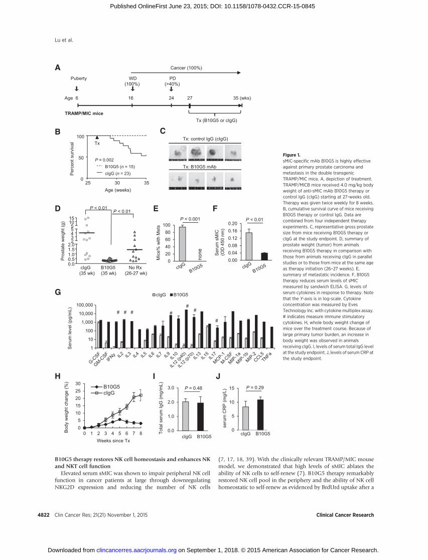
We have previously shown that TRAMP/MIC mice have greaterthan 40% incidence of developing highly invasive PD carcinomaand distant metastasis by 24-weeks of age (7). We also havepreviously determined that the accelerated tumor progression inTRAMP/MICB mice in comparison with TRAMP littermate isassociated with elevated serum sMIC and its immune suppressiveeffects as in cancer patients with advanced diseases (7, 20, 21,23, 27). With the specific aim to treat advanced cancer, we treatedrandom cohorts of 27- to 28-week-old TRAMP/MICmice with theB10G5 mAb or control mouse IgG (cIgG) twice weekly via i.p.route at thedose of 4mg/kg bodyweight (Fig. 1A). Following an8-week therapy, all mice that received B10G5 survived, whereasapproximately 50% of mice in the control group, succumbed tothe disease during the same time framewith the remaining ofmiceshowing severe symptoms of illness (Fig 1B–E and data notshown). Mice that received the 8-week B10G5 therapy had signif-icantly smaller prostate tumor mass in comparison with thosehaving received cIgG or to those at the same disease stage when
treatment began (27–28 week old; Fig. 1C and D). No metas-tasis was found in the B10G5-treated mice. Conversely, over90% of the mice receiving cIgG developed metastasis (Fig. 1E).Systemically, in response to B10G5 therapy, serum levels ofsMIC were significantly reduced (Fig. 1F).
A remarkable serum cytokine and chemokine "storm" that waslikely elicited by multitude activation of cellular immunity wasevident after the 8-week B10G5 therapy (Fig. 1G), as the "storm"was negatively influenced by depletion of respective specificlymphocyte subset during therapy (Supplementary Fig. S2). The"storm" exhibitedmarked elevation ofmajor antitumor cytokinesand immune activation chemokines, for example, IFNg , IL2, IL12,IL9, and CCL5. No cytokine storm was elicited in TRAMP/MICBmice at any other age or tumor development stages or in non-tumor-bearing TRAMP/MICB.A2 mice (ref. 7; data not shown),suggesting that the "storm" is therapy induced. Notably yet, nosignificant systemic toxicities, such as bodyweight loss, increase inserum IgG, or C-reactive protein (CRP) or inflammation in otherorgans (liver, kidney, intestinal, and pancreas) were observed inresponse to B10G5 therapy (Fig. 1H and J and data not shown).
Therapy eliminates invasive prostate tumor cells and primestumor microenvironment to be more immune reactive
Histologic examination revealed that animals havingreceived B10G5 therapy exhibited a mixture of normal prostategland and organ-defined well-differentiated prostate carcino-ma, whereas mice having received cIgG therapy exhibited highfrequency of invasive prostate carcinoma (Fig. 2A). B10G5therapy resulted in markedly reduced proliferation (Ki67þ)and increased apoptosis (C-caspaseþ) of carcinoma cells (Fig.2A). In the primary tumor of majority TRAMP/MIC micereceiving cIgG or no therapy, a distinct population of dissem-inated tumor cells that gained neuroendocrine differentiationmarker synaptophysin was abundantly present in the stroma(SYN; Fig. 2A). Clinically, these tumor cell types were consid-ered to be therapy-resistant and confer a poor prognosis (28,29). B10G5 therapy effectively eliminated the "neuroendo-crine-differentiated" tumor cells.
Inmost solid tumor of human cancers, tumor infiltration ofNKcells correlated with better clinical outcomes or better response totherapy (30–34), suggesting an important role of NK cell incontrolling cancer progression. Resembling human cancer, wehave recently shown that NK cell is rarely found in poorlydifferentiated prostate carcinoma (7). B10G5 therapy evidentlyenriched NK cell infiltration in the prostate tumor parenchyma(NKp46; Fig. 2B). Moreover, CD8 T-cell infiltration to tumorparenchyma was also enriched by B10G5 therapy (CD8; Fig. 2C).
Wehave recently shownthat sMICcan facilitate the expansionofMDSC and skew macrophage into the arginase Iþ alternativelyactivated phenotypes (12). Consistently, TRAMP/MIC had richinfiltrationof arginase Iþ cells in theprostate infiltrates,whichwereeliminated significantly by B10G5 therapy (arginase I; Fig. 2B). Asarginase I canbeproducedbyMDSC(generallydefinedasCD11bþ
Gr-1þ) and/or the alternatively activated tumor-associated mac-rophage (generally defined as CD206þCD11c�; refs. 35–38), wefurther defined these immune suppressive subsets in tumor infil-trates by flow cytometry analyses. The numbers of CD11bþGr-1þ
MDSC and CD206þCD11c� macrophages are both significantlydecreased in response to B10G5 therapy (Fig. 2C andD). Together,these data demonstrate that neutralizing sMIC primed tumormicroenvironment to be more immune active.
Targeting Soluble MIC for Cancer Immunotherapy
www.aacrjournals.org Clin Cancer Res; 21(21) November 1, 2015 4821
on September 1, 2018. © 2015 American Association for Cancer Research. clincancerres.aacrjournals.org Downloaded from
Published OnlineFirst June 23, 2015; DOI: 10.1158/1078-0432.CCR-15-0845
B10G5 therapy restores NK cell homeostasis and enhances NKand NKT cell function
Elevated serum sMIC was shown to impair peripheral NK cellfunction in cancer patients at large through downregulatingNKG2D expression and reducing the number of NK cells
(7, 17, 18, 39). With the clinically relevant TRAMP/MIC mousemodel, we demonstrated that high levels of sMIC ablates theability of NK cells to self-renew (7). B10G5 therapy remarkablyrestored NK cell pool in the periphery and the ability of NK cellhomeostatic to self-renew as evidenced by BrdUrd uptake after a
A
Tx: control IgG (cIgG)
Tx: B10G5 mAb
C
TRAMP/MIC mice
Age 6 16 24 27 35 (wks)
Puberty WD PD(100%) (>40%)
Cancer (100%)
Tx (B10G5 or cIgG)
F
H
05
1015202530
0 1 2 3 4 5 6 7 8
B10G5cIgG
Bod
y w
eigh
t cha
nge
(%)
Weeks since Tx
I
Tota
l ser
um Ig
G(m
g/m
L)
0.0
1.0
2.0
3.0 P = 0.48
cIgG B10G5
E
Mic
e% w
ith M
ets
none
020406080
100
Ser
um s
MIC
(OD
450
nm
)
0.000.040.080.120.160.20
B
Age (weeks)
Per
cent
sur
viva
l
0
50
100
25 30 35
B10G5 (n = 15)cIgG (n = 23)
P = 0.002
Tx
J
0
5
10
15
seru
m C
RP
(mg/
L)
cIgG B10G5
P = 0.29
D
0.00.51.01.52.0
23455
1015
B10G5(35 wk)
cIgG(35 wk)
No Rx(26-27 wk)
Pro
stat
e w
eigh
t (g)
P < 0.01P < 0.01
P < 0.001 P < 0.01
Ser
um le
vel (
pg/m
L) # #
#
##
1
10
100
1,000
10,000
100,000
cIgG B10G5
# #
G
Figure 1.sMIC-specific mAb B10G5 is highly effectiveagainst primary prostate carcinoma andmetastasis in the double transgenicTRAMP/MIC mice. A, depiction of treatment.TRAMP/MICB mice received 4.0 mg/kg bodyweight of anti-sMIC mAb B10G5 therapy orcontrol IgG (cIgG) starting at 27-weeks old.Therapy was given twice weekly for 8 weeks.B, cumulative survival curve of mice receivingB10G5 therapy or control IgG. Data arecombined from four independent therapyexperiments. C, representative gross prostatesize from mice receiving B10G5 therapy orcIgG at the study endpoint. D, summary ofprostate weight (tumor) from animalsreceiving B10G5 therapy in comparison withthose from animals receiving cIgG in parallelstudies or to those from mice at the same ageas therapy initiation (26–27 weeks). E,summary of metastatic incidence. F, B10G5therapy reduces serum levels of sMICmeasured by sandwich ELISA. G, levels ofserum cytokines in response to therapy. Notethat the Y-axis is in log-scale. Cytokineconcentration was measured by EvesTechnology Inc. with cytokine multiplex assay.# indicates measure immune stimulatorycytokines. H, whole body weight change ofmice over the treatment course. Because oflarge primary tumor burden, an increase inbody weight was observed in animalsreceiving cIgG. I, levels of serum total IgG levelat the study endpoint. J, levels of serumCRP atthe study endpoint.
Lu et al.
Clin Cancer Res; 21(21) November 1, 2015 Clinical Cancer Research4822
on September 1, 2018. © 2015 American Association for Cancer Research. clincancerres.aacrjournals.org Downloaded from
Published OnlineFirst June 23, 2015; DOI: 10.1158/1078-0432.CCR-15-0845

consecutive 5-day BrdUrd pulsing (Fig. 3A–D). Moreover, B10G5therapy markedly enhanced NK cell function, illustrated byincreased production of IFNg in response to mitogen stimulationand cytolytic ability against NKG2D ligand-positive target cells(Fig. 3E–G). Together, these data conclude that targeting serumsMIC significantly restores NK cell homeostatic maintenanceand function in MICþ cancer host. The conclusion was furthersupported by adoptively transfer experiments with V450-labeledCD45.1 congenic NK cells to B10G5 or cIgG-treated TRAMP/MIC mice (Supplementary Fig. S3). Furthermore, althoughB10G5 therapy did not significantly affect the pool of NKTcells (data not shown); however, therapy enhanced Th1-likefunctional potential of NKT cells as represented by significantlyincreased IFNg production in response to PMA/I stimulation(Fig. 3H and I).
B10G5 therapy potentiated CD8 and CD4 T cells antitumorresponses
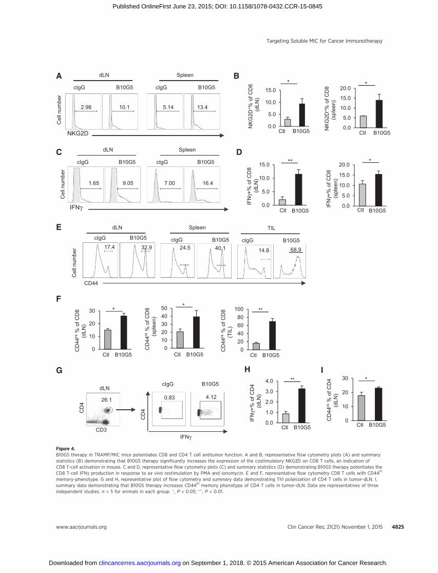
Tumor-derived sMIChas been shown to downregulateNKG2Dexpression on CD8 T cells in cancer patients (16). NKG2D, as a T-cell costimulatory receptor, is expressed by all human CD8 T cellsbut only by activated mouse CD8 T cells (4). B10G5 therapy inTRAMP/MIC mice significantly increased the population and
number of NKG2Dþ CD8 T cells in the periphery (Fig 4A andB), indicating activation of CD8 T cells. When stimulated ex vivowithmitogen, CD8T cells frommice receiving B10G5 therapyhadsignificantly higher frequency andmagnitude of IFNg production(Fig. 4C and D). Moreover, B10G5 therapy significantly increasedthe number of CD8T cells with CD44himemory phenotype in thespleen, tumor-dLN, and tumor infiltrates (Fig. 4E and F). Giventhat anergy and lack of trafficking to tumor sites are the majormechanisms of cytotoxic T dysfunction inmost cancer types, thesedata suggest that targeting sMIC can effectively revive cytotoxicCD8 T-cell antitumor response in cancer host.
NKG2D is rarely expressed by CD4 T cells. However, B10G5therapy potentiated CD4 T cells to Th1 responses in the tumordLNs, shown by the significant increases in IFNgþ CD4 T cells inresponse to mitogenic stimulation (Fig 4G and H) and increasesin T-bet expression (Supplementary Fig. S4). Furthermore, ther-apy also resulted in significant increase inCD4T cellswithCD44hi
memory phenotype (Fig 4I).
Therapy breaks CD8 T-cell tumor antigenic tolerance andaugments antigen-specific responses
Male TRAMP mice express the SV40-T-Antigen (TAg) onco-protein specifically in the prostate to drive spontaneous
B10G5 cIgG
Ki6
7C
.cas
pase
-3S
YN
* *
H&
E
A
Arg
inas
eI
NK
p46
CD
8
B10G5 cIgG
Gr-1
15.83.5
CD11b
B10G5 cIgG
C D
B
18.14.4C
D20
6
CD11c
B10G5 cIgG
TIL, gated on CD45+ F4/80+TIL, gated on CD45+ F4/80+
Figure 2.B10G5 therapy inhibits primary tumorproliferation and survival, eliminatesdissimilated malignant tumor cells,and remodels prostate tumormicroenvironment. A, representativemicrographs demonstrating thatB10G5 therapy inhibits prostatecancer cell proliferation (Ki-67þ),induces tumor cell apoptosis(C-caspase 3þ), eliminates malignantdisseminated neuroendocrine tumorcells from the stroma (SYNþ). B,representative micrographsdemonstrating that B10G5 therapyincreases immune active NK(NKp46þ) and CD8 T cells anddecreases arginase Iþ immunesuppressive cells in tumor infiltrates.C, representative flow cytometryanalyses of MDSC defined byGr-1þCD11bþ in tumor infiltrates. D,representative flow cytometryanalyses of alternatively activatedmacrophages defined by CD206þ
CD11c� in tumor infiltrates.
Targeting Soluble MIC for Cancer Immunotherapy
www.aacrjournals.org Clin Cancer Res; 21(21) November 1, 2015 4823
on September 1, 2018. © 2015 American Association for Cancer Research. clincancerres.aacrjournals.org Downloaded from
Published OnlineFirst June 23, 2015; DOI: 10.1158/1078-0432.CCR-15-0845

prostate tumor development through inactivation of tumorsuppressor genes p53 and Rb pathways (40). Akin to thebiology in cancer patients, the TAgs in male TRAMP mice
serves as a self-antigen and a tumor antigen, leading to toler-ance of TAg and clonal deletion of TAg-specific T cells (41).Adoptively transferred na€�ve CD8 T cells that bear TAg-specific
C
A
EN
K%
(spl
een)
0.00.51.01.52.02.53.03.5
B10G5Ctl
*
B10G5
NK
% (d
LN)
Ctl0.0
0.5
1.0
1.5
2.0 *
Brd
Urd
+%
of s
plen
ic N
K
010203040
B10G5Ctl
**
Brd
Urd
+%
of N
K(b
one
mar
row
)
B10G5Ctl0
10
20
30 **
B10G5IFN
γ+ % o
f spl
enic
NK
0
10
20
30
Ctl
*
B10G5
IFN
γ+ % o
f NK
( d
LN)
0
10
20
30
Ctl
**
02468
10
B10G5Ctl
IFN
γ+ % o
f NK
(b
one
mar
row
)
*
Spleen dLNC
D3
B10G5
NK1.1
2.38
cIgG
0.68
B10G5
0.95
cIgG
0.27
NK
1.1
BrdUrd
Spleen
18.3
B10G5
0.92
cIgG
bone marrow
16.6
B10G5
8.63
cIgG
B
D
dLN
24.9
B10G5
0.90
cIgG
Spleen
24.2
B10G5
8.38
cIgG
Bone marrow
11.4
B10G5
3.73
cIgG
IFN
NK
1.1
F
HG
0
5
10
15
20B10G5cIgG
Spec
ific
lysi
s %
10:1 5:1
**
E:T
IFN
γ+ % o
f spl
enic
NK
T
IFN
γ+ % o
f NK
T (d
LN)
0
10
20
30
40
0
5
10
15
20** **
Ctl B10G5 Ctl B10G5
I
B10G50
5
10
15
20
tota
l NK
(spl
een)
(x10
5 )
Ctl
*
0
20
40
60
Tota
l spl
enic
Brd
Urd
+N
K(x
104 )
Ctl B10G5
0
20
40
60
Tota
l spl
enic
IFN
γ+ NK
(x10
4 )
Ctl B10G5
**
0
20
40
60
Tota
l spl
enic
IFN
γ+ NK
T(x
104 )
Ctl B10G5
**
Figure 3.B10G5 therapy in TRAMP/MIC mice enhances NK cell homeostatic maintenance and function. A and B, B10G5 therapy significantly increased the number of NKcells in the periphery. C and D, B10G5 therapy significantly enhanced NK cell renewal as shown by the increase in the number of NK cells that are BrdUrdþ
after 5-day labeling. E and F, B10G5 therapy significantly enhanced NK cell IFNg production in response to PMA/ionomycin stimulation. G, splenic NK cell cytoxocityagainst NKG2D-Lþ RMA-S-Rae-1–positive cells. E:T ¼ effector:target. H and I, B10G5 therapy increases NKT population and functional potential shown byIFNg production in response to PMA/I stimulation. � , P < 0.05; �� , P < 0.01.
Lu et al.
Clin Cancer Res; 21(21) November 1, 2015 Clinical Cancer Research4824
on September 1, 2018. © 2015 American Association for Cancer Research. clincancerres.aacrjournals.org Downloaded from
Published OnlineFirst June 23, 2015; DOI: 10.1158/1078-0432.CCR-15-0845
D
B
NK
G2D
+ %of
CD
8(s
plee
n)
Ctl0.0
5.0
10.0
15.0
20.0
B10G5
*
NK
G2D
+ % o
f CD
8(d
LN)
B10G5Ctl
*
B10G5Ctl
IFN
γ +%
of C
D8
(spl
een)
0.0
5.0
10.0
15.0
20.0 *
B10G5
IFN
γ+%
of C
D8
(dLN
)
0.0
5.0
10.0
15.0
Ctl
**
Cel
l num
ber
dLN
10.1
B10G5
2.98
cIgG
Spleen
13.4
B10G5
5.14
cIgG
SpleenNLd
9.05
B10G5
1.65
cIgG
16.4
B10G5
7.00
cIgG
Cel
l num
ber
IFNγ
H
B10G5Ctl0.0
1.0
2.0
3.0
4.0
IFN
γ+%
of C
D4
(dLN
)
**
A
C
E
0
10
20
30
CD
44H
i % o
f CD
8(d
LN)
B10G5Ctl
*
01020304050
CD
44H
i % o
f CD
8(s
plee
n)
*
B10G5Ctl
F
G
26.1
CD3
CD
4
dLN
0.0
5.0
10.0
15.0
RX
γ
4.12
B10G5
0.83
cIgG
CD
4
0
10
20
30
CD
44H
i % o
f CD
4(d
LN)
B10G5Ctl
*I
CD44
dLN
32.9
B10G5
17.4
cIgG
Cel
l num
ber
Spleen
40.1B10G5
24.5cIgG
TIL
B10G5cIgG
14.8 68.9
020406080
100 **
B10G5Ctl
CD
44H
i % o
f CD
8(T
IL)
NKG2D
IFN
Figure 4.B10G5 therapy in TRAMP/MIC mice potentiates CD8 and CD4 T cell antitumor function. A and B, representative flow cytometry plots (A) and summarystatistics (B) demonstrating that B10G5 therapy significantly increases the expression of the costimulatory NKG2D on CD8 T cells, an indication ofCD8 T-cell activation in mouse. C and D, representative flow cytometry plots (C) and summary statistics (D) demonstrating B10G5 therapy potentiates theCD8 T-cell IFNg production in response to ex vivo restimulation by PMA and ionomycin. E and F, representative flow cytometry CD8 T cells with CD44hi
memory-phenotype. G and H, representative plot of flow cytometry and summary data demonstrating Th1 polarization of CD4 T cells in tumor-dLN. I,summary data demonstrating that B10G5 therapy increases CD44hi memory phenotype of CD4 T cells in tumor-dLN. Data are representatives of threeindependent studies. n > 5 for animals in each group. � , P < 0.05; �� , P < 0.01.
Targeting Soluble MIC for Cancer Immunotherapy
www.aacrjournals.org Clin Cancer Res; 21(21) November 1, 2015 4825
on September 1, 2018. © 2015 American Association for Cancer Research. clincancerres.aacrjournals.org Downloaded from
Published OnlineFirst June 23, 2015; DOI: 10.1158/1078-0432.CCR-15-0845

TCR became rapidly tolerant in TRAMP mice after initiationexpansion (42).
We sought to address whether anti-sMIC antibody B10G5therapy can enhance antigen-specific CD8 T-cell responsesin TRAMP/MIC mice. Because of the clonal deletion of TAg-specific CD8 T cells (41), we thus adoptively transferred CFSE-labeled na€�ve CD8 T cells from the TCR-I mice that express atransgenic TCR recognizing H-2Db
–restricted epitope I of the TAg(residues 206-215; ref. 26). As examined at day 9 after transfer, asignificantly higher number of H-2Db/epitope I–specific tetra-
mer (Db/I-tetramer) positive CD8 T cells was detected in thespleen, tumor-dLN, and tumor infiltrates of mice receivingB10G5 therapy than those receiving cIgG therapy (Fig. 5A).Notably, endogenous Db/I-tetramerþ CD8 T cells were notdetectable in TRAMP/MIC mice with or without antibodytreatment (Supplementary Fig. S5). The Db/I-tetramerþ CD8T cells are thus originated from adoptively transferred TCR-I cells. Consistent with findings in other studies (26, 43),adoptively transferred na€�ve TCR-I CD8 T cells (identified byDb/I-tetramerþ) underwent initial rapid expansion in all mice
A
B
C mIgG B10G5
IFNγγ
CD
8
Spleen
dLN
TIL
cIgG B10G5
mIgG B10G5C
D8
Spleen
dLN
TIL
Db/I-tetramer
D
0.044 0.28
1.16
8.77
0.40
1.09
2.490.17
2.890.29
0.64
CD
11c
MHCII
1.770.62
CD40
cIgG B10G5
CD86CD80
5.68
Cou
nts
cIgG B10G5
Day 9
Gated on Db/I-tetramer+ CD8 T cells
Day 14 2.34
CFSE
10.3
2.22 6.82CFSEhiCFSElo
4.69 6.27
0.98 0.78
Figure 5.B10G5 therapy breaks tumor antigen-specific CD8 T-cell tolerance. CFSE-labeled TCR-I CD8 T cells bearing TCR specific for H-2Db-restricted TAg epitope I wereadoptively transferred into cohorts of TRAMP/MICB mice (n ¼ 5–7, 2 � 106 cells/mouse) receiving therapy with B10G5 or cIgG. Mice were euthanized at days9 and 14 after transfer for analyses. Because of lack of endogenous CD8 T cells specific for TAg epitope I, adoptively transferred TCR-I–specific CD8 T cellswere identified by positive staining for H-2Db-TAg-epitope I tetramer (Db/I-tetramer). A, representative dot plots demonstrating frequency of Db/I-tetramerþCD8 Tcells in the spleen, tumor-dLN and tumor infiltrates at day 9 after transfer. B, representative dot plots demonstrating proliferation of Db/I-tetramerþ CD8 T cells atdays 9 and 14 after transfer. C, CD8 T cell IFNg production in response to TAg epitope I stimulation. A total of 1�106 bulked splenocytes and single-cell suspensionfrom tumor-dLN were stimulated with 0.5 mmol/L of TAg 206-215 epitope I overnight. IFNg production was assessed by intracellular staining. D, expressionof DC activation markers in tumor-dLN (day 14). Gray fill profile, mice received cIgG treatment. Black filled profile, mice received B10G5 treatment.
Lu et al.
Clin Cancer Res; 21(21) November 1, 2015 Clinical Cancer Research4826
on September 1, 2018. © 2015 American Association for Cancer Research. clincancerres.aacrjournals.org Downloaded from
Published OnlineFirst June 23, 2015; DOI: 10.1158/1078-0432.CCR-15-0845
as shown by low percentage of CFSEhi tetramers when exam-ined at day 9 (Fig. 5B), owing to encountering TAg. However,TCR-I CD8 T cells only continued to expand in mice receivingB10G5 therapy, as shown by marked increase in the percentageof Db/I-tetramerþCFSElo cells when examined at day 14 aftertransfer (Fig. 5B). We further tested antigen-specific responsive-ness of CD8 T cells by stimulating single-cell suspension frombulked splenocytes, tumor-dLN, or tumor digests with TAgepitope I peptide. As shown in Fig. 5C, B10G5 therapy marked-ly enhanced the capacity of CD8 T cells to produce IFNg inresponse to TAg-specific epitope I peptide (TAg 206-215)stimulation.
In vitro studies have elegantly demonstrated that MICþ-tumorcells coated with anti-MIC antibody can opsonize dendritic cells(DC) and enhance antigen cross-presenting to CD8 T cells (44,45). We thus further investigated whether this mechanism maycontribute, at least in part, to the enhanced antigen-specific TCR-IT cells expansion and responses in the current experiment settings.As representatively shown in Fig. 5D, B10G5 therapy not onlyenriched mature DC presence in tumor-dLN not only markedelevated the expression of DC surface activation molecule CD40and costimulatory molecule CD80/86. Altogether, these dataevidently demonstrated that mAb targeting serum sMIC caneffectively overcome tumor antigen-specific CD8 T-cell toleranceand enhance DC priming for antigen-presentation.
Our data in the clinically relevant TRAMP/MIC mouse havedemonstrated that B10G5 neutralizing sMIC can effective pro-voke amultitude of immune responses to suppress tumor growthand metastasis. We further substantiated these findings withexperiments demonstrating that B10G5 effectively retarded thegrowth of transplantable TRAMP-C2 prostate tumor cell lines thatexpress sMICB (TC2-sMICB) using the syngeneicMICB transgenicmouse model (Supplementary Figs. S6 and S7).
As tumors can be highly heterogeneous in differentiation andsurface MIC retention, we further investigated the therapeuticeffect of B10G5 against tumors expressing surfaceMIC.We treatedcohorts of TRAMP/MICB mice of 20 weeks of age, at which timemost of the tumors are well-differentiated and retained surfaceMIC expression as we have shown previously (7). A significanttherapeutic response was also achieved although host immuneresponses were amplified at discrete magnitudes (SupplementaryFig. S8), presumably due to dissimilar mechanisms to whichconferred by neutralizing sMIC.
Therapeutic antitumor effect is conferred byNKandCD8T cellsand CD4 Th1 response is NK-dependent
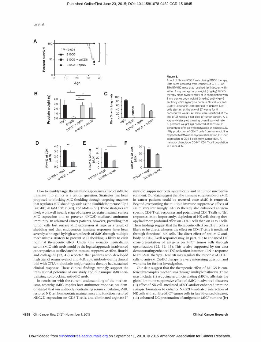
To this end, our data have suggested that therapy with a mAbB10G5 targeting sMIC effectively induces regression ofadvanced MICþ malignancies through restoring innate andadaptive antitumor responses. We further address the contri-bution of NK and CD8 T cells in conferring the therapeuticeffect of B10G5. Depleting NK or CD8 T cells in TRAMP/MICBmice during B10G5 therapy significantly mitigated the thera-peutic effect of B10G5 (Fig. 6), exemplified by a significantlyhigher number of animals succumbed to tumor-related death,owing to larger primary tumor burden and/or development oflung metastasis (Fig. 6A–C). Depletion of CD4 T cells did notpresent a significant impact on therapeutic effect of B10G5(data not shown). Because of the functional heterogeneity ofmultiple CD4 T cells subsets, the outcome is somewhatanticipated.
Intriguingly, with depletion of NK cells, B10G5 therapyfailed to evoke CD4 T cells to generate optimal Th1 immuneresponses and maintaining CD44Hi memory phenotypes(Fig. 6D–F and Supplementary Fig. S9). Depletion of CD8T cells had nominal effect on CD4 T cells (data not shown).These data not only suggest that both NK and CD8 T cells arerequired to generate optimal antitumor responses, but alsosuggest that functional NK cells are essential for generatingoptimal CD4 T-cell responses.
DiscussionTumor-derived sMIC, whether through protease-mediated
shedding or exosome secretion, has been shown to be highlyimmune suppressive in cancer patients throughmultiplemechan-isms (9, 10, 46). However, whether a therapy targeting sMIC canrevitalize host endogenous immune response remains untested.Using a clinically relevant double transgenic TRAMP/MIC mousemodel that closely recapitulates the dynamics of serum sMIC andtumor progression, here we demonstrate that therapy with anonblocking anti-MIC mAb to neutralize serum sMIC effectivelyinduce regression of advanced primary tumors and eliminatedmetastasis through revamping host endogenous innate and adop-tive antitumor immune responses and remodeling tumor micro-environment. Therapy effectuated endogenous antitumorresponses by recuperating the ability of NK cells to self-renewand to be tumor-destructive, enhancing antigen-specific CD8T-cell antitumor responses, and potentiating CD4 T cells to Th1responses. Not only so, therapy also remodels tumor microenvi-ronment demonstrated by enhancing tumor infiltration of NKcells and cytotoxic CD8 T cells and eliminating arginase Iþ
immune suppressive myeloid cells in tumor parenchyma.Remarkably, therapy elicited a systemic cytokine "storm" includ-ing major antitumor cytokines, without inducing systemic auto-immune cytotoxicity. Our study has provided the compellingfirst-in-field preclinical evidence that therapy with a sMIC-neu-tralizing nonblocking anti-MIC mAb alone can obliterateimmune suppressive effective of tumor-derived sMIC and reviveendogenous antitumor immune responses. Our findings define anew effective and feasible translational approach for cancerimmunotherapy.
During oncogenesis, whether due to mutations in tumor sup-pressor genes or oncogenes, cells undergo aberrant DNA replica-tion which initiates DNA repair responses as the cellular checkpoint. This DNA repair response also triggers systemic checkpointby upregulating NKG2D ligand, predominantly MIC, expressionto alert the immune system to eliminate abnormal cells (1, 3).However, this oncoimmunologic coevolutionary process eventu-ally renders tumor immune escape of NKG2D-mediated immunesurveillance and allows tumors to progress. Proteolytic proteasesor exosome-mediated tumor-shedding of sMIC accounted for oneof the major mechanisms for MICþ tumor evasion of NKG2Dimmune surveillance (9, 10, 46). Earlier studies demonstratedthat sMIC induced global NKG2D downmodulation on all sub-sets of antitumor effector cells, such as CD8, NK, NKT, and gd Tcells, in cancer patients (10, 11, 46). Recent studies have furtherdemonstrated that sMIC can produce more profound immunesuppressive effect by perturbingNK cell homeostaticmaintenanceand facilitating the expansion of arginase Iþ myeloid suppressorcells (7, 12). These studies endorsed the viability of blocking sMICpathways in cancer immunotherapy.
Targeting Soluble MIC for Cancer Immunotherapy
www.aacrjournals.org Clin Cancer Res; 21(21) November 1, 2015 4827
on September 1, 2018. © 2015 American Association for Cancer Research. clincancerres.aacrjournals.org Downloaded from
Published OnlineFirst June 23, 2015; DOI: 10.1158/1078-0432.CCR-15-0845
How to feasibly target the immune suppressive effect of sMIC totranslate into clinics is a critical question. Strategies has beenproposed to blocking MIC shedding through targeting enzymesthat regulatesMIC shedding, such as the disulfide isomerase ERp5(47, 48), ADAM 10/17 (49), and MMPs (50). These strategies arelikelyworkwell in early stage of diseases to retainmaximal surfaceMIC expression and to preserve NKG2D-mediated antitumorimmunity. In advanced cancer patients, however, providing thattumor cells lost surface MIC expression at large as a result ofshedding and that endogenous immune responses have beenseverely sabotaged by high serum levels of sMIC throughmultiplemechanisms, strategy to prevent MIC shedding is likely to elicitnominal therapeutic effect. Under this scenario, neutralizingserum sMICwithmAbwould be the logical approach in advancedcancer patients to alleviate the immune suppressive effect. Jinushiand colleagues (22, 45) reported that patients who developedhigh titer of serum levels of anti-MIC autoantibody during clinicaltrial with CTLA-4 blockade and/or vaccine therapy had sustainedclinical response. These clinical findings strongly support thetranslational potential of our study and our unique sMIC-neu-tralizing nonblocking anti-MIC mAb.
In consistent with the current understanding of the mechan-isms, whereby sMIC impairs host antitumor response, we dem-onstrated that our antibody neutralizing serum circulating sMICrestored NK cell homeostatic maintenance and function, restoredNKG2D expression on CD8 T cells, and eliminated arginase Iþ
myeloid suppressor cells systemically and in tumor microenvi-ronment. Our data suggest that the immune suppression of sMICin cancer patients could be reversed once sMIC is removed.Beyond overcoming the multiple immune suppressive effects ofsMIC, very intriguingly, B10G5 therapy also enhanced antigen-specific CD8 T-cell responses and potentiated CD4 T cells to Th1responses. More importantly, depletion of NK cells during ther-apy hadmore profound effect on CD4 T cells than on CD8 T cells.These findings suggest that the therapeutic effect on CD8 T cells islikely to be direct, whereas the effect on CD4 T cells is mediatedthrough functional NK cells. The direct effect of anti-MIC anti-body on CD8 T-cell responses may, in part, due to enhanced DCcross-presentation of antigens on MICþ tumor cells throughopsonization (22, 44, 45). This is also supported by our datademonstrating enhancedDCactivation in tumor-dLN in responseto anti-MIC therapy. HowNKmay regulate the response of CD4 Tcells to anti-sMIC/MIC therapy is a very interesting question andwarrants for further investigation.
Our data suggest that the therapeutic effect of B10G5 is con-ferred by complexmechanisms throughmultiple pathways. Thesemay include: (i) reducing serum circulating sMIC to alleviate theglobal immune suppressive effect of sMIC in advanced diseases;(ii) effect of NK cell–mediated ADCC and/or enhanced immunesynapse formation to enhance NKG2D-mediated interaction ofNK cells with surface MICþ-tumor cells in less advanced diseases;(iii) enhanced DC presentation of antigens onMICþ tumors; (iv)
APe
rcen
t sur
viva
l
0 10 20 30 400
50
100
Age
B10G5
B10G5 + dpCD8
B10G5 + dpNK
* *
* P < 0.001
T-be
t+%
of C
D4
(d
LN)
*D
IFN
γγ+ % o
f CD
4 (d
LN)
**
0.0
1.0
2.0
3.0
4.0
B
Pros
tate
wei
ght (
g)
* **
0.0
1.0
2.0
3.0
4.0C
020406080
100
Mic
e %
with
lung
met
s
none
0
10
20
30E
CD
44H
i%
of C
D4
(d
LN)
F
0
10
20
30
40
Figure 6.Affect of NK and CD8 T cells during B10G5 therapy.Data were obtained from cohorts (n ¼ 5–8) ofTRAMP/MIC mice that received i.p. injection witheither 4 mg per kg body weight (mg/kg) B10G5therapy alone twice weekly or in combination with8 mg per kg body weight (mg/kg) anti-NKp46antibody (BioLegend) to deplete NK cells or anti-CD8a (Cedarlane Laboratories) to deplete CD8 Tcells starting at the age of 27 weeks for 8consecutive weeks. All mice were sacrificed at theage of 35 weeks if not died of tumor burden. A, aKaplan–Meier plot showing overall survival rate.B, prostate weight (g) collected at sacrifice. C,percentage of mice with metastasis at necropsy. D,IFNg production of CD4 T cells from tumor-dLN inresponse to PMA/ionomycin restimulation. E, T-betexpression in CD4 T cells from tumor-dLN. F,memory phenotype CD44hi CD4 T-cell populationin tumor-dLN.
Lu et al.
Clin Cancer Res; 21(21) November 1, 2015 Clinical Cancer Research4828
on September 1, 2018. © 2015 American Association for Cancer Research. clincancerres.aacrjournals.org Downloaded from
Published OnlineFirst June 23, 2015; DOI: 10.1158/1078-0432.CCR-15-0845

potential complex formation of sMIC/B10G5 and subsequentimmune stimulatory effect; (v) enhanced NK-DC cross-talk tobetter prime the adoptive immune responses. The impact of eachmechanism on the therapeutic outcome warrants futureinvestigation.
It is intriguing that antibody targeting sMIC therapy elicit asystemic cytokine and chemokine "storm," but with no detectableautoimmune toxicity. Notably, therapy not only induced highserum levels of antitumor cytokines, such as IL2, IFNg , IL9, IL12,IL17, but also induced cytokines that regulates immune tolerance,such as IL4, IL10, and IL13. The balanced induction of immuneactive and immune tolerance cytokines may explain the absenceof systemic autoimmune cytotoxicity. Given that MIC is a tumor-specific target, the absence of systemic autoimmunity may bereasonably expected.
In conclusion, with a "humanized" genetic engineeredmouse model that recapitulate the dynamic interaction ofhuman NKG2D ligand MIC with tumor progression, we dem-onstrate that an antibody neutralizing sMIC effectively inducedregression of primary tumors and eliminated metastasis inadvanced MICþ malignancy. We further demonstrated that themechanism of tumor suppression and clearance was conferredthrough restoring NK cell homeostatic maintenance and func-tion, overcoming CD8 T-cell tolerance to tumor antigen, andheightening CD4 T cells to Th1 responses, and priming DCs forenhanced antigen presentation and tumor microenvironmentto be more immune reactive. Furthermore, we demonstrate acritical role of NK cells in potentiating adaptive immuneresponses against tumors. Collectively, our study provided thefirst-in-field preclinical evidence demonstrating that an anti-body neutralizing sMIC can reset and revamp endogenousantitumor responses to effectuate elimination of MICþ malig-nancies. Our findings are highly translatable to clinics to treatMICþ cancers with antibodies to neutralize sMIC. Conceptu-
ally, given the global overturning of endogenous antitumorresponses induced by the sMIC-neutralizing antibody, sMICmay be considered as a tumor-specific immune checkpointmolecule.
Disclosure of Potential Conflicts of InterestNo potential conflicts of interest were disclosed.
Authors' ContributionsConception and design: S. Lu, D. Liu, J.D. WuDevelopment of methodology: S. Lu, G. Li, J.D. WuAcquisition of data (provided animals, acquired and managed patients,provided facilities, etc.): S. Lu, J. Zhang, D. Liu, J.D. WuAnalysis and interpretation of data (e.g., statistical analysis, biostatistics,computational analysis): S. Lu, J. Zhang, G. Li, K.F. Staveley-O'Carroll, Z. Li,J.D. WuWriting, review, and/or revision of the manuscript: S. Lu, G. Li, K.F. Staveley-O'Carroll, J.D. WuAdministrative, technical, or material support (i.e., reporting or organizingdata, constructing databases): S. Lu, D. Liu, G. Li, K.F. Staveley-O'Carroll,J.D. WuStudy supervision: K.F. Staveley-O'Carroll, J.D. WuOther (oversaw collaboration): J.D. Wu
Grant SupportThis work was supported by NIH-NCI grant 1R01CA149405 and A. David
Mazzone—Prostate Cancer Foundation Challenge Award (to J. Wu) and, inpart, supported by the Flow Cytometry Core Facility Shared Resource,Hollings Cancer Center, Medical University of South Carolina (P30CA138313).
The costs of publication of this article were defrayed in part by thepayment of page charges. This article must therefore be hereby markedadvertisement in accordance with 18 U.S.C. Section 1734 solely to indicatethis fact.
Received April 5, 2015; revised June 4, 2015; accepted June 12, 2015;published OnlineFirst June 23, 2015.
References1. Gasser S, Orsulic S, Brown EJ, Raulet DH. The DNA damage pathway
regulates innate immune system ligands of the NKG2D receptor. Nature2005;436:1186–90.
2. Groh V, Bahram S, Bauer S, Herman A, Beauchamp M, Spies T. Cell stress-regulated human major histocompatibility complex class I gene expressedin gastrointestinal epithelium. ProcNatl AcadSciUSA1996;93:12445–50.
3. Gasser S, Raulet D. The DNA damage response, immunity and cancer.Semin Cancer Biol 2006;16:344–7.
4. Raulet DH. Roles of the NKG2D immunoreceptor and its ligands. Nat RevImmunol 2003;3:781–90.
5. Diefenbach A, Jensen ER, Jamieson AM, Raulet DH. Rae1 and H60 ligandsof the NKG2D receptor stimulate tumour immunity. Nature 2001;413:165–71.
6. Maasho K,Opoku-Anane J,Marusina AI, Coligan JE, Borrego F.NKG2D is acostimulatory receptor for human naive CD8þ T cells. J Immunol2005;174:4480–4.
7. Liu G, Lu S, Wang X, Page ST, Higano CS, Plymate SR, et al. Perturbation ofNK cell peripheral homeostasis accelerates prostate carcinomametastasis. JClin Invest 2013;123:4410–22.
8. Fang L, Gong J, Wang Y, Liu R, Li Z, Wang Z, et al. MICA/B expression isinhibited by unfolded protein response and associated with poor prog-nosis in human hepatocellular carcinoma. J Exp Clin Cancer Res 2014;33:76.
9. Chitadze G, Bhat J, Lettau M, Janssen O, Kabelitz D. Generation of solubleNKG2D ligands: proteolytic cleavage, exosome secretion and functionalimplications. Scand J Immunol 2013;78:120–9.
10. Salih HR, Holdenrieder S, Steinle A. Soluble NKG2D ligands: prevalence,release, and functional impact. Front Biosci 2008;13:3448–56.
11. Nausch N, Cerwenka A. NKG2D ligands in tumor immunity. Oncogene.2008;27:5944–58.
12. Gang Xiao XW, Jun Sheng, Shengjun Lu, Xuezhong Yu, Jennifer D Wu.Soluble NKG2D ligand promotes MDSC expansion and skews macro-phage to the alternatively activated phenotype. J Hematol Oncol 2015;8:13.
13. Spear P,WuMR, SentmanML, Sentman CL. NKG2D ligands as therapeutictargets. Cancer Immun 2013;13:8.
14. Ullrich E, Koch J, Cerwenka A, Steinle A. New prospects on the NKG2D/NKG2DL system for oncology. Oncoimmunology 2013;2:e26097.
15. Raulet DH, Gasser S, Gowen BG, Deng W, Jung H. Regulation of ligandsfor the NKG2D activating receptor. Annu Rev Immunol 2013;31:413–41.
16. Groh V, Wu J, Yee C, Spies T. Tumour-derived soluble MIC ligandsimpair expression of NKG2D and T-cell activation. Nature 2002;419:734–8.
17. Wu JD, Higgins LM, Steinle A, Cosman D, Haugk K, Plymate SR. Prevalentexpression of the immunostimulatory MHC class I chain-related moleculeis counteracted by shedding in prostate cancer. J Clin Invest 2004;114:560–8.
18. Doubrovina ES, DoubrovinMM, Vider E, Sisson RB,O'Reilly RJ, Dupont B,et al. Evasion from NK cell immunity by MHC class I chain-relatedmolecules expressing colon adenocarcinoma. J Immunol 2003;171:6891–9.
www.aacrjournals.org Clin Cancer Res; 21(21) November 1, 2015 4829
Targeting Soluble MIC for Cancer Immunotherapy
on September 1, 2018. © 2015 American Association for Cancer Research. clincancerres.aacrjournals.org Downloaded from
Published OnlineFirst June 23, 2015; DOI: 10.1158/1078-0432.CCR-15-0845

19. Marten A, von Lilienfeld-Toal M, Buchler MW, Schmidt J. Soluble MIC iselevated in the serum of patients with pancreatic carcinoma diminishinggammadelta T cell cytotoxicity. Int J Cancer 2006;119:2359–65.
20. Holdenrieder S, Stieber P, Peterfi A, Nagel D, Steinle A, Salih HR. SolubleMICB in malignant diseases: analysis of diagnostic significance and cor-relation with soluble MICA. Cancer Immunol Immunother 2006;55:1584–9.
21. Holdenrieder S, Stieber P, Peterfi A, Nagel D, Steinle A, Salih HR. SolubleMICA in malignant diseases. Int J Cancer 2006;118:684–7.
22. Jinushi M, Vanneman M, Munshi NC, Tai YT, Prabhala RH, Ritz J, et al.MHC class I chain-related proteinA antibodies and shedding are associatedwith the progression of multiple myeloma. Proc Natl Acad Sci U S A2008;105:1285–90.
23. Wu JD, Higgins LM, Steinle A, Cosman D, Haugk K, Plymate SR. Prevalentexpression of the immunostimulatoryMHC class I chain-relatedmolecule iscounteracted by shedding in prostate cancer. J Clin Invest 2004;114:560–8.
24. Deng W, Gowen BG, Zhang L, Wang L, Lau S, Iannello A, et al. A shedNKG2D ligand that promotes natural killer cell activation and tumorrejection. Science 2015;348:136–9.
25. Strong RK. Asymmetric ligand recognition by the activating natural killercell receptor NKG2D, a symmetric homodimer. Mol Immunol 2002;38:1029–37.
26. Staveley-O'Carroll K, Schell TD, Jimenez M, Mylin LM, Tevethia MJ,Schoenberger SP, et al. In vivo ligation of CD40 enhances priming againstthe endogenous tumor antigen and promotes CD8þ T cell effector functionin SV40 T antigen transgenic mice. J Immunol 2003;171:697–707.
27. Wu J. NKG2D Ligands in cancer immunotherapy: target or not? Austin JClin Immunol 2014;1:2.
28. MarcuM, Radu E, SajinM.Neuroendocrine transdifferentiation of prostatecarcinoma cells and its prognostic significance. Rom J Morphol Embryol2010;51:7–12.
29. Sun Y, Niu J, Huang J. Neuroendocrine differentiation in prostate cancer.Am J Transl Res 2009;1:148–62.
30. Ishigami S, Natsugoe S, Tokuda K, Nakajo A, Che X, Iwashige H, et al.Prognostic value of intratumoral natural killer cells in gastric carcinoma.Cancer 2000;88:577–83.
31. Coca S, Perez-Piqueras J, Martinez D, Colmenarejo A, Saez MA, Vallejo C,et al. The prognostic significance of intratumoral natural killer cells inpatients with colorectal carcinoma. Cancer 1997;79:2320–8.
32. Desbois M, Rusakiewicz S, Locher C, Zitvogel L, Chaput N. Natural killercells in non-hematopoietic malignancies. Front Immunol 2012;3:395.
33. Takanami I, Takeuchi K, Giga M. The prognostic value of natural killer cellinfiltration in resected pulmonary adenocarcinoma. J Thorac CardiovascSurg 2001;121:1058–63.
34. Villegas FR, Coca S, Villarrubia VG, Jimenez R, Chillon MJ, Jareno J, et al.Prognostic significance of tumor infiltratingnatural killer cells subset CD57in patients with squamous cell lung cancer. Lung Cancer 2002;35:23–8.
35. Nagaraj S, Gabrilovich DI. Tumor escape mechanism governed by mye-loid-derived suppressor cells. Cancer Res 2008;68:2561–3.
36. Rodriguez PC, Ernstoff MS, Hernandez C, Atkins M, Zabaleta J, Sierra R,et al. Arginase I-producing myeloid-derived suppressor cells in renal cell
carcinoma are a subpopulation of activated granulocytes. Cancer Res2009;69:1553–60.
37. Martinez FO, Sica A, Mantovani A, Locati M. Macrophage activation andpolarization. Front Biosci 2008;13:453–61.
38. Martinez FO, Helming L, Gordon S. Alternative activation of macro-phages: an immunologic functional perspective. Annu Rev Immunol2009;27:451–83.
39. Jinushi M, Takehara T, Tatsumi T, Hiramatsu N, Sakamori R, Yamaguchi S,et al. Impairment of natural killer cell and dendritic cell functions by thesoluble form of MHC class I-related chain A in advanced human hepato-cellular carcinomas. J Hepatol 2005;43:1013–20.
40. Deeb KK, Michalowska AM, Yoon CY, Krummey SM, Hoenerhoff MJ,Kavanaugh C, et al. Identification of an integrated SV40 T/t-antigen cancersignature in aggressive human breast, prostate, and lung carcinomas withpoor prognosis. Cancer Res 2007;67:8065–80.
41. Zheng X, Gao JX, Zhang H, Geiger TL, Liu Y, Zheng P. Clonal deletion ofsimian virus 40 large T antigen-specific T cells in the transgenic adenocar-cinoma of mouse prostate mice: an important role for clonal deletion inshaping the repertoire of T cells specific for antigens overexpressed in solidtumors. J Immunol 2002;169:4761–9.
42. Bai A,HighamE, EisenHN,WittrupKD,Chen J. Rapid tolerization of virus-activated tumor-specific CD8þ T cells in prostate tumors of TRAMP mice.Proc Natl Acad Sci U S A 2008;105:13003–8.
43. Shafer-Weaver KA, Watkins SK, Anderson MJ, Draper LJ, Malyguine A,Alvord WG, et al. Immunity to murine prostatic tumors: continuousprovision of T-cell help prevents CD8 T-cell tolerance and activatestumor-infiltrating dendritic cells. Cancer Res 2009;69:6256–64.
44. Groh V, Li YQ, Cioca D, Hunder NN, Wang W, Riddell SR, et al. Efficientcross-priming of tumor antigen-specific T cells by dendritic cells sensitizedwith diverse anti-MICA opsonized tumor cells. Proc Natl Acad Sci U S A2005;102:6461–6.
45. Jinushi M, Hodi FS, Dranoff G. Therapy-induced antibodies to MHCclass I chain-related protein A antagonize immune suppression andstimulate antitumor cytotoxicity. Proc Natl Acad Sci U S A 2006;103:9190–5.
46. Baragano Raneros A, Suarez-Alvarez B, Lopez-Larrea C. Secretory pathwaysgenerating immunosuppressive NKG2D ligands: new targets for therapeu-tic intervention. Oncoimmunology 2014;3:e28497.
47. Kaiser BK, YimD,Chow IT, Gonzalez S,Dai Z,MannHH, et al. Disulphide-isomerase-enabled shedding of tumour-associated NKG2D ligands.Nature 2007;447:482–6.
48. Fonseca C, Soiffer R, Ho V, Vanneman M, Jinushi M, Ritz J, et al. Proteindisulfide isomerases are antibody targets during immune-mediated tumordestruction. Blood 2009;113:1681–8.
49. Waldhauer I, Goehlsdorf D, Gieseke F, Weinschenk T, Wittenbrink M,Ludwig A, et al. Tumor-associated MICA is shed by ADAM proteases.Cancer Res 2008;68:6368–76.
50. Liu G, Atteridge CL, Wang X, Lundgren AD, Wu JD. The membrane typematrix metalloproteinaseMMP14mediates constitutive shedding ofMHCclass I chain-related molecule A independent of A disintegrin and metal-loproteinases. J Immunol 2010;184:3346–50.
Clin Cancer Res; 21(21) November 1, 2015 Clinical Cancer Research4830
Lu et al.
on September 1, 2018. © 2015 American Association for Cancer Research. clincancerres.aacrjournals.org Downloaded from
Published OnlineFirst June 23, 2015; DOI: 10.1158/1078-0432.CCR-15-0845
2015;21:4819-4830. Published OnlineFirst June 23, 2015.Clin Cancer Res Shengjun Lu, Jinyu Zhang, Dai Liu, et al. Eliminates Primary and Metastatic TumorsEndogenous Innate and Adaptive Antitumor Responses and Nonblocking Monoclonal Antibody Targeting Soluble MIC Revamps
Updated version
10.1158/1078-0432.CCR-15-0845doi:
Access the most recent version of this article at:
Material
Supplementary
http://clincancerres.aacrjournals.org/content/suppl/2015/06/24/1078-0432.CCR-15-0845.DC1
Access the most recent supplemental material at:
Cited articles
http://clincancerres.aacrjournals.org/content/21/21/4819.full#ref-list-1
This article cites 50 articles, 18 of which you can access for free at:
Citing articles
http://clincancerres.aacrjournals.org/content/21/21/4819.full#related-urls
This article has been cited by 1 HighWire-hosted articles. Access the articles at:
E-mail alerts related to this article or journal.Sign up to receive free email-alerts
Subscriptions
Reprints and
To order reprints of this article or to subscribe to the journal, contact the AACR Publications Department at
Permissions
Rightslink site. Click on "Request Permissions" which will take you to the Copyright Clearance Center's (CCC)
.http://clincancerres.aacrjournals.org/content/21/21/4819To request permission to re-use all or part of this article, use this link
on September 1, 2018. © 2015 American Association for Cancer Research. clincancerres.aacrjournals.org Downloaded from
Published OnlineFirst June 23, 2015; DOI: 10.1158/1078-0432.CCR-15-0845