non surgica management - guillem...
TRANSCRIPT

Non Surgical management of Cervical Spine Trauma
Guillem Saló Bru, MD, Phd AOSpine Principles Symposium- Cervical SpineOrthopaedic Depatment. Spine Unit.
Hospital del Mar. Barcelona.Associated Professor UAB Barcelona, February 2014
Disclosure information
I have no financial relationships with commercial entities that produce
health-care related products.
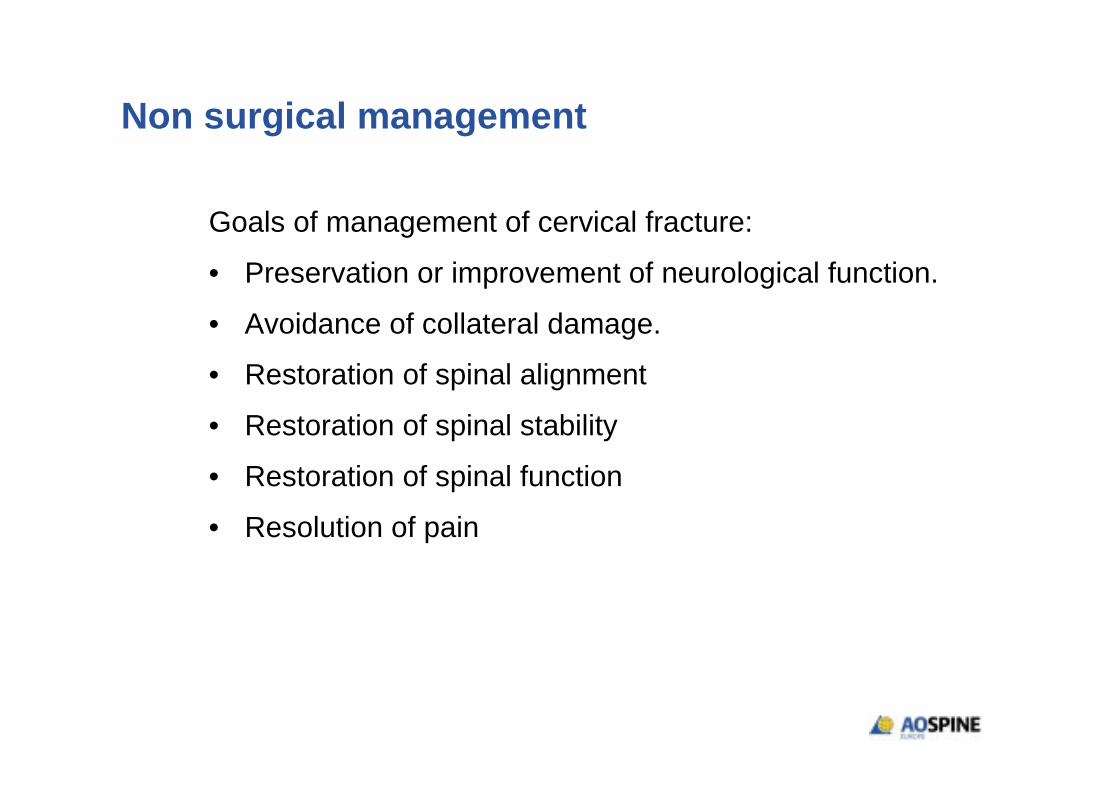
Non surgical management
Goals of management of cervical fracture:
• Preservation or improvement of neurological function.
• Avoidance of collateral damage.
• Restoration of spinal alignment
• Restoration of spinal stability
• Restoration of spinal function
• Resolution of pain

Medical treatment of cervical fracture.
• Painkillers.
• Antiinflamatory drugs.
• Antithrombotic
• Ulcus prevention.
• Antibiotics
• Inmobilization
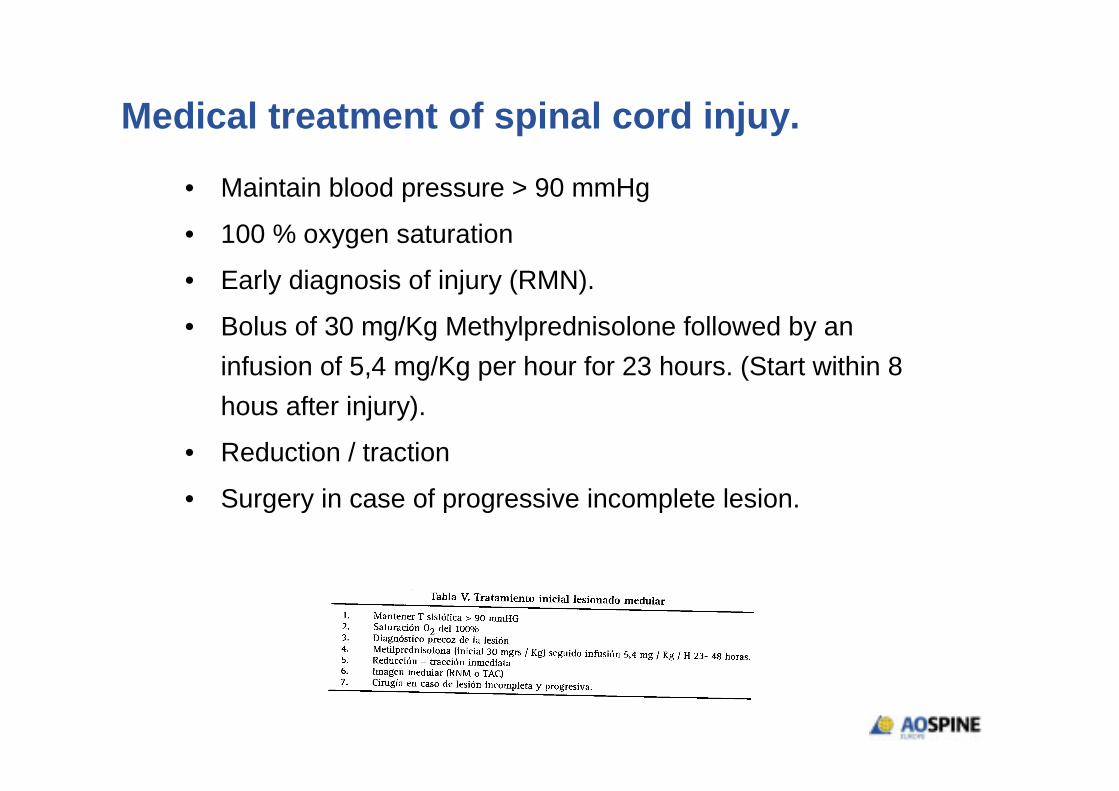
Medical treatment of spinal cord injuy.
• Maintain blood pressure > 90 mmHg
• 100 % oxygen saturation
• Early diagnosis of injury (RMN).
• Bolus of 30 mg/Kg Methylprednisolone followed by an
infusion of 5,4 mg/Kg per hour for 23 hours. (Start within 8
hous after injury).
• Reduction / traction
• Surgery in case of progressive incomplete lesion.
Medical treatment of spinal cord injuy.• The role of steroids in acute spinal cord injury is very controversial.
• Potential side effects of high dose methylprednisolone: such as infections, diabeticdescompensations , pancreatitis , myopathies, psychosis, and lactate acidosis in combination with intravenous adrenaline
• NASCIS (National Acute Spinal Cord Injury Study) II study
• Treatment with methylprednisolone for either 24 or 48 hours is recommended as an option…that should be undertaken only with the knowledge that the evidence suggesting harmful side effects is more consistent than any suggestion of clinical benefit.
• However, many researchers revisited this concern within the evidence-based framework of a critical appraisal of the accumulation of clinical studies and concluded that high-dose methylprednisolone cannot be justified as a standard treatment in acute spinal cord injury within current medical practice.
• We only consider high-dosemethylprednisolone treatment for young patients with a monotrauma of the spine.
Bracken MB, Shepard MJ, Collins WF, Holford TR, Young W, Baskin DS, Eisenberg HM, Flamm E, Leo-Summers L,Maroon J, et al. (1990) A randomized, controlled trial of methylprednisolone or naloxone in the treatment of acute spinal-cord injury. Results of theSecond National Acute Spinal Cord Injury Study. N Engl J Med 322:1405–11Short D (2001) Is the role of steroids in acute spinal cord injury now resolved? Curr Opin Neurol 14:759–63

Non surical management: inmobilization
• In the initial stage, as a temporally treatment.
• Later on as an adjunct to surgery.
• As the definitive treatment.
•Cervical brace (four categories)• Soft collars: provides minimal motion restriction.• Rigid collars: Philadelphia, Aspen, Miami, etc• Poster braces (connection to the torso by two or four
metal struts) and cervicothoracic orthoses: SOMI• Minerva cevical brace.
•Cast: uncomfortable for the patient.•Traction.•Halo inmobilization (cast, jacket or pelvic).
Johnson RM, Hart DL, Simmons EF, Ramsby GR, Southwick WO (1977) Cervical orthoses. J Bone Joint Surg (Am) 59-A:3

Rigid collar: Philadelphia collar
• The Philadelphia collar is a two-piece semirigid orthosis made of Platazote, reinforced with anterior and posterior plastic struts.
• The Philadelphia collar has been shown to control neck motion, especially in the flexion/extension.
• Restriction in flexion/extension is 71%, lateral bending 34%, andaxial rotation 56% (1).
• Disadvantages of the Philadelphia collar are the lack of control for flexion/extension control in the upper cervical region andlateral bending and axial rotation.
• Further, the Philadelphia collar was shown to elicit increasedoccipital pressure, which may result in scalp ulcers, particularly in ederly or comatose patients.
• Indications:can be used to treat stable cervical fractures, or in the postoperative period. In the absence of both neurological abnormality and compression to neural structures observed in CT/MRI, treatment with the Philadelphia collar alone is safe, cost-effective and easily applicable for many cases of upper cervical injury (2).
1. Podolsky S, Baraff LJ, Simon RR,Hoffman JR, Larmon B, AblonW(1983) Efficacy of cervical spine immobilization methods. J Trauma 23:461–5.2. Cosan, T.E.; Tel, E.; Arslantas, A.; Vural, M.; Gunter. Indications of Philadelphia collar in the treatment of upper cervical injuries., A.I.European Journal of Emergency Medicine. 8(1):33-37, March 2001.

Cervicothoracic orthoses: Sternal-Occipital-Mandibular-Immobilizer (SOMI)
• By incorporating the upper torso into the construct, these braces limit theamount of pivoting compared with a conventional collar
• Adjustabitily to immobilizes head in prescribed position• Dorsal section allows patient to lie flat• Chin support is easily removed as needed (for eating, i.e.)• Ease of fitting in supine position ensures minimal disturbance.• Cervical flexion is limited by 93%, Lateral bending is limited by 66% and
Rotation is limited by 66%• Extension is limitted only 42%: The SOMI controls extension less effectively
than do other orthoses.• Compared with cervical collars, a cervicothoracic orthosis provides better
restriction of motion of the midand low-cervical spine (C5-C7).
• Indications: can be indicated in relatively stable injuries to the lowercervical spine or in the treatment of cervicothoracic injuries, or postoperativelyin patients with a questionable fixation.
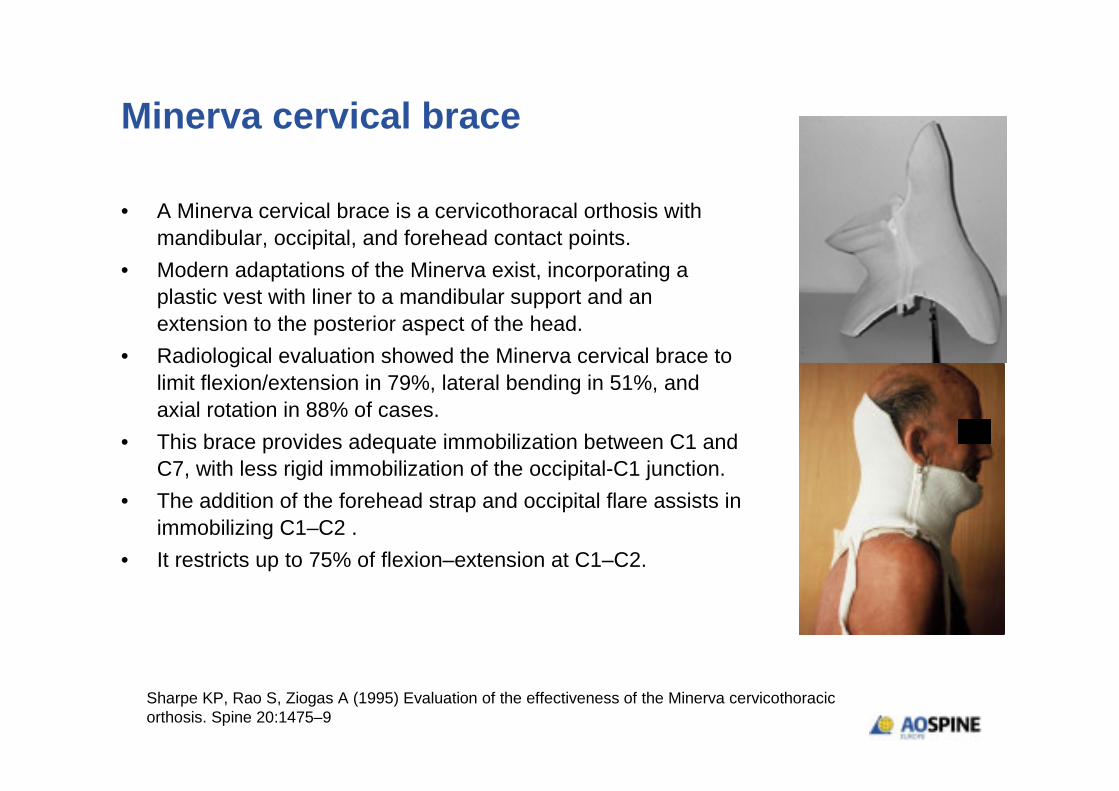
Minerva cervical brace
• A Minerva cervical brace is a cervicothoracal orthosis withmandibular, occipital, and forehead contact points.
• Modern adaptations of the Minerva exist, incorporating a plastic vest with liner to a mandibular support and anextension to the posterior aspect of the head.
• Radiological evaluation showed the Minerva cervical brace tolimit flexion/extension in 79%, lateral bending in 51%, andaxial rotation in 88% of cases.
• This brace provides adequate immobilization between C1 andC7, with less rigid immobilization of the occipital-C1 junction.
• The addition of the forehead strap and occipital flare assists in immobilizing C1–C2 .
• It restricts up to 75% of flexion–extension at C1–C2.
Sharpe KP, Rao S, Ziogas A (1995) Evaluation of the effectiveness of the Minerva cervicothoracicorthosis. Spine 20:1475–9

Minerva cervical brace
• We prefer a customized Minerva castmade of a Scotch cast,
which can be individually molded and provides a reliable
fixation which the patient cannot simply take off
• the use of thermoplastic materials and custom-made braces
further enhances comfort, compliance and will thus better
meet the ultimate goal of brace treatment.
• Indications: orthosis of choice when rigid immobilization is
required of an unstable cervical spine injury. Stable fractures
in C1-C2 segment.
Sharpe KP, Rao S, Ziogas A (1995) Evaluation of the effectiveness of the Minerva cervicothoracicorthosis. Spine 20:1475–9
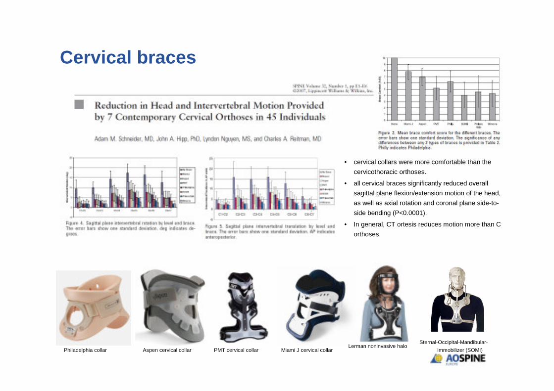
Cervical braces
PMT cervical collar Aspen cervical collar Philadelphia collar Miami J cervical collar Lerman noninvasive halo
Sternal-Occipital-Mandibular-
Immobilizer (SOMI)
• cervical collars were more comfortable than the
cervicothoracic orthoses.
• all cervical braces significantly reduced overall
sagittal plane flexion/extension motion of the head,
as well as axial rotation and coronal plane side-to-
side bending (P<0.0001).
• In general, CT ortesis reduces motion more than C
orthoses

Traction (Gardner-Wells tongs or halo)TECNIQUE.• The Gardner-Wells tongs can be applied using local anesthesia.• Trendelenburg position with shoulder straps attached to the
footend of the table.• The device should be tightened until 1 mm of the spring-loaded
stylet protrudes, which corresponds to an average of 13.5 kg of compressive force.
• The average force necessary to penetrate the inner table withcadaveric specimens with the tong pin was 73 kg, indicating a large safety margin.
• Contraindicated in atlanto-occipital dislocation or complete discoligamentous injuries because of the inherent risk of rapid neurological deterioration, which can be irreversible
• The initial weight should not exceed 5–7 kg (depending on body weight) and increases incrementally (30–60 min) only after control imaging.
• After tongs aplication, new radiographs are mandatory• If reduction cannot be obtained, or in cases of increasing
neurologic deficit, urgent surgical intervention is necessary.
Lerman JA, Dickman CA, Haynes RJ (2001) Penetration of cranial inner table withGardner-Wells tongs. J Spinal Disord 14:211–3

Traction (Gardner-Wells tongs or halo)Indications• As a temporally treatement is mainly indicated in cases of facet
subluxation or dislocation, and in burst-type fractures, to stabilize andrealign the cervical spine.
• Early application and attempt at reduction is advocated in patients with a spinal cord injury.
• Controversy mainly exists in those cases of a neurologically intact orcognitively impaired patient, recent literature supporting the safety of earlyreduction before magnetic resonance imaging (MRI) investigation
• When the patient is awake, closed reduction with skull tongs is a safe procedure, and MRI is not mandatory in this situation.
• However, if the patient has to undergo general anesthesia for a closed or open reduction, then MRI scan is absolutely indicated.
• long-term skull traction has a poor tolerance for the patient and isassociated with morbidity, it can be part of a treatment plan to avoid fusionin complex fractures, considering conversion to a halo vest after a 6-weekto 3-month period.
Tracion 7 Kg

Halo• Frank Bloom (1943)
• Apparatus for stabilization of facial fractures• “Maxillofacial surgeon”• World War II: treated pilots with inwardly displaced
facial fractures• Nickel (1968)
• Similar design • Incomplete ring with 3 pin tiara• originally developed to immobilize the unstable
cervical spine for surgical arthrodesis in patients with poliomyelitis.
O'Donnell,P.W.; Anavian,J.; Switzer,J.A.; Morgan,R.A. The history of the halo skeletal fixator. Spine, 2009, 34, 16, 1736-1739
Nickel VL, Perry J, Garrett A, HeppenstallM(1968) The halo. A spinal skeletal traction fixation device. J Bone Joint Surg Am 50:1400–9
Nickel VL, Perry J, Garrett A, HeppenstallM(1989) The halo. A spinal skeletal traction fixation device. In: Nickel VL, Perry J, Garrett A, Heppenstall M, 1968. Clin Orthop Relat Res:4–11

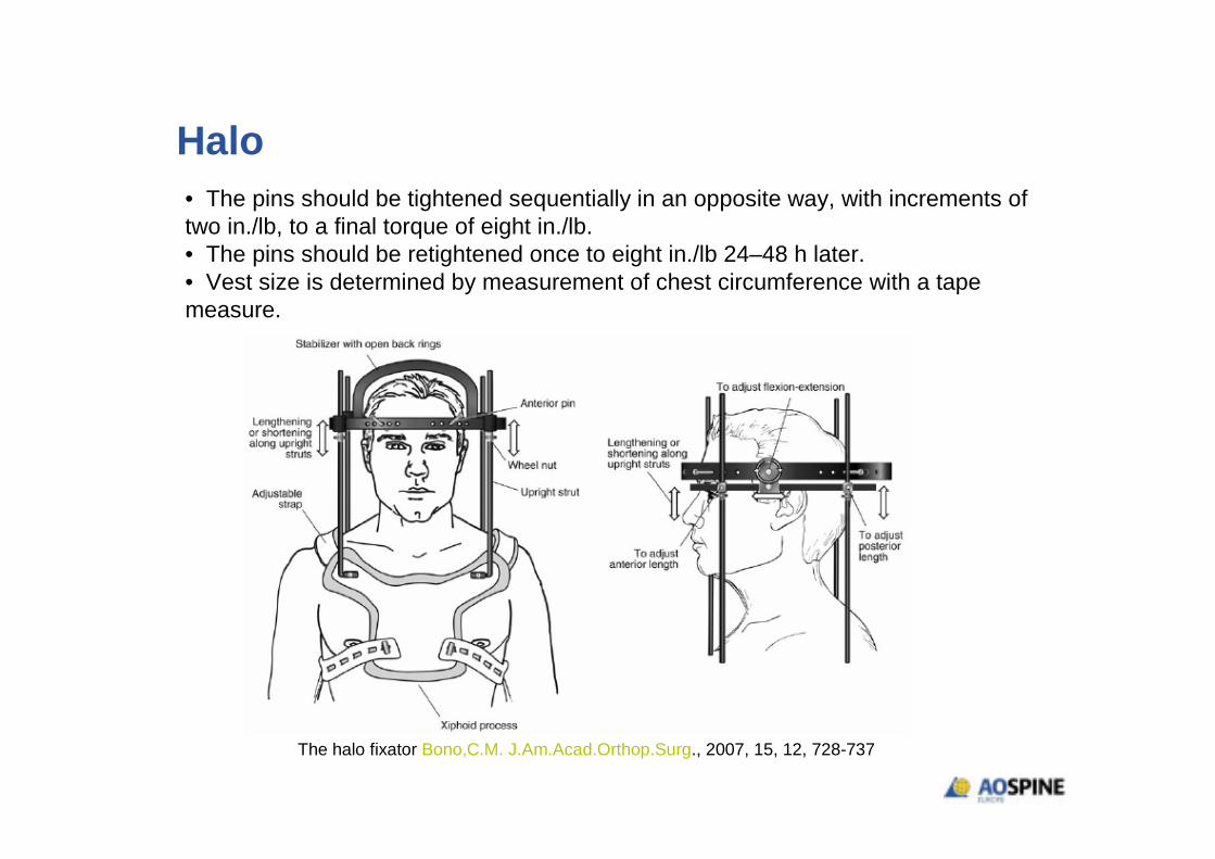
Halo: Pin Placement
The halo fixator Bono,C.M. J.Am.Acad.Orthop.Surg., 2007, 15, 12, 728-737
• The optimal position for anterior halo pin placement is 1 cm superior to theorbital rim(eyebrow), above the lateral two-thirds of the orbit, and below thegreatest circumference of the skull. Thisarea can be considered as a relatively“safe zone”
• Ring or crown size is determined by selection of a ring that provides 1–2 cm clearance around every aspect of the head perimeter
Halo
The halo fixator Bono,C.M. J.Am.Acad.Orthop.Surg., 2007, 15, 12, 728-737
• The pins should be tightened sequentially in an opposite way, with increments oftwo in./lb, to a final torque of eight in./lb.• The pins should be retightened once to eight in./lb 24–48 h later.• Vest size is determined by measurement of chest circumference with a tape measure.

Halo
• A halo vest is the most effective way to immobilise thecervical spine externally and is superior to braces.
• Affords control and positioning in cervical flexion, extension, tilt, and rotation as well as longitudinal distraction forces.
• It is the stiffest immobilization, restricting up to 75% offlexion–extension in the upper cervical spine.
• It also provides the best control of rotation and lateral bending.
• The use of halo vest may allow in shortening the hospital stay, and is also a relatively cheap method of treatment.
• When a vest has been applied both the supine and uprightX-rays must be performed to detect eventual loss ofreduction in standing or sitting position.
Halo
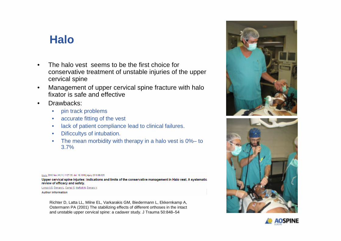
• The halo vest seems to be the first choice for conservative treatment of unstable injuries of the upper cervical spine
• Management of upper cervical spine fracture with halo fixator is safe and effective
• Drawbacks:• pin track problems• accurate fitting of the vest• lack of patient compliance lead to clinical failures.• Dificcultys of intubation.• The mean morbidity with therapy in a halo vest is 0%– to
3.7%
Richter D, Latta LL, Milne EL, Varkarakis GM, Biedermann L, Ekkernkamp A, Ostermann PA (2001) The stabilizing effects of different orthoses in the intactand unstable upper cervical spine: a cadaver study. J Trauma 50:848–54

Halo.Indications: • A halo vest or jacket can be used as definitive treatment,
as an adjunct to surgery, or as treatment for non-contiguous fractures.
• Upper cervical spine (C0-C2) isolated Jefferson fractures, hangman's fractures, odontoid type III and type I fractures, with a low dislocation rate
• Lower cervical Spine (C3-C7). is mainly indicated in cancellous bony injuries with limited displacement.
• The duration of treatment varies between 6 weeks and 4 months. Overall, its use is limited to the treatment of a minority of cervical fractures.
Contraindications: is relatively contraindicated:• In patients with severe cachexia• in patients with severe deformity (ankylosing
spondylitis or scoliosis).• in morbid obese patients• In the elderly• In non-compliant or tetraplegic patients.

Halo in Elderly
• Tashijan J. Trauma 2006• 78 patients, age > 65yo• Type II or III odontoid fractures• Increased early morbidity and mortality
• Compared with treatment using operative fixation or rigid collar
• Van Middendorp JBJS 2009• 239 patients• All ages in halo• No increased risk of pneumonia or death in
patients >65 years old
Halo vest immobilization in the elderly: a death sentence? Majercik,S.; Tashjian,R.Z.; Biffl,W.L.; Harrington,D.T.; Cioffi,W.G. J.Trauma,2005, 59, 2, 350-6; discussion 356-8
Incidence of and risk factors for complications associated with halo-vest immobilization: a prospective, descriptive cohort study of 239 patients van Middendorp,J.J.; Slooff,W.B.; Nellestein,W.R.; Oner,F.C. J.Bone Joint Surg.Am., 2009, 91, 1, 71-79

Halo Immobilization: complications
Applying a halo ring and vest requires the availability of a trained team

Conclusions. Take at home message.
• The decision-making in choosing the most appropriate
treatment modality for a cervical trauma involves many
considerations, including injury type, neurologic status,
risk of displacement, patient’s body habitus and eventual
deformity, location of the fracture, and compliance.
• The choice of one modality over the other should be made
on an individual basis, taking the above-mentioned factors
into consideration.
• Conservative treatment still has a role as a temporally
treatment or as a definitive treatment in cervical fractures.
